Embed Size (px)
Citation preview
214 Korean J Radiol 6(4), December 2005
Screen-Film Mammography and Soft-Copy Full-Field DigitalMammography: Comparison in thePatients with Microcalcifications
Objective: We wanted to compare the ability of screen-film mammography(SFM) and soft-copy full-field digital mammography (s-FFDM) on two differentmonitors to detect and characterize microcalcifications.
Materials and Methods: The images of 40 patients with microcalcifications(three patients had malignant lesion and 37 patients had benign lesion), whounderwent both SFM and FFDM at an interval of less than six months, were inde-pendently evaluated by three readers. Three reading sessions were undertakenfor SFM and for FFDM on a mammography-dedicated review workstation (RWS,2K 2.5K), and for FFDM on a high-resolution PACS monitor (1.7K 2.3K). Theimage quality, breast composition and the number and conspicuity of the micro-calcifications were evaluated using a three-point rating method, and the mammo-graphic assessment was classified into 4 categories (normal, benign, low con-cern and moderate to great concern).
Results: The image quality, the number and conspicuity of the microcalcifica-tions by s-FFDM (on the RWS, PACS and both) were superior to those by SFM in85.0%, 80.0% and 52.5% of the cases, respectively (p < 0.01), and those by thes-FFDM on the two different monitors were similar in 15.0%, 12.5% and 35.0% ofthe cases, respectively (p > 0.01). The mammographic assessment category forthe microcalcifications in the three reading sessions was similar.
Conclusion: s-FFDM gives a superior image quality to SFM and it is better atevaluating microcalcifications. In addition, s-FFDM with the PACS monitor iscomparable to s-FFDM with the RWS for evaluating microcalcifications.
maging the microcalcifications in the breast is very important for detect-ing non-palpable early breast cancer. Clustered microcalcifications arethe primary mammographic abnormality that occurs in approximately
40% of all the patients with non-palpable breast cancer (1).Mammography is the best method for detecting these early-stage breast cancers and
conventional screen-film mammography (SFM) has been shown to have a highsensitivity and specificity for the detection of breast cancer. However, there areseveral technical limitations for performing SFM that can affect the image quality andhide the fine details.
There has been a great deal of improvement in mammography over the last threedecades. Recently, full-field digital mammography (FFDM) systems have beendeveloped and they are being increasingly used to replace conventional SFM (2, 3).However, there is general concern that the lower spatial resolution of FFDM might bean obstacle for the detection and characterization of microcalcifications. Furthermore,people are becoming more concerned about the soft-copy readings, which are
Hye Seong Kim, MD1
Boo-Kyung Han, MD1
Ki-Seok Choo, MD1
Yong Hwan Jeon, MD1
Jung-Han Kim, MD2
Yeon Hyeon Choe, MD1
Index terms:Radiography, digitalMammography, MicrocalcificationScreens and filmsBreast radiographySoft copy reading
Korean J Radiol 2005;6:214-220Received February 24, 2005; accepted after revision August 29, 2005.
Departments of 1Radiology and theCenter for Imaging Science and 2Surgery,Samsung Medical Center, SungkyunkwanUniversity School of Medicine
This study was supported by a grant ofthe Korea Health 21 R&D Project,Ministry of Health & Welfare, Republic ofKorea (02-PJ3-PG6-EV06-0002)
Address reprint requests to:Boo-Kyung Han, MD, Department ofRadiology, Samsung Medical Center,Sungkyunkwan University School ofMedicine, 50 Ilwon-dong, Kangnam-gu,Seoul 135-710, Korea.Tel. (822) 3410-2518Fax. (822) 3410-2559e-mail: [email protected]
I
dependent on the quality of the viewing monitors. The aim of this retrospective study was to compare the
ability of SFM and soft-copy FFDM (s-FFDM), with usingtwo different monitors [a mammography-dedicated reviewworkstation (RWS) and a high-resolution PACS monitor]to detect and characterize microcalcifications.
MATERIALS AND METHODS
Patient SelectionFrom April to June 2003, 3,015 women underwent
FFDM in a screening or diagnostic setting. Among them,40 patients with microcalcifications in a single localizedarea (clustered, segmental or regional) had received asimultaneous or recent SFM (< 6 months). Additionalmammograms with FFDM were obtained before themagnification mammography in nine patients, and thesepatients had undergone SFMs less than three months priorto the magnification mammography (1-3 month). All themammograms were done with the patients’ consent. Theremaining 31 patients underwent additional mammogramswith FFDM during the six-month follow-up for suspectedbenign calcifications. This study included only thosepatients who had all four standard views. Ten lesions werehistopathologically confirmed by surgery or stereotaxicmammotome biopsy. Three lesions were malignant (ductalcarcinoma in situ in two patients and infiltrating ductalcarcinoma in one patient) and seven lesions were benign.The benign pathological findings were as follows: calcifica-tions in a benign duct in one patient, fibrocystic changes intwo patients, stromal fibrosis in three patients and ductalhyperplasia with dystrophic calcification in one patient.
Mammography SystemsA conventional SFM system (Senographe 600T) and a
FFDM unit (Senographe 2000D, General Electric MedicalSystems, Buc, France) were used and compared in thisstudy. This FFDM unit uses an amorphous silicon-flat-paneldetector with Cesium iodide (CsI) as the scintillator. Thepixel size was 100 (m, which gave a spatial resolution ofapproximately 5 lp/mm (4), and the depth of the bit was a14-bits; this resulted in 16,384 gray levels. The FFDMimages were reviewed on a soft-copy display system withusing both a 2 2.5 K mammography-dedicated RWS(General Electric Medical Systems, Buc, France) and a 1.7
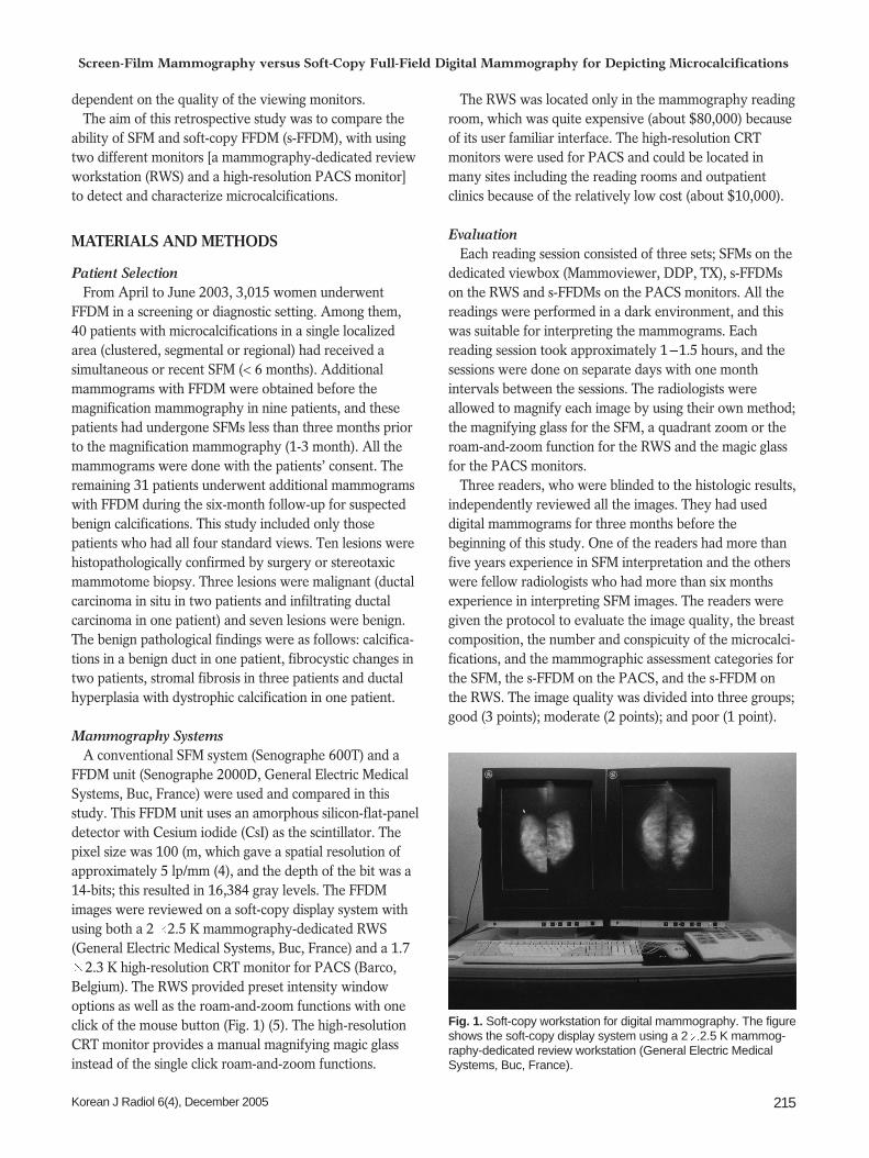
2.3 K high-resolution CRT monitor for PACS (Barco,Belgium). The RWS provided preset intensity windowoptions as well as the roam-and-zoom functions with oneclick of the mouse button (Fig. 1) (5). The high-resolutionCRT monitor provides a manual magnifying magic glassinstead of the single click roam-and-zoom functions.
The RWS was located only in the mammography readingroom, which was quite expensive (about $80,000) becauseof its user familiar interface. The high-resolution CRTmonitors were used for PACS and could be located inmany sites including the reading rooms and outpatientclinics because of the relatively low cost (about $10,000).
EvaluationEach reading session consisted of three sets; SFMs on the
dedicated viewbox (Mammoviewer, DDP, TX), s-FFDMson the RWS and s-FFDMs on the PACS monitors. All thereadings were performed in a dark environment, and thiswas suitable for interpreting the mammograms. Eachreading session took approximately 1 1.5 hours, and thesessions were done on separate days with one monthintervals between the sessions. The radiologists wereallowed to magnify each image by using their own method;the magnifying glass for the SFM, a quadrant zoom or theroam-and-zoom function for the RWS and the magic glassfor the PACS monitors.
Three readers, who were blinded to the histologic results,independently reviewed all the images. They had useddigital mammograms for three months before thebeginning of this study. One of the readers had more thanfive years experience in SFM interpretation and the otherswere fellow radiologists who had more than six monthsexperience in interpreting SFM images. The readers weregiven the protocol to evaluate the image quality, the breastcomposition, the number and conspicuity of the microcalci-fications, and the mammographic assessment categories forthe SFM, the s-FFDM on the PACS, and the s-FFDM onthe RWS. The image quality was divided into three groups;good (3 points); moderate (2 points); and poor (1 point).
Screen-Film Mammography versus Soft-Copy Full-Field Digital Mammography for Depicting Microcalcifications
Korean J Radiol 6(4), December 2005 215
Fig. 1. Soft-copy workstation for digital mammography. The figureshows the soft-copy display system using a 2 2.5 K mammog-raphy-dedicated review workstation (General Electric MedicalSystems, Buc, France).
The breast composition was categorized into one of fourpatterns according to the American College of RadiologyBreast Imaging and Reporting Database System (ACR BI-RADS). The number of microcalcifications was assigned toone of the following groups: 0 5, 6 10, 11 20, 21 40and more than 40 particles. The conspicuity, including themargin of the microcalcifications, was also divided intothree groups; clear (3 points), moderate (2 points), andindistinct (1 point). The microcalcifications were character-ized using the BI-RADS assessment categories (6, 7). Theassessment categories were divided into four groups:category 1 was normal, category 2 or 3 was benign,category 4a was low concern and category 4b or 4c wasmoderate to great concern. The category 5 microcalcifica-tions were not included in this study group. Category 1was assigned when the readers could not identify thecalcifications at the reading session even though the initialreading had suggested the presence of calcifications andother readers had also observed them.
The results of the image quality, as well as the numberand conspicuity of the microcalcifications obtained fromthe three readers, were averaged. The mammographicpattern of the breast composition and the mammographicassessment category were determined according to themajor opinion of the interpretation results. The results ofthe s-FFDM on the RWS were compared with those of theSFM and also with those of the s-FFDM on the PACS.
Statistical analysisThe data, except for the mammographic pattern of the
breast composition and the assessment category, wereanalyzed statistically using the mixed model method (8).Statistical analyses using a generalized estimating equationwere used because the data concerning the mammographicpattern and the category of the microcalcifications werenumerical (absolute) values.
Kim et al.
216 Korean J Radiol 6(4), December 2005
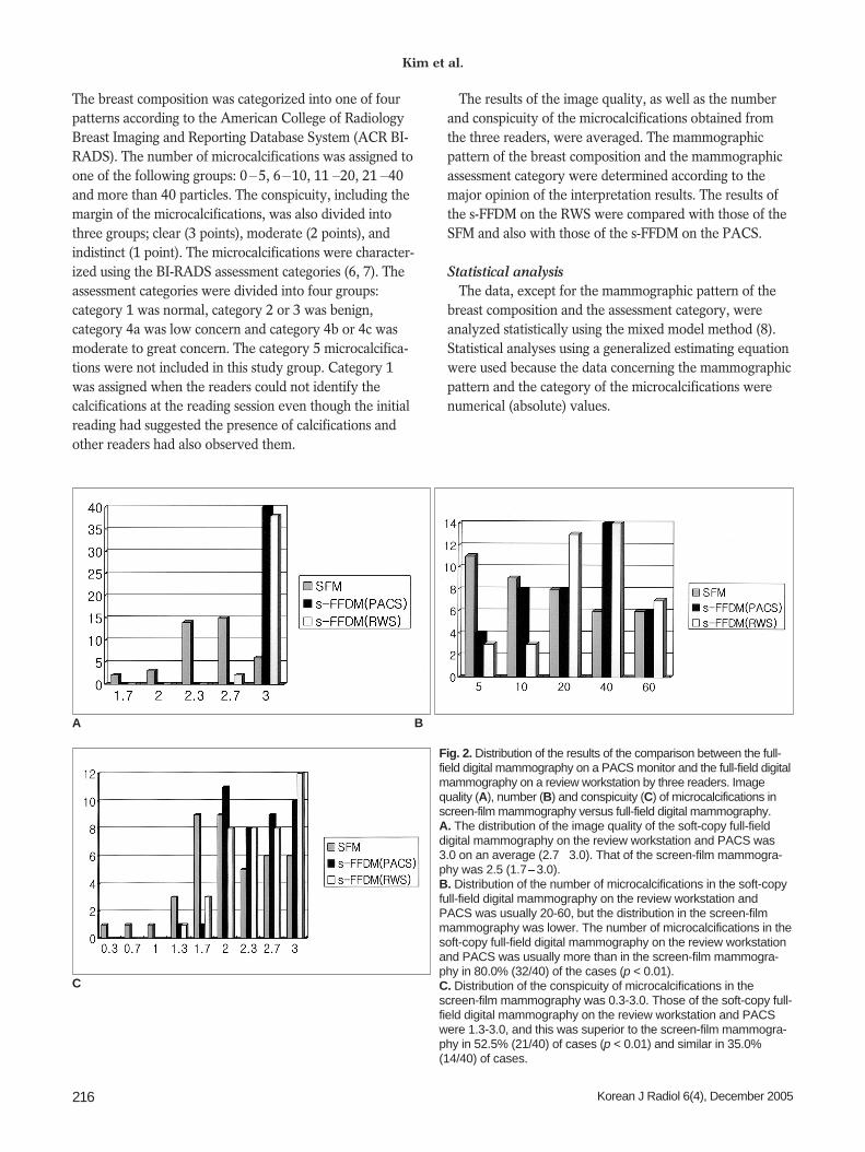
Fig. 2. Distribution of the results of the comparison between the full-field digital mammography on a PACS monitor and the full-field digitalmammography on a review workstation by three readers. Imagequality (A), number (B) and conspicuity (C) of microcalcifications inscreen-film mammography versus full-field digital mammography. A. The distribution of the image quality of the soft-copy full-fielddigital mammography on the review workstation and PACS was3.0 on an average (2.7 3.0). That of the screen-film mammogra-phy was 2.5 (1.7 3.0).B. Distribution of the number of microcalcifications in the soft-copyfull-field digital mammography on the review workstation andPACS was usually 20-60, but the distribution in the screen-filmmammography was lower. The number of microcalcifications in thesoft-copy full-field digital mammography on the review workstationand PACS was usually more than in the screen-film mammogra-phy in 80.0% (32/40) of the cases (p < 0.01).C. Distribution of the conspicuity of microcalcifications in thescreen-film mammography was 0.3-3.0. Those of the soft-copy full-field digital mammography on the review workstation and PACSwere 1.3-3.0, and this was superior to the screen-film mammogra-phy in 52.5% (21/40) of cases (p < 0.01) and similar in 35.0%(14/40) of cases.
A B
C
RESULTS
The distribution of the image quality of the mammogra-phy according to the three readers was as follows: 2.5(1.7 3.0) on the SFM, 3.0 on the PACS and 3.0 (2.7 3.0)on the RWS (Fig. 2). The image quality of the s-FFDM onthe RWS was superior to that of the SFM in 85.0% (34/40)of the cases (p < 0.01) and it was equal to the SFM in15.0% (6/40) of the cases (Fig. 3). There was no casewhere the image quality of the s-FFDM was inferior to the
SFM. The image quality of the s-FFDM, RWS and PACSwas similar (p > 0.01). The mammographic pattern of thebreast composition of the s-FFDM on the RWS and SFMand the s-FFDM on the RWS and PACS monitor wassimilar (p > 0.01).
The number of microcalcifications detected by thevarious techniques was as follows: 18.2 (0.7 70.0) onSFM, 24.4 (1.0 70.0) on the PACS and 24.8 (1.0 70.0)on the RWS. The number of microcalcifications detected inthe s-FFDM on the RWS was significantly higher than inthe SFM in 80.0% (32/40) of cases (p < 0.01) (Fig. 4), and
Screen-Film Mammography versus Soft-Copy Full-Field Digital Mammography for Depicting Microcalcifications
Korean J Radiol 6(4), December 2005 217
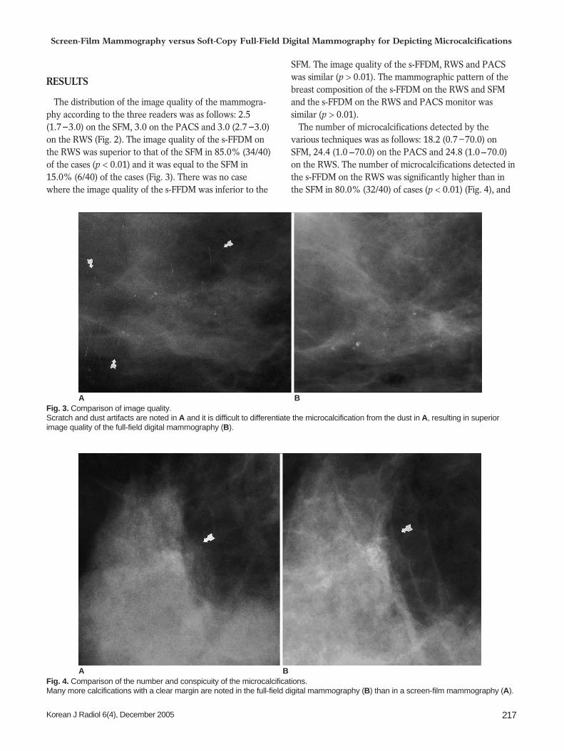
Fig. 3. Comparison of image quality. Scratch and dust artifacts are noted in A and it is difficult to differentiate the microcalcification from the dust in A, resulting in superiorimage quality of the full-field digital mammography (B).
A B
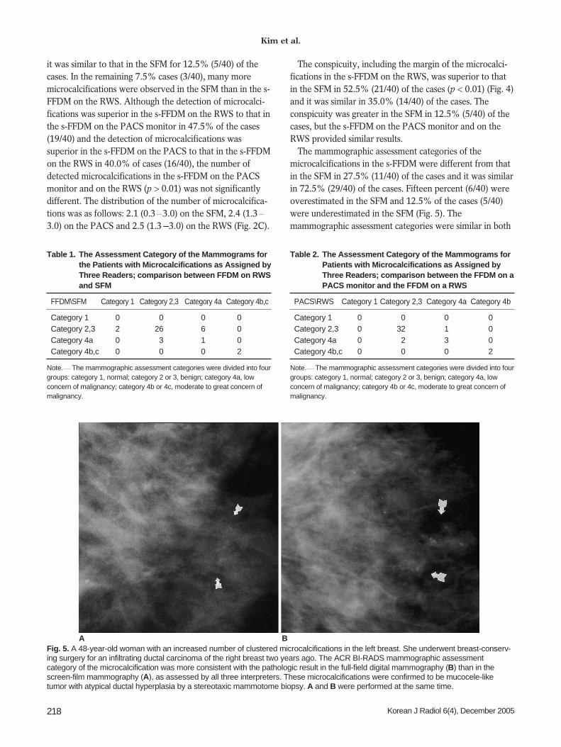
Fig. 4. Comparison of the number and conspicuity of the microcalcifications.Many more calcifications with a clear margin are noted in the full-field digital mammography (B) than in a screen-film mammography (A).
A B
it was similar to that in the SFM for 12.5% (5/40) of thecases. In the remaining 7.5% cases (3/40), many moremicrocalcifications were observed in the SFM than in the s-FFDM on the RWS. Although the detection of microcalci-fications was superior in the s-FFDM on the RWS to that inthe s-FFDM on the PACS monitor in 47.5% of the cases(19/40) and the detection of microcalcifications wassuperior in the s-FFDM on the PACS to that in the s-FFDMon the RWS in 40.0% of cases (16/40), the number ofdetected microcalcifications in the s-FFDM on the PACSmonitor and on the RWS (p > 0.01) was not significantlydifferent. The distribution of the number of microcalcifica-tions was as follows: 2.1 (0.3 3.0) on the SFM, 2.4 (1.33.0) on the PACS and 2.5 (1.3 3.0) on the RWS (Fig. 2C).
The conspicuity, including the margin of the microcalci-fications in the s-FFDM on the RWS, was superior to thatin the SFM in 52.5% (21/40) of the cases (p < 0.01) (Fig. 4)and it was similar in 35.0% (14/40) of the cases. Theconspicuity was greater in the SFM in 12.5% (5/40) of thecases, but the s-FFDM on the PACS monitor and on theRWS provided similar results.
The mammographic assessment categories of themicrocalcifications in the s-FFDM were different from thatin the SFM in 27.5% (11/40) of the cases and it was similarin 72.5% (29/40) of the cases. Fifteen percent (6/40) wereoverestimated in the SFM and 12.5% of the cases (5/40)were underestimated in the SFM (Fig. 5). Themammographic assessment categories were similar in both
Kim et al.
218 Korean J Radiol 6(4), December 2005
Table 1. The Assessment Category of the Mammograms forthe Patients with Microcalcifications as Assigned byThree Readers; comparison between FFDM on RWSand SFM
FFDM\SFM Category 1 Category 2,3 Category 4a Category 4b,c
Category 1 0 00 0 0Category 2,3 2 26 6 0Category 4a 0 03 1 0Category 4b,c 0 00 0 2
Note. The mammographic assessment categories were divided into fourgroups: category 1, normal; category 2 or 3, benign; category 4a, lowconcern of malignancy; category 4b or 4c, moderate to great concern ofmalignancy.
Table 2. The Assessment Category of the Mammograms forPatients with Microcalcifications as Assigned byThree Readers; comparison between the FFDM on aPACS monitor and the FFDM on a RWS
PACS\RWS Category 1 Category 2,3 Category 4a Category 4b
Category 1 0 00 0 0Category 2,3 0 32 1 0Category 4a 0 02 3 0Category 4b,c 0 00 0 2
Note. The mammographic assessment categories were divided into fourgroups: category 1, normal; category 2 or 3, benign; category 4a, lowconcern of malignancy; category 4b or 4c, moderate to great concern ofmalignancy.
Fig. 5. A 48-year-old woman with an increased number of clustered microcalcifications in the left breast. She underwent breast-conserv-ing surgery for an infiltrating ductal carcinoma of the right breast two years ago. The ACR BI-RADS mammographic assessmentcategory of the microcalcification was more consistent with the pathologic result in the full-field digital mammography (B) than in thescreen-film mammography (A), as assessed by all three interpreters. These microcalcifications were confirmed to be mucocele-liketumor with atypical ductal hyperplasia by a stereotaxic mammotome biopsy. A and B were performed at the same time.
A B

the s-FFDM on the PACS monitor and in the s-FFDM onthe RWS (Tables 1, 2).
DISCUSSION
The National Cancer Institute in the USA has designateddigital mammography as the imaging technology with thehighest potential for improving the detection and diagnosisof breast cancer (9). Direct digital mammography was firstperformed for stereotaxic biopsies and localizations withusing systems that were based on charge-coupled devicechips. However, these chips could not be used for full-fieldimaging on account of the limited detector size. The nextattempt in digital mammography was the use of digitizedstorage phosphor systems. However, this technique waslimited due to the low quantum efficiency and the lowspatial resolution (6, 10). A combination of a directmagnification technique using a microfocus tube andstorage phosphor plates was tried so as to increase thespatial resolution. A later study demonstrated that magnifi-cation mammography with using this technique wassuperior to conventional film-screen magnificationmammography (11). There have been further develop-ments in these combinations, and high-resolution digitizedstorage phosphor systems have recently become available.
The full-field digital mammography system used in thisstudy is based on a flat amorphous silicon array and a CsIscintillator. The potential limitation of this system is thelower spatial resolution of 5 line pairs (lp)/mm comparedwith that of 12 15 lp/mm for the SFM. The minimal pixelsize required for digital mammography is still a subject ofdebate. The spatial resolution of the flat-panel and the CRsystems is determined by this pixel size of 5 lp/mm.However, the flat-panel system has a much higher modula-tion transfer function at all spatial frequencies below thislimit than does the computed radiography systems. Thishigher resolution is a result of the CsI phosphor producinghigher-resolution images than the storage phosphors (3).Although the full-field digital system presented in thispaper does not meet the current standards and guidelinesthat are defined for conventional screen-film mammogra-phy (limited spatial resolution), FFDM was shown to beslightly better for detecting and characterizing microcalci-fications than the SFM that we used (12, 13). This is due tothe increased contrast-detail detection rate of the digitalsystem, which allows better visualization of small high-contrast structures. There is also optimized image process-ing for the detailed visualization that is needed in digitalmammography. The image processing technique optimizesthe image quality of the soft-copy display as well as thehard-copy images. The other advantages of the digital
technique are the wider dynamic range, the highercontrast-detail detectability and the superior detectivequantum efficiency (4, 9, 14). These factors cause bettervisualization of the peripheral breast structures such as theskin, subcutaneous tissue and retromammary space, as wellas the parenchymal structures that are seen in the FFDMrather than in the SFM (6). Dust and other artifacts that aredue to the film developer often appear in the SFM, butthey do not exist in the FFDM. These artifacts can hinderthe diagnosis of breast cancer. Repeated mammograms dueto the over- or underexposure are no longer necessarywhen using the FFDM (3).
This study also showed a higher image quality andsuperior detectability, as well as the better characterizationof the microcalcifications in the s-FFDM than in the SFM.We thinks that this was also due to the improved contrastrecolution in the digital system.
The results of this study show that the SFM interpreta-tion can give an underestimation; for example, themicrocalcifications interpreted as category 4a in the s-FFDM were interpreted as being category 3 in the SFM. Inaddition, the s-FFDM on the RWS with its better quality isnot available to all clinicians. Therefore, this studycompared the s-FFDM on the RWS to the s-FFDM on thePACS monitor. The RWS that provided preset intensitywindowing options and roam-and-zoom functions with aclick of a mouse button was located only in a mammogra-phy reading room. This workstation is quite expensivebecause it has a user familiar interface. In contrast, thehigh-resolution CRT monitors for the PACS are located atmultiple sites, including reading rooms and outpatientclinics, because of their relatively low cost.
Many hospitals have implemented or are consideringimplementing a PACS. The filmless environment createdby the PACS enables more efficient storage, retrieval andtransmission of images. It has improved radiology servicesby allowing complete control of the runaway film problem,i.e., reducing the number of lost films and also reducing therate of unreported films. Ideally, all the imaging techniquesshould be connected to the PACS for a hospital’s PACSsystem to be cost-effective. The obvious advantage ofdigital mammography systems is that they can be directlyinterfaced with the PACS. Introducing mammography intoa PACS system is complicated by the necessity ofexpensive, high-resolution monitors for the soft-copyreporting of digital mammograms. At the least, 2Kmonitors are needed for viewing digital mammograms atfull resolution (15 17).
This study suggests that digital mammography readingson a PACS monitor and on a RWS have equivalentdiagnostic accuracy. This indicates that communication
Screen-Film Mammography versus Soft-Copy Full-Field Digital Mammography for Depicting Microcalcifications
Korean J Radiol 6(4), December 2005 219

Kim et al.
220 Korean J Radiol 6(4), December 2005
between the radiologist and clinicians will be facilitatedthrough the PACS system.
There were some limitations in this study. All the SFMsand FFDMs were not taken at the same time, the studyinvolved only a small number of cases and some variationsin mammographic compression are inevitable. In addition,most of the selected cases were biased because they werebeing followed up for the category 3 lesions and only 10 ofthe studied lesions were confirmed histopathologically.Therefore, a prospective study on a large population willbe needed to supplement the results of this study.
In conclusion, the s-FFDM has superior image qualityand a superior ability to evaluate microcalcifications ascompared with the SFM. In addition, the s-FFDM on aRWS gives an equivalent evaluation of microcalcificationsto that obtained on a PACS monitor.
References1. Sickels EA. Mammographic features of 300 consecutive nonpal-
pable breast cancers. AJR Am J Roentgenol 1986;46:661-6632. Feig SA, Yaffe MJ. Digital mammography, computer-aided
diagnosis, and telemammography. Radiol Clin North Am1995;33:1205-1230
3. Obenauer S, Luftner-Nagel S, von Heyden D, Munzel U, BaumF, Grabbe E. Screen film vs. full-field digital mammography:image quality, detectability and characterization of lesions. EurRadiol 2002;12:1697-1702
4. Hermann KP, Hundertmark C, Funke M, Brenndorff AV,Grabbe E. Digital mammography in direct magnificationtechnique using a large-area amorphous silicon X-ray detector.Rofo 1999;170:503-506
5. Pisano ED, Cole EB, Kistner EO, Muller KE, Hemminger BM,Brown ML, et al. Interpretation of digital mammograms:comparison of speed and accuracy of soft-copy versus printed-film display. Radiology 2002;223:483-488
6. Fischer U, Baum F, Obenauer S, Luftner-Nagel S, Heyden D,Vosshenrich R, et al. Comparative study in patients with
microcalcifications: full-field digital mammography vs. screen-film mammography. Eur Radiol 2002;12:2679-2683
7. American College of Radiology. Breast imaging reporting anddata system (BI-RADSTM), 3rd ed. American College ofRadiology, Reston, Va., 1998
8. Brown H, Prescott R. Applied mixed model in medicine. NewYork: John Wiley & Sons, 1999:199-260
9. Shtern F. Digital mammography and related technologies: aperspective from the national cancer institute. Radiology1992;183:629-630
10. Kheddache S, Thilander-Klang A, Lanhede B, Mansson LG,Bjurstam N, Ackerholm P, et al. Storage phosphor and film-screen mammography: performance with differentmammographic techniques. Eur Radiol 1999;9:591-597
11. Funke M, Hermann KP, Breiter N, Hundertmark C, Sachs J,Gruhl T, et al. Digital storage phosphor mammography in amagnification technic: experimental studies for spatial resolutionand for detection of microcalcifications. Rofo 1997;167:174-179
12. Lewin JM, Hendrick RE, Dorsi CJ, Isaacs PK, Moss LJ, KarellasA, et al. Comparison of full-field digital mammography withscreen-film mammography for cancer detection: results of 4945paired examinations. Radiology 2001;218:873-880
13. Venta LA, Hendrick RE, Adler YT, DeLeon P, Mengoni PM,Scharl AM, et al. Rates and causes of disagreement in interpreta-tion of full-field digital mammography and film-screenmammography in a diagnostic setting. AJR Am J Roentgenol2000;176:1241-1248
14. Hermann KP, Obenauer S, Grabbe E. Radiation exposure infull-field digital mammography with a flat-panel X-ray detectorbased on amorphous silicon in comparison with conventionalscreen-film mammography. Rofo 2000;172:940-945
15. Leung JW. New modalities in breast imaging: digital mammog-raphy, positron emission tomography, and sestamibi scintimam-mography. Radiol Clin North Am 2002;40:467-482
16. James JJ. The current status of digital mammography. ClinRadiol 2004;59:1-10
17. Hayt DB, Alexander S, Drakakis J, Berdebes N. Filmless in 60days: the impact of picture archiving and communicationssystems within a large urban hospital. J Digit Imaging2001;14:62-71