Embed Size (px)
Citation preview
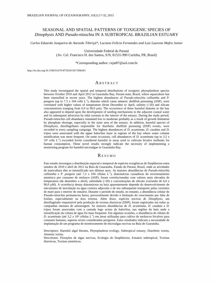
BRAZILIAN JOURNAL OF OCEANOGRAPHY, 63(1):17-32, 2015
SEASONAL AND SPATIAL PATTERNS OF TOXIGENIC SPECIES OF
Dinophysis AND Pseudo-nitzschia IN A SUBTROPICAL BRAZILIAN ESTUARY
Carlos Eduardo Junqueira de Azevedo Tibiriçá*, Luciano Felício Fernandes and Luiz Laureno Mafra Junior
Universidade Federal do Paraná
(Av. Cel. Francisco H. dos Santos, S/N, 81531-990 Curitiba, PR, Brasil)
*Corresponding author: [email protected]
http://dx.doi.org/10.1590/S1679-87592015071906301
A B S T R A C T
This study investigated the spatial and temporal distributions of toxigenic phytoplankton species between October 2010 and April 2012 in Guaratuba Bay, Paraná state, Brazil, where aquaculture has
been intensified in recent years. The highest abundances of Pseudo-nitzschia calliantha and P.
pungens (up to 7.3 x 10⁴ cells L-1), diatoms which cause amnesic shellfish poisoning (ASP), were correlated with higher values of temperature (from December to April, salinity (>20) and silicate
concentrations (ranging from 6.0 to 90.0 µm). The occurrence of these harmful diatoms in the bay
also appeared to depend upon the development of seeding mechanisms in the adjacent coastal water and its subsequent advection by tidal currents to the interior of the estuary. During the study period,
Pseudo-nitzschia cell abundance remained low to moderate probably as a result of growth limitation
by phosphate shortage, especially in the outer area of the estuary. In addition, harmful species of Dinophysis, dinoflagellates responsible for diarrhetic shellfish poisoning (DSP) events, were
recorded in every sampling campaign. The highest abundances of D. acuminata, D. caudata and D.
tripos were associated with the upper halocline layer in regions of the bay where water column stratification was more frequent. On some occasions, cell abundances of D. acuminata (up to 3.2 x
10³ cells L-1) exceeded levels considered harmful in areas used to cultivate bivalve mollusks for
human consumption. These novel results strongly indicate the necessity of implementing a monitoring program for harmful microalgae in Guaratuba Bay.
R E S U M O
Este estudo investigou a distribuição espacial e temporal de espécies toxigênicas de fitoplâncton entre
outubro de 2010 e abril de 2012 na Baía de Guaratuba, Estado do Paraná, Brasil, onde as atividades
de maricultura têm se intensificado nos últimos anos. As maiores abundâncias de Pseudo-nitzschia calliantha e P. pungens (até 7,3 x 10⁴ células L-1), diatomáceas causadoras do envenenamento
amnésico por consumo de molusco (ASP), foram correlacionadas com valores mais elevados de
temperatura (de dezembro a abril), salinidade (>20) e concentrações de silicato (variando de 6,0 a 90,0 µM). A ocorrência destas diatomáceas na baía aparentemente depende do desenvolvimento de
mecanismo de inoculação na água costeira adjacente e de seu subsequente transporte pelas correntes
de maré para o interior do estuário. Durante o período de estudo, no entanto, a abundância celular de Pseudo-nitzschia permaneceu baixa, provavelmente devido à limitação do crescimento por falta de
fosfato, especialmente na área externa. Além disso, espécies nocivas de Dinophysis, um
dinoflagelado responsável pela produção de toxinas diarreicas (DSP), foram registradas em todas as campanhas mensais de amostragem. As maiores abundâncias de D. acuminata, D. caudata e D.
tripos foram associadas com a camada logo acima da haloclina, nas regiões da baía onde a
estratificação da coluna de água foi mais frequente. Em algumas ocasiões, a abundância de células de D. acuminata (até 3,2 x 10³ células L-1) em áreas utilizadas para cultivo de moluscos bivalves para
consumo humano, superou níveis considerados perigosos. Estes resultados indicam a necessidade de
implantação de um programa de monitoramento de microalgas nocivas na Baía de Guaratuba.
Descriptors: Harmful algal blooms, Phytoplankton ecology, Subtropical estuary, Diarrhetic toxins,
Amnesic toxins. Descritores: Florações de algas nocivas, Ecologia do fitoplâncton, Estuário subtropical, Toxinas
diarreicas, Toxinas amnésicas.
INTRODUCTION
Guaratuba Bay is a relatively pristine
estuary, located in the subtropical region of Brazil,
where oyster farming (Crassostrea spp.) has grown
substantially in recent years (BALDAN;
BENDHACK, 2009). The dependence of traditional
communities on fishery products as well as an increase
in the consumption of bivalve mollusks from this bay
raises a concern about human poisoning due to the
consumption of products contaminated with algal
toxins (PERL et al., 1990; CARVALHO et al., 1998;
DARANAS et al., 2001; REIZOPOULOU et al., 2008;
ETHERIDGE, 2010). The fear that such events could
occur in Guaratuba Bay is reinforced by the presence
of toxin-producing microalgae in nearby locations,
including that of species that cause diarrhetic shellfish
poisoning (DSP) and amnesic shellfish poisoning
(ASP) (MAFRA Jr. et al., 2006; ODEBRECHT et al.,
2002; PROCOPIAK et al., 2006; PROENÇA, 2006;
FERNANDES; BRANDINI, 2010; FERNANDES et
al., 2013).
The main toxins responsible for DSP —
okadaic acid (OA) and its analogs, the
dinophysistoxins (DTXs) — are produced by
dinoflagellates of the genera Dinophysis and
Prorocentrum (DARANAS et al., 2001;
TOYOFUKU, 2006; GERSSEN et al., 2010). These
toxins affect humans through the consumption of
bivalve mollusks, which accumulate the toxic
compounds in their tissues (REIZOPOULOU et al.,
2008). Importantly, even at low cell densities (i.e.,
1000 cells L-1), Dinophysis spp. can produce toxin
concentrations sufficient to cause intoxication
symptoms, such as nausea, abdominal cramps and
diarrhea, in people who consume the contaminated
bivalves (TOYOFUKU, 2006). In humans, DSP has
been documented since the 1960s, and currently, high
levels of OA are repeatedly found in coastal regions
worldwide (GERSSEN et al., 2010). This toxin has
already been found in the soft tissues of mollusks
collected along the southeastern coast of Brazil
(PROENÇA; MAFRA Jr., 2005; MARINÉ et al.,
2009).
In addition to the DSP-causing toxins,
domoic acid (DA) – the main toxin which causes ASP
outbreaks, produced by several species of the diatom
Pseudo-nitzschia (JEFFERY et al., 2004; GRANT et
al., 2010) – has also been recorded in South Brazil
(PROENÇA; MAFRA Jr., 2005). Domoic acid, which
is responsible for massive mortalities of birds and
marine mammals following the ingestion of
contaminated fish (BELTRÀN et al., 1997;
LEFEBVRE et al., 1999), can also intoxicate humans
through the consumption of bivalve mollusks that
accumulate the toxin in their tissues, mainly in the
hepatopancreas and viscera (PERL et al., 1990;
GRANT et al., 2010; MAFRA et al., 2010). Although,
only in special episodes, when a large number of
Pseudo-nitzschia spp. cells containing high DA
concentrations occur in coastal waters, are bivalve
mollusks capable of accumulating sufficient toxin to
affect humans, causing gastrointestinal and
neurological symptoms (JEFFERY et al., 2004;
GRANT et al., 2010). During the major 1987 Pseudo-
nitzschia multiseries bloom on Prince Edward Island,
Canada, more than 100 people were hospitalized, and
four patients died (PERL et al., 1990).
In the coastal region and other estuaries
surrounding Guaratuba Bay, harmful Dinophysis spp.
and Pseudo-nitzschia species have already been
recorded in abundances considered dangerous by
monitoring programs (ODEBRECHT et al., 2002;
MAFRA Jr. et al., 2006; PROENÇA et al., 2011;
CIDASC, 2012). This bay is surrounded by seaside
resorts and small- to medium-sized cities that can
become densely populated in summer. The local
economy is widely tied to small-scale fishing and
tourism activities, which causes the bay area to
become an ecosystem highly vulnerable to syndromes
caused by microalgal toxins. Despite this vulnerability
and the nearby occurrence of toxin-producing algal
species, this study is the first to investigate the spatio-
temporal variation of the potentially harmful
Dinophysis spp. and Pseudo-nitzschia spp. in
Guaratuba Bay.
MATERIAL AND METHODS
Study Area
Guaratuba Bay is located on the coast of
Paraná State, Brazil (25º52’S, 48º39’W). This region
has a subtropical, humid, mesothermic climate,
presenting a hot summer and lacking a dry season
(Cfa-type in the Köppen classification). The driest
period extends from May to August, with a mean
monthly rainfall of 250 to 350 mm and a mean
temperature of 16 to 17ºC (IAPAR, 2012). Much of
the inner edge of the bay is unpopulated, being
surrounded by mangrove forests (SANTOS et al.,
2008). This semi-enclosed coastal system is mainly
influenced by the tide (up to 1.5 m), but is also
strongly affected by river discharge (MARONE et al.,
2006). The main rivers draining into the bay are the
Cubatão and São João, which together contribute a
runoff of about 80 m s-1 (MARONE et al., 2006).
There are over 20 other minor rivers, of which the
Parati, located on the north-south axis of the bay
where oyster farming is concentrated, is the most
important (Fig. 1). The shallow depths, the narrow but
deep connection to the ocean and the extensive
drainage basin (of approximately 1,700 km²)
contribute to the occurrence of water column
stratification, mainly in the periods subject to neap
18 BRAZILIAN JOURNAL OF OCEANOGRAPHY, 63(1), 2015

tides (MARONE et al., 2006). In fact, this bay is
considered a partially stratified estuary according to
the classification of DAY JR. et al. (1989).
Sampling
Sa ling a aigns ere n erta en
a ro i atel onthl et een to er 2 an
ril 2 2, o ering t o s essi e s ring an
s er seasons, the ost a ora le or the
roli eration o har l algae in so thern Brazil
( et al., 998 r. et al., 2006). Seven
sampling stations were selected (Fig. 1) in Guaratuba
Bay covering areas with commercial oyster farms
and/or areas with the potential for the future expansion
of such facilities. At each georeferenced station, the
seawater was sampled with a Van Dorn bottle at the
surface, near the bottom and, depending on the depth
and the transparency of the water, in an intermediate
stratum located at the base of the euphotic zone.
Salinity, temperature, chlorophyll fluorescence and
turbidity were measured along a vertical depth profile
(at every 20 cm) using a CTD (conductivity,
temperature and depth meter) device (JFE ALEC Co.
Ltd.®, model COMPAC-CTD). Vertical and/or
oblique hauls were carried out using a phytoplankton
net with a 20-μm mesh size.
Analysis of Physical and Chemical Parameters
Samples collected with the Van Dorn bottle
were filtered in 200-ml aliquots through a vacuum
filtration system and used to determine the
concentrations of dissolved nutrients by
spectrophotometry and of chlorophyll-a by
fluorometry (HOLM-HANSEN et al., 1965). The
latter analysis was only applied to selected samples
and was used to calibrate the measurements obtained
by a chlorophyll fluorescence sensor attached to the
CTD. Additionally, dissolved ammonium and
phosphate concentrations were determined according
to STRICKLAND and PARSONS' method (1972),
and the analysis of silicate, nitrite and nitrate
concentrations followed the recommendations of
APHA (1999). Concentrations of nitrite, nitrate and
ammonium were summed up and expressed as
dissolved inorganic nitrogen (DIN). Finally, silicate to
DIN (Si:N) and DIN to phosphate (N:P) ratios were
calculated.
Phytoplankton Composition and Abundance
Samples from the phytoplankton net were
preserved in a formaldehyde solution at a final
concentration of 4.0%, and observed under a BX-30
light microscope coupled to an Olympus® DP-71
digital camera. Whenever Pseudo-nitzschia spp. cells
were abundant, samples were prepared according to
HASLE and FRYXELL (1970) for examination in a
transmission electron microscope (TEM, model
JEOL® JM1200 EXII).
For quantitative analysis, samples collected
with the Van Dorn bottle were fixed with neutral
L gol’s sol tion at a inal on entration o
approximately 1%. Each sample was processed using
the sedimentation method of ÜTERMOHL (1958).
The cells were counted in accordance with Edler and
Elbrächter's methodology (2010), covering the whole
counting chamber to estimate the total abundance of
cells in a sample volume ranging from 10 to 25 mL.
Fig. 1. Map of Guaratuba Bay, located in the state of Paraná (PR), Brazil. The sampling
stations were designated E1 to E7. The following main rivers flowing into the bay are indicated with dotted lines: São João River (R-SJ), Cubatão River (R-C) and Parati River (R-P). Image
source: USGS (http://www.usgs.gov/).
TIBIRICÁ ET AL.: Dinophysis AND Pseudo-nitzschia IN GUARATUBA BAY 19
Treatment of Data and Statistical Analysis
Descriptive analyses were used in the
interpretation of results, complemented by canonical
correspondence analysis (CCA). The CCA was
performed with all the microphytoplankton taxa, the
environmental variables of salinity, temperature,
turbidity, and the concentrations of chlorophyll-a and
dissolved nutrients. The abundance data were
transformed using the fourth root prior to the analysis.
Monte Carlo permutations were performed to test the
significance (p < 0.05) of each environmental factor,
with stepwise inclusion (forward selection) of only the
significant factors in the model generated. For this
analysis, the Canoco software for Windows 4.5
(Microcomputer Power, Inc., Ithaca, NY, USA) was
used (TER BRAAK; SMILAUER, 2002).
RESULTS
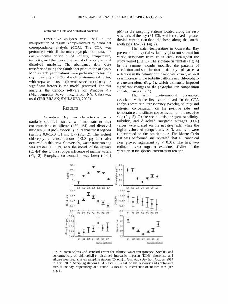
Guaratuba Bay was characterized as a
partially stratified estuary, with moderate to high
concentrations of silicate (>30 µM) and dissolved
nitrogen (>10 µM), especially in its innermost regions
(salinity 0.0-15.0, E1 and E7) (Fig. 2). The highest
chlorophyll-a concentrations (>3.0 µg L-1) also
occurred in this area. Conversely, water transparency
was greater (>1.3 m) near the mouth of the estuary
(E3-E4) due to the stronger influence of marine waters
(Fig. 2). Phosphate concentration was lower (< 0.5
µM) in the sampling stations located along the east-
west axis of the bay (E1-E3), which received a greater
fluvial contribution than did those along the south-
north axis (E5-E7) (Fig. 2).
The water temperature in Guaratuba Bay
presented little spatial variability (data not shown) but
varied seasonally from 16 to 30ºC throughout the
study period (Fig. 3). The increase in rainfall (Fig. 4)
in the summer months modified the patterns of
circulation and stratification in the bay and caused a
reduction in the salinity and phosphate values, as well
as an increase in the turbidity, silicate and chlorophyll-
a concentrations (Fig. 3), which ultimately imposed
significant changes on the phytoplankton composition
and abundance (Fig. 5).
The main environmental parameters
associated with the first canonical axis in the CCA
analysis were rain, transparency (Secchi), salinity and
nitrogen concentration on the positive side, and
temperature and silicate concentration on the negative
side (Fig. 5). On the second axis, the greatest salinity,
turbidity, and dissolved inorganic nitrogen (DIN)
values were placed on the negative side, while the
higher values of temperature, Si:N, and rain were
concentrated on the positive side. The Monte Carlo
test was performed and revealed that all canonical
axes proved significant (p < 0.01). The first two
ordination axes together explained 51.6% of the
variation in the species-environment relation.
Fig. 2. Mean values and standard errors for salinity, water transparency (Secchi), and concentrations of chlorophyll-a, dissolved inorganic nitrogen (DIN), phosphate and
silicate measured at seven sampling stations (X-axis) in Guaratuba Bay from October 2010
to April 2012. Sampling stations E1-E3 and E5-E7 fall on the east-west and north-south axes of the bay, respectively, and station E4 lies at the intersection of the two axes (see
Fig. 1).
20 BRAZILIAN JOURNAL OF OCEANOGRAPHY, 63(1), 2015

Fig. 3. Minimum, mean and maximum values for temperature, salinity, concentration of
chlorophyll-a, dissolved inorganic nitrogen (DIN), phosphate, silicate and the ratio of nitrogen
to phosphate (N:P) based on the 15 sampling campaigns (X axis) performed in Guaratuba Bay. The Y axis for N:P is on logarithmic scale.
Fig. 4. Daily rainfall (left y-axis) recorded in Guaratuba Bay, PR, from September
2010 to April 2012. Markers on the bottom of the plot indicate the sampling dates,
and the shaded bars indicate the monthly rainfall (right y-axis). Data from INMET
(2012).
TIBIRICÁ ET AL.: Dinophysis AND Pseudo-nitzschia IN GUARATUBA BAY 21
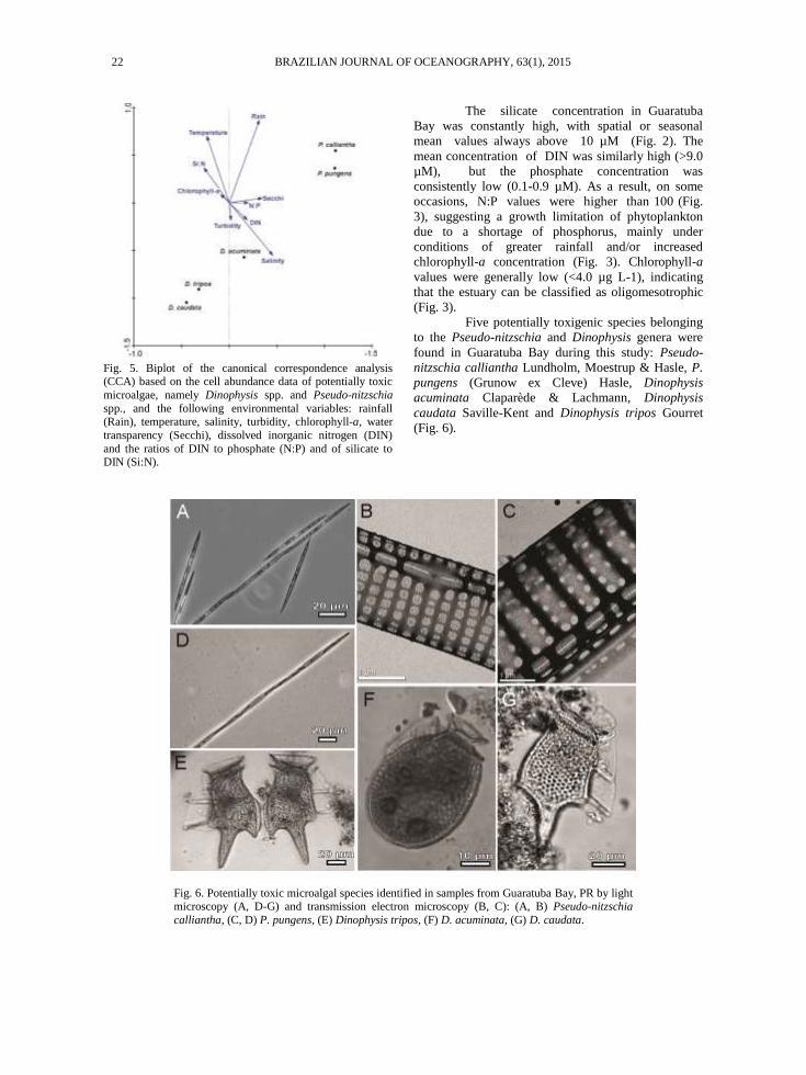
Fig. 5. Biplot of the canonical correspondence analysis (CCA) based on the cell abundance data of potentially toxic
microalgae, namely Dinophysis spp. and Pseudo-nitzschia
spp., and the following environmental variables: rainfall (Rain), temperature, salinity, turbidity, chlorophyll-a, water
transparency (Secchi), dissolved inorganic nitrogen (DIN)
and the ratios of DIN to phosphate (N:P) and of silicate to DIN (Si:N).
The silicate concentration in Guaratuba
Bay was constantly high, with spatial or seasonal
mean values always above 10 µM (Fig. 2). The
mean concentration of DIN was similarly high (>9.0
µM), but the phosphate concentration was
consistently low (0.1-0.9 µM). As a result, on some
occasions, N:P values were higher than 100 (Fig.
3), suggesting a growth limitation of phytoplankton
due to a shortage of phosphorus, mainly under
conditions of greater rainfall and/or increased
chlorophyll-a concentration (Fig. 3). Chlorophyll-a
values were generally low (<4.0 µg L-1), indicating
that the estuary can be classified as oligomesotrophic
(Fig. 3).
Five potentially toxigenic species belonging
to the Pseudo-nitzschia and Dinophysis genera were
found in Guaratuba Bay during this study: Pseudo-
nitzschia calliantha Lundholm, Moestrup & Hasle, P.
pungens (Grunow ex Cleve) Hasle, Dinophysis
acuminata Claparède & Lachmann, Dinophysis
caudata Saville-Kent and Dinophysis tripos Gourret
(Fig. 6).
Fig. 6. Potentially toxic microalgal species identified in samples from Guaratuba Bay, PR by light microscopy (A, D-G) and transmission electron microscopy (B, C): (A, B) Pseudo-nitzschia
calliantha, (C, D) P. pungens, (E) Dinophysis tripos, (F) D. acuminata, (G) D. caudata.
22 BRAZILIAN JOURNAL OF OCEANOGRAPHY, 63(1), 2015
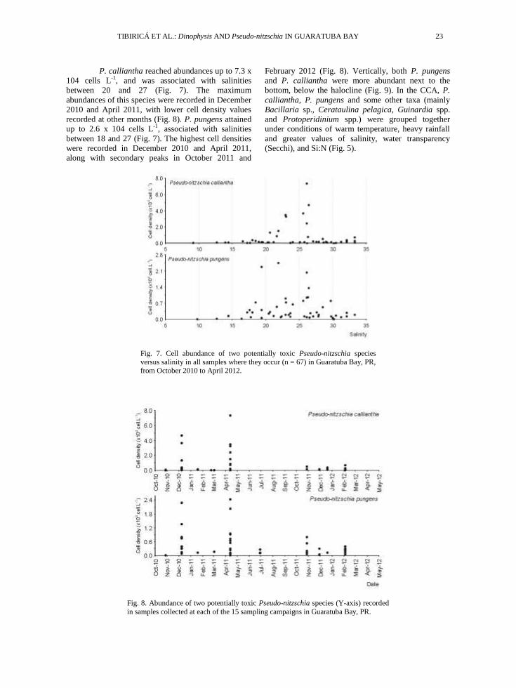
P. calliantha reached abundances up to 7.3 x
104 cells L-1, and was associated with salinities
between 20 and 27 (Fig. 7). The maximum
abundances of this species were recorded in December
2010 and April 2011, with lower cell density values
recorded at other months (Fig. 8). P. pungens attained
up to 2.6 x 10⁴ cells L-1, associated with salinities
between 18 and 27 (Fig. 7). The highest cell densities
were recorded in December 2010 and April 2011,
along with secondary peaks in October 2011 and
February 2012 (Fig. 8). Vertically, both P. pungens
and P. calliantha were more abundant next to the
bottom, below the halocline (Fig. 9). In the CCA, P.
calliantha, P. pungens and some other taxa (mainly
Bacillaria sp., Cerataulina pelagica, Guinardia spp.
and Protoperidinium spp.) were grouped together
under conditions of warm temperature, heavy rainfall
and greater values of salinity, water transparency
(Secchi), and Si:N (Fig. 5).
Fig. 7. Cell abundance of two potentially toxic Pseudo-nitzschia species
versus salinity in all samples where they occur (n = 67) in Guaratuba Bay, PR,
from October 2010 to April 2012.
Fig. 8. Abundance of two potentially toxic Pseudo-nitzschia species (Y-axis) recorded
in samples collected at each of the 15 sampling campaigns in Guaratuba Bay, PR.
TIBIRICÁ ET AL.: Dinophysis AND Pseudo-nitzschia IN GUARATUBA BAY 23

Fig. 9. Vertical profiles of salinity, temperature (ºC), chlorophyll-a concentration
(µg L-1) and abundance (cells L-1) of Pseudo-nitzschia calliantha and P. pungens at selected sampling dates in Guaratuba Bay.
D. acuminata reached up to 3.2 x 10³ cells
L-1, with the maximum abundance values coinciding
with salinities from 10 to 25 (Fig. 10) and occurring
between August and December (Fig. 11). Canonical
analysis grouped D. acuminata with certain nontoxic
species that were moderately frequent in the samples
(e.g. Asterionellopsis glacialis, Chaetoceros spp. and
Thalassionema nitzschioides), under conditions of
greater salinity, turbidity and DIN concentration
(Fig. 5). However, greater cell densities of D.
acuminata were found near the surface and above
the haline stratification of the water column, unlike
the co-occurring diatoms that were observed at
higher abundances mainly below the haline
stratification (Fig. 12).
Fig. 10. Cell abundance of three potentially toxic Dinophysis species versus salinity in all samples where they occur (n = 73) in Guaratuba Bay, PR, from October 2010 to
April 2012.
24 BRAZILIAN JOURNAL OF OCEANOGRAPHY, 63(1), 2015

D. caudata reached up to 1.0 x 10³ cells L-1
at a salinity of 30, and occurred only in samples with a
salinity greater than 22 (Fig. 10), in September 2011
and April 2012, when total phytoplankton abundances
were higher (Fig. 11). Additionally, low abundances
of D. tripos (up to 200 cells L-1) were reported mainly
in samples with salinity between 20 and 28 (Fig. 10).
Similar to D. caudata, D. tripos occurred only in
September 2011 and April 2012 (Fig. 11). In the CCA,
D. caudata, D. tripos and two species of marine
diatoms (Helicotheca sp. and Thalassiothrix sp.) were
grouped together under conditions of low rainfall,
which were associated with samples collected in April
2012 (Fig. 5). In addition, greater cell densities of D.
caudata were found at the surface of the water
column, whereas D. tripos was dominant either at the
surface or associated with deeper high-chlorophyll-a
layers (Fig. 13).
Fig. 11. Cell abundance of Dinophysis spp. in samples collected at each of the
15 sampling campaigns in Guaratuba Bay, PR.
Fig. 12. Vertical profiles of salinity, temperature (ºC), chlorophyll-a concentration
(µg L-1), and abundance (cells L-1) of Dinophysis acuminata (X-axis) in selected samples of Guaratuba Bay.
TIBIRICÁ ET AL.: Dinophysis AND Pseudo-nitzschia IN GUARATUBA BAY 25

Fig. 13. Vertical profiles of salinity, temperature (ºC), chlorophyll-a concentration (µg L-1) and abundance (cells L-1) of Dinophysis tripos (A–C) and
Dinophysis caudata (D) in selected samples from Guaratuba Bay.
DISCUSSION
Pseudo-nitzschia spp.
In Guaratuba Bay, the maximum abundances
of P. calliantha and P. pungens were detected from
October to April, and were typically associated with
an increase in temperature at locations with higher
salinity (between 18 and 27, predominantly above 22)
and a greater silicate availability. The higher rainfall
observed in Guaratuba Bay during that period most
likely increased the silicate input to the bay through
continental drainage. As shown in the CCA, the more
saline waters exhibited higher Pseudo-nitzschia spp.
cell densities, especially when considering the
absolute values measured near the bottom, where cells
accumulated. This genus also occurred in conditions
of lower salinity (16-25) during periods of high
rainfall, but always near the bottom and in the estuary
region with the greatest marine influence (the outer
sector).
In other places in the world, similar results
were found regarding the distribution of Pseudo-
nitzschia spp. as a function of temperature and
salinity. THESSEN et al. (2005), who investigated the
seasonal dynamics of Pseudo-nitzschia in different
environments (estuaries, bays and the outer shelf)
along the coast of Louisiana, USA, demonstrated the
affinity of P. calliantha for more saline waters. In that
study, P. calliantha (identified as P.
pseudodelicatissima) and P. pungens were abundant in
samples with salinities ranging from 20 to 35,
although occurring in lower salinity areas as well. P.
pungens was particularly more abundant in waters
with salinities higher than 30. In general, when
isolated cells were tested in laboratory growth
experiments, the optimal range of salinity (15 to 30)
closely overlapped that observed in the field
(THESSEN et al., 2005). In the high-salinity (33-38)
waters of Lim Bay, in the Adriatic Sea, P. calliantha
blooms (up to 1.6 x 10⁶ cells L-1) are mainly
associated with a temperature increase during the
transition from spring to summer, aside from other
factors, such as phosphorus and ammonium inputs,
especially at the end of summer, when aquaculture
activities are more intense (LJUBESIC et al., 2011).
In the present study, Pseudo-nitzschia
calliantha and P. pungens o rre re erentiall in
s er an at the eginning o the all, et een
e e er an ril, a re rrent seasonal attern in
so thern Brazil ( et al., 998 r. et
al., 2006; TAVARES et al., 2009). During that period,
temperatures varied from 22 to 32ºC, with the greatest
cell densities observed in temperatures around 24ºC.
26 BRAZILIAN JOURNAL OF OCEANOGRAPHY, 63(1), 2015
The species found in this study are regarded as
eurythermic (THESSEN; STOECKER, 2008;
QUIJANO-SCHEGGIA et al., 2008; LIEFER et al.,
2009; ANDERSON et al., 2010; LJUBESIC et al.,
2011). The euryhaline and eurythermic features of
both species pose serious potential implications for
aquaculture. Our results, supported by the literature
data, suggest that Pseudo-nitzschia spp. are potential
threats to local shellfish farms, since they occur at
harmful cell abundances mainly at the end of summer,
coincidently with the peak of tourism and shellfish
sales in the region.
Simultaneously with the variations in
salinity and temperature during the rainy season,
increasing silicate concentrations (and slightly higher
nitrogen concentrations) were recorded during the
peaks of Pseudo-nitzschia spp. abundance in
Guaratuba Bay. The formation of a haline
stratification in this estuary is also more evident in
summer due to the increased freshwater input (our
results; MIZERKOWSKI et al., 2012), which indicates
the relevance of rainfall in the population dynamics of
the Pseudo-nitzschia spp. in Guaratuba Bay.
SPATHARIS et al. (2007) also correlated P.
calliantha blooms in Kalloni Gulf, Greece, to periodic
freshwater inputs, which bring silicate and reduce the
salinity to a level closer to the optimum for the growth
of these algae. As such, blooms of Pseudo-nitzschia
spp. are common after large nutrient inputs, even if
these blooms occur occasionally during local
oscillations in salinity (MAFRA Jr. et al., 2006;
LIEFER et al., 2009; PIEDRAS; ODEBRECHT,
2012).
On some occasions, macronutrients can
become limiting for the growth of Pseudo-nitzschia
spp. For example, SPATHARIS et al. (2007) found a
marked increase in P. calliantha abundance
immediately after a period of intense rainfall in
Kalloni Gulf, Greece. The higher concentrations of
nutrients, particularly phosphorus, were attributed to
the intensification of agricultural activities and
resulted from the application of fertilizers to cultivated
lands before the most intense period of rainfall. In
other marine areas, an inverse relation between
macronutrient concentration and phytoplankton
abundance has also been found; however, this
apparently contradictory result can be explained by the
excessive consumption of phosphorus and nitrogen by
rapidly growing phytoplankton (FISHER et al., 1988).
In Chesapeake Bay, USA, FISHER et al. (1988)
observed that the silicate, nitrate and phosphate
concentrations depleted mainly by virtue of the
increase in phytoplankton biomass, whereas the nitrite
and ammonia concentrations were more affected by
regeneration and nitrification. For phosphate, a similar
pattern was observed in Guaratuba Bay, probably
leading to growth limitation as a result of a shortage of
this nutrient.
The highest cell densities of Pseudo-
nitzschia were found at the sampling stations located
in the outer sector of the bay, decreasing drastically
towards the inner sectors. Similar results were found
in other environments with comparable
geomorphological characteristics, such as bays and
coastal inlets (THESSEN et al., 2005; SPATHARIS et
al., 2007). For example, in Chesapeake Bay, USA, the
most abundant toxin-producing species, P. calliantha,
P. pungens and P. multiseries, occurred in elevated
cell densities near the estuary mouth; but they were
absent in areas with salinity < 6.0 in the inner sectors
(THESSEN; STOECKER, 2008). This pattern
suggests that the abundance of Pseudo-nitzschia spp.
near Guaratuba Bay should increase mainly outside
the estuary, in the adjacent coastal zone (BRANDINI;
FERNANDES, 1996), and that the cells are advected
by coastal waters into the estuary at certain periods,
which explains the higher abundances found below the
halocline and in the outer sector of the estuary, where
coastal water prevails. Although Pseudo-nitzschia spp.
are usually observed close to the mouth of estuaries
(SPATHARIS et al., 2007; THESSEN; STOECKER,
2008), they can also occur at lower salinities, as was
the case in several areas of Chesapeake Bay with
salinities up to 6 (THESSEN; STOECKER, 2008) and
in the present study, where they were observed at
salinities as low as 14.
In Guaratuba Bay, two widely dominant
Pseudo-nitzschia species showed a typical annual
variation, with maximum abundances ranging from
2.6 to 7.3 x 10⁴ cells L-1 between December 2010 and
April 2011. These densities are not sufficient to be
characterized as blooms, but are already considered
dangerous because of the potential risk for shellfish
contamination (MAFRA Jr. et al., 2006). Such cell
ensities an in i ate an “alert” sit ation in
monitoring programs during which the frequency and
number of samples collected for cell counts should be
intensified (CIDASC, 2012). When cell abundance
reaches 10 x 104 cells L-1, toxin analysis in bivalve
mollusks is also recommended because at such a level,
DA concentrations in mollusk tissues can already be
sufficient to poison humans, if the cells are producing
and retaining reasonable amounts of toxins (MAFRA
Jr. et al., 2006; PROENÇA et al., 2011).
Despite the low Pseudo-nitzschia
concentrations usually measured in the bay, we cannot
rule out the risk of blooms' existing, since other
triggering environmental conditions such as adequate
temperature, salinity and silicate levels are
simultaneously present in the bay. Moreover, it is
likely that high cell abundances (> 106 cells L-1) can
be achieved and transported from the adjacent coastal
waters into the outer portion of the estuary. Indeed,
TIBIRICÁ ET AL.: Dinophysis AND Pseudo-nitzschia IN GUARATUBA BAY 27

blooms of Pseudo-nitzschia have previously been
recorded in areas near Guaratuba Bay. In Paranaguá
Bay, approximately 40 km north of Guaratuba Bay,
Pseudo-nitzschia comprises an important fraction of
the phytoplankton biomass, sometimes dominating the
community (BRANDINI; THAMM, 1994; MAFRA
Jr. et al., 2006). Additionally, blooms of P. multiseries
and P. calliantha, both toxin-producing species, were
recorded in the coastal areas in front of Guaratuba
Bay in early summer in 1997 and 1999, from
December to February in both years (FERNANDES;
BRANDINI, 2010). During those periods, the
intrusion of nutrient-rich waters resulting from the
upwelling of the South Atlantic Central Water was
regarded as responsible for the blooms (BRANDINI et
al., 2007). Farther south in the state of Santa Catarina,
the entire coastal region from São Francisco do Sul to
the south of Santa Catarina Island has been identified
as a hotspot for this genus (FERNANDES et al.,
2013). That is, the area presents high species diversity,
frequent blooms and previous reports of domoic acid
accumulation in cultivated mollusks (PROENÇA et
al., 2011). Cell concentrations as high as 21 x 106
cells L-1, associated with increased DA levels in
mollusk tissues (up to 98.5 µg g-1 in the mussel Perna
perna), were recorded in some areas in January and
February of 2009 (FERNANDES et al., 2013). The
entire coastal area around Guaratuba Bay thus
constitutes a potential seeding area for Pseudo-
nitzschia cells, which can be transported into the bay
by tides and coastal currents, the main exchange
mechanisms between the bay and the adjacent coastal
waters (MARONE et al., 2006).
Dinophysis spp.
In this study, three toxin-producing species
of Dinophysis (D. acuminata, D. caudata and D.
tripos) were recorded, with D. acuminata being the
dominant one, occurring frequently throughout the
study period at abundances of up to 3.2 x 10³ cells L-1.
Because of its high cellular diarrhetic toxin content
(i.e., cell quota), D. acuminata can be harmful to
humans in abundances as low as 1.0 x 10³ cells L-1
(reviewed in Reguera et al., 2014). Even lower
abundances can cause episodes of intoxication (e.g.,
100-200 cells L-1; YASUMOTO et al., 1985),
especially in situations that favor the retention of high
toxin quotas in the cells, such as growth limitation due
to the lack of prey or light (NIELSEN et al., 2012;
MAFRA et al., 2014). Thus, although D. acuminata
did not dominate the phytoplankton assemblage at any
particular point, on several occasions the abundance of
this species was greater than the alert threshold
established by monitoring programs such as Brazil's
(CIDASC, 2012). It is recommended, therefore, that
toxin analyses of bivalve mollusks should be carried
out systematically in Guaratuba Bay. The cell
densities recorded in the present study suggest that the
concentration of toxins in mollusk tissues from
Guaratuba Bay may occasionally be sufficient to
intoxicate humans (PROENÇA et al., 2011).
D. acuminata showed an irregular pattern of
seasonal variation, occurring in different months
during winter, spring and summer. The CCA results
and the additional analyses indicated that none of the
parameters evaluated could alone adequately explain
the seasonal variation of D. acuminata in Guaratuba
Bay. This finding can possibly be explained by the
complex mode of energy uptake of this species. D.
acuminata is mixotrophic and captures its prey, the
ciliate Myrionecta rubra, by inserting a peduncle that
immobilizes it and, after a certain time, sucks the cell
content of the prey through that same structure (PARK
et al., 2006). Thus, the main factors affecting the
growth of this species appears to be the quality and
quantity of prey (TONG et al., 2010; REGUERA et
al., 2012), although pH (PARK et al., 2006),
temperature (TONG et al., 2010) and light may also be
factors of great relevance for the development of D.
acuminata (REGUERA et al., 2012). In the present
study, M. rubra was observed in 67% of the samples
collected (data not shown); however, the cell counts of
this ciliate would not be sufficiently accurate because
of the sampling preservation methodology employed
in this study. Because of its frequency of occurrence in
the samples, M. rubra is most likely an important food
source for Dinophysis spp. in Guaratuba Bay, and its
availability probably affects D. acuminata distribution
and abundance patterns.
Under suitable conditions, growth of D.
acuminata in the laboratory can be equated with that
of other mixotrophic dinoflagellates (KAMIYAMA;
SUZUKI, 2009), although D. acuminata may normally
be characterized as a slow-growing species. However,
this species can further reduce its rate of cell division
and cell size to enable it to persist in the water column
in the absence of prey, demonstrating a high adaptive
capacity (REGUERA et al., 2012). Thus, Dinophysis
is a genus with highly persistent species that can easily
multiply in the presence of light and prey, but can also
slow their growth rate considerably when food is
lacking (REGUERA et al., 2012). These ecological
strategies could, therefore, explain the occurrence of
D. acuminata all year round in Guaratuba Bay.
Additionally, in the present study, no regular
pattern of spatial distribution was observed for D.
acuminata, since high cell abundances were recorded
across all sampling stations. The species was mainly
recorded in salinities between 10 and 25. Considering
the quite distinct environmental conditions of the
stations sampled and the strong dynamics of
Guaratuba Bay (MARONE et al., 2006), the use of
fixed sampling points (geographic coordinates) can
28 BRAZILIAN JOURNAL OF OCEANOGRAPHY, 63(1), 2015

hinder the interpretation of spatial data over time.
Even with the precaution of always sampling during
the same astronomical tide condition, as adopted in
this study (in this case at high tide), variations in the
rainfall, tidal amplitude, winds and meteorological
tides among the different sampling campaigns can
potentially result in different current patterns,
suggested by the variability in stratification throughout
the study (e.g., Figs. 12 and 13). The variation in
surface and subsurface currents is highly relevant
because the transport of blooms is one of the factors
determining the occurrence of harmful events
involving Dinophysis (TAVARES et al., 2009;
REGUERA et al., 2012). Any monitoring program in
this area that includes this important toxigenic species
should take these factors into account. The frequent
occurrence of D. acuminata above the halocline,
mainly at stations E1, E2 and E3, suggests that
stratification of the water column helps in the growth
of this species, as has been revealed in other studies.
D. acuminata has been studied in a fjord in Chile,
where its occurrence has mainly been associated with
stratification and stabilization of the water column
(DIAZ et al., 2011). In another study in Portugal, high
abundances of this species occurred whenever the
river runoff was low, associated with high salinities
and water column stability (VALE; SAMPAYO,
2003). Other investigations have also confirmed that
cell densities of Dinophysis spp. begin to increase as
the water column stabilizes (REGUERA et al., 2012).
The present study highlights the importance of water
column stratification in promoting the observed cell
peaks of D. acuminata, which has also been evidenced
by a study carried out in the nearby estuarine complex
of Paranaguá (MAFRA Jr. et al., 2006). This finding is
important not only to understand the ecology of the
species but also to determine the best methodology for
its detection and enumeration, given that Dinophysis
cells can migrate through the water column and
concentrate in thin subsurface layers (ESCALERA et
al., 2012). Aside from the use of stratified samples for
better quantification, whole water column sampling,
net samples and the analysis of toxins in the tissues of
bivalves are recommended for a successful monitoring
program focused on Dinophysis spp.
In addition to D. acuminata, D. caudata and
D. tripos can also produce OA, DTXs and
pectenotoxins (PTXs) (TAYLOR et al., 2004;
FERNÁNDEZ et al., 2006; REGUERA et al., 2012).
Of the three species, only D. acuminata and D.
caudata have been associated with DSP events in
various coastal water areas around the world
(REGUERA et al., 2012). D. caudata occurred
occasionally in Guaratuba Bay at up to 1.0 x 10³ cells
L-1 in the fall and spring. This species has already been
recorded between inter an s er in so thern
Brazil, altho gh ne er at high ell ensities ( et
al., 1998; SCHMITT; PROENÇA, 2000; TAVARES
et al., 2009; HARAGUCHI; ODEBRECHT, 2010).
The risk of a harmful event caused by this species is
low in Guaratuba Bay; however, the co-occurrence of
D. caudata can potentiate the bloom toxicity of other
toxic species, such as D. acuminata. The third species,
D. tripos, also occurred occasionally in the bay at a
maximum abundance of 0.2 x 10³ cells L-1 during the
fall and spring months. This species is present all year
round in southern Brazil, although harmful events
have not been recorded to date (TAVARES et al.,
2009; HARAGUCHI; ODEBRECHT, 2010). Thus,
similarly to D. caudata, D. tripos should be monitored
in Guaratuba Bay because of its potential to cause
DSP (TAYLOR et al., 2004), mainly in multi-species
blooms. In addition, D. tripos cells are capable of
retaining greater toxin quotas because they are much
larger (~130 µm in cell length) than D. acuminata
(~50 µm) and D. caudata (~90 µm).
CONCLUSIONS
Based on the novel results obtained, we may
affirm that Dinophysis spp. can occur throughout the
year in Guaratuba Bay, with their growth most likely
depending on food quality, light and water column
stratification. Furthermore, the production of toxins by
D. acuminata depends on the environmental
conditions (LINDAHL et al., 2007; HACKETT et al.,
2009). Although relatively low, the D. acuminata cell
abundances reported herein can occasionally be
sufficient for the contamination of bivalve mollusks
cultivated in the bay, making the monitoring for
diarrhetic toxins highly recommendable in this area.
Increases in the temperature, salinity (>20)
and silicate concentration favor the growth of Pseudo-
nitzschia spp. in this estuary. However, phosphate
limitation likely prevented these species from attaining
cell densities high enough to threaten fishing and
aquaculture activities in the region. Higher cell
densities of Pseudo-nitzschia spp. within the estuary
depend on the existence of a cell seeding mechanism
in the adjacent coastal region and its advection into the
bay. The monitoring of these species in Guaratuba Bay
is desirable since blooms of these harmful diatoms
have previously been recorded in adjacent waters.
ACKNOWLEDGEMENTS
The authors would like to thank the National
Council of Scientific and Technological Development
(CNPq, project no. 481759/2010-7) and the Araucária
Foundation for the financial support provided for L. L.
M. Jr. In addition, this study was partially funded by
the CNPQ/PROTAX under contract no. 562151/2010-
9. Our gratitude is also extended to the Paraná Institute
TIBIRICÁ ET AL.: Dinophysis AND Pseudo-nitzschia IN GUARATUBA BAY 29

of Technical Assistance and Rural Extension
(EMATER-PR) for their assistance during sampling
campaigns and to Dr. Eunice da Costa Machado and
the technician Liciane Siqueira of the Center for
Marine Studies (CEM), Federal University of Paraná,
for their assistance with nutrient analysis. Electron
microscope assistance was provided by the Center of
Electron Microscopy - UFPR.
REFERENCES
ANDERSON, C. R.; SAPIANO, M. R. P.; PRASAD, M. B.
K.; LONG, W.; TANGO, P. J.; BROWN, C. W.;
MURTUGUDDE, R. Predicting potentially toxigenic
Pseudo-nitzschia blooms in the Chesapeake Bay. J.
Mar. Syst., v. 83, p. 127–140, 2010.
APHA - American Public Health Association. Standard
Methods for the Examination of Water and
Wastewater. Washington: American Public Health
Association, 1496 pp., 1999. BALDAN, A. P.; BENDHACK, F. Maricultura sustentável
no litoral do Paraná, Brasil: atualidades e perspectivas.
Rev. Acad. Ciênc. Agrár. Ambient., v. 7, n. 4, p. 491-497, 2009.
BELTRÁN, A. S.; PALAFOX-URIBE, M.; GRAJALES-MONTIEL, J.; CRUZ-VILLACORTA, A.; OCHOA, J.
L. Sea bird mortality at Cabo San Lucas, Mexico:
evidence that toxic diatom blooms are spreading. Toxicon, v. 35, n. 3, p. 447-453, 1997.
BRANDINI, F. P.; THAMM, C. A. C. Variações diárias e
sazonais do fitoplâncton e parâmetros ambientais na Baía de Paranaguá. Nerítica, v. 8, n. 2, p. 55-72, 1994.
BRANDINI, F. P.; FERNANDES, L. F. Microalgae of the
continental shelf off Paraná State, southeastern Brazil: a review of studies. Rev. bras. oceanogr., v. 44, n. 1, p.
69-80, 1996.
BRANDINI, F. P.; SILVA, A. S.; SILVA, E. T.; KOLM, H. Sources of Nutrients and Seasonal Dynamics of
Chlorophyll on the Inner Shelf off Paraná State—South
Brazil Bight. J. Coast. Res., v. 23, n. 5, p. 1131-1140, 2007.
CARVALHO, M.; JACINTO, J.; RAMOS, N.; OLIVEIRA,
V.; MELO, T. P.; SÁ, J. Paralytic shellfish poisoning: clinical and electrophysiological observations. J.
Neurol., v. 245, p. 551–554, 1998.
CIDASC – COMPANHIA INTEGRADA DE
DESENVOLVIMENTO AGRÍCOLA DE SANTA
CATARINA. Monitoramento de Algas Nocivas.
Disponível em: Acesso em: 06 ago. 2012. DARANAS, A. H.; NORTE, M.; FERNÂNDEZ, J. J. Toxic
marine microalgae. Toxicon, v. 39, p. 1101-1132, 2001.
DAY JR., J. W.; HALL, C. A. S.; KEMP, W. M.; YÁÑEZ-ARANCIBIA, A (Ed.). Estuarine ecology. John Wiley,
1989, 558 p.
DIAZ, P.; MOLINET, C.; CACERES M. A.; VALLE-LEVINSON, A. Seasonal and intratidal distribution of
Dinophysis spp. in a Chilean fjord. Harmful Algae, v.
10, p. 155–164, 2011. EDLER, L.; ELBRÄCHTER, M., The Utermöhl method for
quantitative phytoplankton analysis. In: KARLSON, B.;
CUSACK, C.; BRESNAN, E. (Eds). Microscopic and
molecular methods for quantitative phytoplankton
analysis. Paris: Intergovernmental Oceanographic Commission of UNESCO, 2010, p. 13-20.
ESCALERA, L.; PAZOS, Y.; DOVAL, M. D.;
REGUERA, B. A comparison of integrated and discrete depth sampling for monitoring toxic species of
Dinophysis. Mar. Pollut. Bull., v. 64, p. 106–113,
2012. ETHERIDGE, S. M. Paralytic shellfish poisoning: Seafood
safety and human health perspectives. Toxicon, v. 56, p.
108–122, 2010. FERNANDES, L. F.; BRANDINI, F. P. The potentially toxic
diatom Pseudo-nitzschia H. Peragallo in the Paraná and
Santa Catarina States, Southern Brazil. Iheringia, Ser. Bot., v. 65, n. 1, p. 47-62, 2010.
FERNANDES, L. F.; CAVALCANTE, K. P.; PROENÇA,
L. A. O.; SCHRAMM, M. A. Blooms of Pseudo-
nitzschia pseudodelicatissima and P. calliantha and
associated domoic acid accumulation in shellfish from
the South Brazilian coast. Diatom Research, v. 28:381-393, 2013.
FERNÁNDEZ, M. L.; REGUERA, B.; GONZÁLEZ-GIL,
S.; MÍGUEZ, A. Pectenotoxin-2 in single-cell isolates of Dinophysis caudata and Dinophysis acuta from the
Galician Rías (NW Spain). Toxicon, v. 48, p. 477–490,
2006. FISHER, T. R.; HARDING JR, L. W.; STANLEY, D. W.;
WARD, L. G. Phytoplankton, Nutrients, and Turbidity in
the Chesapeake, Delaware, and Hudson Estuaries. Estuar. Coast. Shelf Sci, v. 27, p. 61-93, 1988.
GERSSEN, A.; POL-HOFSTAD, I. E.; POELMAN, M.;
MULDER, P. P. J.; TOP, H. J. VAN DEN; BOER, J. Marine Toxins: Chemistry, Toxicity, Occurrence and
Detection, with Special Reference to the Dutch Situation.
Toxins, v. 2, p. 878-904, 2010. GRANT, K. S.; BURBACHER, T.M.; FAUSTMAN, E.M.;
GRATTTAN, L. Domoic acid: Neurobehavioral
consequences of exposure to a prevalent marine biotoxin. Neuro Tox. Teratol., v. 32, p. 132–141, 2010.
HACKETT, J. D.; TONG, M.; KULIS, D. M.; FUX, E.;
HESS, P.; BIRE, R.; ANDERSON, D. M. DSP toxin production de novo in cultures of Dinophysis acuminate
(Dinophyceae) from North America. Harmful Algae, v.
8, p. 873–879, 2009. HARAGUCHI, L.; ODEBRECHT, C. Dinophysiales
(Dinophyceae) no extremo Sul do Brasil (inverno de
2005, verão de 2007). Biota Neotrop., v. 10, n. 3, p. 101-114, 2010.
HASLE, G. R.; FRYXELL, G. A.; Diatoms: cleaning and
mounting for light and electron microscopy. Trans. Am.
Microscop. Soci., v. 89, p. 469-474, 1970.
HOLM-HANSEN, O.; LORENZEN, C. J.; HOLMES, R. W.;
STRICKLAND, J. D. H. Fluorimetric determination of chlorophyll. ICES J. Mar. Sci., v. 30, n. 1, p. 3-15,
1965. IAPAR – INSTITUTO AGRONÔMICO DO PARANÁ.
Cartas Climáticas do Paraná. Disponível em: Acesso
em: 06 dez. 2012. INMET – INSTITUTO NACIONAL DE
METEOROLOGIA. BDMEP: Banco de Dados
Meteorólogicos para Ensino e Pesquisa. Disponível em: Acesso em: 06 dez. 2012.
JEFFERY, B.; BARLOW, T.; MOIZER, K.; PAUL, S.;
BOYLE, C. Review: Amnesic shellfish poison. Food
Chem. Toxicol., v. 42, p. 545–557, 2004.
30 BRAZILIAN JOURNAL OF OCEANOGRAPHY, 63(1), 2015
KAMIYAMA, T.; SUZUKI, T. Production of dinophysistoxin-1 and pectenotoxin-2 by a culture of
Dinophysis acuminata (Dinophyceae). Harmful Algae,
v. 8, p. 312–317, 2009. LEFEBVRE, K. A.; POWELL, C. L.; BUSMAN, M.;
DOUCETTE, G. J.; MOELLER, P. D. R.; SILVER, J.
B.; MILLER, P. E.; HUGHES, M. P.; SINGARAM, S.; SILVER, M. W.; TJEERDEMA, R. S. Detection of
Domoic Acid in Northern Anchovies and California Sea
Lions Associated with an Unusual Mortality Event. Natural Toxins, v. 7, p. 85-92, 1999.
LIEFER, J. D.; MACINTYRE, H. L.; NOVOVESKÁ, L.;
SMITH, W. L.; DORSEY, C. P. Temporal and spatial variability in Pseudo-nitzschia spp. in Alabama
oastal aters: ‘‘hot s ot’’ lin e to s arine
groundwater discharge?. Harmful Algae, v. 8, p. 706–
714, 2009.
LINDAHL, O; LUNDVE, B; JOHANSEN, M. Toxicity of
Dinophysis spp. in relation to population density and environmental conditions on the Swedish west coast.
Harmful Algae, v. 6, p. 218–231, 2007.
LJUBESIC, Z.; BOSAK, S.; VILICIC, D.; BOROJEVIC, K. K.; MARIC, D.; GODRIJAN, J.; UJEVIC, I.;
PEHAREC, P.; DAKOVAC, T. Ecology and taxonomy
of potentially toxic Pseudo-nitzschia species in Lim Bay (north-eastern Adriatic Sea). Harmful Algae, v. 10, p.
713–722, 2011.
MAFRA JR, L. L.; FERNANDES, L. F.; PROENÇA, L. A. O. Harmful Algae and Toxins in Paranaguá Bay, Brazil:
Bases for Monitoring. Braz. J. Oceanogr., v. 54, n. 1, p.
107-121, 2006. MAFRA JR., L. L. ; BRICELJ, V. M.; OUELLETTE, C.;
BATES, S. S. Feeding mechanics as the basis for
differential uptake of the neurotoxin domoic acid by oysters, Crassostrea virginica, and mussels, Mytilus
edulis. Aquatic Toxicology, v. 97, p. 160-171, 2010.
MAFRA JR., L. L.; TAVARES, C. P. S.; SCHRAMM, M. A. Diarrheic toxins in field-sampled and cultivated
Dinophysis spp. cells from southern Brazil. J. applied
Phycol., v. 26, n. 4, p. 1727-1739, 2014. MARINÉ, G. F.; SILVA, P. P. O.; OLIVEIRA, G. M.;
FERREIRA, V. M. Detecção de ácido ocadaico em
cultivo de mexilhões Perna perna, Angra dos Reis, RJ. Cienc. Rural, v. 40, n.1, Santa Maria, 2009.
MARONE, E.; NOERNBERG, M. A.; SANTOS, I.;
LAUTERT, L. F.; ANDREOLI, O. R.; BUBA, H.; FILL, H. D. Hydrodynamics of Guaratuba Bay, PR, Brazil. J.
Coast. Res., v. 39, p. 1879-1883, 2006.
MIZERKOWSKI, B. D.; MACHADO, E. C.; BRANDINI, N.; NAZARIO, M. G.; BONFIM, K. V. Environmental
water quality assessment in Guaratuba Bay, State of
Paraná, Southern Brazil. Braz. J. Oceanogr., v. 60, n. 2, p. 107-118, 2012.
NIELSEN, L. T.; KROCK, B.; HANSEN, P. J. Effects of light and food availability on toxin production, growth
and photosynthesis in Dinophysis acuminata. Mar. Ecol.
Prog. Ser., v. 471, p. 37-50, 2012. ODEBRECHT, C.; AZEVEDO, S. M. F. O.; GARCIA, V.
M. T.; HUSZAR, V. L. M.; MAGALHAES, V. F.;
MENEZES, M.; PROENÇA, L. A. O.; RÖRIG, L. R.; TENENBAUM, D. R.; VILLAC, M. C.; YUNES, J. S.
Floraciones de microalgas nocivas en Brasil: estado del
arte y proyectos en curso. In: SAR, E. A.; FERRARIO,
M. E.; REGUERA, B. (Ed.). Floraciones Algales
Nocivas en el Cono Sur Americano. Vigo: Instituto Español de Oceanografia, 2002, p. 217-233.
PARK, M. G.; KIM, S.; KIM, H. S.; MYUNG, G.; KANG,
Y. G.; YIH, W. First successful culture of the marine dinoflagellate Dinophysis acuminate. Aquat. Microb.
Ecol., v. 45, p. 101–106, 2006.
PERL, T. M.; BÉDARD, L.; KOSATSKY, T.; HOCKIN, J. C.; TODD, E. C. D.; REMIS, R. S. An outbreak of toxic
encephalopathy caused by eating mussels contaminated
with domoic acid. The New England Journal of
Medicine, v. 322, n. 25, p. 1775-1780, 1990.
PIEDRAS, F. R.; ODEBRECHT, C. The response of surf-
zone phytoplankton to nutrient enrichment (Cassino Beach, Brazil). J. exp. Mar. Biol. Ecol., v. 432-433, p.
156–161, 2012.
PROCOPIAK, L. K.; FERNANDES, L. F.; MOREIRA
FILHO, H. Diatomáceas (Bacillariophyta) marinhas e
estuarinas do Paraná, Sul do Brasil: lista de espécies com
ênfase em espécies nocivas. Biota Neotrop., v. 6, n. 3, 28 pp., 2006.
PROENÇA, L. A. O.; MAFRA JR, L. L. Ocorrência
de ficotoxinas na costa brasileira. In: Reunião Brasileira de Ficologia, X. Anais. (Série Livros do Museu
Nacional, 10). Rio de Janeiro: Museu Nacional, 2005, p.
57-77. PROENÇA, L. A. O. Algal blooms in coastal zones:
examples of harmful impacts from the Brazilian coast. J.
Coast. Res., v. 39, p. 76 - 78, 2006. PROENÇA, L. A. O.; FONSECA, R. S.; PINTO, T. O.
Microalgas em área de cultivo do litoral de Santa
Catarina. São Carlos: Rima, 2011, 80 p. QUIJANO-SCHEGGIA, S.; GARCÉS, E.; SAMPEDRO, N.;
VAN LENNING, K.; FLO, E.; ANDREE, K.;
FORTUÑO, J.; CAMP, J. Identification and characterisation of the dominant Pseudo-nitzschia
species (Bacillariophyceae) along the NE Spanish coast
(Catalonia, NW Mediterranean). Scientia Marina, v. 72, n. 2, p. 343-359, 2008.
REGUERA, B.; VELO-SUAREZ, L.; RAINE, R.; PARK, M.
G. Harmful Dinophysis species: A review. Harmful
Algae, v. 14, p. 87–106, 2012.
REGUERA, B.; RIOBÓ, P.; RODRÍGUEZ, F.; DÍAZ, P. A.;
PIZARRO, G.; PAZ, B.; FRANCO, J. M.; BLANCO, J. Dinophysis Toxins: Causative Organisms, Distribution
and Fate in Shellfish. Mar. Drugs, v. 12, p. 394-461,
2014. REIZOPOULOU, S.; STROGYLOUDI, E.;
GIANNAKOUROU, A.; PAGOU, K.;
HATZIANESTIS, I.; PYRGAKI, C.; GRANELI, E. Okadaic acid accumulation in macrofilter feeders
subjected to natural blooms of Dinophysis acuminate.
Harmful Algae, v. 7, p. 228–234, 2008. , L. . S, S. . P. L L , . .
PROENÇA, L. A. O.; MANZONI, G. C.; MARENZI, A. C. Monitorização de Microalgas Planctônicas
Potencialmente Tóxicas na Área de Maricultura da
Enseada de Armação de Itapocoroy - Penha - SC. Notas
Téc. Facimar, v. 2, p. 71-79, 1998.
SANTOS, P. R. N. M.; KOLM, H. E.; SAUTTER, K. D.
Bactérias em sedimentos da região entre-marés da Baía de Guaratuba, Paraná, Brasil. Braz. J. Aquat. Sci.
Technol., v. 12, n. 1, p. 9-17, 2008.
SCHMITT, F.; PROENÇA, L. A. O. Ocorrência de
Dinoflagelados do Gênero Dinophysis (Enrenberg, 1839)
TIBIRICÁ ET AL.: Dinophysis AND Pseudo-nitzschia IN GUARATUBA BAY 31
na Enseada de Cabeçudas (Verão e Outono de 1999). Notas Téc. Facimar, v. 4, p. 49-59, 2000.
SPATHARIS, S.; DANIELIDIS, D. B.; TSIRTSIS, G.
Recurrent Pseudo-nitzschia calliantha (Bacillariophyceae) and Alexandrium insuetum
(Dinophyceae) winter blooms induced by agricultural
runoff. Harmful Algae, v. 6, p. 811–822, 2007. STRICKLAND, J. D. H.; PARSONS, T. R. A practical
handbook of seawater analysis. Bull. Fish. Res. bd.
Canada, Ottawa, v. 167, p. 1 - 205, 1972. TAVARES, J. F.; PROENÇA, L. A. O.; ODEBRECHT, C.
Assessing the Harmful Microalgae Occurrence and
Temporal Variation in a Coastal Aquaculture Area, Southern Brazil. Atlântica, v. 31, n. 2, p. 129-144, 2009.
TAYLOR, F. J. R.; FUKUYO, Y.; LARSEN, J.;
HALLEGRAEFF, G. M. Taxonomy of harmful
dinoflagellates. In: HALLEGRAEFF, G. M.;
ANDERSON, D. M.; CEMBELLA, A. D. (Ed.).
Manual on harmful marine microalgae. Paris: Monographs on oceanographic methodology 11,
UNESCO, 2004, p. 389-432.
TER BRAAK, C. J. F.; SMILAUER, P. CANOCO
Reference manual and CanoDraw for Windows
user’s guide: Software for Canonical Community
Ordination (version 4.5). Ithaca: Microcomputer Power, 2002, 500p.
THESSEN, A. E.; DORTCH, Q.; PARSONS, M. L.;
MORRISON, W. Effect of salinity on Pseudo-nitzschia species (Bacillariophyceae) growth and distribution. J.
Phycol., v. 41, p. 21–29, 2005.
THESSEN, A. E.; STOECKER, D. K. Distribution, Abundance and Domoic Acid Analysis of the Toxic
Diatom Genus Pseudo-nitzschia from the Chesapeake
Bay. Estuaries and Coasts, 9 pp., 2008. TONG, M.; ZHOU, Q.; KULIS, M. D.; JIANG, T.; QI, Y.;
ANDERSON, M. D. Culture techniques and growth
characteristics of Dinophysis acuminata and its prey. Chinese J. Oceanol. Limnol., v. 28, n. 6, p. 1230-1239,
2010.
TOYOFUKU, H. Joint FAO/WHO/IOC activities to provide scientific advice on marine biotoxins (research report).
Marine Pollution Bulletin, v. 52, p. 1735–1745, 2006.
ÜTERMOHL, I. Zur Vervolkommnung der quantitativen Phytoplankton-Methodik. Mitt. Int. Ver. Theor.
Angew. Limnol., v. 9, p. 1-38, 1958.
VALE, P.; SAMPAYO, M. A. M. Seasonality of diarrhetic
shellfish poisoning at a coastal lagoon in Portugal:
rainfall patterns and folk wisdom. Toxicon, v. 41, p.187–
197, 2003. YASUMOTO, T.; MURATA, M.; OSHIMA, Y.; SANO, M.
Diarrhetic Shellfish Toxins. Tetrahedron, v. 41, n. 6, p.
1019-1025, 1985.
(Manuscript received 22 November 2013; revised
28 September 2014; accepted 29 September 2014)
32 BRAZILIAN JOURNAL OF OCEANOGRAPHY, 63(1), 2015