Embed Size (px)
Citation preview

Secreted cysteine proteases of the carcinogenic liver fluke,Opisthorchis viverrini: Regulation of cathepsin F activation byautocatalysis and trans-processing by cathepsin BSripa, J., Laha, T., To, J., Brindley, P. J., Sripa, B., Kaewkes, S., Dalton, J., & Robinson, M. (2010). Secretedcysteine proteases of the carcinogenic liver fluke, Opisthorchis viverrini: Regulation of cathepsin F activation byautocatalysis and trans-processing by cathepsin B. Cellular Microbiology, 12(6), 781-795.https://doi.org/10.1111/j.1462-5822.2010.01433.x
Published in:Cellular Microbiology
Document Version:Peer reviewed version
Queen's University Belfast - Research Portal:Link to publication record in Queen's University Belfast Research Portal
General rightsCopyright for the publications made accessible via the Queen's University Belfast Research Portal is retained by the author(s) and / or othercopyright owners and it is a condition of accessing these publications that users recognise and abide by the legal requirements associatedwith these rights.
Take down policyThe Research Portal is Queen's institutional repository that provides access to Queen's research output. Every effort has been made toensure that content in the Research Portal does not infringe any person's rights, or applicable UK laws. If you discover content in theResearch Portal that you believe breaches copyright or violates any law, please contact [email protected].
Download date:11. Mar. 2022

1
Secreted cysteine proteases of the carcinogenic liver fluke,
Opisthorchis viverrini: regulation of cathepsin F activation by
autocatalysis and trans-processing by cathepsin B.
Jittiyawadee Sripa1, Thewarach Laha
1, Joyce To
2, Paul J. Brindley
3, Banchob Sripa
4,
Sasithorn Kaewkes1, John P. Dalton
5 and Mark W. Robinson
2
Departments of Parasitology1 and Pathology
4, Faculty of Medicine, Khon Kaen
University, Khon Kaen 40002 Thailand; 2Institute for the Biotechnology of Infectious
Diseases (IBID), University of Technology Sydney (UTS), Level 6, Building 4,
Corner of Thomas and Harris Street, Ultimo, Sydney, NSW 2007, Australia;
3Department of Microbiology, Immunology and Tropical Medicine, The George
Washington University Medical Center, Washington, DC 20037, USA; 5Institute of
Parasitology, McDonald Campus, McGill University, 21111 Lakeshore Road, St.
Anne de Bellevue, Quebec H9X 3V9, Canada.
Running title: O. viverrini secretory protease network
Address correspondence to Dr. Mark W. Robinson. Institute for the Biotechnology of
Infectious Diseases, University of Technology Sydney, Building 4, Harris Street,
Ultimo, Sydney, New South Wales 2007, Australia. Tel: 61-2-95141789; Fax: 61-2-
95148206; Email: [email protected]
Note: Nucleotide sequence data reported in this paper are available in the Genbank
,
database under the accession numbers GQ303559 and GQ303560.

2
SUMMARY
Opisthorchis viverrini is an important helminth pathogen of humans that is endemic in
Thailand and Laos. Adult flukes reside within host bile ducts and feed on epithelial
tissue and blood cells. Chronic opisthorchiasis is associated with severe hepatobiliary
diseases such as cholangiocarcinoma. Here we report that adult O. viverrini secrete
two major cysteine proteases: cathepsin F (Ov-CF-1) and cathepsin B1 (Ov-CB-1).
Ov-CF-1 is secreted as an inactive zymogen that auto-catalytically processes and
activates to a mature enzyme at pH 4.5 via an intermolecular cleavage at the
prosegment-mature domain junction. Ov-CB-1 is also secreted as a zymogen but, in
contrast to Ov-CF-1, is fully active against peptide and macromolecular substrates
despite retaining the N-terminal prosegment. The active Ov-CB-1 zymogen was
capable of trans-activating Ov-CF-1 by proteolytic removal of its prosegment at pH
5.5, a pH at which the Ov-CF-1 zymogen cannot auto-catalytically activate. Both
cathepsins hydrolyse human haemoglobin but their combined action more efficiently
degrades haemoglobin to smaller peptides than each enzyme alone. Ov-CF-1 degraded
extracellular matrix proteins more effectively than Ov-CB-1 at physiological pH. We
propose that Ov-CB-1 regulates Ov-CF-1 activity and that both enzymes work
together to degrade host tissue contributing to the development of liver fluke-
associated cholangiocarcinoma.

3
INTRODUCTION
The helminth parasite Opisthorchis viverrini is an important food-borne pathogen of
humans. The disease caused by this parasite, opisthorchiasis or liver fluke disease, is
endemic to mainland Southeast Asia, predominantly Northeast Thailand, Laos,
Cambodia and central Vietnam where at least 79.8 million people live at risk of
infection (Jongsuksuntigul and Imsomboon, 2003; Keiser and Utzinger, 2005; Sripa et
al., 2007, 2008; Hotez et al., 2008). Humans become infected by eating raw or under-
cooked fish containing the infective stage (metacercariae) which excyst in the gut and
migrate via the ampulla of Vater into the bile ducts of the liver where they develop
and become sexually mature adult flukes. While many O. viverrini infections are
asymptomatic, chronic disease is associated with a range of hepatobiliary
complications, including inflammation, epithelial desquamation, goblet cell
metaplasia, adenomatous hyperplasia and periportal/periductal fibrosis (Kaewpitoon
et al., 2008). The most serious symptoms, however, are cholangitis, cholecystitis and
the development of bile duct cancer (cholangiocarcinoma; CCA) (Chai et al., 2005;
Sripa et al., 2007). CCA is highly prevalent throughout East Asia where there is a
strong link between infection with this parasite and human cancer (Sripa et al., 2007).
O. viverrini is one of only three metazoan pathogens of humans that is considered a
Group 1 carcinogen by the World Health Organization’s International Agency for
Research on Cancer (Parkin, 2006; Sripa et al., 2007; Bouvard et al., 2009).
Several factors may enhance the development of CCA including mechanical
damage, prolonged immunopathological damage and the continual action of parasite-
secreted molecules (Kaewpitoon et al., 2008; Sripa and Pairojkul, 2008). It has been
demonstrated that proteins secreted by adult O. viverrini induce proliferation of cells
in culture suggesting that the parasites liberate carcinogenic molecules (Thuwajit et

4
al., 2004; Smout et al., 2009). Amongst these secretions are proteases that may
contribute to the pathologies associated with O. viverrini–induced hepatobiliary
abnormalities by degrading macromolecules and damaging cells of the bile duct wall
(e.g., Suttiprapa et al., 2009; Pinlaor et al., 2009).
In the current study, we report on the two major cysteine proteases secreted by
adult O. viverrini, a cathepsin F (Ov-CF-1), the discovery of which we reported earlier
(Pinlaor et al., 2009), and a newly discovered cathepsin B1 (Ov-CB-1). Ov-CF-1 is
secreted as an inactive precursor enzyme, or zymogen, that auto-catalytically
processes to a fully active mature enzyme at low pH (pH 4.5) via a specific inter-
molecular cleavage at the juncture between the N-terminal prosegment and mature
protease domain. Ov-CB-1 is also secreted as a zymogen but in contrast to Ov-CF-1
and despite retaining its N-terminal prosegment, this is fully active and, therefore,
does not need undergo processing for activation. The active Ov-CB-1 zymogen can
trans-activate the Ov-CF-1 zymogen by proteolytic removal of its prosegment at pH
5.5, a pH at which Ov-CF-1 cannot autocatalytically activate. Analysis of the substrate
specificity of the proteases using fluorogenic peptides and the physiologically relevant
substrate haemoglobin demonstrated that Ov-CF-1 and Ov-CB-1 exhibit overlapping
and distinct specificities for peptide bonds. While the two proteases can hydrolyse
human haemoglobin to small peptides this is far more effective when both Ov-CF-1
and Ov-CB-1 function in concert. Ov-CF-1 and Ov-CB-1 degrade extracellular matrix
proteins fibronectin and laminin at near physiological pH, although Ov-CF-1
exhibited greater activity against these substrates. Our observations suggest that Ov-
CF-1 and Ov-CB-1 are primary tissue-degrading proteases secreted by adult O.
viverrini and that the hydrolytic activity of Ov-CF-1 is regulated by trans-processing
by Ov-CB-1, presumably in the low pH milieu of the trematode gut.

5
EXPERIMENTAL PROCEDURES
Materials
Z-Phe-Arg-NHMec, Z-Leu-Arg-NHMec, Z-Pro-Arg-NHMec, Z-Arg-Arg-NHMec
and Z-Val-Val-Arg-NHMec were obtained from Bachem (St. Helens, UK). E-64,
DTT, EDTA, trypsin (proteomics grade), fibronectin and laminin were obtained from
Sigma-Aldrich (Sydney, Australia). Restriction enzymes were obtained from New
England Biolabs (UK) Ltd. (Hitchin, UK). Primers were obtained from Sigma-
Genosys (Pampisford, UK). The pPIC ZαA vector and Pichia pastoris strain X33
were obtained from Invitrogen Corp. (San Diego, CA, USA). Ni-NTA agarose and
columns were obtained from Qiagen (Crawley, UK). Pre-cast NuPage 4-12 % Bis-
Tris gels and pre-stained molecular weight markers were purchased from Invitrogen
(Australia).
O. viverrini RNA extraction and RT-PCR
O. viverrini metacercariae were obtained by digesting the flesh of naturally infected
cyprinoid fish (collected from an endemic area of Khon Kaen province, Thailand)
with pepsin. About 100 metacercariae of O. viverrini were used to infect hamsters,
Mesocricetus auratas, by stomach intubation as previously described (Pinlaor et al.
2004) using procedures approved by the Animal Ethics Committee of Khon Kaen
University. Hamsters were euthanized at either three weeks or six weeks post-
infection from which the 3 week-old juvenile flukes or adult O. viverrini respectively
were recovered by perfusing the bile ducts with phosphate-buffered saline (PBS), pH
7.2. Eggs of O. viverrini were recovered from tissue culture medium where they had
been discharged from adult worms (Suttiprapa et al. 2008). Total RNA was prepared

6
from O. viverrini eggs, metacercariae, 3 week-old juveniles and adult flukes using
Trizol reagent (Invitrogen) according to the manufacturer`s instructions.
Contaminating genomic DNA was removed by treatment with DNAse I (Promega).
Reverse transcription was performed with 1 g total RNA using the RevertAid™ First
Strand cDNA Synthesis Kit (Fermentas). Aliquots of the resulting cDNA from each
life-cycle stage (200 ng) were subjected to PCR amplification under the following
conditions: 94°C for 1 minute, 55°C for 1 minute and 72°C for 2 minutes with a final
extension at 72°C for 10 minutes. A total of 35 cycles were performed. The following
gene-specific primers were used: Ov-CB forward 5’-
GGAACAATGGCCTCACTGTC and reverse 5`-CCGCAGTGACTTCATCTTCA-3`
and Ov-CF-1 forward 5`-TCGGACCAGTATTGGACCAAG-3` and reverse 5`-
TACGCTGGAAAGCACACAACG-3`. RT-PCR amplification of constitutively
expressed O. viverrini -actin was performed as a positive control. PCR products
were separated by 0.8 % agarose gel electrophoresis and stained with ethidium
bromide.
Expression and purification of recombinant O. viverrini peptidases in yeast
Recombinant O. viverrini procathepsin F (Ov-CF-1) was produced in yeast as
previously described (Pinlaor et al., 2009). Two O. viverrini expressed sequence tag
(EST) clones encoding cathepsin B-like sequences (EST identifier OVAE615
designated Ov-CB-1 and EST identifier OVAE532 designated Ov-CB-2) that were
previously identified by Laha et al., (2007) were used to design specific primers for
PCR. Using the forward primer 5’-
GCGCGCGAATTCGGAGAACTTGAAGATGTA and reverse primer 5’-
GCGCGCGCGGCCGCTCCTTTCTCACCCCAGTC both procathepsin B coding

7
sequences immediately downstream of the predicted N-terminal signal peptides were
amplified from the lambda TriplEx2 plasmids using Taq polymerase (Invitrogen).
Cycling conditions were: 95°C for 1 minute, 60°C for 1 minute and 72°C for 2
minutes with a final extension at 72°C for 10 minutes. A total of 35 cycles were
performed. PCR products were cloned into the pCR2.1-TOPO vector (Invitrogen)
according to the manufacturer’s instructions and sequenced at the Bioservice Unit
(Bangkok, Thailand) to ensure congruence with the original cDNA. Inserts were
digested from plasmid preparations with NotΙ and EcoRΙ restriction enzymes and
inserted in-frame with the yeast alpha-factor at the NotΙ/EcoRΙ site of Pichia pastoris
expression vector pPIC ZαA (Invitrogen). Constructs were linearized with SacI and
the digestion products employed to transform competent X33 P. pastoris cells using
the Pichia EasyComp™ Kit (Invitrogen) according to the manufacturer’s instructions.
P. pastoris yeast transformants were cultured in 500 ml BMGY broth,
buffered to pH 8.0, in 5 L baffled flasks at 30°C until an OD600 of 2-6 was reached
(Collins et al., 2004). Cells were harvested by centrifugation at 2000 x g for 5 min and
protein expression induced by resuspending in 100 ml BMMY broth, buffered at pH
6.0 containing 1% methanol (Dowd et al., 1997). Recombinant proteins were affinity
purified from yeast using Ni-NTA-agarose (Qiagen). Recombinant propeptidases
were dialysed against phosphate buffered saline (PBS) and stored at –20oC.
Autocatalytic Processing and Activation of Ov-CB-1 and Ov-CF-1
To determine whether the recombinant Ov-CB-1 and Ov-CF-1 proenzymes were
capable of autocatalytic activation, 20 l of each enzyme (100 g) was added to 100
l activation buffer (0.1 M sodium acetate, pH 4.5 or 5.5; 0.1 M sodium phosphate,
pH 6.5 each containing 1 mM DTT and 1 mM EDTA) and incubated for up to 6 h at

8
37oC. Aliquots of 10 l were removed at various times and transferred into tubes
containing 1 l of 1 mM E-64 to halt the enzymatic reaction. After separation on 4 –
12 % Bis-Tris NuPage gels, auto-activated Ov-CB-1 and Ov-CF-1 proteins were
transferred to polyvinylidene fluoride (PVDF) immobilon-P membranes (Millipore) at
120 mA for 45 min. The membranes were washed with distilled water and stained
with 0.025 % Coomassie Brilliant Blue R-250 in 40 % methanol, 10 % acetic acid.
Selected protein bands were subjected to 5 cycles of N-terminal (Edman) sequencing
using an Applied Biosystems 494 Procise Protein Sequencing System at the
Australian Proteome Analysis Facility (Sydney, Australia).
The trans-processing of Ov-CF-1 was carried out by mixing 50 g of the
purified recombinant with 5.0 g of recombinant Ov-CB-1. The mixtures were
incubated in 0.1 M sodium acetate (pH 5.5) containing 1 mM EDTA and 1 mM DTT
for 1 h at 37°C, and samples were removed at various time points for analysis on 4 –
12 % Bis-Tris NuPage gels. Following SDS-PAGE, Coomassie blue-stained protein
bands corresponding to Ov-CF-1 that had been trans-processed by Ov-CB-1 were
excised and analysed by mass spectrometry as described (Robinson et al., 2008b;
Robinson et al., 2009). Briefly, individual gel bands were cut into smaller pieces
(approximately 1 mm2) and reduced and alkylated with 5 mM tributylphosphine and
20 mM acrylamide (Sigma) in 100 mM NH4HCO3 for 90 min. The excised sections
were in-gel digested with trypsin (Sigma Proteomics grade) and the peptides
solubilised with 2 % formic acid (Sigma) prior to analysis by nano liquid
chromatography electrospray ionisation tandem mass spectrometry (nanoLC-ESI-
MS/MS) using a Tempo nanoLC system (Applied Biosystems) with a C18 column
(Vydac) coupled to a QSTAR Elite QqTOF mass spectrometer running in IDA mode
(Applied Biosystems). Peak list files generated by the Protein Pilot v1.0 software

9
(Applied Biosystems) using default parameters were exported to a local PEAKS
(Bioinformatics Solutions Inc.) search engine and employed as queries to search a
custom-made database containing only Ov-CF-1 and Ov-CB-1 protein sequences. The
present analysis was performed to identify peptides released from the N-terminal of
trans-processed Ov-CF-1 following digestion with trypsin. Accordingly, the enzyme
specificity of the PEAKS search engine was set to “no enzyme” in order to match
peptides generated by Ov-CB-1 cleavage at the N-terminal and by trypsin cleavage at
the C-terminal (cuts after Arg/Lys). Propionamide (acrylamide) modification of
cysteines was used as a fixed parameter and oxidation of methionines was set as a
variable protein modification. The mass tolerance was set at 0.1 Da for both precursor
and fragment ions and only one missed cleavage was allowed. Only high-scoring (>
60 %) peptides were considered to be significant.
Enzyme assays with fluorogenic peptide substrates
Assays to monitor auto-activation and trans-processing of the cathepsin zymogens
and activity of the mature enzymes were performed as previously described (Stack et
al., 2008). Briefly, initial rates of hydrolysis of the fluorogenic dipeptide substrates
were monitored by the release of the fluorogenic leaving group, NHMec, at an
excitation wavelength of 380 nm and an emission wavelength of 460 nm using a Bio-
Tek KC4 microfluorometer. Ov-CF-1 and Ov-CB-1 (0.1 nM) were incubated in a
range of 100 mM buffers: glycine-HCl (pH 2.0 – 3.0), formate (pH 3.0 – 4.0), sodium
acetate (pH 4.0 – 5.5), sodium phosphate (pH 5.5 – 8.0), and sodium borate (pH 8.0 –
9.0), each containing 1 mM DTT and 1 mM EDTA in the presence of 2 M substrate.
Preparation of red blood cell lysates and Hb digestion assays

10
Human red blood cells were washed three times by resuspending 0.25 ml of whole
blood in 5 ml PBS and centrifugation at 5000 rpm. The supernatant with the buffy
coat was removed each time. After the final wash, the cells were lysed to release
haemoglobin (Hb) by adding 1 ml ice-cold distilled H2O for 10 min, after which the
suspension was centrifuged at 15000 rpm to remove insoluble material (Brady et al.,
1999). To remove any free amino acids or low molecular mass material, Hb was
dialysed twice against 1.5 L phosphate-buffered saline (PBS), pH 7.3, for 3 h using a
dialysis membrane with a 3000 Da molecular mass cut-off (Sigma, Australia). Hb was
quantified using an extinction coefficient of 125 000 M-1
cm-1
at 414 nm (Gabay and
Ginsburg, 1993) and was in good agreement with the total protein in lysates measured
by the Lowry method (Lowry et al., 1951) using BSA as standard.
Hb (1.8 nmoles) was incubated with either Ov-CB-1 or Ov-CF-1 as well as
both enzymes together (0.2 nM) in 100 mM sodium acetate (pH 4.0) containing 1 mM
DTT for 6 h at 37C. Control reactions contained no enzyme. The reactions were
stopped at 0, 15, 30, 60, 90, 120, 240 or 360 min by addition of 1 l 1 mM E-64 to the
tube. Aliquots were analysed on 4 - 12 % NuPage gels under reducing conditions.
Gels were visualised by staining with Flamingo fluorescent protein stain (Bio-Rad)
and images of the gels documented using the PharosFX laser imaging system (Bio-
Rad).
Analysis of Hb hydrolysis by nanoLC-ESI-MS/MS
Hb digests (15 min samples) were spun at 13,000 rpm for 15 min to remove
particulates and were concentrated to a final volume of 15 l using a Concentrator
5301 (Eppendorf). Peptides were analysed by nanoLC-ESI-MS/MS using a Tempo
nanoLC system (Applied Biosystems) with a C18 column (Vydac) coupled to a

11
QSTAR Elite QqTOF mass spectrometer running in IDA mode (Applied Biosystems).
Peak list files generated by the Protein Pilot v1.0 software (Applied Biosystems) were
exported to local MASCOT (Matrix Science) and PEAKs (Bioinformatics Solutions
Inc.) search engines for protein database searching. MS/MS data was used to search
3239079 entries in the MSDB (20060809) database using MASCOT whereas PEAKs
software was used to search a custom-made database containing only human Hb-alpha
and Hb-beta sequences. The enzyme specificity was set to “no enzyme” and
propionamide (acrylamide) modification of cysteines was used as a fixed parameter
and oxidation of methionines was set as a variable protein modification. The mass
tolerance was set at 100 ppm for precursor ions and 0.2 Da for fragment ions. Only
one missed cleavage was allowed. For MASCOT searches, matches with a MOWSE
score > 70 were considered to be significant (Robinson and Connolly, 2005; Robinson
et al., 2007); and matched peptides achieving a score > 60 % were accepted during
PEAKs searches (Robinson et al., 2009). The matching peptides were then mapped
onto the primary amino acid sequences of human Hb-alpha and Hb-beta to identify
Ov-CB-1 and Ov-CF-1 cleavage sites and to plot P1-P4 preference for each enzyme.
Digestion of extracellular matrix (ECM) proteins by Ov-CB-1 and Ov-CF-1
Fibronectin and laminin (both dissolved in distilled water at 1 mg/ml) were dialysed
for two days against 0.1 M sodium acetate (pH 4.5) or PBS (pH 6.5). Digestion
reactions contained 2.0 nM of dialysed ECM protein substrates, 1 mM DTT and 1
mM EDTA and 0.2 nM activated peptidase in a final volume of 100 μl of one of the
above buffers. Reactions were performed for 3 h at 37 °C, after which they were
stopped by addition of E-64 to 10 μM. ECM protein digests were analyzed on

12
reducing 4 – 12 % NuPage gels and visualised by staining with Flamingo fluorescent
stain (Bio-Rad).
RESULTS
Autocatalytic activation of Ov-CF-1
We reported previously the expression of Ov-CF-1 in the yeast P. pastoris and
isolation by affinity chromatography (Pinlaor et al, 2009). In that study we found that
the Ov-CF-1 proenzyme in the yeast medium was present as two major protein bands
migrating at 41 kDa and 47 kDa as a result of differential addition of N-linked
glycans. Moreover, the recombinant Ov-CF-1 zymogen was unable to auto-
catalytically activate under standard conditions (pH 4.5 for 3 h) that result in auto-
activation of other trematode cathepsins (Stack et al., 2008). However, we have now
determined that the Ov-CF-1 zymogen can autocatalytically process upon prolonged
incubation (6 h) at pH 4.5 by inter-molecular cleavage and removal of the prosegment
to release a fully mature and active enzyme (Figure 1). Analysis of the in vitro auto-
activation process by 4 -12 % SDS-PAGE shows that following 6 h incubation at pH
5.5 and pH 6.5, the 47 kDa and 41 kDa bands were evident but the 41 kDa band was
much more prominent at pH 5.5. N-terminal sequencing showed that the 41 kDa
species (pH 5.5) represented an intermediate Ov-CF-1 that had undergone partial
prosegment removal via cleavage at Phe-26
-Lys-25Thr
-24, leaving 24 residues of the
prosegment still attached to the mature enzyme. In contrast, when incubated at pH 4.5,
the 47 kDa and 41 kDa bands were hydrolytically reduced to a single major band of ~
30 kDa. N-terminal sequencing confirmed that the 30 kDa band represents the mature
enzyme with no prosegment attached generated by cleavage at Val-2
-Thr-1
Met1.
This was the same cleavage site observed when Ov-CF-1 was trans-activated in vitro

13
upon addition of F. hepatica cathepsin L1 (Pinlaor et al., 2009). Peptides representing
products of the cleaved prosegment were observed below the 6 kDa molecular mass
standard (see Figure 1).
The rate of formation of the active mature enzyme from the inactive zymogen
was monitored between pH 4.5 – 6.5 by performing the auto-catalytic reaction in the
presence of the fluorogenic substrate Z-Leu-Arg-NHMec and monitoring the release
of -NHMec over time. The rate of hydrolysis of Z-Leu-Arg-NHMec, and hence the
rate of activation from proOv-CF-1 to mature Ov-CF-1 increased with time and
occurred much more rapidly at pH 4.5 than at either pH 5.5 or 6.5 indicating that auto-
catalytic activation occurs much more efficiently in an acidic environment.
Interestingly, when Ov-CF-1 was incubated at pH 5.5 or pH 6.5, the enzyme showed
only modest activity against Z-Leu-Arg-NHMec even after prolonged incubation
periods of up to 6 h. This indicates that the 24-residue section of the C-terminal end of
the Ov-CF-1 prosegment that remained attached to the mature domain of the 41 kDa
intermediate at pH 5.5 inhibits optimal processing of the zymogen.
The fully activated Ov-CF-1 cleaved fluorogenic -NHMec substrates over a
wide pH range - pH 3.5 - 8.5 with an optimum at pH 6.5 – (Figure 4B) with the
preference: Val-Leu-Arg > Leu-Arg > Phe-Arg > Arg-Arg with little or no activity
against Pro-Arg or Pro-Lys substrates (Figure 4A). Collectively, the data show that
autocatalytic cleavage of the proOv-CF-1 at Val-2
-Thr-1
Met1
generates a 30 kDa
mature active enzyme capable of cleaving small and large substrates.
Characterisation and expression of the Ov-CB-1 and Ov-CB-2 transcripts
The full length Ov-CB-1 (OvAE615 clone) and Ov-CB-2 (OvAE532 clone)
transcripts were isolated from an adult O. viverrini cDNA library (Laha et al., 2007).

14
For Ov-CB-1, the resulting 1102 bp cDNA (GenBank accession number GQ303560)
comprised a 1011 bp open reading frame encoding a 337 amino acid protein. The
encoded protein contained a 16-residue N-terminal signal peptide predicted by the
SignalP algorithm (Bendtsen et al., 2004). The 1053 bp Ov-CB-2 cDNA (GenBank
GQ303559) comprised a 939 bp open reading frame encoding a 313 amino acid
protein. The deduced Ov-CB-2 protein contained a putative 22-residue N-terminal
signal peptide predicted by SignalP. The molecular mass of the Ov-CB-1 and Ov-CB-
2 zymogens (without the predicted N-terminal signal peptides) were calculated as
36.0 kDa and 32.6 kDa with theoretical pI values of 5.28 and 5.95, respectively. The
conceptually translated cathepsin B cDNAs (sharing ~ 62 % amino acid sequence
identity) showed identity to cathepsin B proteases from other pathogenic trematodes
including Clonorchis sinensis (86 %), Trichobilharzia regenti (52 %) and
Schistosoma japonicum (51 %). Primary sequence alignments showed that both Ov-
CB-1 and Ov-CB-2 contained the conserved active site dyad residues Cys108
and
His277
as well as conserved catalytic Gln102
and Asn297
residues. The predicted mature
cathepsin B domain of Ov-CB-1 (molecular mass 28.7 kDa; theoretical pI 5.51) and
Ov-CB-2 (molecular mass 25.6 kDa; theoretical pI 5.59) contained two putative N-
linked glycosylation sites: Asn126
and Asn226
.
The relative levels of Ov-CB-1, Ov-CB-2 and Ov-CF-1 gene expression in O.
viverrini eggs, metacercariae, immature worms and mature adults were determined
using RT-PCR (Figure 2). Ov-CF-1 was constitutively expressed at similar levels
throughout the life-cycle stages analysed. In contrast, Ov-CB-1 and Ov-CB-2 showed
little or no expression in O. viverrini eggs but were co-expressed with Ov-CF-1 at
similar levels in the other developmental stages tested.

15
The un-processed Ov-CB-1 zymogen exhibits full enzymatic activity
The Ov-CB-1 zymogen was expressed in the yeast P. pastoris and purified from
culture supernatants as a single major band migrating at ~ 44 kDa which was
confirmed by N-terminal sequencing as the un-processed zymogen (with the addition
of Glu-Phe at the N-terminal that was introduced by the EcoRI cloning site used in the
pPIC ZαA expression vector). Since the theoretical molecular mass of the zymogen is
36 kDa, the additional 8 kDa likely resulted from addition of N-linked glycans on the
recombinant enzyme, a phenomenon consistent with the smearing observed by SDS-
PAGE. Attempts to produce recombinant Ov-CB-2 in P. pastoris were unsuccessful
so detailed biochemical analysis was performed using Ov-CB-1 only. In order to
determine if Ov-CB-1 undergoes autocatalytic activation, the recombinant protease
was incubated at pH 4.5 for 6 h and the reaction monitored by SDS-PAGE. At pH 4.5,
the 44 kDa Ov-CB-1 zymogen was clipped to bands migrating at ~ 43 kDa and ~ 40
kDa after 1 h and 6 h incubation respectively (Figure 3). However, N-terminal
sequencing showed that only the first six residues at the N-terminal of the zymogen
were removed by cleavage at Phe-64
-Glu-63
Val-62
and then at Thr-61
-Gly-60
Ser-59
.
The prosegment of Ov-CB-1 was not removed even after extended incubation periods
in a range of buffers (pH 4.0 – 8.0) for up to 6 h (not shown).
The potential activation of the Ov-CB-1 zymogen was examined in a
continuous fluorescence assay by mixing the enzyme with substrate Z-Leu-Arg-
NHMec at pH 4.5 and monitoring the release of -NHMec over time. As shown in
Figure 3B, the enzyme activity increases rapidly in the first 60 min and although the
rate slows slightly after this, the activity continues to increase until the 6 h time point.
The data show that, unlike most other papain-like cysteine proteases, the Ov-CB-1
zymogen is highly active even when the N-terminal prosegment is still attached to the

16
mature enzyme domain. The recombinant Ov-CB-1 cleaved fluorogenic -NHMec
peptide substrates over a wide pH range (pH 3.5 - 8.5 with an optimum of pH 5.0;
Figure 4B) whether the enzyme had been pre-incubated at pH 4.5 or not and showed
the preference: Gly-Pro-Arg > Gly-Pro-Lys ≥ Arg-Arg > Ala-Gly-Pro-Arg > Leu-Arg
> Phe-Arg > Val-Pro-Arg. There was little or no detectable activity against Pro-Arg,
Val-Leu-Lys or Val-Val-Arg (Figure 4A).
Ov-CF-1 is trans-processed and activated by Ov-CB-1
Ov-CF-1 does not undergo autocatalytic processing and activation at pH values
greater than 4.5 (Figure 1). However, it is possible that the Ov-CF-1 zymogen is
trans-processed and activated by other O. viverrini enzymes such as Ov-CB-1. To
investigate this, Ov-CF-1 was incubated in the absence or presence of Ov-CB-1 at pH
5.5 (the Ov-CF-1 zymogen is inactive at pH 5.5) for 1 h at 37C. The reaction
mixtures were then assayed for specific proteolytic activity against the fluorogenic
substrate Z-Leu-Arg-NHMec as described above (Figure 5). In these studies, Ov-CF-1
which had been incubated in the absence of Ov-CB-1 at pH 5.5 displayed only
minimal activity against Z-Leu-Arg-NHMec which is consistent with the low activity
of the enzyme at pH 5.5 shown in Figure 1. At the low concentrations used, Ov-CB-1
alone also displayed very low activity against the Z-Leu-Arg-NHMec substrate at pH
5.5. However, pre-incubation of Ov-CF-1 with Ov-CB-1 led to a marked increase in
the specific activity of the cathepsin F against Z-Leu-Arg-NHMec, which increased
over the course of the experiment (Figure 5B).
Aliquots of the Ov-CF-1/Ov-CB-1 pre-incubation reaction were removed at
various time points and analysed by SDS-PAGE. During the reaction, Ov-CF-1 was
progressively clipped to faster migrating bands and by 24 h a profile consistent with
17
the molecular sizes of the Ov-CF-1 mature enzyme, 30 kDa, and liberated prosegment
was observed (Figure 5C). This SDS-PAGE profile was identical to that observed
when Ov-CF-1 was trans-activated by FhCL1 via cleavage of the prosegment at Val-2
-
Thr-1Met
1 (Pinlaor et al., 2009). However, for undetermined reasons N-terminal
sequencing attempts to identify the cleavage site were not successful.
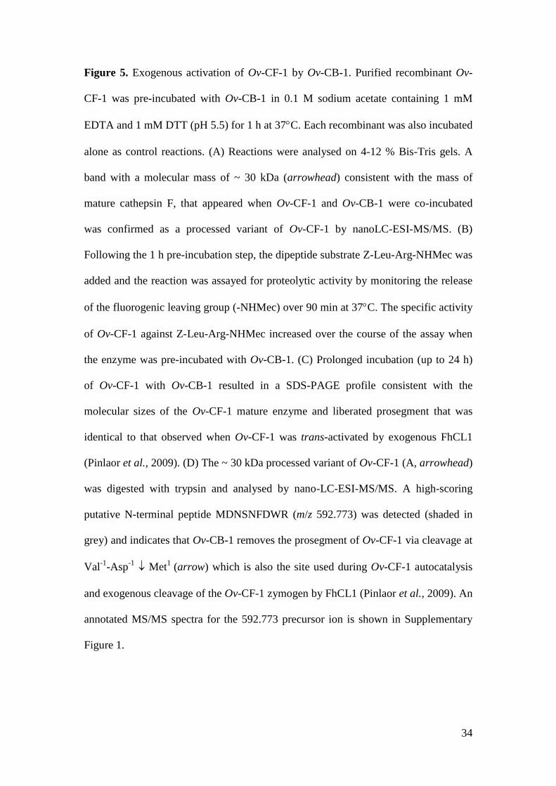
An alternative approach was therefore undertaken. The 30 kDa band was
excised, digested with trypsin and analysed by nanoLC-ESI-MS/MS. A high-scoring
doubly charged ion matching with a peptide corresponding to the putative N-terminal
of Ov-CF-1 following trans-processing by Ov-CB-1 was identified (Figure 5C;
Supplementary file 1). Matched peptide MDNSNFDWR (m/z 592.773),
corresponding to residues 1-9 of the mature domain could not be generated by tryptic
digest alone since the amino acid preceding this sequence in Ov-CF-1 is Thr (trypsin
can only cleave after Lys or Arg). Thus, this peptide is likely to form the N-terminus
of the ~ 30 kDa species that appeared when Ov-CF-1 was trans-processed by Ov-CB-
1 making the cleavage site Val-2
-Thr-1Met
1: this site is used by Ov-CF-1 during auto-
processing at pH 4.5 (Figure 1) and is the site of exogenous cleavage of the Ov-CF-1
prosegment by FhCL1 (Pinlaor et al., 2009).
Ov-CF-1 and Ov-CB-1 can work in concert to hydrolyse haemoglobin at low pH
Since blood is a major source of nutrition for O. viverrini the ability of Ov-CF-1 and
Ov-CB-1 to hydrolyse Hb was investigated. To examine the process of Hb
degradation by Ov-CF-1 and Ov-CB-1, Hb was mixed with each protease individually
as well as together at pH 4.0 for up to 360 minutes at 37C. Reactions were stopped at
several time points by addition of E-64 (an irreversible inhibitor of cysteine proteases)
and the degradation products analysed by SDS-PAGE. When incubated at pH 4.0 the

18
Hb molecule migrates as a major band at 15 kDa representing the Hb-alpha and Hb-
beta monomers (Lowther et al., 2009). However, this band was gradually degraded to
smaller peptides in the molecular size region of 3 – 10 kDa following incubation with
Ov-CF-1 or Ov-CB-1 (Figure 6A). Strikingly, when both proteases were incubated
together, Hb was rapidly digested to smaller protein bands (3 – 10 kDa) within the
first 15 minutes of the reaction and almost completely degraded between 240 and 360
min (Figure 6A).
To identify the cleavage sites for Ov-CF-1 and Ov-CB-1 within Hb, the 15 min
reaction aliquots were analysed by nanoLC-ESI-MS/MS to determine the masses and
sequence identities of the resulting hydrolytic products. Liberated peptides were
mapped onto the primary amino acid sequences of human Hb-alpha and Hb-beta to
identify the cleavage sites of the O. viverrini proteases (Figure 6B). By 15 min, Ov-
CF-1 cleaved Hb-alpha at 64 sites and Hb-beta at 44 sites while Ov-CB-1 cleaved Hb-
alpha at 53 sites and Hb-beta at 45 sites. When Ov-CF-1 and Ov-CB-1 were added
together, Hb-alpha was cleaved at 70 sites and Hb-beta at 48 sites.
Within a 15 min time-frame Ov-CF-1 and Ov-CB-1 could both generate small
peptides (ranging from 4 - 34 amino acids) from Hb but not dipeptides or free amino
acids. The average length of the released peptides (from both the Hb alpha and beta
chains) was 23 amino acids for Ov-CF-1 with 26-residue peptides occurring most
frequently and 19 amino acids for Ov-CB-1 with 12-residue peptides occurring most
frequently. When both enzymes digested Hb together, the average length of the
released peptides was 12 amino acids with 10-residue peptides occurring most often
(Figure 6C). It is unlikely that Ov-CB-1 and Ov-CF-1 cleave all Hb molecules in the
same manner and, thus, the cleavage map shown in Figure 6B represents a composite
of cleavage sites.

19
Ov-CB-1 and Ov-CF-1 cleavage sites within Hb indicate different substrate
specificities
Substrate residues present at the P2 position from the scissile bond interact with the
S2 subsite of the active site of papain-like cysteine proteases and determine the
efficiency by which the bond is cleaved (Schechter and Berger, 1968). Therefore, we
examined the frequency of each amino acid in the P2 site of the proteolytic cleavage
site identified in aliquots of the 15 min Hb digest described above. Consistent with
our previous findings using fluorogenic peptide substrates (Pinlaor et al., 2009) Ov-
CF-1 preferentially cleaved Hb at peptide bonds where the P2 position was occupied
with hydrophobic residues such as Leu, Ala and Phe. However, Ov-CF-1 could also
accommodate a range of other amino acids at the P2 position, notably Gly and to a
lesser extent Pro, Met, Lys, Ser and Thr (Figure 7B). In contrast, Ov-CB-1 showed a
more specific P2 preference within Hb. Ov-CB-1 also digested at bonds where Leu
and Ala occupied the P2 position but also showed a marked preference for other
residues including Tyr, Val and His that was not evident in the Ov-CF-1 digests
(Figure 7B). Finally, in similarity to our recent findings that used mass spectrometry
to map Hb cleavage sites for F. hepatica cathepsin L1 (Lowther et al., 2009), the P1
position in human Hb could be occupied by many amino acids but most preferentially
Leu or Ala for both O. viverrini proteases (not shown).
Differential degradation of extracellular matrix proteins by Ov-CB-1 and Ov-CF-1
The ability of Ov-CB-1 and Ov-CF-1 to hydrolyse two major components of the
extracellular matrix (ECM) was investigated as an indication of their potential roles in
tissue invasion and nutrition (Figure 8). Fibronectin was digested by both O. viverrini

20
proteases. At pH 4.5, the ~ 200 kDa band corresponding to co-migrating fibronectin
- and -chains was partially digested by Ov-CB-1 to a major band of approximately
150 kDa and a number of smaller degradation products. In contrast, Ov-CF-1
completely digested the fibronectin - and -chains at pH 4.5 into a large number of
well-defined fragments. This pattern of fibronectin digestion was repeated by Ov-CF-
1 at pH 6.5 whereas at this pH Ov-CB-1 displayed negligible activity against
fibronectin. Ov-CB-1 was not capable of degrading laminin at either pH 4.5 or 6.5. In
contrast, both high molecular mass bands (~ 200 kDa) representing the laminin B1-
and B2-chains were readily hydrolysed by Ov-CF-1 at both pH 4.5 and pH 6.5 to a
range of breakdown products indicated by the smearing shown by SDS-PAGE.
DISCUSSION
Papain-like cathepsin cysteine proteases are predominant molecules secreted by
trematode pathogens and perform a number of functions in parasite-host interactions
including facilitating tissue penetration (Curwen et al., 2006; McGonigle et al., 2008;
Robinson et al., 2008a), obtaining nutrients (Dalton et al., 2004; Na et al., 2008;
Lowther et al., 2009) and disarming the soluble and cellular arms of the host immune
system (Dalton et al,, 2003). However, there are clear differences in the array of
cathepsins expressed by each trematode species; for example, parasites of the genus
Fasciola, which burrow through host liver tissue and reside in the bile ducts, express a
large family of cathepsin L proteases and several cathepsin B proteases (reviewed in
Robinson et al., 2008b), while those of the genus Schistosoma, that live in the blood
vessels, secrete a mixture of cathepsin L, cathepsin F, cathepsin B and cathepsin C
cysteine proteases (Dvorak et al., 2008). As presented in this study, the major
proteases expressed by O. viverrini are cathepsin F and cathepsin B, which is in

21
agreement with reports for the related fish-borne trematodes including Clonorchis
sinensis and Paragonimus westermani (Park et al., 2001; Na et al., 2008; Pinlaor et
al., 2009). These differences likely have a biological significance that relate to the
organ or tissue site in which the parasites reside, and hence the protein
macromolecules they consume. Understanding the complexity and specificity of the
proteases secreted by these flukes should expose critical features in host-parasite
relationships and stimulate novel means by which we can devise future control
mechanisms.
Papain-like cathepsin proteases are synthesised as inactive zymogens
consisting of a mature enzyme domain with an N-terminal extension, or prosegment
that lies within the active site cleft of the enzyme and prevents unwanted proteolysis
during folding, trafficking and storage. The proteases become active following
removal of the prosegment to produce a mature protease with a substrate-accessible
active site (Coulombe et al. 1996; Stack et al. 2008). Much of our knowledge of the
synthesis, secretion and activation of cysteine protease zymogens in trematodes is
inferred from studies of the cathepsin L protease family from the liver fluke, Fasciola
hepatica (Robinson et al., 2008a). Our present data shows that Opisthorchis cathepsin
F and Fasciola cathepsin L proteases share similar mechanisms of secretion and auto-
catalytic activation. In F. hepatica cathepsin Ls are synthesised within specialised
gastrodermal epithelial cells and are stored in secretory vesicles as inactive zymogens
(Collins et al., 2004). Upon secretion, the prosegment is removed by specific auto-
catalytic processing events, facilitated by the low pH environment of the parasite gut
lumen, to release a mature active enzyme in the digestive milieu (Robinson et al.,
2008a). Ov-CF-1 is also secreted by gastrodermal cells surrounding the gut of O.
viverrini (Pinlaor et al., 2009), and here we have shown that recombinant Ov-CF-1
22
undergoes auto-catalytic activation in the range pH 4.5 – 6.5, with the rate of auto-
activation increasing with decreasing pH. It is likely that auto-catalysis rather than
trans-processing is the major mechanism of Ov-CF-1 activation within O. viverrini
eggs given the absence of cathepsin B transcripts (Figure 2).
When incubated at pH 5.5 for 6 hours Ov-CF-1 underwent an autocatalytic
process that produced a 41 kDa intermediate via cleavage at Phe-26
-Lys-25Thr
-24.
However, at pH 4.5 the prosegment was completely removed via cleavage at Val-2
-
Thr-1Met
1 to release a 30 kDa fully mature enzyme that efficiently cleaves synthetic
peptides and macromolecular substrates. This process of auto-activation of O.
viverrini cathepsin F is similar to that reported for F. hepatica cathepsin L1 (FhCL1)
(Collins et al., 2004; Stack et al., 2007; Lowther et al., 2009). Notably, the
prosegment of Ov-CF-1 contains a conserved GXTXFXD motif similar to the
GXNXFXD motif found FhCL1 and in other cathepsin L-like proteases. This motif is
implicated in triggering the pH-dependent intra-molecular cleavage of the prosegment
whereby the interaction of conserved charged Asp with residues on the mature portion
of the enzyme is perturbed by reduced pH (Vernet et al., 1995). This triggering event
may occur in Ov-CF-1 at pH 5.5 allowing the initial cleavage at Phe-26
-Lys-25Thr
-24
which is C-terminal to the GXNXFXD motif. Primary sequence alignments and
comparative analysis with the atomic structure of FhCL1 (Stack et al., 2008; not
shown) show that the 24 residue C-terminal region of the Ov-CF-1 prosegment that
remains attached to the mature domain of the enzyme lies within the active site cleft
of the enzyme. This would explain why retention of this 41 kDa intermediate cleavage
form of Ov-CF-1 exhibited a reduced (~ 20 %) activity.
A number of studies have shown that as well as being capable of autocatalytic
activation helminth cathepsin proteases can be trans-activated by other proteases that

23
cleave at residues that lie at the junction between the prosegment and mature enzyme
domain (Dalton and Brindley, 1996; Sajid et al., 2003; Beckham et al., 2006;
Delcroix et al., 2006). Here, pre-incubation of proOv-CF-1 with exogenously added
active Ov-CB-1 zymogen at pH 5.5 for 60 minutes was sufficient to trans-activate the
cathepsin F and release a fully mature and active enzyme. However, the activity of the
cathepsin B-trans-activated Ov-CF-1 against the fluorogenic substrate Z-Leu-Arg-
NHMec was very low when measured immediately after pre-incubation with Ov-CB-
1 (time zero). This suggests that only a small population of Ov-CF-1 zymogens are
initially trans-activated by Ov-CB-1. Nevertheless, this exogenous proteolysis appears
to have generated sufficient amounts of mature Ov-CF-1 to trans-activate other
cathepsin F zymogens since a rapid increase in Z-Leu-Arg-NHMec hydrolysis occurs
as the reaction proceeds. Indeed, when the Ov-CF-1/Ov-CB-1 reactions were analysed
by SDS-PAGE, Ov-CF-1 did not show a significant shift in molecular mass; a discreet
band of ~ 30 kDa appeared following 60 minutes at pH 5.5 that was absent when Ov-
CF-1 was incubated alone. The presence of peptide MDNSNFDWR (m/z 592.773) in
tryptic digests of the ~ 30 kDa product shows that this represents a fully matured
protein formed by exogenous cleavage of the cathepsin F prosegment at Val-2
-Thr-1
Met1. This was the same cleavage site observed when Ov-CF-1 was trans-activated by
exogenously-added FhCL1 (Pinlaor et al., 2009) and when the enzyme was auto-
catalytically activated at pH 4.5 (see above). Therefore, our data shows that the Val-2
-
Thr-1 Met
1 cleavage
site represents a critical protease-susceptible region between the
prosegment and mature domain, which serves to regulate cathepsin F activation.
In contrast to cathepsin F, the major secreted cathepsin B of O. viverrini did
not undergo typical auto-processing events that lead to removal of the prosegment.
This contrasts with a number of cathepsin Bs that are secreted from related trematode

24
species including F. hepatica, S. mansoni and T. regenti which release the prosegment
following auto-activation at low pH (Gotz et al., 1992; Dvorak et al., 2005; Beckham
et al., 2006). However, although the prosegment was still covalently attached to the
mature domain of Ov-CB-1, the zymogen exhibited activity against a range of
fluorogenic –NHMec substrates in the pH range 3.5 - 8.5, and, at pH 4.5, efficiently
hydrolysed haemoglobin to small peptides. These data demonstrate that the Ov-CB-1
zymogen can cleave physiologically relevant substrate molecules and, in addition, its
ability to trans-activate cathepsin F suggests that one of its functions is to regulate the
protease network in O. viverrini.
Atypical active human procathepsin B has been reported previously
(Pungercar et al., 2009) and crystallographic studies indicate that cathepsin B
prosegments (which are considerably shorter than their cathepsin L and cathepsin F
counterparts) are susceptible to acid-induced conformational changes making them
less efficient inhibitors of the mature domain (Cygler et al., 1996; Coulombe et al.,
1996; Groves et al., 1998). Thus the low-pH environment of the Opisthorchis gut may
loosen the tertiary fold of Ov-CB-1 increasing the mobility of the prosegment which
then dissociates from the mature domain, albeit still attached. This would allow entry
of the protease-susceptible region of the Ov-CF-1 zymogen into the Ov-CB-1 active
site cleft for trans-activation. Thus, bringing all of our observations together we can
propose a model of cathepsin F activation which involves initial processing of a small
population of cathepsin F zymogens by an active cathepsin B zymogen followed by
rapid activation of additional cathepsin F zymogens by these active mature molecules
(Figure 9).
Trematodes produce prodigious numbers of eggs, which requires a reliable
source of amino acids for anabolism of eggshell proteins. Within the bile ducts, adult

25
F. hepatica is an obligate blood feeder and liberates 30,000 eggs per hour. To obtain
nutrient, the liver fluke secretes the FhCL1 protease that has evolved an active site
with a strong preference for hydrophobic amino acids, Leu, Ala, Phe and Val, which
comprise 42 % of haemoglobin (Robinson et al., 2008b; Lowther et al., 2009). Adult
O. viverrini also use blood as a food source and likely employ the Ov-CF-1 and Ov-
CB-1 to digest human haemoglobin. Although both proteases can degrade this
substrate into short peptides the digestion process is much more rapid and complete
when both enzymes act together. It is noteworthy that the SDS-PAGE profile of
haemoglobin digestion by Ov-CF-1 and Ov-CB-1 together was similar to the pattern
of digestion when haemoglobin was digested with FhCL1 alone (Lowther et al.,
2009). Thus, adult O. viverrini may require both Ov-CF-1, Ov-CB-1, and possibly a
cathepsin D-like aspartic protease (Ov-APR-1) (Suttiprapa et al. 2009), to accomplish
complete digestion of haemoglobin into small peptides that can be used for protein
anabolism. Such multi-enzyme networks have been reported in other blood-feeding
parasites including the human blood fluke S. mansoni (Delcroix et al., 2006) and the
hookworm Ancylostoma caninum (Williamson et al., 2004) representing a mechanism
common to evolutionarily distant haemotophagous parasites.
In addition to blood, O. viverrini flukes also graze on bile duct epithelial cells
and mucus (Rim, 2005; Sripa et al., 2007). These food preferences would require the
ability to cleave a variety of macromolecular substrates and may explain why Ov-CF-
1 and/or Ov-CB-1 exhibit different substrate specificity to FhCL1. We observed that a
broader range of haemoglobin residues can be accommodated at the P2 site by the two
O. viverrini cathepsins compared with the more specific usage of hydrophobic P2
amino acids by FhCL1 (Stack et al., 2008; Lowther et al., 2009). The broader
specificity of Ov-CF-1 and Ov-CB-1, and overlapping pH optima for activity (pH 6.5

26
and 4.5, respectively), may allow them to work in concert to digest a number of
physiologically relevant extracellular matrix (ECM) proteins. Ov-CF-1 effectively
digested fibronectin and laminin close to physiological pH (pH 6.5) and under acidic
conditions (pH 4.5). In contrast, Ov-CB-1 partially degraded fibronectin at low pH but
could not digest either of the ECM proteins at pH 6.5. The combined action of the
secreted O. viverrini cathepsins would result in the degradation of interstitial laminin
and fibronectin within the bile duct, and lead to disruption of the cellular integrity of
the cholangiocytes thus allowing the parasite to access underlying liver cells. A
cathepsin F (CsCF-6) secreted by adult Clonorchis sinensis displayed similar
biochemical properties to Ov-CF-1 and also degraded a range of host macromolecules
including collagen, fibronectin and haemoglobin (Na et al., 2008). The evolution of
ECM-degrading activity in trematodes is significant and likely to be pivotal to the
ability of these pathogens to infect and survive within their mammalian hosts.
Adult O. viverrini express both cathepsin F and B endoproteases at similar
levels; an analysis of ~ 5000 O. viverrini ESTs using Ov-CF-1 and Ov-CB-1 primary
sequences as queries gave 60 and 50 significant (e < 1) matches, respectively. The
Ov-CF-1 and Ov-CB-1 combination clearly constitutes effective molecular machinery
for tissue degradation. One of the most intriguing aspects of O. viverrini biology is the
link between liver fluke infection and the development of CCA. Given the potential of
Ov-CF-1 and Ov-CB-1 to cause tissue destruction it is likely that these proteases
contribute to CCA progression. Since local changes in ECM microenvironment
contribute to the induction of intra-hepatic CCA (Farazi et al., 2006) it is likely that
degradation of ECM proteins by the battery of proteases secreted by O. viverrini has a
similar effect during the aetiology of liver fluke-associated CCA. As degradation of
the ECM is a prerequisite step for the invasion and metastasis of cancer cells, O.

27
viverrini infection may even promote the spread of invasive carcinoma as a result of
ECM and basement membrane instability within the bile ducts (Mon et al., 2009;
Yasoshima et al., 2009).
ACKNOWLEDGEMENTS
This research was supported by a grant from the Strategic Scholarships for Frontier
Research Network (Thai Doctoral degree) from the Office of the Higher Education
Commission, Thailand and by grants from the Sandler Family Foundation and from
the National Institute of Allergy and Infectious Diseases (award number
UO1AI065871): the content is solely the responsibility of the authors and does not
necessarily represent the official views of the NIAID or the NIH. JPD is supported by
a grant from the Natural Sciences and Engineering Research Council (NSERC) of
Canada. MWR is supported by a UTS Chancellor`s Postdoctoral Fellowship.
REFERENCES
Beckham, S.A., Law, R.H., Smooker, P.M., Quinsey, N.S., Caffrey, C.R., McKerrow, J.H., et
al (2006) Production and processing of a recombinant Fasciola hepatica cathepsin B-like
enzyme (FhcatB1) reveals potential processing mechanisms in the parasite. Biol Chem 387:
1053-1061.
Bendtsen, J.D., Nielsen, H., von Heijne, G., and Brunak, S. (2004) Improved prediction of
signal peptides: SignalP 30. J Mol Biol 340: 783-795.
Bouvard, V., Baan, R., Straif, K., Grosse, Y., Secretan, B., El Ghissassi, F., et al (2009) A
review of human carcinogens - Part B: biological agents. Lancet Oncol 10: 321-322.
Brady, M. T., O’Neill, S. M., Dalton, J. P., and Mills, K. H. G. (1999) Fasciola hepatica
suppresses a protective Th1 response against Bordetella pertussis. Infect Immun 67: 5372–
5378.
Chai, J.Y., Murrell, K.D. and Lymbery, A.J. (2005) Fish-borne parasitic zoonoses: status and
issues. Int J Parasitol 35: 1233-1254.
Collins, P.R., Stack, C.M., O'Neill, S.M., Doyle, S., Ryan, T., Brennan, G.P., et al (2004)
Cathepsin L1, the major protease involved in liver fluke (Fasciola hepatica) virulence:
propetide cleavage sites and autoactivation of the zymogen secreted from gastrodermal cells.
J Biol Chem 279: 17038-17046.

28
Coulombe, R., Grochulski, P., Sivaraman, J., Menard, R., Mort, J.S. and Cygler, M. (1996)
Structure of human procathepsin L reveals the molecular basis of inhibition by the
prosegment. EMBO J 15: 5492-5503.
Curwen, R.S., Ashton, P.D., Sundaralingam, S., and Wilson, R.A. (2006) Identification of
novel proteases and immunomodulators in the secretions of schistosome cercariae that
facilitate host entry. Mol Cell Proteomics 5: 835-844.
Cygler, M., Sivaraman, J., Grochulski, P., Coulombe, R., Storer, A.C., and Mort, J.S. (1996)
Structure of rat procathepsin B: model for inhibition of cysteine protease activity by the
proregion. Structure. 4(4): 405-416.
Dalton, J.P. and Brindley, P.J. (1996) Schistosome asparaginyl endopeptidase Sm 32 in
hemoglobin digestion. Parasitol Today 12 : 125.
Dalton, J.P., Neill, S.O., Stack, C., Collins, P., Walshe, A., Sekiya, M., et al (2003) Fasciola
hepatica cathepsin L-like proteases: biology, function, and potential in the development of
first generation liver fluke vaccines. Int J Parasitol 33: 1173-1181.
Dalton, J.P., Skelly, P. and Halton, D.W. (2004) Role of the tegument and gut in nutrient
uptake by parasitic platyhelminths. Can J Zool 82: 211-232.
Delcroix, M., Sajid, M., Caffrey, C.R., Lim, K.C., Dvorák, J., Hsieh, I., et al., (2006) A
multienzyme network functions in intestinal protein digestion by a platyhelminth parasite. J
Biol Chem 281: 39316-39329.
Dowd, A. J., Tort, J., Roche, L., Ryan, T., and Dalton, J. P. (1997) Isolation of a cDNA
encoding Fasciola hepatica cathepsin L2 and functional expression in Saccharomyces
cerevisiae. Mol Biochem Parasitol 88: 163-174.
Dvorák, J., Delcroix, M., Rossi, A., Vopálenský, V., Pospísek, M., Sedinová, M., et al (2005)
Multiple cathepsin B isoforms in schistosomula of Trichobilharzia regenti: identification,
characterisation and putative role in migration and nutrition. Int J Parasitol 35(8): 895-910.
Dvorák J, Mashiyama ST, Braschi S, Sajid M, Knudsen GM, Hansell E, Lim KC, et al (2008)
Differential use of protease families for invasion by schistosome cercariae. Biochemie 90:
345-358.
Farazi, P.A., Zeisberg, M., Glickman, J., Zhang, Y., Kalluri, R., and DePinho, R.A. (2006)
Chronic bile duct injury associated with fibrotic matrix microenvironment provokes
cholangiocarcinoma in p53-deficient mice. Cancer Res 66(13): 6622-6627.
Gabay, T., and Ginsburg, H. (1993) Hemoglobin denaturation and iron release in acidified red
blood cell lysate – a possible source of iron for intraerythrocytic malaria parasites. Exp
Parasitol 77: 261-272.
Götz, B., Felleisen, R., Shaw, E., and Klinkert, M.Q. (1992) Expression of an active cathepsin
B-like protein Sm31 from Schistosoma mansoni in insect cells. Trop Med Parasitol 43(4):
282-284.
Groves, M.R., Coulombe, R., Jenkins, J., and Cygler, M. (1998) Structural basis for
specificity of papain-like cysteine protease proregions toward their cognate enzymes. Proteins
32(4): 504-514.

29
Hotez, P.J., Brindley, P.J., Bethony, J.M., King, C.H., Pearce, E.J. and Jacobson, J. (2008)
Helminth infections: The great neglected tropical diseases. J Clin Invest 118(4): 1311–1321.
Jongsuksuntigul, P. and Imsomboon, T. (2003) Opisthorchiasis control in Thailand. Acta Trop
88(3): 229-232.
Kaewpitoon, N., Kaewpitoon, S.J., Pengsaa, P. and Sripa, B. (2008) Opisthorchis viverrini:
the carcinogenic human liver fluke. World J Gastroenterol 14(5): 666-674.
Keiser, J. and Utzinger, J. (2005) Emerging foodborne trematodiasis. Emerg Infect Dis 11:
1507-1514.
Laha, T., Pinlaor, P., Mulvenna, J., Sripa, B., Sripa, M., Smout, M.J., et al (2007) Gene
discovery for the carcinogenic human liver fluke, Opisthorchis viverrini. BMC Genomics 8:
189.
Lowry, O.H., Rosebrough, N.J., Farr, A.L. and Randall, R.J. (1951) Protein measurement
with the Folin phenol reagent. J Biol Chem 193: 265-275.
Lowther, J., Robinson, M.W., Donnelly, S.M., Xu, W., Stack, C.M., Matthews, J.M. and
Dalton. J.P. (2009) The importance of pH in regulating the function of Fasciola hepatica
cathepsin L1 cysteine protease. PLoS Negl Trop Dis 3 (1): e369.
McGonigle, L., Mousley, A., Marks, N.J., Brennan, G.P., Dalton, J.P., Spithill, T.W., et al
(2008) The silencing of cysteine proteases in Fasciola hepatica newly excysted juveniles
using RNA interference reduces gut penetration. Int J Parasitol 38: 149-155.
Mon, N.N., Kokuryo, T., and Hamaguchi, M. (2009) Inflammation and tumor progression: a
lesson from TNF-alpha-dependent FAK signaling in cholangiocarcinoma. Methods Mol Biol
512: 279-293.
Na, B.K., Kang, J.M., and Sohn, W.M. (2008) CsCF-6, a novel cathepsin F-like cysteine
protease for nutrient uptake of Clonorchis sinensis. Int J Parasitol 38(5): 493-502.
Park, H., Hong, K.M., Sakanari, J.A., Choi, J.H., Park, S.K., Kim, K.Y., Hwang, H.A., et al
(2001) Paragonimus westermani: cloning of a cathepsin F-like cysteine proteinase from the
adult worm. Exp Parasitol 98: 223-227.
Parkin, D.M. (2006) The global health burden of infection-associated cancers in the year
2002. Int J Cancer 118(12): 3030-3044.
Pinlaor, S., Sripa, B., Sithithaworn, P., Yongvanit, P. (2004) Hepatobiliary changes, antibody
response, and alteration of liver enzymes in hamsters re-infected with Opisthorchis viverrini.
Exp Parasitol 108(1-2): 32-39.
Pinlaor, P., Kaewpitoon, N., Laha, T., Sripa, B., Kaewkes, S., Morales, M.E., Mann, V.H., et
al (2009) Cathepsin F cysteine protease of the human liver fluke, Opisthorchis viverrini.
PLoS Negl Tropl Dis 3 (3): e398.
Pungercar, J.R., Caglic, D., Sajid, M., Dolinar, M., Vasiljeva, O., Pozgan, U., et al (2009)
Autocatalytic processing of procathepsin B is triggered by proenzyme activity. FEBS J
276(3): 660-668.
Rim, H.J. (2005) Clonorchiasis: An update. J Helminthol 79: 269–281.

30
Robinson, M.W. and Connolly, B. (2005) Proteomic analysis of the excretory-secretory
proteins of the Trichinella spiralis L1 larva, a nematode parasite of skeletal muscle.
Proteomics 5: 4525-4532.
Robinson, M.W, Greig, R., Beattie, K., Lamont, D. and Connolly, B. (2007) Comparative
analysis of the excretory-secretory proteome of the muscle larva of Trichinella pseudospiralis
and Trichinella spiralis. Int J Parasitol 37: 139-148.
Robinson, M.W., Dalton, J.P. and Donnelly, S. (2008a) Helminth pathogen cathepsin
proteases: it's a family affair. Trends Biochem Sci 33(12): 601-608.
Robinson, M.W., Tort, J.F., Wong, E., Donnelly, S.M., Lowther, J., Xu, W., Stack, C.M., et al
(2008b) Proteomic and phylogenetic analysis of the cathepsin L protease family of the
helminth pathogen, Fasciola hepatica: expansion of a repertoire of virulence-associated
factors. Mol Cell Proteomics 7: 1111-1123.
Robinson, M.W., Menon, R., Donnelly, S.M., Dalton, J.P. and Ranganathan, S. (2009) An
integrated transcriptomic and proteomic analysis of the secretome of the helminth pathogen,
Fasciola hepatica: proteins associated with invasion and infection of the mammalian host.
Mol Cell Proteomics 8(8):1891-1907.
Sajid, M., McKerrow, J.H., Hansell, E., Mathieu, M.A., Lucas, K.D., Hsieh, I., et al (2003)
Functional expression and characterization of Schistosoma mansoni cathepsin B and its trans-
activation by an endogenous asparaginyl endopeptidase. Mol Biochem Parasitol 131: 65-75.
Schechter, I, and Berger, A. (1968) On the size of the active site in proteases. I. Papain.
Biochem Biophys Res Commun 27(2): 157-162.
Smout, M.J., Laha, T., Mulvenna, J., Sripa, B., Suttiprapa, S., Jones, A., et al (2009) A
granulin-like growth factor secreted by the carcinogenic liver fluke, Opisthorchis viverrini,
promotes proliferation of host cells. PLoS Pathog 5(10): e1000611
Sripa, B., and Pairojkul, C. (2008) Cholangiocarcinoma: lessons from Thailand. Curr Opin
Gastroenterol 24: 349-356.
Sripa, B., Kaewkes, S., Sithithaworn, P., Mairiang, E., Laha, T., Smout, M. et al (2007) Liver
fluke induces cholangiocarcinoma. PLoS Med 4(7): e201.
Sripa, B. (2008) Concerted action is needed to tackle liver fluke infections in Asia. PLoS Negl
Trop Dis 2(5): e232.
Stack, C.M., Donnelly, S., Lowther, J., Xu, W., Collins, P.R., Brinen, L.S. and Dalton, J.P.
(2007) The major secreted cathepsin L1 protease of the liver fluke, Fasciola hepatica: a Leu-
12 to Pro-12 replacement in the nonconserved C-terminal region of the prosegment prevents
complete enzyme autoactivation and allows definition of the molecular events in prosegment
removal. J Biol Chem 282: 16532-16543.
Stack, C.M., Caffrey, C.R., Donnelly, S.M., Seshaadri, A., Lowther, J., Tort, J.F., et al (2008)
Structural and functional relationships in the virulence-associated cathepsin L proteases of the
parasitic liver fluke, Fasciola hepatica. J Biol Chem 283: 9896-9908.

31
Suttiprapa, S., Loukas, A., Laha, T., Wongkham, S., Kaewkes, S., Gaze, S., et al (2008)
Characterization of the antioxidant enzyme, thioredoxin peroxidase, from the carcinogenic
human liver fluke, Opisthorchis viverrini. Mol Biochem Parasitol 160(2): 116-122.
Suttiprapa, S., Mulvenna, J., Huong, N.T., Pearson, M.S., Brindley, P.J., Laha, T., et al (2009)
Ov-APR-1, an aspartic protease from the carcinogenic liver fluke, Opisthorchis viverrini:
functional expression, immunolocalization and subsite specificity. Int J Biochem Cell Biol 41:
1148-1156.
Thuwajit, C., Thuwajit, P., Kaewkes, S., Sripa, B., Uchida, K., Miwa, M. and Wongkham, S.
(2004) Increased cell proliferation of mouse fibroblast NIH-3T3 in vitro induced by
excretory/secretory product(s) from Opisthorchis viverrini. Parasitology 129: 455-464.
Vernet, T., Berti, P.J., de Montigny, C., Musil, R., Tessier, D.C., Menard, R., et al (1995)
Processing of the papain precursor. The ionization state of a conserved amino acid motif
within the Pro region participates in the regulation of intramolecular processing. J Biol Chem
270: 10838-10846.
Williamson, A.L., Lecchi, P., Turk, B.E., Choe, Y., Hotez, P.J., McKerrow, J.H., et al (2004)
A multi-enzyme cascade of hemoglobin proteolysis in the intestine of blood-feeding
hookworms. J Biol Chem 279(34): 35950-35957.
Yasoshima, M., Sato, Y., Furubo, S., Kizawa, K., Sanzen, T., Ozaki, S., et al (2009) Matrix
proteins of basement membrane of intrahepatic bile ducts are degraded in congenital hepatic
fibrosis and Caroli's disease. J Pathol 217(3): 442-451.

32
Figure 1. Auto-activation of Ov-CF-1 at pH 4.5. (A) Purified recombinant Ov-CF-1
(50 g) was incubated in either 0.1 M sodium acetate (pH 4.5 or 5.5) or 0.1 M sodium
phosphate (pH 6.5) for 6 h. Aliquots of the reaction mixtures were removed after 6 h,
the hydrolysis stopped by addition of E-64 on ice, and analyzed on 4-12 % Bis-Tris
NuPage gels (Invitrogen). At pH 6.5, Ov-CF-1 migrated as a major band of 47 kDa
representing the unprocessed zymogen (Pinlaor et al., 2009). Following incubation at
pH 5.5, the majority of the enzyme migrated as an intermediate band with a molecular
mass of 41 kDa. At pH 4.5, Ov-CF-1 had been fully processed and migrated as a
single band at 30 kDa with low molecular mass prosegment peptides (< 6 kDa) clearly
visible. (B) The Ov-CF-1 auto-activation reactions shown in (A) were assayed for
peptidolytic activity against the fluorogenic dipeptide substrate Z-Leu-Arg-NHMec
(measured by monitoring the release of the fluorogenic leaving group (-NHMec) over
360 min (6 h) at 37C. (C) N-terminal sequences obtained for the Ov-CF-1 samples
marked with asterisks in (A). The cleavage sites (arrows) identified by N-terminal
sequencing are also mapped onto the primary amino acid sequence of the Ov-CF-1
prosegment (bottom). The EF found at the N-terminal was introduced by the EcoRI
cloning site used in the pPIC ZαA expression vector. 1Theoretical molecular mass of
the Ov-CF-1 polypeptides calculated by Compute pI/MW.
Figure 2. RT-PCR analysis of Ov-CB-1, Ov-CB-2 and Ov-CF-1 transcripts.
RT-PCR analysis of Ov-CB-1, Ov-CB-2 and Ov-CF-1 expression in (1) O. viverrini
eggs, (2) metacercariae, (3) immature worms, (4) adult worms, and (5) an adult worm
cDNA library (5). Amplification of constitutively expressed O. viverrini -actin was
used as a control transcript.

33
Figure 3. Ov-CB-1 is expressed as an active zymogen. (A) Purified recombinant Ov-
CB-1 (50 g) was incubated in 0.1 M sodium acetate, pH 4.5 for 6 h. Aliquots of the
reaction mixtures were removed at time 0 h, 1 h and 6 h, halted with E-64 on ice, and
analyzed on 4-12 % Bis-Tris NuPage gels (Invitrogen). The recombinant enzyme
showed a progressive decrease in molecular mass during this incubation period
suggesting that partial auto-processing had occurred. (B) The auto-activation of Ov-
CB-1 shown in (A) was analysed by following the initial rates of hydrolysis of the
fluorogenic dipeptide substrate Z-Leu-Arg-NHMec, measured by monitoring the
release of the fluorogenic leaving group (-NHMec) over 360 min (6 h) at 37C. (C) N-
terminal sequences obtained for the 0 h, 1 h and 6 h Ov-CB-1 samples shown in (A).
The cleavage sites (arrows) identified by N-terminal sequencing are also mapped onto
the primary amino acid sequence of the Ov-CB-1 prosegment (bottom). The EF found
at the N-terminal was introduced by the EcoR I cloning site used in the pPIC ZαA
expression vector and the open arrow represents the predicted juncture of the
prosegment and mature enzyme domain. 1Theoretical molecular mass of the Ov-CB-1
polypeptides calculated by Compute pI/MW.
Figure 4. Activity of Ov-CB-1 and Ov-CF-1 against a panel of diagnostic fluorescent
peptides. (A) The relative activity of Ov-CB-1 and Ov-CF-1 (enzymes were pre-
incubated in 0.1 M sodium acetate, pH 4.5 for 6 h) against a range of fluorescent
substrates was determined by measured by monitoring the release of the fluorogenic
leaving group (-NHMec) over 1 h at pH 5.5. (B) Initial rates of hydrolysis of Z-Leu-
Arg-NHMec by Ov-CB-1 and Ov-CF-1 were measured over 1 h min at 37C in a
variety of buffers (in the range pH 2-10).
34
Figure 5. Exogenous activation of Ov-CF-1 by Ov-CB-1. Purified recombinant Ov-
CF-1 was pre-incubated with Ov-CB-1 in 0.1 M sodium acetate containing 1 mM
EDTA and 1 mM DTT (pH 5.5) for 1 h at 37C. Each recombinant was also incubated
alone as control reactions. (A) Reactions were analysed on 4-12 % Bis-Tris gels. A
band with a molecular mass of ~ 30 kDa (arrowhead) consistent with the mass of
mature cathepsin F, that appeared when Ov-CF-1 and Ov-CB-1 were co-incubated
was confirmed as a processed variant of Ov-CF-1 by nanoLC-ESI-MS/MS. (B)
Following the 1 h pre-incubation step, the dipeptide substrate Z-Leu-Arg-NHMec was
added and the reaction was assayed for proteolytic activity by monitoring the release
of the fluorogenic leaving group (-NHMec) over 90 min at 37C. The specific activity
of Ov-CF-1 against Z-Leu-Arg-NHMec increased over the course of the assay when
the enzyme was pre-incubated with Ov-CB-1. (C) Prolonged incubation (up to 24 h)
of Ov-CF-1 with Ov-CB-1 resulted in a SDS-PAGE profile consistent with the
molecular sizes of the Ov-CF-1 mature enzyme and liberated prosegment that was
identical to that observed when Ov-CF-1 was trans-activated by exogenous FhCL1
(Pinlaor et al., 2009). (D) The ~ 30 kDa processed variant of Ov-CF-1 (A, arrowhead)
was digested with trypsin and analysed by nano-LC-ESI-MS/MS. A high-scoring
putative N-terminal peptide MDNSNFDWR (m/z 592.773) was detected (shaded in
grey) and indicates that Ov-CB-1 removes the prosegment of Ov-CF-1 via cleavage at
Val-1
-Asp-1
Met1
(arrow) which is also the site used during Ov-CF-1 autocatalysis
and exogenous cleavage of the Ov-CF-1 zymogen by FhCL1 (Pinlaor et al., 2009). An
annotated MS/MS spectra for the 592.773 precursor ion is shown in Supplementary
Figure 1.

35
Figure 6. Hydrolysis of human haemoglobin by Ov-CB-1 and Ov-CF-1 and analysis
of digests by nanoLC-ESI-MS/MS. (A) Purified human haemoglobin (Hb) was
digested by Ov-CB-1 and Ov-CF-1 in 0.1 M sodium acetate buffer (pH 4.0),
containing 1 mM DTT and 1mM EDTA at 37C. Reactions were stopped at time 0
and at various time-points (10, 15, 30, 60, 90, 120, 240 and 360 min) by the addition
of the cysteine protease inhibitor E-64 and aliquots analysed on 4-12 % Bis-Tris
NuPage gels. (B) Map of Hb - and -chains indicating sites of Ov-CB-1 and Ov-CF-
1 cleavage. Cleavage sites within Hb present in 15 min reactions as determined by
nanoLC-ESI-MS/MS are shown. Arrows, cleavage sites shared by Ov-CB-1 and Ov-
CF-1; open arrowheads, Ov-CB-1-specific cleavage sites; filled arrowheads, Ov-CF-
1-specific cleavage sites. (C) Frequency of peptides of varying length released
following hydrolysis of Hb alpha and beta chains by Ov-CB-1 and Ov-CF-1.
Figure 7. P2 residues in peptides released from Hb following digestion by Ov-CB-1
and Ov-CF-1 were determined by nanoLC-ESI-MS/MS analysis of digest samples.
The frequency by which amino acids occur at the P2 positions of Hb - and -chains
(converted to a percentage of the total) are plotted for the 15 min reactions shown in
(Figure 6A).
Figure 8. Digestion of extracellular matrix proteins by Ov-CB-1 and Ov-CF-1.
Fibronectin and laminin were incubated with Ov-CB-1 and Ov-CF-1 at pH 4.5 and pH
6.5 at 37 ºC for 3 hours. F, fibronectin alone; L, laminin alone; CB, Ov-CB-1 digest;
CF, Ov-CF-1 digest.

36
Figure 9. Proposal for a three-step mechanism of cathepsin F trans-activation in O.
viverrini. Step 1: under the acidic microenvironment of the O. viverrini gut, the short
pH-sensitive prosegment of cathepsin B undergoes conformational relaxation and
dissociates from the mature domain sufficient to allow the zymogen to become active.
Step 2: subsequently, in a bi-molecular process, a small number of cathepsin F
zymogens are trans-activated by the active cathepsin B zymogens which remove the
cathepsin F prosegment via cleavage at Val-1
-Asp-1Met
1 at the junction of the
prosegment and mature domain. Step 3: the rapid cleavage of prosegments from other
cathepsin F zymogens by the trans-activated cathepsin F molecules again through
cleavage at Val-1
-Asp-1Met
1. Molecular models of Ov-CB-1 and Ov-CF-1 were
established (for illustrative purposes only) by the SWISS-MODEL homology
modelling pipeline (http://swissmodel.expasy.org) using the atomic structures of
human procathepsin B (PBD ID: 3PBH) and F. hepatica cathepsin L1 (PBD ID:
2O6X) as templates, respectively. Figures were produced with Pymol
(http://www.pymol.org).


![ASequentialModelofHostCellKillingandPhagocytosisby …downloads.hindawi.com/journals/jpr/2011/926706.pdf · 2019. 7. 31. · immunity [6]. Cysteine proteases that are known to degrade](https://img.pdfslide.net/doc/110x75/5fd741032cd3ff17140678f5/asequentialmodelofhostcellkillingandphagocytosisby-2019-7-31-immunity-6.jpg)