Embed Size (px)
Citation preview
Secretion machinery at the cell plasma membraneBhanu P Jena
Secretion is a fundamental cellular process involving the
regulated release of intracellular products from cells.
Physiological functions such as neurotransmission, or the
release of hormones and digestive enzymes, are all governed
by cell secretion. Three critical activities occur at the cell
plasma membrane to ensure secretion. Membrane-bound
secretory vesicles dock, fuse, and expel their contents to the
outside via specialized and permanent plasma membrane
structures, called porosomes or fusion pores. In recent years,
significant progress has been made in our understanding of
these three key cellular activities required for cell secretion. The
molecular machinery and mechanism involving them is
summarized in this article.
Addresses
Department of Physiology, Wayne State University School of Medicine,
5245 Scott Hall, 540 E. Canfield Avenue, Detroit, MI 48201-4177, USA
Corresponding author: Jena, Bhanu P ([email protected])
Current Opinion in Structural Biology 2007, 17:437–443
This review comes from a themed issue on
Membranes
Edited by Graham Shipley and Declan Doyle
Available online 30th August 2007
0959-440X/$ – see front matter
# 2007 Elsevier Ltd. All rights reserved.
DOI 10.1016/j.sbi.2007.07.002
IntroductionCell secretion has been the subject of intense research for
decades, however only recently, the general molecular
machinery and mechanism of the process has emerged. In
particular, considerable knowledge on cell secretion has
been gained from in vivo studies complemented by invitro work, using atomic force microscopy (AFM) com-
bined with conventional imaging, biochemical, electro-
physiological, and molecular approaches. Understanding
the structural assembly and interaction of membrane
proteins has always been difficult, especially their crystal-
lization and hence the determination of their native
structure–function. Similarly the resolution limit of the
light microscope (250 nm in lateral and much less in
depth resolution) had precluded determination of live
cellular structure–function at the nanometer scale and in
real time. The AFM has helped overcome these difficul-
ties and limitations in studying membrane proteins and
their complexes, especially those involved in the
secretory process in cells.
www.sciencedirect.com
Secretion is a precisely regulated and universal cellular
process, involving delivery of intracellular products to the
extracellular space. Cellular cargo destined for secretion
are packaged and stored in membranous sacs or vesicles,
which on demand dock and fuse at specialized plasma
membrane structures called porosomes [1–6]. The estab-
lishment of continuity between secretory vesicle mem-
brane and the porosome membrane requires calcium and
the assembly of a molecular complex [7] formed when
specific proteins [8–11] in opposing bilayers interact. At
the nerve terminal, for instance, target membrane
proteins SNAP-25 and syntaxin (t-SNAREs) associated
at the base of porosomes [4], and synaptic vesicle-associ-
ated protein VAMP or v-SNARE, interact in a circular
array [7] to form conducting channels in the presence of
calcium [7,12]. During cell secretion, swelling of secretory
vesicles result in a build-up of intravesicular pressure,
required for the expulsion of vesicular contents through
porosomes to the outside [13]. The extent of vesicle
swelling dictates the amount of vesicular contents
expelled [13]. In the past two decades, besides discovery
of the porosome [1–6], the discovery of SNAREs [14–16],
the molecular assembly [7,17��] and disassembly [18,19�]of the membrane-associated t-/v-SNARE complex
[7,17��,18], in addition to the molecular mechanism of
secretory vesicle swelling [20–22] required for the
regulated release of intravesicular contents, has been
determined using, among other approaches, the AFM.
Porosome: the universal secretionmachineryElectrophysiological measurements of mast cells [23–27]
as well as adrenal chromaffin cells [28] suggested the
presence of fusion pores at the cell plasma membrane as
‘dynamic entities’, originating following a secretory stim-
ulus [29]. Results from earlier studies on cell capacitance
and conductance measurements, suggested that following
stimulation of secretion, the ‘exocytotic fusion pore’
abruptly appears as a 1–2 nm in diameter pore at the cell
plasma membrane, with conductance similar to a large ion
channel [29]. During cell secretion, the fusion pore either
irreversibly expands or closes [24,29–31]. The latter pro-
cess, where the fusion pore opens allowing secretory
vesicles to fuse momentarily and subsequently close, has
been referred to as ‘transient fusion’ [24,26,30,31]. Exper-
imental data from these and other studies, and from
theoretical considerations, gave rise to a hypothetical work-
ing model of the exocytotic fusion pore [29,32] in cells.
According to the model, the fusion pore is formed by the
regulatory function of a group of proteins responsible for
bringing the plasma membrane into a highly curved dim-
ple, close to a tense secretory vesicle membrane [29].
Current Opinion in Structural Biology 2007, 17:437–443
438 Membranes
However, the discovery of the porosome (Figure 1) [1–6]
as permanent (not induced) and dynamic structures at the
cell plasma membrane, brought to light a molecular un-
derstanding of the secretory process in cells [33]. Old
Figure 1
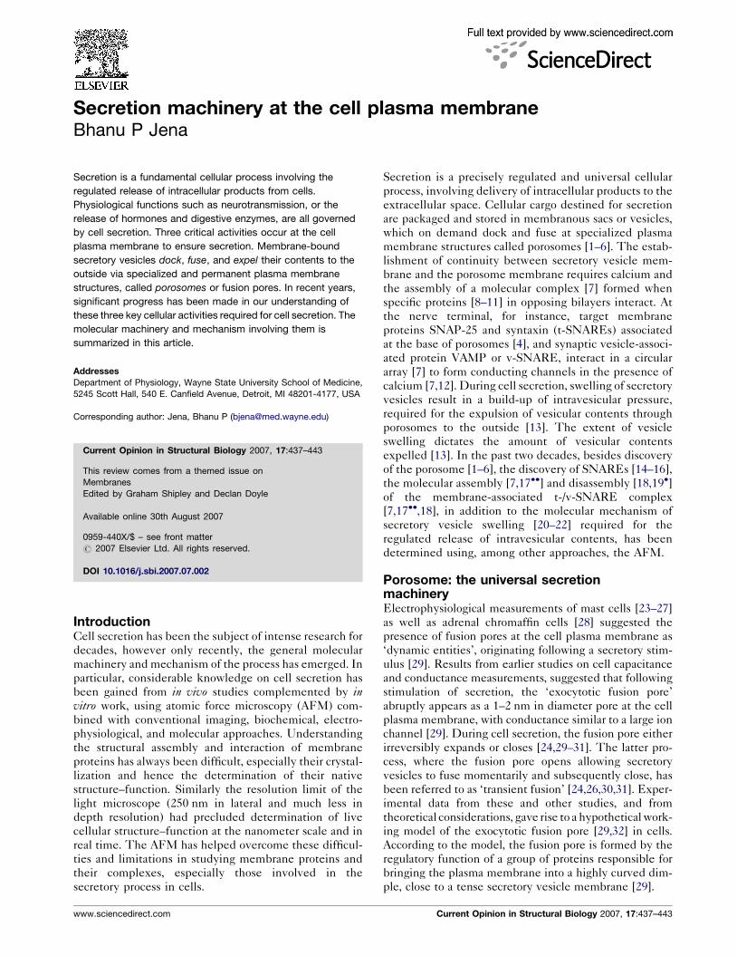
Porosomes at the plasma membrane in pancreatic acinar cells and the
neuron. (a) AFM micrograph depicting ‘pits’ (yellow arrow) and
‘porosomes’ within (blue arrow), at the apical plasma membrane in a live
pancreatic acinar cell. (b) To the right is a schematic drawing depicting
porosomes at the cell plasma membrane (PM), where membrane-bound
secretory vesicles called zymogen granules (ZG), dock and fuse to
release intravesicular contents. (c) A high resolution AFM micrograph
shows a single pit with four 100–180 nm porosomes within. (d) An
electron micrograph depicting a porosome (red arrowhead) close to a
microvilli (MV) at the apical plasma membrane (PM) of a pancreatic
acinar cell. Note association of the porosome membrane (yellow
arrowhead), and the zymogen granule membrane (ZGM) (red arrow
head) of a docked ZG (inset). Cross-section of a circular complex at the
mouth of the porosome is seen (blue arrow head). (e) The bottom left
panel shows an electron micrograph of a porosome (red arrowhead) at
the nerve terminal, in association with a synaptic vesicle (SV) at the
presynaptic membrane (pre-SM). Notice a central plug at the neuronal
porosome opening. (f) The bottom right panel is an AFM micrograph of a
neuronal porosome in physiological buffer, also showing the central plug
(red arrowhead) at its opening. It is believed that the central plug in
neuronal porosomes may regulate its rapid close–open conformation
during neurotransmitter release. The neuronal porosome is an order of
magnitude smaller (10–15 nm) in comparison to porosome in the
exocrine pancreas.
Current Opinion in Structural Biology 2007, 17:437–443
questions such as the appearance of partially empty
vesicles following cell secretion were answered [33].
Porosomes in pancreatic acinar (Figure 1a–d) [4–6] or
growth hormone-secreting cells are permanent structures
at the cell plasma membrane, with a 100–180-nm
diameter opening. Membrane-bound secretory vesicles
ranging in size from 0.2 to 1.2 mm in diameter dock and
fuse at porosomes to release vesicular contents. Following
fusion of secretory vesicles at porosomes, only a 25–45%
increase in porosome diameter is demonstrated [1–3]. It is
therefore reasonable to conclude that secretory vesicles
‘transiently’ dock and fuse at the base of porosomes,
instead of complete merger of the secretory vesicle mem-
brane at the site. In contrast to accepted belief, if
secretory vesicles were to completely incorporate at poro-
somes, the plasma membrane structure would distend
much greater than what is observed. Furthermore, if
secretory vesicles were to completely merge at the cell
plasma membrane, there would be a loss in vesicle
number following cell secretion. Examination of
secretory vesicles within cells before and after secretion
demonstrates that the total number of secretory vesicles
remains constant following cell secretion. However, the
number of empty and partially empty vesicles increases
significantly, supporting the occurrence of transient
fusion [33]. In agreement, it has been demonstrated that
secretory granules are recaptured largely intact following
stimulated exocytosis in cultured endocrine cells [33].
Further support of transient fusion is provided by the
presence of neurotransmitter transporters at the synaptic
vesicle membrane. Synaptic vesicle-associated transpor-
ters would be of little use if vesicles were to fuse and
merge completely at the cell plasma membrane, to be
endocytosed at a later time. Similarly in neurons, 10–
15 nm porosomes at the presynaptic membrane
(Figure 1e and f) allow for the transient docking and
fusion of synaptic vesicles, during neurotransmission.
Studies report that single synaptic vesicles fuse transi-
ently and successively without loss of vesicle identity
[33].
Over the years, the term ‘fusion pore’ has been loosely
referred to plasma membrane dimples that originate
following a secretory stimulus, or to the continuity or
channel established between opposing lipid membrane
during membrane fusion. Hence for clarity, the term
‘porosome’ was assigned to this newly discovered struc-
ture at the cell plasma membrane, where secretory
vesicles transiently dock and fuse to release their con-
tents. In the past decade, studies demonstrate that poro-
somes are present in all secretory cells examined, such as
acinar cells of the exocrine pancreas, neurons, chromaffin
cells, mast cells, growth hormone cells of the pituitary
gland, b-cells of the endocrine pancreas [1–6,33], and
recently in astrocytes (personal observation). Further-
more, the porosome has been isolated, its composition
determined [4,6], and it has been both structurally and
www.sciencedirect.com
Secretion machinery at the cell plasma membrane Jena 439
functionally reconstituted in artificial lipid membrane
[5,6].
As previously discussed, in the exocrine pancreas and
neuroendocrine cells, porosomes range in size from 100
to 180 nm, whereas in neurons and astrocytes, they
measure merely 10–15 nm. Results from both AFM and
EM studies demonstrate porosomes in every cell type
examined (exocrine pancreas, to neuroendocrine cells, to
neurons), to possess a cup-shaped basket-like morphology.
During cell secretion, the porosome opening dilates 25–
45% to facilitate expulsion of intravesicular contents,
returning to its resting size following completion of
secretion [1]. Porosomes are supramolecular lipoprotein
structures, composed of among other proteins [4,6], SNAP-
23/25, syntaxin, synaptotagmin, the ATPase NSF, cyto-
skeletal proteins (actin, a-fodrin, and vimentin), calcium
channels b3 and a1c, chloride ion channels ClC2 and ClC3,
and in some cases their isoforms [4]. Recent studies further
demonstrate cholesterol to be an integral component of the
porosome complex, required for retaining its integrity and
intramolecular interactions [34]. Apart from the fact that t-
SNAREs and calcium channels are present at the base of
porosomes [4], and the likely association of actin at the
neck region of the structure [1] to regulate opening and
closing to the outside, the localization of other proteins
within the porosome complex remains to be determined.
The size and complexity of the membrane-associated
porosome has precluded an understanding of its atomic
structure, which will ultimately provide further structural
and functional details about the complex.
Secretory vesicle fusion at plasmamembrane-associated porosomesA primary understanding of membrane fusion in cells was
made possible following discovery of a N-ethylmaleimide-
sensitive factor (NSF) [35] and SNARE proteins [14–16],
and their involvement in membrane fusion [10,36]. Target
membrane proteins, SNAP-25 and syntaxin (t-SNARE)
and secretory vesicle-associated membrane protein
(VAMP or v-SNARE), are part of the conserved protein
complex involved in fusion of opposing bilayers [36].
VAMP and syntaxin are both integral membrane proteins,
with the soluble SNAP-25 associating with syntaxin.
Hence, the key to our understanding of SNARE-induced
membrane fusion requires determination of the atomic
arrangement and interaction between membrane-associ-
ated v-SNAREs and t-SNAREs. Ideally, the atomic coor-
dinates of membrane-associated SNARE complex using
X-ray crystallography would provide atomic details of the
structure and its involvement in membrane fusion. So far
such atomic details of membrane-associated t-/v-SNARE
complex has not been possible, primarily due to solubility
problems of membrane SNAREs, compounded with the
fact that v-SNARE and t-SNAREs need to reside in
opposing membranes when they meet, to form the appro-
priate physiologically relevant SNARE complex. The
www.sciencedirect.com
remaining option has been the use of nuclear magnetic
resonance spectroscopy (NMR), however the NMR
approach too has not been successful, primarily due to
the molecular size limitation of the NMR. Regardless of
these set backs and limitations, AFM force spectroscopy
has provided for the first time at nanometer resolution, an
understanding of the structure, assembly, and disassembly
of the membrane-associated t-/v-SNARE complex in phys-
iological buffer solution [7,12,17��,18].
The structure and arrangement of SNAREs associated
with lipid bilayers were first examined using AFM [7]. A
bilayer electrophysiological assay allowed measurements
of membrane conductance and capacitance, before and
after t-SNARE-reconstitution or v-SNARE-reconstitu-
tion, and following exposure to v-SNARE-reconstituted
or t-SNARE-reconstituted lipid vesicles. Results from the
study demonstrate that t-SNAREs and v-SNARE when
present in opposing bilayers, interact in a circular array,
and in the presence of calcium they form conducting
channels (Figure 2) [7,17��]. The interaction of t-/v-
SNARE proteins to form a conducting channel is strictly
dependent on the presence of t-SNAREs and v-SNARE
in opposing bilayers. Addition of purified recombinant v-
SNARE to a t-SNARE-reconstituted lipid membrane,
results in an increase only in the size of the globular
t-SNARE oligomer, and is without influence on the
electrical properties of the membrane [7]. However, when
t-SNARE vesicles are added to v-SNARE-reconstituted
membrane (Figure 2a–e), SNAREs assemble in a ring
configuration (Figure 2c and d), and a stepwise increase in
capacitance and conductance is observed, demonstrating
the establishment of continuity between the opposing
bilayers (Figure 2e). These results demonstrate that t-
SNARE and v-SNARE are required to reside in opposing
bilayers (Figure 2f), to allow appropriate t-/v-SNARE
interactions leading to membrane fusion in the presence
of calcium [7]. Studies using SNARE-reconstituted lipo-
somes and bilayers [12] demonstrate: first, a low fusion
rate (t = 16 min) between t-SNARE-reconstituted and v-
SNARE-reconstituted liposomes in the absence of Ca2+
and second, exposure of t-/v-SNARE liposomes to Ca2+
drives vesicle fusion on a near physiological relevant
time-scale t � 10 s), demonstrating Ca2+ and SNAREs
in combination to be the minimal fusion machinery in
cells [12]. Native and synthetic vesicles exhibit a signifi-
cant negative surface charge primarily due to the polar
phosphate head groups. These polar head groups produce
a repulsive force, preventing aggregation and fusion of
opposing vesicles. SNAREs bring opposing bilayers clo-
ser, to within a distance of 2–3 A [12], enabling Ca2+
bridging. The bound Ca2+ then leads to the expulsion of
water between the bilayers at the bridging site, leading to
lipid mixing and membrane fusion. Hence SNAREs,
besides bringing opposing bilayers closer, dictate the
site and size of the fusion area during cell secretion.
The size of the t-/v-SNARE complex forming the
Current Opinion in Structural Biology 2007, 17:437–443
440 Membranes
Figure 2
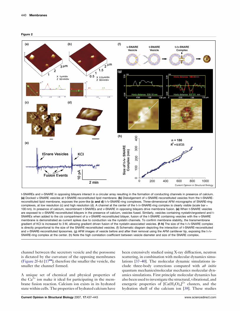
t-SNAREs and v-SNARE in opposing bilayers interact in a circular array resulting in the formation of conducting channels in presence of calcium.
(a) Docked v-SNARE vesicles at t-SNARE-reconstituted lipid membrane. (b) Dislodgement of v-SNARE-reconstituted vesicles from the t-SNARE-
reconstituted lipid membrane, exposes the pore-like (c and d) t-/v-SNARE-ring complexes. Three-dimensional AFM micrographs of SNARE-ring
complexes, at low resolution (c) and high resolution (d). A channel at the center of the t-/v-SNARE-ring complex is clearly visible (scale bar =
100 nm). In presence of calcium, recombinant t-SNAREs and v-SNARE in opposing bilayers drive membrane fusion. (e) When t-SNARE vesicles
are exposed to v-SNARE-reconstituted bilayers in the presence of calcium, vesicles fused. Similarly, vesicles containing nystatin/ergosterol and t-
SNAREs when added to the cis compartment of a v-SNARE-reconstituted bilayer, fusion of the t-SNARE containing vesicles with the v-SNARE
membrane is demonstrated as current spikes due to conduction via the nystatin channels. To confirm membrane stability, the transmembrane
gradient of KCl is increased to 3 M, allowing gradient driven fusion of the nystatin-associated vesicles. (f–h) The size of the t-/v-SNARE complex
is directly proportional to the size of the SNARE-reconstituted vesicles. (f) Schematic diagram depicting the interaction of t-SNARE-reconstituted
and v-SNARE-reconstituted liposomes. (g) AFM images of vesicle before and after their removal using the AFM cantilever tip, exposing the t-/v-
SNARE-ring complex at the center. (h) Note the high correlation coefficient between vesicle diameter and size of the SNARE complex.
channel between the secretory vesicle and the porosome
is dictated by the curvature of the opposing membranes
(Figure 2f–h) [17��]; therefore the smaller the vesicle, the
smaller the channel formed.
A unique set of chemical and physical properties of
the Ca2+ ion make it ideal for participating in the mem-
brane fusion reaction. Calcium ion exists in its hydrated
state within cells. The properties of hydrated calcium have
Current Opinion in Structural Biology 2007, 17:437–443
been extensively studied using X-ray diffraction, neutron
scattering, in combination with molecular dynamics simu-
lations [37–40]. The molecular dynamic simulations in-
clude three-body corrections compared with ab initioquantum mechanics/molecular mechanics molecular dyn-
amics simulations. First principle molecular dynamics has
also been used to investigate the structural, vibrational, and
energetic properties of [Ca(H2O)n]2+ clusters, and the
hydration shell of the calcium ion [38]. These studies
www.sciencedirect.com
Secretion machinery at the cell plasma membrane Jena 441
demonstrate that hydrated calcium [Ca(H2O)n]2+ has more
than one shell around the Ca2+, with the first hydration
shell having six water molecules in an octahedral arrange-
ment [38]. In studies using light scattering and X-ray
diffraction of SNARE-reconstituted liposomes, it has been
demonstrated that fusion proceeds only when Ca2+ ions are
available between the t-SNARE-apposed and v-SNARE-
apposed proteoliposomes [12,33].
Mixing of t-SNARE and v-SNARE proteoliposomes in
the absence of Ca2+ leads to a diffuse and asymmetric
diffractogram in X-ray diffraction studies, a typical charac-
teristic of short-range ordering in a liquid system [41]. In
contrast, when t-SNARE and v-SNARE proteoliposomes
in the presence of Ca2+ are mixed, it leads to a more
structured diffractogram with approximately a 12%
increase in X-ray scattering intensity, suggesting an
increase in the number of contacts between opposing
bilayers, established presumably through calcium–phos-
phate bridges, as previously suggested [12,33,42]. The
ordering effect of Ca2+ on interbilayer contacts observed
in X-ray studies [12] is in good agreement with light,
AFM, and spectroscopic studies, suggesting close apposi-
tion of PO-lipid head groups in the presence of Ca2+,
followed by formation of Ca2+–PO bridges between the
adjacent bilayers [12,33,43]. X-ray diffraction studies
show that the effect of Ca2+ on bilayers orientation and
interbilayer contacts is most prominent in the area of 3 A,
with additional appearance of a new peak at position
2.8 A, both of which are within the ionic radius of Ca2+
[12]. These studies further suggest that the ionic radius of
Ca2+ may make it an ideal player in the membrane fusion
reaction. Hydrated calcium [Ca(H2O)n]2+ however, with a
hydration shell having six water molecules and measuring
�6 A would be excluded from the t-/v-SNARE apposed
interbilayer space, hence calcium has to be present in the
buffer solution when t-SNARE vesicles and v-SNARE
vesicles meet. Indeed, studies demonstrate that if t-
SNARE and v-SNARE vesicles are allowed to mix in a
calcium-free buffer, there is no fusion following post
addition of calcium [44]. How does calcium work?
Calcium bridging of apposing bilayers may lead to the
release of water from the hydrated Ca2+ ion, leading to
bilayer destabilization and membrane fusion. Addition-
ally, the binding of calcium to the phosphate head groups
of the apposing bilayers may also displace the loosely
coordinated water at the PO-lipid head groups, resulting
in further dehydration, leading to destabilization of the
lipid bilayer and membrane fusion.
We have discussed the possible mechanism of SNARE and
calcium triggered membrane fusion, which brings us to the
question — is there a physiological blocker of membrane
fusion in cells? Recent studies demonstrate that indeed
there is such a clamp or blocker in — ‘complexins’ [45��].Complexins are 20 kDa proteins which can associate with
the SNARE complex but not individual SNAREs. At the
www.sciencedirect.com
cell plasma membrane, the principal calcium sensor in the
membrane fusion reaction is the calcium-binding protein
synaptotagmin. It is suggested that when calcium binds to
the calcium sensor — synaptotagmin, the clamp is
released, allowing membrane fusion to proceed [45��]. It
has been demonstrated that over expression of complexin
blocks membrane fusion in cells [45��].
Expulsion of intravesicular contents at thecell plasma membraneStudies demonstrate that vesicle swelling is required for
the expulsion of intravesicular content from cells during
secretion [13]. It has been demonstrated [13] that the
extent of swelling is directly proportional to the amount
of intravesicular contents expelled, hence providing cells
with the ability to precisely regulate the release of secretory
products during cell secretion. Direct observation in live
cells of the requirement of secretory vesicle swelling in cell
secretion [13] explains the appearance of empty and par-
tially empty vesicles following cell secretion [46,47].
Live pancreatic acinar cells in near physiological buffer,
when imaged using the AFM, reveal the size of secretory
vesicles called zymogen granules (ZGs) lying immedi-
ately beneath the surface of the apical plasma membrane.
Within 2.5 min of exposure to a secretory stimulus, a
majority of ZGs within cells swell, followed by a decrease
in ZG size, and a concomitant release of secretory pro-
ducts [13]. These studies demonstrated for the first time
in live cells that there is intracellular swelling of secretory
vesicles following stimulation of secretion, and vesicle
deflation following partial discharge of their contents.
Measurements of intracellular ZG size further reveal that
the extent of swelling differs between vesicles following a
secretory stimulus. This differential swelling among ZGs
within the same cell, may explain why following stimu-
lation of secretion, some intracellular ZGs demonstrate
the presence of less vesicular content than others.
The precise role of secretory vesicle swelling in the
expulsion of intravesicular contents during cell secretion
has been determined using an electrophysiological
bilayer fusion assay employing porosome-reconstituted
lipid membrane [13]. It has been demonstrated that
exposure of isolated secretory vesicles to GTP results
in their swelling, similar to the observations made in live
cells. Different secretory vesicles respond differently to
the same swelling stimulus. This differential response to
GTP has been further assessed, by measuring changes in
volume of different size vesicles. Addition of isolated
secretory vesicles to the cis compartment of the bilayer
electrophysiological apparatus, demonstrates vesicle
fusion at the porosome-reconstituted lipid membrane.
However, even after 15 min of vesicle addition, little or
no release of intravesicular content is detected in the transcompartment of the bilayer chamber. In contrast, addition
of GTP to the cis compartment of the chamber induces
Current Opinion in Structural Biology 2007, 17:437–443
442 Membranes
vesicle swelling, resulting in a robust expulsion of intra-
vesicular contents into the trans compartment of the
bilayer chamber. This holds true for both a slow secretory
cells like the acinar cells of exocrine pancreas, and in the
fast secreting neuron [13]. These studies demonstrate
that during cell secretion, secretory vesicle swelling is a
requirement for the expulsion of intravesicular contents.
Our understanding of the molecular mechanism of
secretory vesicle swelling has greatly advanced in the
last decade. Isolated secretory vesicles and reconstituted
swelling-competent proteoliposomes have been utilized
[13,20–22,48] to determine the mechanism and regulation
of vesicle swelling. As previously discussed, isolated ZGs
from the exocrine pancreas swell rapidly in response to
GTP [20], suggesting rapid water gating into ZGs following
exposure to GTP. Results from studies demonstrate the
presence of the water channel aquaporin-1 (AQP-1) at the
ZG membrane [21] and its participation in GTP-mediated
water entry and vesicle swelling. Further, the molecular
regulation of AQP-1 at the ZG membrane has been studied
[48], providing a general mechanism of secretory vesicle
swelling. Detergent-solubilized ZGs immunoprecipitated
with monoclonal AQP-1 antibody, coisolates AQP-1,
PLA2, Gai3, potassium channel IRK-8, and the chloride
channel ClC-2 [48]. Exposure of ZGs to either the potass-
ium channel blocker glyburide, or the PLA2 inhibitor
ONO-RS-082, blocks GTP-induced ZG swelling. Red
blood cells known to possess AQP-1 at the plasma mem-
brane also swell on exposure to the GaI agonist mastoparan,
and responds similarly to ONO-RS-082 and glyburide, as
do ZGs [47]. Artificial liposomes reconstituted with the
AQP-1 immunoisolated complex from solubilized ZGs also
swell in response to GTP. Glyburide or ONO-RS-082 also
abrogates the GTP effect in reconstituted liposomes. AQP-
1 immunoisolate-reconstituted planar lipid membrane
demonstrate conductance, which is sensitive to glyburide
and an AQP-1-specific antibody. These results demon-
strate a Gai3-PLA2-mediated pathway and potassium
channel involvement in AQP-1 regulation at the ZG mem-
brane [48], contributing to ZG swelling. Similarly, AQP-6
involvement has been demonstrated in GTP-induced and
Go-mediated synaptic vesicle swelling in neurons [22].
ConclusionThe findings outlined in this review provide our current
understanding of the molecular machinery involved, and
the mechanism of secretion operating at the cell plasma
membrane. Porosomes are specialized plasma membrane
structures present in all secretory cells, from exocrine and
endocrine cells, to neuroendocrine cells and neurons.
Since porosomes in exocrine and neuroendocrine cells
measure 100–180 nm, and only 25–45% increase in poro-
some diameter is demonstrated following the docking and
fusion of 0.2–1.2 mm in diameter vesicles, it is reasonable
to conclude that secretory vesicles ‘transiently’ dock and
fuse at the porosome complex to release their contents to
Current Opinion in Structural Biology 2007, 17:437–443
the outside. The discovery of the porosome, an under-
standing of SNARE-induced membrane fusion, and the
requirement of secretory vesicle swelling for expulsion of
intravesicular contents has progressed our understanding
of secretion in cells. It has become clear that the secretory
process in cells is coordinated by a highly regulated and
fine-tuned orchestra.
AcknowledgementsI thank Won Jin Cho for help in preparation of the figures andformatting the manuscript, and members of my laboratory for theirhelpful discussions and suggestions. Supported by Grants DK-56212 andNS-39918 from the National Institutes of Health (BPJ).
References and recommended readingPapers of particular interest, published within the annual period ofreview, have been highlighted as:
� of special interest�� of outstanding interest
1. Schneider SW, Sritharan KC, Geibel JP, Oberleithner H, Jena BP:Surface dynamics in living acinar cells imaged by atomic forcemicroscopy: identification of plasma membrane structuresinvolved in exocytosis. Proc Natl Acad Sci USA 1997, 94:316-321.
2. Cho SJ, Quinn AS, Stromer MH, Dash S, Cho J, Taatjes DJ,Jena BP: Structure and dynamics of the fusion pore in livecells. Cell Biol Int 2002, 26:35-42.
3. Cho SJ, Jeftinija K, Glavaski A, Jeftinija S, Jena BP, Anderson LL:Structure and dynamics of the fusion pores in live GH-secreting cells revealed using atomic force microscopy.Endocrinology 2002, 143:1144-1148.
4. Jena BP, Cho SJ, Jeremic A, Stromer MH, Abu-Hamdah R:Structure and composition of the fusion pore. Biophys J 2003,84:1337-1343.
5. Jeremic A, Kelly M, Cho SJ, Stromer MH, Jena BP: Reconstitutedfusion pore. Biophys J 2003, 85:2035-2043.
6. Cho WJ, Jeremic A, Rognlien KT, Zhvania MG, Lazrishvili I,Tamar B, Jena BP: Structure, isolation, composition andreconstitution of the neuronal fusion pore. Cell Biol Int 2004,28:699-708.
7. Cho SJ, Kelly M, Rognlien KT, Cho J, Hoerber JKH, Jena BP:SNAREs in opposing bilayers interact in a circular array toform conducting pores. Biophys J 2002, 83:2522-2527.
8. Malhotra V, Orci L, Glick BK, Block MR, Rothman JE: Role of an N-ethylmaleimide-sensitive transport component in promotingfusion of transport vesicles with cisternae of the Golgi stack.Cell 1988, 54:221-227.
9. Clary DO, Griff IC, Rothman JE: SNAPs, a family of NSFattachment proteins involved in intracellular membrane fusionin animals and yeast. Cell 1990, 61:709-721.
10. Wilson DW, Whiteheart SW, Wiedmann M, Brunner M,Rothman JE: A multisubunit particle implicated in membranefusion. J Cell Biol 1992, 117:531-538.
11. Sollner T, Whiteheart SW, Brunner M, Erdjument-Bromage H,Geromanos S, Tempst P, Rothman JE: SNAP receptorsimplicated in vesicle targeting and fusion. Nature 1993,362:318-324.
12. Jeremic A, Kelly M, Cho WJ, Cho SJ, Horber JKH, Jena BP:Calcium drives fusion of SNARE-apposed bilayers. Cell Biol Int2004, 28:19-31.
13. Kelly M, Cho WJ, Jeremic A, Abu-Hamdah R, Jena BP: Vesicleswelling regulates content expulsion during secretion.Cell Biol Int 2004, 28:709-716.
14. Trimble WS, Cowam DM, Scheller RH: VAMP-1: a synapticvesicle-associated integral membrane protein. Proc Natl AcadSci USA 1988, 85:4538-4542.
www.sciencedirect.com
Secretion machinery at the cell plasma membrane Jena 443
15. Oyler GA, Higgins GA, Hart RA, Battenbarg M, Bloom FE,Wilson MC: The identification of a novel synaptosomal-associated protein, SNAP-25, differentially expressed byneuronal subpopulations. J Cell Biol 1989, 109:3039-3052.
16. Bennett K, Calakos N, Scheller RH: Syntaxin: a synaptic proteinimplicated in docking of synaptic vesicles at presynapticactive zones. Science 1992, 257:255-259.
17.��
Cho WJ, Jeremic A, Jena BP: Size of supramolecular SNAREcomplex: membrane-directed self-assembly. J Am Chem Soc2005, 127:10156-10157.
In an earlier study, it had been demonstrated that membrane-associatedt-SNAREs and membrane-associated v-SNARE interact in a circulararray to form conducting channels in the presence of calcium, thusestablishing continuity between opposing bilayers [7]. In the absenceof membrane, no such SNARE-ring/channel complexes are formed. Thisstudy reports for the first time that the size of the SNARE-ring complexformed by the interaction of membrane-associated t-SNAREs and v-SNARE is directly proportional to the size of the vesicle the v-SNARE hasbeen reconstituted in. Small proteoliposomes produce small SNARE-ringcomplexes, hence vesicle curvature regulates the size of the SNAREcomplex.
18. Jeremic A, Quinn AS, Cho WJ, Taatjes DJ, Jena BP: Energy-dependent disassembly of self-assembled SNARE complex:observation at nanometer resolution using atomic forcemicroscopy. J Am Chem Soc 2006, 128:26-27.
19.�
Cho WJ, Jena BP: N-Ethylmaleimide-sensitive factor is aright-handed molecular motor. J Biomed Nanotechnol 2007,3:209-211.
The SNARE complex is an SDS-resistant and an extremely stable com-plex. Exposure of t-/v-SNARE complex to the ATPase N-ethylmaleimide-sensitive factor (NSF) and ATP results in the disassembly of the complex,which can be imaged at near nanometer resolution using AFM. This studyshows that t-/v-SNAREs form left-handed supercoils, which are uncoiledby NSF, thus demonstrating for the first time that NSF is a right-handedmolecular motor.
20. Jena BP, Schneider SW, Geibel JP, Webster P, Oberleithner H,Sritharan KC: Gi regulation of secretory vesicle swellingexamined by atomic force microscopy. Proc Natl Acad Sci USA1997, 94:13317-13322.
21. Cho SJ, Sattar AK, Jeong EH, Satchi M, Cho JA, Dash S,Mayes MS, Stromer MH, Jena BP: Aquaporin 1 regulates GTP-induced rapid gating of water in secretory vesicles. Proc NatlAcad Sci USA 2002, 99:4720-4724.
22. Jeremic A, Cho WJ, Jena BP: Involvement of water channels insynaptic vesicle swelling. Exp Biol Med 2005, 230:674-680.
23. Breckenridge LJ, Almers W: Currents through the fusionpore that forms during exocytosis of a secretory vesicle.Nature 1987, 328:814-817.
24. Breckenridge LJ, Almers W: Final steps in exocytosis observedin a cell with giant secretory granules. Proc Natl Acad Sci USA1987, 84:1945-1949.
25. Alvarez de Toledo G, Fernandez JM: The events leading tosecretory granule fusion. Cell Physiology of Blood. RockefelerUniversity Press; 1988:. 334–344.
26. Spruce AE, Breckenridge LJ, Lee AK, Almers W: Properties of thefusion pore that forms during exocytosis of a mast cellsecretory vesicle. Neuron 1990, 4:643-654.
27. Alvarez de Toledo G, Fernandez-Chaocn R, Fernandez JM:Release of secretory products during transient vesicle fusion.Nature 1993, 363:554-558.
28. Chow RH, von Ruden R, Neher E: Delay in vesicle fusionrevealed by electrochemical monitoring of single secretoryevents in adrenal chromaffin cells. Nature 1992, 356:60-63.
29. Monck JR, Oberhauser AF, Fernandez JM: The exocytotic fusionpore interface: a model of the site of neurotransmitter release.Mol Membr Biol 1995, 12:151-156.
30. Monck JR, Alvarez de Toledo G, Fernandez JM: Tension insecretory granule membranes causes extensive membrane
www.sciencedirect.com
transfer through the exocytotic fusion pore. Proc Natl Acad SciUSA 1990, 87:7804-7808.
31. Fernandez JM, Neher E, Gomperts BD: Capacitancemeasurements reveal stepwise fusion events in degranulatingmast cells. Nature 1984, 312:453-455.
32. Monck JR, Fernandez JM: The exocytotic fusion pore andneurotransmitter release. Neuron 1994, 12:707-716.
33. Jena BP: Molecular machinery and mechanism of cellsecretion. Exp Biol Med 2005, 230:307-319.
34. Jeremic A, Cho WJ, Jena BP: Cholesterol is critical to theintegrity of neuronal porosome/fusion pore. Ultramicroscopy2006, 106:674-677.
35. Block MR, Glick BS, Wilcox CA, Wieland FT, Rothman JE:Purification of an N-ethylmaleimide-sensitive proteincatalyzing vesicular transport. Proc Natl Acad Sci USA 1988,85:7852-7856.
36. Weber T, Zemelman BV, McNew JA, Westerman B, Gmachi M,Parlati F, Sollner TH, Rothman JE: SNAREpins: minimalmachinery for membrane fusion. Cell 1998, 92:759-772.
37. Chialvo AA, Simonson JM: The structure of CaCl2 aqueoussolutions over a wide range of concentration. Interpretation ofdiffraction experiments via molecular simulation. J Chem Phys2003, 119:8052-8061.
38. Bako I, Hutter J, Palinkas G: Car–Parrinello molecular dynamicssimulation of the hydrated calcium ion. J Chem Phys 2002,117:9838-9843.
39. Schwenk CF, Loeffler HH, Rode BM: Molecular dynamicssimulations of Ca2+ in water: comparison of a classicalsimulation including three-body corrections and Born–Oppenheimer ab initio and density functional theory quantummechanical/molecular mechanics simulations. J Chem Phys2001, 115:10808-10813.
40. Licheri G, Piccaluga G, Pinna G: X-ray diffraction study of theaverage solute species in CaCl2 aqueous solutions. J ChemPhys 1976, 64:2437-2446.
41. McIntosh TJ: Short-range interactions between lipid bilayersmeasured by X-ray diffraction. Curr Opin Struct Biol 2000,10:481-485.
42. Portis A, Newton C, Pangborn W, Papahadjopoulos D: Studies onthe mechanism of membrane fusion: evidence for anintermembrane Ca2+–phospholipid complex, synergism withMg2+, and inhibition by spectrin. Biochemistry 1979, 18:780-790.
43. Laroche G, Dufourc EJ, Dufoureq J, Pezolet M: Structure anddynamics of dimyristoylphosphatidic acid/calcium complexby 2H NMR, infrared, spectroscopies and small-angle x-raydiffraction. Biochemistry 1991, 30:3105-3114.
44. Jeremic A, Cho WJ, Jena BP: Membrane fusion: what maytranspire at the atomic level. J Biol Phys Chem 2004, 4:139-142.
45.��
Giraudo CG, Eng WS, Melia TJ, Rothman JE: A clampingmechanism involved in SNARE-dependent exocytosis.Science 2006, 313:676-680.
This study reports for the first time that complexins, a previously identifiedfamily of 20 kDa proteins, act as blockers of SNARE-mediated andcalcium-mediated membrane fusion in cells.
46. Cho SJ, Cho J, Jena BP: The number of secretory vesiclesremains unchanged following exocytosis. Cell Biol Int 2002,26:29-33.
47. Lee JS, Mayes MS, Stromer MH, Scanes CG, Jeftinija S,Anderson LL: Number of secretory vesicles in growth hormonecells of the pituitary remains unchanged after secretion.Exp Biol Med 2004, 229:291-302.
48. Abu-Hamdah R, Cho WJ, Cho SJ, Jeremic A, Kelly M, Ilie AE,Jena BP: Regulation of the water channel aquaporin-1:isolation and reconstitution of the regulatory complex. Cell BiolInt 2004, 28:7-17.
Current Opinion in Structural Biology 2007, 17:437–443


![Ranu Bhanu - Sunil Gangopadhyay [Amarboi.com]](https://img.pdfslide.net/doc/110x75/577c7c1e1a28abe054995b03/ranu-bhanu-sunil-gangopadhyay-amarboicom.jpg)