Embed Size (px)
Citation preview

J. exp. Biol. 134, 155-171 (1988) 155Printed in Great Britain © The Company of Biologists Limited 1988
SECRETORY POTENTIAL AND IONIC TRANSPORT INTHE POSTERIOR SILK GLANDS OF BOMBYX MORI
BY I. NAKAGAKI AND S. SASAKI
Department of Physiology, Osaka Medical College, Takatsuki, Osaka 569, Japan
Accepted 15 July 1987
SUMMARY
1. The concentrations of Na, Mg, P, S, Cl, K and Ca in the cytoplasm and lumenof the posterior silk gland cells of Bombyx mori were measured by X-ray microprobeanalysis of freeze-dried thin sections. The basal and luminal membrane potentials ofthe gland cells were measured using microelectrode techniques. The input resistanceof the luminal plasma membrane was simultaneously measured by injecting electriccurrent via an intracellular microelectrode.
2. The basolateral membrane potential was —47 ± l-8mV (s.E.) (iV = 46), andthe glands exhibited lumen-negative voltages of —6± 0-1 mV (s.E.) (iY= 40) in thenormal state. Increasing the extracellular K+ concentration depolarized the basolat-eral membrane potential, whereas the membrane potential hyperpolarized when Cl~concentration in the extracellular fluid was increased. There were no significanteffects on the membrane potential when Na+, Mg2+ and Ca2+ concentrations in theextracellular fluid were changed.
3. The representative X-ray spectra showed high K and phosphorus peaks, andlow Cl and Mg peaks in the cytoplasm of the normal posterior silk gland cells. Thenormal glandular lumen showed relatively high K, and low Cl, sulphur, Ca and Mgpeaks. Quantitative microprobe values were, for the cytoplasm (mmolkg"1 wetmass, A'=30) Na, 5; Mg, 14; phosphorus, 168; sulphur, 16; Cl, 12; K, 168;Ca, 0 5 ; and for the lumen (A'= 10) Na, 3; Mg, 25; phosphorus, 42; sulphur, 24;Cl, 38; K, 133; Ca, 9-4 in the normal glands.
4. The basal plasma membrane potential was hyperpolarized by 7mV afterstimulation with 5X 10~5 mmol I"1 5-hydroxytryptamine (5-HT). Microprobevalues for the cytoplasm were (mmol kg"1 wet mass, AT=15) Na, 4; Mg, 13;phosphorus, 160; sulphur, 17; Cl, 8; K, 187; Ca, 0-6 in the stimulated glands. Thecytoplasmic [K] increased after stimulation with 5-HT.
5. The basal membrane potential of the gland cells was depolarized by 3 mV afterapplication of a juvenoid, methoprene (10~smol 1~'). X-ray microprobe values forthe cytoplasm were (mmol kg"1 wet mass, A' = 15) Na, 7; Mg, 11; P, 170; S, 14;Cl, 23; K, 130; Ca, 3-4 in the treated glands. The cytoplasmic [Ca] and [Cl]increased, while the [K] decreased with methoprene stimulation.
6. The luminal membrane potential of the gland cells was depolarized by 8mVand a simultaneous decrease of luminal membrane resistance was apparent afterstimulation with an anti-microfilament reagent, cytochalasin D (2x 10~6mol 1~').
Key words: secretory potential, ionic transport, silk gland.

156 I. NAKAGAKI AND S. SASAKI
X-ray microprobe values for the cytoplasm became (mmolkg"1 wet mass, i\ = 15)Na, 10; Mg, 28; P, 194; S, 22; Cl, 24; K, 148; Ca, 1-2; and for the lumen (A'= IS)Na, 14; Mg, 13; phosphorus, 31; sulphur, 30; Cl, 92; K, 122; Ca, 1-1 in thestimulated glands. The cytoplasmic [Ca] and [Cl] increased and [K] decreased,whereas the luminal [Cl] and [sulphur] increased and [Ca] and [Mg] decreased aftercytochalasin D stimulation.
7. The reaction products of adenosine triphosphatase activity were found on theluminal and lateral plasma membranes of the posterior silk gland cells.
8. The possible routes of ion transport into the lumen are discussed.
INTRODUCTION
The relationship between the intracellular ionic environments and the secretion ofexocrine glands has been extensively studied. Calcium is known to have a central rolein the secretory process, and physiological stimulants leading to enzyme and fluidsecretion exert marked changes on the bioelectrical properties of the cell membraneof exocrine glands (Kanno, 1972; Matthews, Petersen & Williams, 1973; Berridge,Lindleg & Prince, 1975; Petersen, 1980; Ginsborg & House, 1980).
It has also been suggested that microtubules and/or microfilaments play some rolein the secretion process of various organs such as the pancreas (Lacy, Howell, Young& Fink, 1968; Baudin, Stock, Vincent & Grenier, 1975), salivary gland (Rossignol,Herman & Kerger, 1972; Nakagaki, Goto, Sasaki & Imai, 1978) and adrenal medulla(Douglas & Sorimachi, 1972; Poisner & Banerjee, 1971).
The posterior silk gland of the silkworm is a tubular epithelium which is large anddifferentiated for the synthesis of the silk protein fibroin, and it secretes a protein-rich fluid into the glandular lumen. These large cells have a radial microtubularsystem and a circular microtubule— microfilament system (Fig. 5A,B). Thesesystems are concerned with the intracellular transport and secretion of fibroin (Sasaki& Tashiro, 1976). We reported that, in the presence of colchicine or vinblastine, thesecretion of fibroin was suppressed and that there was a marked accumulation of thesecretory granules of fibroin in the Golgi regions. The secretion was accelerated andextensive invagination of the luminal surface was observed in the presence ofcytochalasin B or D (Sasaki, Nakajima, Fujii-Kuriyama & Tashiro, 1981).
Some investigators reported that a suitable dose of juvenile hormone (methoprene)stimulated the RN A and fibroin synthesis of the posterior silk gland cells and broughtabout an increase in weight of the cocoon shell, due to an enhancement of fibroinsecretion into the lumen (Henrick el at. 1975; Akai, Kiguchi & Mori, 1971, 1973).
There is general agreement that the intracellular transport of ions and water isdriven by active ion transport systems on the luminal plasma membranes of insectexocrine gland cells. This transport is enhanced by exposure to the neurotransmitterserotonin (5-hydroxytryptamine) (Gupta, Berridge, Hall & Moreton, 1978).
We investigated properties of the secreting system in the posterior silk glands ofBombvx mori, using electrophysiological and electron microprobe techniques.Special reference was made to changes in the basolateral and luminal plasma

Silk secretory potential and ionic transport 157
membrane potentials, and cytoplasmic and luminal ion concentrations in thesecretorv stimulated states.
MATERIALS AND METHODS
Materials
The strain of silkworm used is a hybrid of Shunrei and Shogetsu. The posteriorsilk gland cells of the silkworm, Bombyx mori, are very large hexagonal cells, each ofwhich extends for as much as half the circumference of the gland, the cell dimensionsbeing (approximately) 1-3 mm X 1-6 mm X 0-17 mm at the maturation stage of thefifth instar. Their function is to synthesize and secrete a large amount of fibroin intothe lumen, the diameter of which is about 0-7 mm.
Micivelectrode measurements
The posterior silk glands in the later stages of the fifth larval instar are about 15 cmlong. They can be removed from the abdomen of the silkworms, and incubated inWyatt's (1956) insect tissue culture medium, or in artificial haemolymph (pH6-8)which contains (mmolP1) K+, 30; Na+, 11; Ca2+, 12; Mg2+, 23; Cl", 79;NaHCC>3, 4; NaH2PC>4, 7. The osmolarity was adjusted to 300mosmol P 1 by addingmannitol, and the medium was gassed continuously with 95% O2 and 5% CO2,maintained at 37°C. The Na+, K+ and CP concentrations were altered by varioussalts: Na+ and K+ were replaced with Tris or tetraethylammonium and CP wasreplaced with sulphate. The lumen of the posterior silk glands was perfused with amodified solution and the lumen was separated from the bath compartment. Themodified luminal solution used mimicked the luminal contents, as estimated from theX-ray microanalysis and/or flame photometry. The concentrations were (mmol P 1 ) :Na+, 10; Mg2+, 25; CP, 42; K+, 84; Ca2+, 9-4.
Glass microelectrodes filled with 3molP ' KC1, with a tip resistance between 5and 20 MQ, were used. The microelectrode was connected to a differential typeamplifier (Nihon Kohden MEZ-7101) allowing for current injection and simul-taneous measurements of membrane potential and input resistance. The potentialand resistance changes were recorded using an oscilloscope and a pen recorder. Themicroelectrode was advanced into the cell and further into the lumen, then retractedinto the cell and further out into the extracellular space. Sometimes the reference wasalso taken into the lumen.
Serotonin (5-HT) (Merck) was added to the bath solution at a final concentrationof 5X 10~°mol P 1 . Methoprene (Otsuka Chemical Co., Japan) was dissolved inethanol (1-25 mgmP1) and added to the bath solution at a final concentration oflO"3 mol P . Cytochalasin D (Sigma) was dissolved in dimethyl sulphoxide(DMSO) ( lmgmP1) and added to the bath solution at a final concentration of2x l0~ 6 molP ' . Addition of ethanol or DMSO at the same concentrations used inthe experiments did not produce any significant change in the electrical and ionicproperties of the silk glands.

158 I. NAKAGAKI AND S. SASAKI
We recorded changes in the membrane potential of the basolateral and luminalplasma membranes, the resistance of the luminal membrane and the transcellularpotential differences.
Elements in whole tissues
The Na+, K+ and Cl~ concentrations in the whole glands, cells and lumen of theposterior silk glands were determined by flame photometry (Eppendolf) and bychloridometer (Buchler, Inc.), respectively.
X-ray microanalysis
For electron probe X-ray microanalysis, the posterior silk glands in the resting andtreated (1-30 min after the additions of reagents) states were put on copper specimenholders and rapidly frozen by pressing them against the wall of a copper block whichhad been precooled in liquid nitrogen. 100-200 nm thick cryosections were cut on thefrozen thin sectioner (FTS) of a Poter Blum MT-2 ultramicrotome maintained at— 150°C, and the sections were mounted on gold or titanium grids and transferred toan FTS freeze-drier (FTS Systems, Inc., Stone Ridge, NY, USA). The prep-arations were dried at 10" — 10~4mmHg overnight, followed by carbon coating. TheX-ray microanalysis was done using a Hitachi H-500 electron microscope interfacedwith a Kevex Si(Li) detector and 5100 multi-channel analyser. The microanalyserwas operated at 75 kV. A probe current of 10~9-10~luA was used and the analysiswas usually for 100 s. For the estimation of local dry mass fractions, an analysis wasmade of the frozen sections. The grids with frozen sections were set in the coolingspecimen holder of a Hitachi H-500 electron microscope (H5001C) in theultramicrotome and the X-ray microanalysis was carried out immediately (Sasaki,Nakagaki, Mori & Imai, 1983). We used only the spectra in which the gold peak waslow. The X-ray energy spectra and further data processing to obtain the finalconcentration values were performed using an on-line computer system (HitachiMB-MA 16003, CPU: 8088, MS-DOS). The special utility programs included somestatistical analysis. Details of the procedures have been reported elsewhere (Naka-gaki, Sasaki, Shiguma & Imai, 1984).
Cytochemical demonstration of ATPase
The cytochemical localization of ATPase was carried out according to the methodsof Wachstein & Meisel (1957) and Marchesi & Palade (1967), as reported previously(Nakagaki et at. 1978).
RESULTS
Membrane potentials of the posterior silk gland cells
Fig. 1 shows a representative intracellular in vitro recording of the membrane andtransepithelial potential, with reference to the extracellular bath solution. Theaverage basolateral membrane potential was —47 ± l-8mV (S.E.) (A' = 46), and the

Silk secretory potential and ionic transport 159
u
ifer
ence
(n
8•5« -30
iten
ti
°- -40
jimerl
— .30s
Cell Cell
Fig. 1. Profiles of transmembrane and transepithelial potential differences across theposterior silk gland cells.
average transcellular potential difference was 6 ± 0-1 mV (S.E.) (N = 40). The glandsexhibited lumen-negative voltages of — 6mV.
The effects of various extracellular ions on the basolateral membrane potentialwere investigated (Fig. 2). Increasing extracellular [K+] depolarized the membraneand the slope was 10 mV for a ten-fold change in [K+]. The membrane hyperpolar-ized when [CP] in the extracellular fluid was increased. The slope of the lineobtained was 30 mV for a ten-fold change in [CP]. There were no significant effectson the membrane potential when Na+, Mg2"1" and Ca2+ concentrations in theextracellular fluid were changed (data not shown). The lines of basal membranepotentials at different concentrations of potassium and chloride in the extracellularsolution lay between the Nernst plots for potassium and chloride, estimated from thecytoplasmic K+ and CP concentrations given in Table 1 and those found in theextracellular fluid (Fig. 2).
The effects of 5-HT, methoprene and cytochalasin on potential responses
Serotonin acts as a secretagogue and enhances ion secretion in insect gland cells(Gupta et al. 1978). We found that the basal membrane potential of the posterior silkgland cells was hyperpolarized to 7mV by treatment with 5-HT (SxlO-'mmolP1)(Fig. 3A; Table 1).
A suitable dose of juvenoid stimulates fibroin synthesis in the posterior silk glandcells and the cocoon shell increases in mass (Akai et al. 1971). In our experiments,the basal membrane potential was depolarized by application of a juvenoid,methoprene (10~3mmolP1) (Fig. 3B). The mean change in basolateral membranepotential was 5 mV following the application of methoprene (Table 1).
Cytochalasin D, an anti-microfilament reagent, effectively facilitated the secretionof fibroin and caused extensive invagination of the luminal surface. These events

160 I. NAKAGAKI AND S. SASAKI
were attributed to the serial exocytosis of fibroin globules in the posterior silk glandcells (Sasaki et al. 1981). Addition of cytochalasin D (2x 10~6mmoir') to the bathsolution caused a depolarization of 80 mV in the luminal membrane, and there was asimultaneous decrease in membrane resistance (Fig. 4; Table 2). The mean restinginput resistance was 970 ± 210 kQ (AT= 10) and the mean input resistance of glandstreated with cytochalasin D was 360 ± 140 kQ (AT = 7) across the luminal membrane.
-140
-120
-100
-80
-60
-40
>Er -20
8. oo 1
K +
100 200 300
[K+Wmmoir1)
-80
-60
-40
-20
0
20
K+
CI-
100 200 300
Fig. 2. Dependence of the posterior silk gland cell membrane potential on external K+
(A) and C\~ (B) concentrations. The dashed lines indicate the K+ and Cl~ equilibriumpotentials calculated from the Nernst equation.

Silk secretoiy potential and ionic transport 161
Table 1. Effects of semtonin (5-HT) and a juvenoid (methoprene) on the basalmembrane potential (mean ± S.E.; mV) and cytoplasmic elemental concentration(mean ± S.E.; mmolkg*1 wet mass) of silk gland cells measured by electron probe
X-ray micmanalysis of freeze-dried sections
Basal membranepotential
A'
ElementCvtoplasm
"NaMgPSClKCa
A'
*P< 0-005, comparing
Control
-47 ±1-8
46
5 ±0-514± 1-5
168 ±6-816 ±0-612± 1-6
168 ±3-30-5 ±0-1
30
control and treated cells.
5-HT
-54± 1-5*
32
4±0-813 ±2
160 ±817 ± 1-38± 1-3
187 ±5-4*0-610-1
15
Methoprene
-42 ±0-9*
25
7±l-311 ±2-5
170 ±3-414 + 0-823±l-5#
130 ±3-6*3-4 ±0-5*
15
The basal membrane potential is depolarized after exposure to cytochalasin D(Sasaki & Nakagaki, 1979).
Cytochemistry ofATPase
Electron microscopy reveals the radial microtubule and circular microtubule-microfilament systems in perinuclear and laminal cytoplasm, respectively
A0r-i
>E
iV
-10
-20
-30
-40
-50
-60
Br— 0 1
-10
-20
-30
-40
-50
lmin
\Methoprene
,-sFig. 3. Basal membrane potential changes induced by serotonin (5-HT) (1x10moll"1) (A) and a juvenoid (methoprene) ( lxl0~5mol l~') (B) in gland cells. At thearrows, one drop of the secretagogue was added to the bath solution. Addition of ethanolonly (used as a solute for methoprene) produced no significant changes.

162 I. NAKAGAKI AND S. SASAKI
>E
-10
~ -20
8.
1 -30
-40
-50Cytochalasin D 1 min
Fig. 4. Luminal membrane potential and resistance changes induced by cytochalasin Din the posterior silk gland cells. At the arrow cytochalasin D (1X 10~3 mol 1~') was addedto the bath solution. Current pulses (SnA) were injected through the recordingmicroelectrode. The addition of DMSO (the solute for cytochalasin D) produced nosignificant changes.
(Fig. 5A,B), and the extensively'developed basal infoldings of the plasma mem-branes of the posterior silk gland cells (Fig. 6). ATPase reaction products were notpresent on these membranes. Golgi apparatus, fibroin globules and endoplasmicreticulum were present in the cytoplasm (Fig. 6A). The reaction products of ATPaseactivity were found on the lateral and luminal plasma membranes and on themembrane of luminal microvilli (Fig. 6B).
Micivprobe analysis
Scanning electron micrographs of freshly freeze-dried thin sections revealednuclei, nucleoli and cytoplasm (Fig. 5C).
Representative X-ray spectra over the cytoplasm and lumen of dehydrated sectionsare shown in Figs 7 and 8. There are high K and P peaks, and low Cl and Mg peaksover the basal cytoplasm of the normal glands. The apical cytoplasm shows higher Cland lower P peaks as compared with the basal cytoplasm, and fibroin globules andendoplasmic reticulum contain Ca (spectra not shown). The peripheral glandularlumen shows relatively high K, and Cl, S, Ca and Mg peaks. The concentrations ofthese elements in the cytoplasm and lumen are shown in Tables 1 and 2. The Kconcentration in the cytoplasm was over 168 mmol kg~' wet mass, which is higherthan in other exocrine cells such as blowfly salivary gland (Gupta et al. 1978; Gupta& Hall, 1983) and mammalian salivary gland and pancreas (Sasaki et al. 1983;Nakagaki et al. 1984). The bathing medium and the haemolymph in vivo have30 mmol I"1 K+ as compared with 10 mmol I"1 in the blowfly and only about3mmoll~' in mammalian systems. The intracellular and extracellular K+ concen-trations in the silk glands measured by ion-selective microelectrodes were 177 ± 21-5
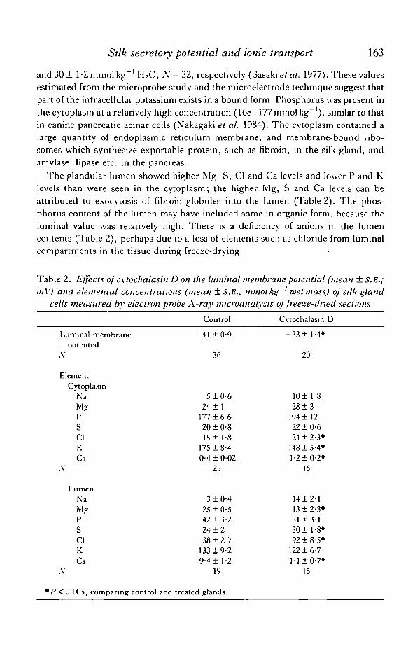
Silk secretory potential and ionic transport 163
and 30 ± 1-2 mmol kg"1 H2O, A'= 32, respectively (Sasaki et al. 1977). These valuesestimated from the microprobe study and the microelectrode technique suggest thatpart of the intracellular potassium exists in a bound form. Phosphorus was present inthe cytoplasm at a relatively high concentration (168—177 mmol kg" ), similar to thatin canine pancreatic acinar cells (Nakagaki et al. 1984). The cytoplasm contained alarge quantity of endoplasmic reticulum membrane, and membrane-bound ribo-somes which synthesize exportable protein, such as fibroin, in the silk gland, andamylase, lipase etc. in the pancreas.
The glandular lumen showed higher Mg, S, Cl and Ca levels and lower P and Klevels than were seen in the cytoplasm; the higher Mg, S and Ca levels can beattributed to exocytosis of fibroin globules into the lumen (Table 2). The phos-phorus content of the lumen may have included some in organic form, because theluminal value was relatively high. There is a deficiency of anions in the lumencontents (Table 2), perhaps due to a loss of elements such as chloride from luminalcompartments in the tissue during freeze-drying.
Table 2. Effects of cytochalasin D on the luminal membrane potential (mean ± S.E.;mV) and elemental concentrations (mean ±S.E.; mmol kg~' wet mass) of silk gland
cells measured by electron probe X-ray microanalysis of freeze-dried sections
Luminal membranepotential
A'
ElementCytoplasm
NaMgPSClKCa
A'
LumenNaMgPSClKCa
A-
*P<0-005, comparing control
Control
-41 ±0-9
36
5 ±0-624 ±1
177 ±6-620 ±0-815 ± 1-8
175 ±8-40-4 ±002
25
3 ±0-425 ±0-542 ±3-224 ±238 ±2-7
133 ±9-29-4 ±1-2
19
and treated glands.
Cytochalasin D
-33 ±1-4*
20
10 ± 1-828±3
194 ±1222 ±0-624 ± 2-3*
148 ±5-4*l-2±0-2*
15
14±2113 ±2-3*31 ± 3-130 ±1-8*92 ±8-5*
122 ±6-711 ±0-7*
15

164 I. NAKAGAKI AND S. SASAKI
Fig. S. Conventional electron micrographs of perinuclear cytoplasm (A) and luminalcytoplasm (B) of posterior silk gland cells, and scanning transmission electronmicrograph of a freeze-dried thin section of posterior silk gland cells (C). (A)Microtubules run from basal to luminal membrane in the cytoplasm, and mitochondria(m) and fibroin globules (g) can be seen in close proximity to the microtubules(arrowheads); n, nucleus. X 13 500. (B) A circular microtubule system (arrowheads) canbe seen in the luminal cytoplasm, and numerous fibroin globules (g) are visible; /, lumen.X6500. (C) Scanning electron micrographs of freshly frozen, dried, thin sections of aposterior silk gland cell showing nucleus (/;), nucleolus and cytoplasm (c). X4S00.
In the X-ray spectra of the cytoplasm, the K concentration increased when thegland was treated with serotonin (5-HT) and, following treatment with methoprene,the concentration of cytoplasmic Ca increased slightly whereas the K concentrationdecreased (Fig. 7; Table 1).
After cytochalasin D treatment, [Cl] and [Ca] in the cytoplasm increased, and[Cl] in the lumen increased (Fig. 8; Table 2). These results suggest that thetranscellular Cl~ flow from the basal extracellular space to the lumen of the posteriorsilk gland occurs as a result of the secretory state of the gland cells.
The dry mass fraction was estimated to be 25 % in the cytoplasm and 34% in theglandular lumen, using frozen hydrated and dehydrated sections of the posterior silk

Silk secretory potential and ionic transport 165
Fig. 6. Electron micrographs of the basal (A) and luminal (B) portion of the posteriorsilk gland cells stained by the adenosine triphosphatase cytochemical reaction. Reactionproducts of the ATPase activity are found on the membrane of microvilli in the luminalportion of the cell and can also be seen on the lateral membrane of the cells (B inset).There are no reaction products on the basal plasma membrane (A), bm, basementmembrane; m, mitochondria; /, lumen. A X17000; B X17000; inset, XllOOO.

166 I. NAKAGAKI AND S. SASAKI
glands, in the resting state. The dry mass fraction of the 5-HT-treated cytoplasmincreased slightly, and this increase was less than that seen in the blowfly salivaryglands (Gupta & Hall, 1981). The dry mass fractions of cytoplasm treated withmethoprene and cytochalasin and of lumen treated with cytochalasin remainedunchanged.
NaMgPSCI KCa• I i t ' ' i
-7ANaMg PSCI KCa NaMg PSCI KCa
03 04keVPig. 7. Energy dispersive X-ray spectra from cytoplasm of normal (A), and 5-HT- (B)and methoprene- (C) treated gland cells. Non-fixed, freeze-dried thin sections wereobtained from freshly frozen posterior silk glands. Note the higher K peak in thespectrum obtained from the gland treated with 5-HT and the lower K peak in thespectrum obtained from the methoprene-treated gland.
NaMg PSCI KCa NaMg PSCI KCaiiiiilmtIlllllllll
NaMg PSCI KCa NaMgPSCI KCa
04 0 01 02 03 04keV
Fig. 8. Energy dispersive X-ray spectra of the cytoplasm (A,B) and glandular lumen(C,D) of freshly freeze-dried thin sections from normal (A,C) and cytochalasin-treated(B,D) gland cells. Note the higher Cl peaks in the spectra of cytoplasm and lumen treatedwith cvtochalasin D.

Silk secretory potential and ionic transport 167
Measurements of elements in whole tissues
Using the flame photometer and a chloridometer, we measured the Na+, K+ andCl~ concentrations in the resting posterior silk glands. Levels in the whole glandswere 13 ±2-1, 108 ± 6-5 and 20 ± 3-5 (±s.D.) mmol kg"1 wet tissue, ,Y= 14, re-spectively. The lumen is partitioned into regions occupied by newly dischargedfibroin, a thin layer of fibroin, and a columnar fibroin filled with material of moderatedensity (Sasaki & Tashiro, 1976). The ion concentrations in the part of the lumenfilled with columnar fibroin were: Na+, 16 ± 3-4; K+, 35 ±5-7; Cl", 45 ± 5-3 mmolkg"1. The K+ value given here is very low compared with the data for the lumenmeasured by X-ray microanalysis. This large discrepancy indicates that themicroprobe analysis measured the peripheral parts of the lumen close to the apicalcytoplasm: as a result, some of the microvilli were included and/or this layer mayhave a large K+ concentration gradient. The concentrations in the cellular part,which remains after removal of columnar fibroin from the gland, were Na+, 6 ± 1-4;K+, 163±5-8;C1", 18 ± 2-3 (±S.D.) mmol kg"1 wet tissue, AT= 14.
DISCUSSION
The posterior silk gland consists of very large cells highly specialized for synthesis,intracellular transport and secretion of protein (Sasaki & Tashiro, 1976; Sasaki et al.1981). There are many similarities between protein production in the silk gland andin the pancreatic exocrine gland. Both preparations have all the structural features ofa typical protein-secreting gland (Palade, 1975). The posterior silk gland has a largelumen, which facilitates direct measurements of membrane potentials and ionicconcentrations in the various compartments, such as cytoplasm and lumen, duringsecretory states. We investigated the mechanism of secretion of the posterior silkgland, using electron probe X-ray microanalysis, coupled with studies usingmicroelectrode techniques.
Intracellular and transcellular recordings from the posterior silk gland cells in thenormal state suggest that the plasma membrane of the gland cells is permeable to K+
and Cl~, and that an anion such as Cl~ is transported passively and/or secondarilyactively from the basal extracellular space to the gland lumen, in normal secretorystates. The presence of adenosine triphosphatase activity, which is located on theluminal plasma membrane, also suggests that the posterior silk gland cells may have acation pump, such as Na+,K+-ATPase and/or H + ,K+-ATPase, on the luminalsurface, as the extracellular [Na+] was low and the luminal pH was shown to beacidic using ion-selective microelectrodes: cytoplasm, pH7-l±0-09 (±S.D.); lu-men, pH 6-2 ±0-1 (±S.D.) A' = 5. This cation pump would create the driving forcefor secondary active Cl~ transport. The K+ channel on the luminal membranesupplies K+ for this pump. Gupta & Hall (1983) proposed that in cockroach salivaryglands, a cation pump, such as Na+,K+-ATPase, is located on the luminal plasmamembrane and supplies the driving force for intracellular Cl" transport from theserosal portion to the lumen of the acinar P-cells. However, the ionic composition inthe luminal contents of posterior silk gland is similar to that of the goblet cavity in the

168 I. NAKAGAKI AND S. SASAKI
K+-secreting cells of the larval midgut in Manduca sexta (Dow, Gupta, Hall &Harvey, 1984; Gupta, Dow, Hall & Harvey, 1985). The evidence suggests that theremay be an electrogenic K+ pump on the apical membrane and a conventionalNa+,K+-pump on the basolateral plasma membrane. Greger, Schlatter & Lang(1983) proposed that in the thick ascending limbs of Henle's loop, the intracellularCP transport of tubular cells could be explained by a secondary active process andthat the Na+, K+-ATPase located on the peritubular plasma membrane would supplythe driving force for CP transport. Some workers, studying parietal cells of the rat orrabbit gastric mucosa, have found that H+,K+-ATPase, which is located on thetubulovesicular membrane and/or plasma membrane of microvilli of the secretorycanaliculus, could provide the driving force for CP transport into the lumen (Rabonet al. 1983). Although the parietal cells of the vertebrate stomach secrete isotonicHC1 at pH 2-0, the gastric mucosa also generates a potential difference that is lumen-negative, as is the case in the posterior silk gland. The membranes of tubulovesiclessurrounding the secretory canaliculi of gastric parietal cells have H+,K+-ATPase,K+ and CP channels. It is thought that stimulation of these channels opens andactivates the pump, and that Ca2+ is simultaneously released from an intracellularstore, such as the tubulovesicular structures and/or the luminal membrane of thesecretory canaliculi. Finally, this Ca2+ release enhances the exocytosis of thetubulovesicles into the canaliculus lumen, a process in which the cytoskeletal systemis involved (Tsunoda & Mizuno, 1985).
In our study, which used 5-HT as the stimulus, the basal membrane washyperpolarized and the intracellular K+ concentration was increased. We attributethese events to increases in the K+ permeability of the basal cell membrane and in theactive transport of K+, following stimulation of the K+-pump on the apicalmembrane and the Na + /K + exchange pump on the basal cell membrane. This cationpump may be present in other epithelia, such as Calliphora salivary glands andMalpighian tubules (Berridgee* al. 1975; Gupta, Hall, Maddrell & Moreton, 1976).
The basal membrane potential of the posterior silk gland cells became depolarizedin the presence of the juvenile hormone, methoprene. This agent effectivelyincreases synthesis and secretion of fibroin, resulting in the production of largecocoons at the final stage of the fifth instar. There are similarities between thechemical formula of methoprene and of unsaturated fatty acids (Henrick et al. 1975);this hormone may act on the gland cells in a manner similar to that of someunsaturated fatty acids such as arachidonic acid and/or some prostaglandins.Methoprene may affect the exocrine gland cells by acting as a transmitter and/or apeptide hormone, as do acetylcholine and gastrin-CCK-PZ. In the present micro-probe study using methoprene, we observed an influx of Ca2+, an efflux of K+, andan increase in CP transport into the lumen in silk gland cells. The Ca2+ influx andthe CP efflux via apical membranes explain the depolarization of the plasmamembrane and the lumen-negative transepithelial potential. The rise in intracellularCa2+ concentration also enhances the exocytotic mechanism of the secretory granulesof fibroin in the luminal plasma membrane, which is carried out by the microtubule-microfilament systems of the cells. These data also suggest that the Ca2+-regulated

Silk secretory potential and ionic transport 169
Cl~ transport into the lumen is stimulated by treatment with the juvenile hormone,methoprene, following which the voltage-dependent and Ca2+-regulated K+ chan-nels in the basolateral plasma membranes are opened (Maruyama, Gallacher &Petersen, 1983; Petersen & Maruyama, 1984; Trautman & Marty, 1984; Marty, Tan& Trautman, 1984).
Cytochalasin B or D accelerates exocytosis of the secretory granules of fibroin intothe luminal portion of the gland cells and enhances the secretion of fibroin into thelumen of the posterior silk gland (Sasaki et al. 1981). In this and previous studies,intracellular recordings of the luminal plasma membrane potential of the gland cellsshowed a depolarization and the input resistance of the membrane decreased whenthe gland was treated with cytochalasin B or D (Sasaki & Nakagaki, 1979). In thepresent study, we have shown that the intracellular Ca2+ concentration increased andthat Cl~ transport into the lumen occurred in gland cells treated with cytochalasin D.The increase in cytoplasmic Ca + concentration may be important in the regulationby Ca + of transcellular Cl~ transport. Moreover, the increase in cytoplasmic [Ca2+]and the decrease in luminal [Ca +] indicate that Ca + is released from fibroinglobules, which serve as an intracellular store for Ca . Further, this Cl~ transport isthought to be a secondary active process, the driving force for which is derived fromNa+,K+-ATPase located on the lateral plasma membrane or a K+ pump such asH+,K+-ATPase, and an electrogenic pump on the luminal plasma membrane, as inthe case of salivary glands and gastric mucosa (Berridge et al. 1975; Gupta & Hall,1983; Rabon et al. 1983). This Cl~ transport system would explain our electro-physiological data and may be regulated by cytoplasmic Ca2+, derived mainly fromintracellular Ca + stores such as secretory granules and/or endoplasmic reticulum(Sasaki el al. 1983; Streb, Irvine, Berridge & Shultz, 1983; Nakagaki et al. 1984;Berridge & Irvine, 1984). This Ca2+ would enhance exocytotic mechanisms, and alsoaffect the microtubule-microfilament systems in the luminal portion of the glandcells.
We thank Dr Y. Tashiro of Kansai Medical University and Drs Y. Imai and M.Fujimoto of Osaka Medical College for advice and encouragement, Drs Y. Okadaand W. Tsuchiya of Kyoto University and Dr M. Kitano of Kyoto University ofIndustrial Arts and Textile Fibers and Dr B. Sakaguchi of Kyushu University foruseful discussion and the gifts of specimens. This work was supported by a grant-in-aid for scientific research from the Ministry of Education, Science and Culture,Japan.
REFERENCESAKAI, H., KlGUCHl, K. & MORI, K. (1971). Increased accumulation of silk protein accompanying
JH-induced prolongation of larval life in Bombyx mori L. Appl. Ent. Zool. 6, 218-220.AKAI, H., KIGUCHI, K. & MORI, K. (1973). The influence of juvenile hormone on the growth and
metamorphosis of Bombyx larvae. Bull, sericult. Exp. Slat. 25, 287-305 (in Japanese).BAUDIN, H., STOCK, K., VINCENT, D. & GRENIER, J. F. (1975). Microfilaments system and
secretion of enzyme in the exocrine pancreas. Effect of cytochalasin B._7. CellBiol. 66, 165-181.

170 I. NAKAGAKI AND S. SASAKI
BERRIDGE, M. J. & IRVINE, R. F. (1984). Inositol trisphosphate, a novel second messenger incellular signal transduction. Sature, Land. 312, 315-321.
BERRIDGE, M. J., LINDLEG, B. D. & PRINCE, \V. T. (1975). Membrane permeability changesduring estimation of isolated salivary glands of Calliphora by 5-hydroxytryptamine. jf. Phvswl.,Lond. 244, 549-567.
DOUGLAS, W. \V. & SORIMACHI, M. (1972). Colchicine inhibits adrenal medullary secretion evokedby potassium. Br.J. Phannac. 45, 129-132.
Dow, J. A. T., GUPTA, B. L., HALL, T. A. & HARVEY, \V. R. (1984). X-ray microanalysis ofelements in frozen-hydrated sections of an electrogenic K+-transport system. The posteriormidgut of tobacco hornworm (Manduca sexta) in vivo andin vitro. J. Membr. Biol. 77, 223-241.
GlNSBORG, B. L. & HOUSE, C. R. (1980). Stimulus-response coupling in gland cells. .4. Rev.Biophys. Bioeng. 9, 55-80.
GREGER, R., SCHLATTER, E. & LANG, F. (1983). Evidence for electroneutral sodium chloridecotransport in the cortical thick ascending limb of Henle's loop of rabbit kidney. Pflugers Arch.ges. Physiol. 396, 308-314.
GUPTA, B. L., BERRIDGE, M. J., HALL, T. A. & MORETON, R. B. (1978). Electron microprobc andion-selective microelectrode studies of fluid secretion in the sahvan' glands of Calliphora. J. exp.Biol. 72, 261-284.
GUPTA, B. L., DOW, J. A. T., HALL, T. A. & HARVEY, \V. R. (1985). Electron probe X-raymicroanalysis of the effects of Bacillus thuringiensis var kurstaki crystal protein insecticide onions an electrogenic K-transporting epithelium of the larval midgut in the lepidopteran,Manduca sexta, in vitro. J. Cell Sci. 74, 137-152.
GUPTA, B. L. & HALL, T. A. (1981). Microprobe analysis of fluid-transporting epithelia. Evidencefor local osmosis and solute recycling. In Water Transport Acivss Epithelia. Alfred BenzonSymposium 15 (ed. H. H. Ussing & N. A. Thorn), pp. 17-35. Copenhagen, Tokyo:Munksgaard.
GUPTA, B. L. & HALL, T . A. (1983). Ionic distribution in dopainine-stimulated NaCI fluid-secreting cockroach salivary glands. Am.jf. Physiol. 244, R176—R186.
GUPTA, B. L., HALL, T. A., MADDRELL, S. H. P. & MORETON, R. B. (1976). Distribution of ionsin a fluid-transporting epithelium determined by electron probe X-ray microanalysis. Sature,Lond. 264, 284-287.
HENRICK, C. A., WILLY, W. E., MCKEAN, D. R., BAGGIOLINI, E. & SIDDALL, B. (1975).
Approaches to the synthesis of the insect juvenile hormone analog ethyl-3,7,ll-trimethyl-2,4,-dodecadienoate and its photochemistry. J. org. Client. 40, 8-14.
KANNO, T. (1972). Calcium dependent amylase release and elcctrophysiological measurements incells of the pancreas, jf. Physiol., Lond. 226, 353-371.
LACY, P., HOWELL, S. L., YOUNG, D. A. & FINK, C. J. (1968). New hypothesis of insulinsecretion. Sature, Lond. 219, 1177-1179.
MARCHESI, V. T. & PALADE, G. E. (1967). The localization of Mg-Na-K activated adenosinetriphosphatase on red cell ghost membranes. J. Cell Biol. 35, 385-404.
MARTY, A., TAN, Y. P. & TRAUTMANN, A. (1984). Three types of calcium-dependent channels inrat lacrimal glands. J. Physiol., Lond. 357, 293-325.
MARUYAMA, Y., GALLACHER, D. V. & PETERSEN, O. H. (1983). Voltage and Ca2+-activated K+
channel in baso-lateral acinar cell membranes of mammalian salivary glands. Sature, Lond. 302,827-829.
MATTHEWS, E. K., PETERSEN, O. H. & WILLIAMS, J. A. (1973). Pancreatic acinar cellsacetylcholine-induced membrane depolarization, calcium efflux and amylase release, jf. Physiol.,Lond. 234, 689-701.
NAKAGAKI, I., GOTO, T., SASAKI, S. & IMAI, Y. (1978). Histochemical and cytochemicallocalization of (Na+-K+)-activated adenosine triphosphatase in the acini of dog submandibulargland. J. Histochem. Cytochem. 26, 835-845.
NAKAGAKI, I., SASAKI, S., SHIGUMA, M. & IMAI, Y. (1984). Distribution of elements in thepancreatic exocrine cells determined by electron probe X-ray microanalysis. Pfliigers Arch. ges.Physiol. 401, 340-345.
PALADE, G. E. (1975). Intracellular aspects of the process of protein synthesis. Science 189,347-358.

Silk secretory potential and ionic transport 171
PETERSEN, O. H. (1980). The electrophysiology of gland cells. Monographs of the PhysiologicalSociety, no. 36. London: Academic Press.
PETERSEN, O. H. & MARUYAMA, Y. (1984). Calcium-activated potassium channels and their role insecretion. Xature, Loud. 307, 693-6%.
PoiSNER, A. M. & BANERJEE, K. (1971). A possible role of microtubules on catecholamine releasefrom the adrenal medulla: effect of chochicine, vinca alkaloids and deuterium oxide. J. Phannac.exp. Then 177, 102-108.
RABON, E., CUPPOLETTI, J., MALINOWSKA, D., SMOLKA, A., HELANDER, H. F., MENDLEIN, J. &
SACHS, G. (1983). Proton secretion by the gastric parietal cell. J. exp. Biol. 106, 119-133.ROSSIGNOL, B., HERMAN, G. & KERGER, G. (1972). Inhibition by colchicine of carbamylcholine
induced glyoprotein secretion by submaxillary gland. A possible mechanism of cholinergicinduced protein secretion. FEBS Letts 21, 189-194.
SASAKI, S., MORI, H., IMAI, Y., TSUCHYA, \Y\, SHIMAZU, Y. & TASHIRO, Y. (1977). Fibroin
secretion and the membrane potential changes of posterior silk gland cells. J'. phvsiol. Soc. Japan39, 238 (in Japanese).
SASAKI, S. & NAKAGAKI, I. (1979). Secretory mechanism of fibroin, a silk protein, in the posteriorsilk gland cells of Bom byx won. Membrane Biochem. 3, 37-47.
SASAKI, S., NAKAGAKI, I., MORI, H. &IMAI, Y. (1983). Intracellular calcium store and transport ofelements in acinar cells of the salivary gland determined by electron probe X-ray microanalysis.Jap. J. Phvsiol. 33, 69-83.
SASAKI, S., NAKAJIMA, E., FUJII-KURIYAMA, Y. & TASHIRO, Y. (1981). Intracellular transport andsecretion of fibroin in the posterior silk gland of the silkworm Bombyx won. J. Cell Sci. 50,19-44.
SASAKI, S. &TASHIRO, Y. (1976). Studies on the posterior silk gland of the silkworm Bombyx mori.VI. Distribution of microtubules in the posterior silk gland cells. J . Cell Biol. 71, 565-574.
STREB, H., IRVINE, R. F., BERRIDGE, M. J. & SCHULTZ, I. (1983). Release of Ca2+ from anonmitochondrial intracellular store in pancreatic acinar cells by inositol-l,4,5-trisphosphate.Xature, Loud. 306, 67-69.
TRAUTMANN, A. & MARTY, A. (1984). Activation of Ca-dependent channels by carbamylcholine inrat lacnmal glands. Pivc. natn. Acad. Sci. LJ.SA. 81, 611-615.
TSUNODA, Y. & MIZUNO, T. (1985). Participation of the microtubular-microfilamentous systemin intracellular Ca2+ transport and acid secretion in dispersed parietal cells. Biochim. biophvs.Ada 855, 186-188.
WACHSTEIN, M. & MEISEL, E. (1957). Histochemistry of hepatic phosphatases at a physiologicalpH. Am. J. din. Path. 27, 13-23.
WYATT, S. S. (1956). Culturein vitro of tissue from the silkworm, Bombvx mori, L.J. i>en. Phvsiol.39,841-852.