Embed Size (px)
Citation preview
8/3/2019 Sect 2.3B Tissue Healing
http://slidepdf.com/reader/full/sect-23b-tissue-healing 1/24
Properties of Ligamentous and Tendinous Tissues
Change in Tissue Length
(Strain or % Deformation)
Deformation Force(Sress or Load)
x cross sectional area
2x cross sectional area
2x length
x length
slope
8/3/2019 Sect 2.3B Tissue Healing
http://slidepdf.com/reader/full/sect-23b-tissue-healing 2/24
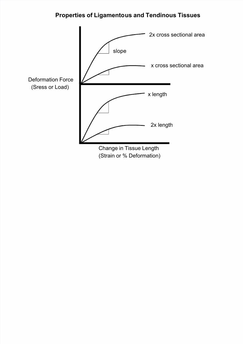
Ligament Injury
Ligament - fibrous dense connective tissue - binds bones
injuries to these structures may be a precursor to osteoarthritis
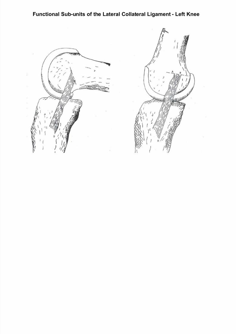
has functional subunits that tighten or loosen depending on joint position
is not densely innervated or densely vascularized
do contain some blood vessels and nerves in outer covering (epiligament)
do contain proprioceptors
do transmits pain signals via type C fibersin bone-ligament-bone structures, ligament is the weakest link
weakest near ligament insertion (adolescent & osteoperotic exceptions)
ligaments are not readily weakened by inactivity (takes many weeks)
ligaments show only a 10% - 20% u in tensile strength with exercise
It is currently not known whether any modalities aid in ligament healingsurgical repair not done unless ends are significantly far apart
length of repair scar does not affect final functionality or tensile strength
unless ends are far apart: r extra-long scar r d joint stability & u joint laxity
ACL tears most often result in ends unopposed r surgery required
surgical repair restores only about 80% - 90% of original tensile strength
8/3/2019 Sect 2.3B Tissue Healing
http://slidepdf.com/reader/full/sect-23b-tissue-healing 3/24
Functional Sub-units of the Lateral Collateral Ligament - Left Knee
8/3/2019 Sect 2.3B Tissue Healing
http://slidepdf.com/reader/full/sect-23b-tissue-healing 4/24
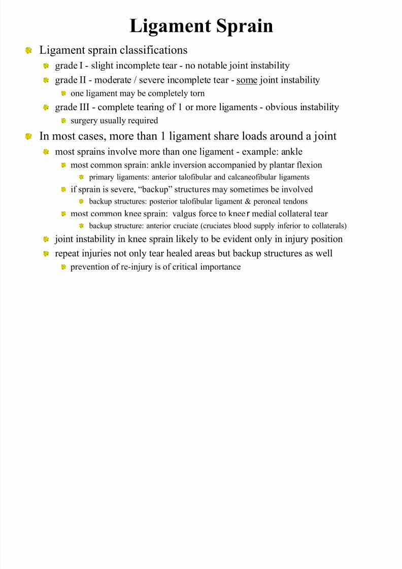
Ligament SprainLigament sprain classifications
grade I - slight incomplete tear - no notable joint instabilitygrade II - moderate / severe incomplete tear - some joint instability
one ligament may be completely torn
grade III - complete tearing of 1 or more ligaments - obvious instability
surgery usually required
In most cases, more than 1 ligament share loads around a joint
most sprains involve more than one ligament - example: ankle
most common sprain: ankle inversion accompanied by plantar flexion
primary ligaments: anterior talofibular and calcaneofibular ligaments
if sprain is severe, ³backup´ structures may sometimes be involved
backup structures: posterior talofibular ligament & peroneal tendons
most common knee sprain: valgus force to knee r medial collateral tear
backup structure: anterior cruciate (cruciates blood supply inferior to collaterals)
joint instability in knee sprain likely to be evident only in injury position
repeat injuries not only tear healed areas but backup structures as well
prevention of re-injury is of critical importance
8/3/2019 Sect 2.3B Tissue Healing
http://slidepdf.com/reader/full/sect-23b-tissue-healing 5/24
tibia
tibia
fibula
navicular
navicular
calcaneus
calcaneus
talus
cuniforms
metatarsals
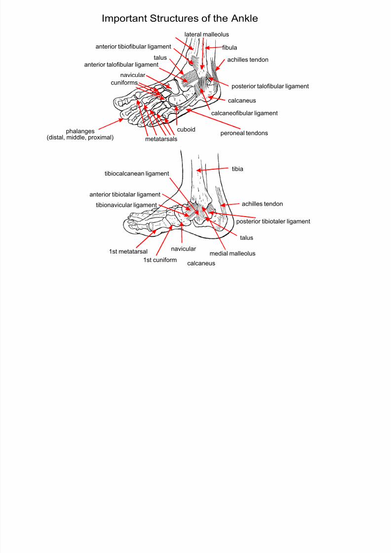
lateral malleolus
anterior talofibular ligament
calcaneofibular ligament
achilles tendon
achilles tendon
cuboid peroneal tendons
posterior talofibular ligament
anterior tibiofibular ligament
phalanges(distal, middle, proximal)
medial malleolus
1st metatarsal
1st cuniform
talus
anterior tibiotalar ligament
tibionavicular ligament
tibiocalcanean ligament
posterior tibiotaler ligament
Important Structures of the Ankle
8/3/2019 Sect 2.3B Tissue Healing
http://slidepdf.com/reader/full/sect-23b-tissue-healing 6/24
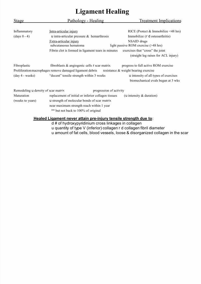
Stage Pathology - Healing Treatment Implications
Inflammatory Intra-articular injury RICE (Protect & Immobilize <48 hrs)(days 0 - 4) u intra-articular pressure & hemarthrosis Immobilize (r d osteoarthritis)
Extra-articular injury NSAID drugs
subcutaneous hematoma light passive ROM exercise (>48 hrs)
Fibrin clot is formed in ligament tears in minutes exercises that ³cross´ the joint
(straight leg raises for ACL injury)
Fibroplastic fibroblasts & angiogenic cells r scar matrix progress to full active ROM exerciseProliferationmacrophages remove damaged ligament debris resistance & weight bearing exercise
(day 4 - weeks) ³decent´ tensile strength within 3 weeks u intensity of all types of exercises
biomechanical evals began at 3 wks
Remodeling u density of scar matrix progression of activity
Maturation replacement of initial or inferior collagen tissues (u intensity & duration)
(weeks to years) u strength of molecular bonds of scar matrix
near maximum strength reach within 1 year
** but not back to 100% of original
Ligament Healing
Healed Ligament never attain pre-injury tensile strength due to:
d # of hydroxypyridinium cross linkages in collagen
u quantity of type V (inferior) collagen r d collagen fibril diameter
u amount of fat cells, blood vessels, loose & disorganized collagen in the scar
8/3/2019 Sect 2.3B Tissue Healing
http://slidepdf.com/reader/full/sect-23b-tissue-healing 7/24
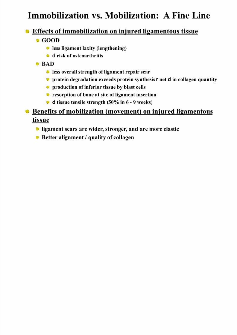
Immobilization vs. Mobilization: A Fine Line
Effects of immobilization on injured ligamentous tissue
GOOD
less ligament laxity (lengthening)
d risk of osteoarthritis
BAD
less overall strength of ligament repair scar
protein degradation exceeds protein synthesis r net d in collagen quantity
production of inferior tissue by blast cells
resorption of bone at site of ligament insertion
d tissue tensile strength (50% in 6 - 9 weeks)
Benefits of mobilization (movement) on injured ligamentous
tissue
ligament scars are wider, stronger, and are more elastic
Better alignment / quality of collagen
8/3/2019 Sect 2.3B Tissue Healing
http://slidepdf.com/reader/full/sect-23b-tissue-healing 8/24
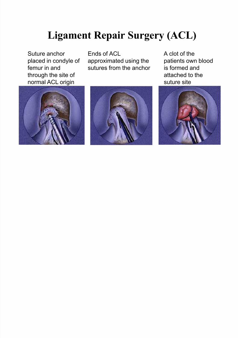
Ligament Repair Surgery (ACL)Suture anchor
placed in condyle of
femur in and
through the site of
normal ACL origin
Ends of ACL
approximated using the
sutures from the anchor
A clot of the
patients own blood
is formed and
attached to the
suture site
8/3/2019 Sect 2.3B Tissue Healing
http://slidepdf.com/reader/full/sect-23b-tissue-healing 9/24
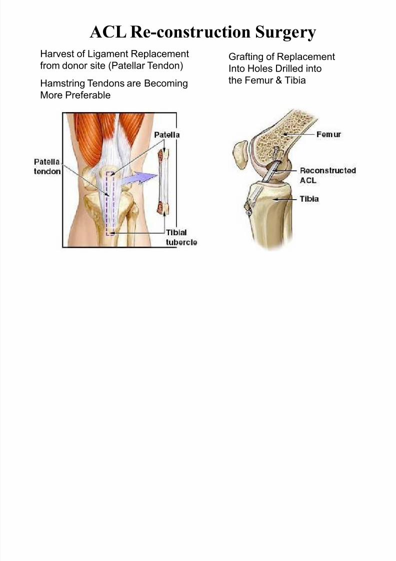
ACL Re-construction SurgeryHarvest of Ligament Replacement
from donor site (Patellar Tendon)
Hamstring Tendons are Becoming
More Preferable
Grafting of Replacement
Into Holes Drilled intothe Femur & Tibia
8/3/2019 Sect 2.3B Tissue Healing
http://slidepdf.com/reader/full/sect-23b-tissue-healing 10/24
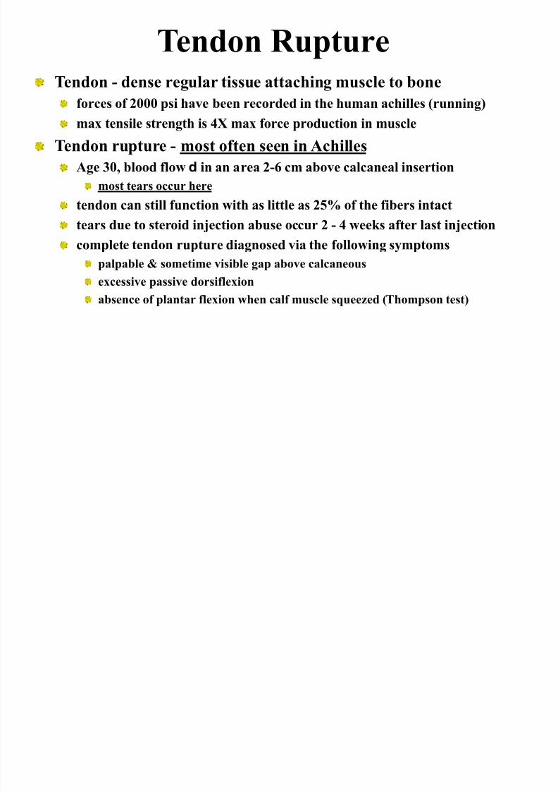
Tendon RuptureTendon - dense regular tissue attaching muscle to bone
forces of 2000 psi have been recorded in the human achilles (running)
max tensile strength is 4X max force production in muscle
Tendon rupture - most often seen in Achilles
Age 30, blood flow d in an area 2-6 cm above calcaneal insertion
most tears occur here
tendon can still function with as little as 25% of the fibers intact
tears due to steroid injection abuse occur 2 - 4 weeks after last injection
complete tendon rupture diagnosed via the following symptoms
palpable & sometime visible gap above calcaneous
excessive passive dorsiflexion
absence of plantar flexion when calf muscle squeezed (Thompson test)
8/3/2019 Sect 2.3B Tissue Healing
http://slidepdf.com/reader/full/sect-23b-tissue-healing 11/24
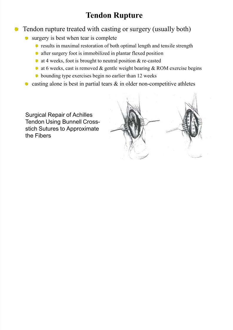
Tendon Rupture
Tendon rupture treated with casting or surgery (usually both)
surgery is best when tear is completeresults in maximal restoration of both optimal length and tensile strength
after surgery foot is immobilized in plantar flexed position
at 4 weeks, foot is brought to neutral position & re-casted
at 6 weeks, cast is removed & gentle weight bearing & ROM exercise begins
bounding type exercises begin no earlier than 12 weekscasting alone is best in partial tears & in older non-competitive athletes
Surgical Repair of AchillesTendon Using Bunnell Cross-
stich Sutures to Approximate
the Fibers
8/3/2019 Sect 2.3B Tissue Healing
http://slidepdf.com/reader/full/sect-23b-tissue-healing 12/24
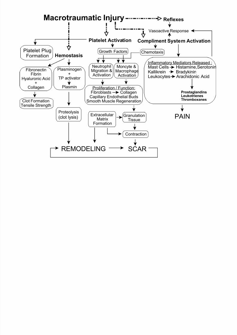
Macrotraumatic Injury
FibronectinFibrin
Hyaluronic Acid+
Collagen
Platelet Activation
Chemotaxis
Plasminogen+
TP activator
Plasmin
Growth Factors
NeutrophilMigration & Activation
Moncyte &Macrophage Activation
Proliferation / Function:Fibroblasts Collagen
Capillary Endothelial BudsSmooth Muscle Regeneration
GranulationTissue
Contraction
SCAR
Proteolysis
(clot lysis) Extracellular MatrixFormation
REMODELING
Hemostasis
Compliment System Activation
Inflammatory Mediators Released :
Mast Cells Histamine,SerotoninKallikrein Bradykinin
Leukocytes Arachidonic Acid
ProstaglandinsLeukotrienesThromboxanes
Vasoactive Response
PAIN
Reflexes
Platelet PlugFormation
Clot FormationTensile Strength
8/3/2019 Sect 2.3B Tissue Healing
http://slidepdf.com/reader/full/sect-23b-tissue-healing 13/24
Bone Fractures Most fractures occur to the shaft of long bones
Bone is well vascularized and highlyinnervated
Heals relatively rapidly when ends are
well approximated (6 weeks or less) Healed bone often stronger than original
due to external calcification
8/3/2019 Sect 2.3B Tissue Healing
http://slidepdf.com/reader/full/sect-23b-tissue-healing 14/24
FractureTypes
simple (closed) - little or no bone displacement
compound - fracture ruptures the skin & bone protrudes
green stick - occurs mostly in children whose bones have not calcified or hardened
transverse - crack perpendicular to long axis of the bone - displacement may occur
oblique - diagonal crack across the long axis of the bone - u chance of displacement
spiral - diagonal crack involving a "twisting" of the bone about the longitudinal axis
(occurs in skiing when bindings are too tight)
comminuted (blowout) - "crushing" fracture - more common in elderly - may require
screws, rods, & wires - may cause permanent discrepancy in leg length
impacted - one end of bone is driven up into the other - may result in length discrepancy
depressed - broken bone is pressed inward (skull fracture)
avulsion - fragment of bone is pulled away by tendon (Hip flexors, adductors)
8/3/2019 Sect 2.3B Tissue Healing
http://slidepdf.com/reader/full/sect-23b-tissue-healing 15/24
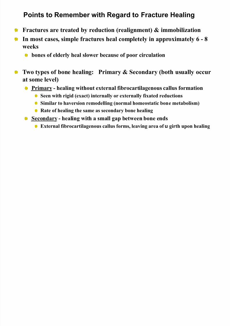
Points to Remember with Regard to Fracture Healing
Fractures are treated by reduction (realignment) & immobilization
In most cases, simple fractures heal completely in approximately 6 - 8weeks
bones of elderly heal slower because of poor circulation
Two types of bone healing: Primary & Secondary (both usually occur
at some level)
Primary - healing without external fibrocartilagenous callus formation
Seen with rigid (exact) internally or externally fixated reductions
Similar to haversion remodelling (normal homeostatic bone metabolism)
Rate of healing the same as secondary bone healing
Secondary - healing with a small gap between bone ends
External fibrocartilagenous callus forms, leaving area of u girth upon healing
8/3/2019 Sect 2.3B Tissue Healing
http://slidepdf.com/reader/full/sect-23b-tissue-healing 16/24
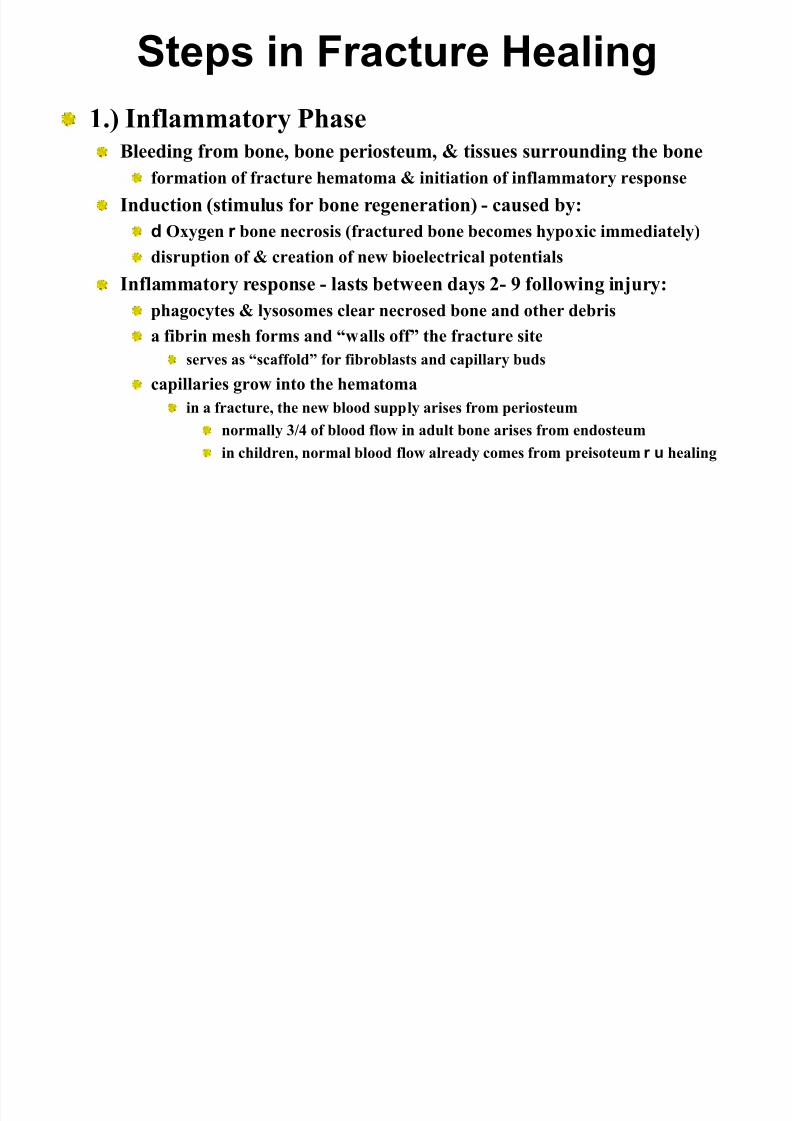
Steps in Fracture Healing
1.) Inflammatory PhaseBleeding from bone, bone periosteum, & tissues surrounding the bone
formation of fracture hematoma & initiation of inflammatory response
Induction (stimulus for bone regeneration) - caused by:
d Oxygen r bone necrosis (fractured bone becomes hypoxic immediately)
disruption of & creation of new bioelectrical potentialsInflammatory response - lasts between days 2- 9 following injury:
phagocytes & lysosomes clear necrosed bone and other debris
a fibrin mesh forms and ³walls off´ the fracture site
serves as ³scaffold´ for fibroblasts and capillary buds
capillaries grow into the hematomain a fracture, the new blood supply arises from periosteum
normally 3/4 of blood flow in adult bone arises from endosteum
in children, normal blood flow already comes from preisoteumr u healing
8/3/2019 Sect 2.3B Tissue Healing
http://slidepdf.com/reader/full/sect-23b-tissue-healing 17/24
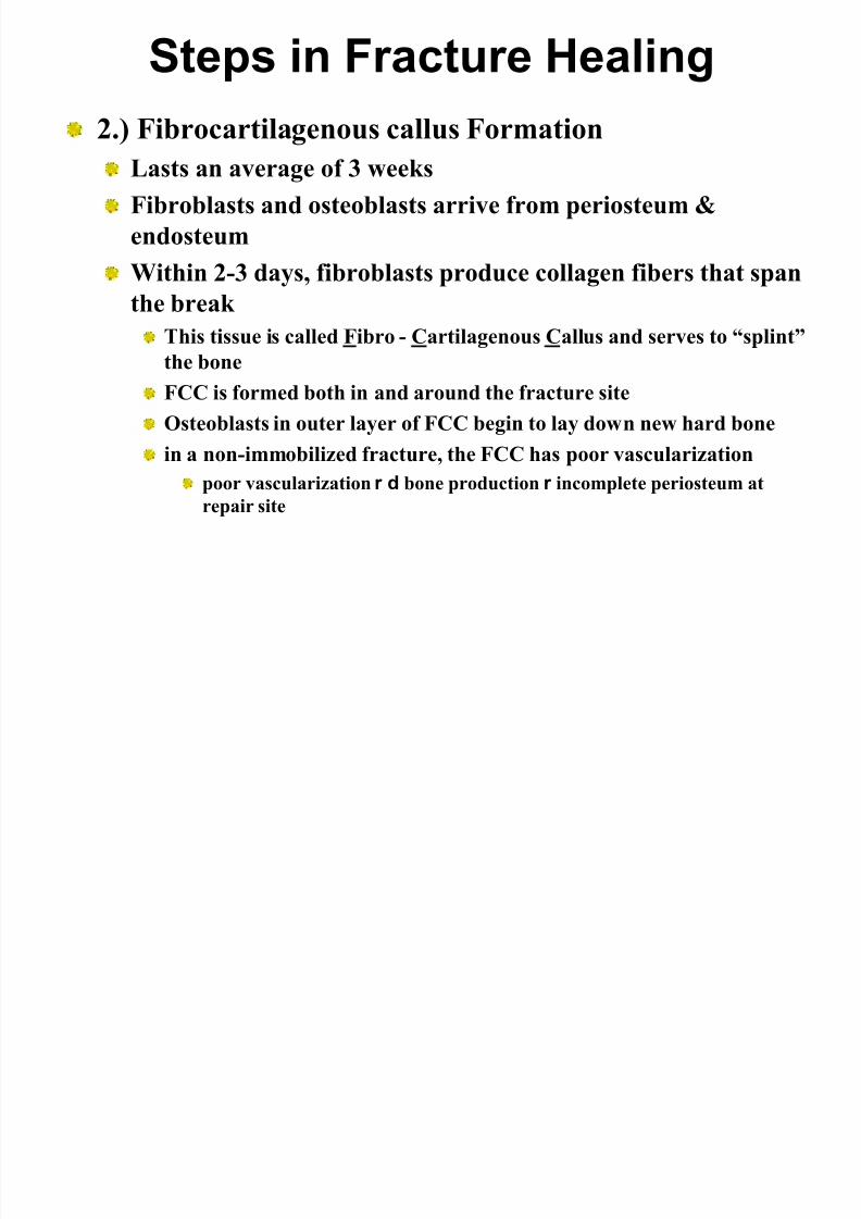
2.) Fibrocartilagenous callus FormationLasts an average of 3 weeks
Fibroblasts and osteoblasts arrive from periosteum &
endosteum
Within 2-3 days, fibroblasts produce collagen fibers that spanthe break
This tissue is called Fibro - Cartilagenous Callus and serves to ³splint´
the bone
FCC is formed both in and around the fracture site
Osteoblasts in outer layer of FCC begin to lay down new hard bonein a non-immobilized fracture, the FCC has poor vascularization
poor vascularization r d bone production r incomplete periosteum at
repair site
Steps in Fracture Healing
8/3/2019 Sect 2.3B Tissue Healing
http://slidepdf.com/reader/full/sect-23b-tissue-healing 18/24
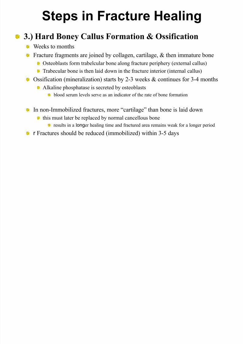
3.) Hard Boney Callus Formation & OssificationWeeks to months
Fracture fragments are joined by collagen, cartilage, & then immature bone
Osteoblasts form trabelcular bone along fracture periphery (external callus)
Trabecular bone is then laid down in the fracture interior (internal callus)
Ossification (mineralization) starts by 2-3 weeks & continues for 3-4 monthsAlkaline phosphatase is secreted by osteoblasts
blood serum levels serve as an indicator of the rate of bone formation
In non-Immobilized fractures, more ³cartilage´ than bone is laid down
this must later be replaced by normal cancellous boneresults in a longer healing time and fractured area remains weak for a longer period
r Fractures should be reduced (immobilized) within 3-5 days
Steps in Fracture Healing
8/3/2019 Sect 2.3B Tissue Healing
http://slidepdf.com/reader/full/sect-23b-tissue-healing 19/24
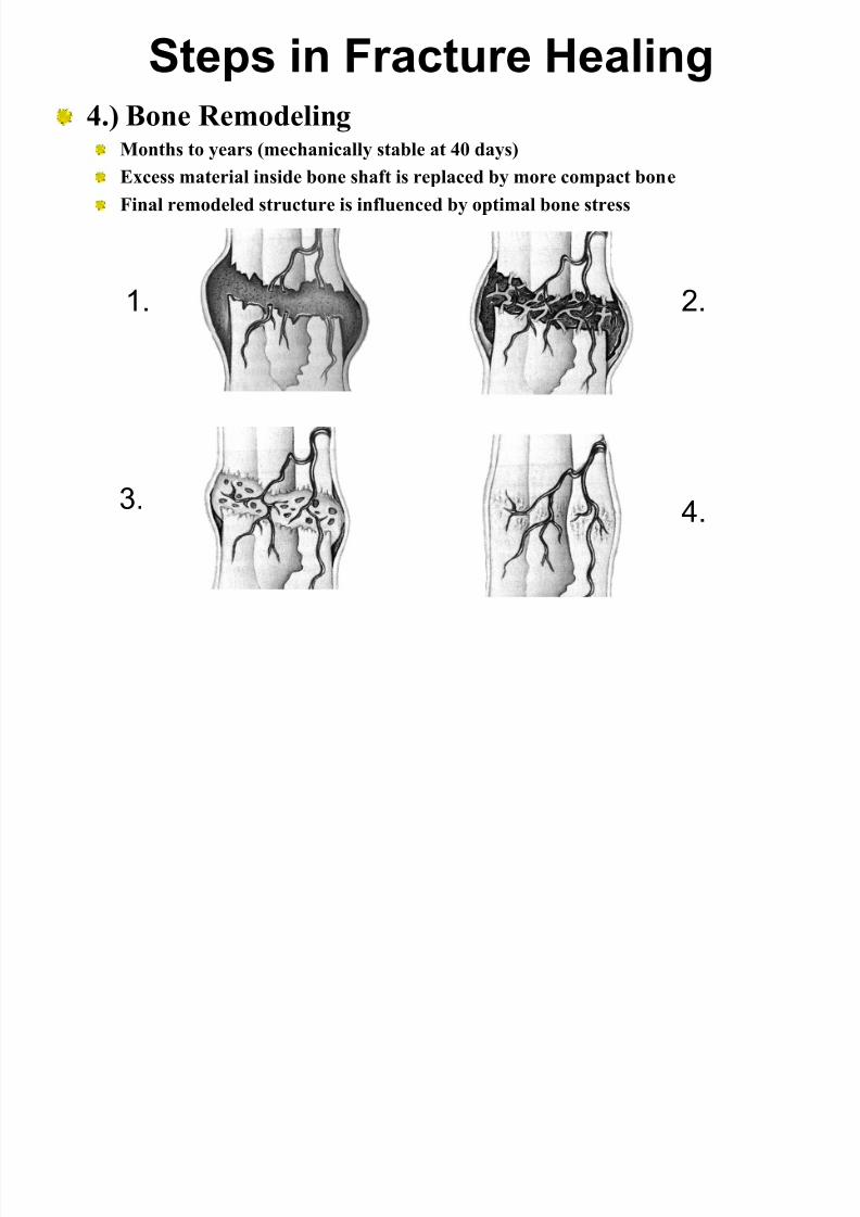
4.) Bone RemodelingMonths to years (mechanically stable at 40 days)
Excess material inside bone shaft is replaced by more compact bone
Final remodeled structure is influenced by optimal bone stress
Steps in Fracture Healing
1. 2.
3. 4.
8/3/2019 Sect 2.3B Tissue Healing
http://slidepdf.com/reader/full/sect-23b-tissue-healing 20/24
Bioelectricity and Fracture HealingBioelectric Factors in Bone Repair & Nonunion
FracturesAreas of growth & repair in fractures have shown to be electronegative
play a major role in induction
stimulate osteoblast activity
compression of fractured bone ends seems to u electronegativity
u electronegativity r u rate of hard bone deposition
strong case for using internal or external fixator
Non-union fractures (fractures that fail to heal within 5 months)
caused by excessive age, contamination (infection), motion at fracture site
treatment 1. electrical stimulation (20 amps for 12 weeks)
implantation of electrodes in the fibrous tissue at fracture site or under skin
treatment 2. bone grafting
harvesting small quantities of bone from a non-critical area (ex: pelvis)
implanting the harvested bone at non-union fracture site
8/3/2019 Sect 2.3B Tissue Healing
http://slidepdf.com/reader/full/sect-23b-tissue-healing 21/24
Immobilization: Cast Disease
Most changes are reversible
Muscle Atrophy
d calcium content in surrounding bone
resorption and weakening of tissues at sites of ligament
attachmentsno stress forces on an immobilized joint r thinning of
articular cartilage
Adhesions r joint stiffness
loss of peripheral autonomic vascular control r hair loss
-shiny mottled skin
sensory dissociation (light touches interpreted as
painful)
8/3/2019 Sect 2.3B Tissue Healing
http://slidepdf.com/reader/full/sect-23b-tissue-healing 22/24
Therapeutic Implications for
Treating FracturesActive ROM exercises to joints above and below
immobilized region
Resistive ROM exercises to muscle groups that are not
immobilized
Once the cast or immobilization device has been
removed:
gentle but progressive resistance exercises of all immobilized
joints
evaluate strength of joint(s) and compare to non-injured
counterparts
return to vigorous activity only after strength discrepency < 15%
8/3/2019 Sect 2.3B Tissue Healing
http://slidepdf.com/reader/full/sect-23b-tissue-healing 23/24
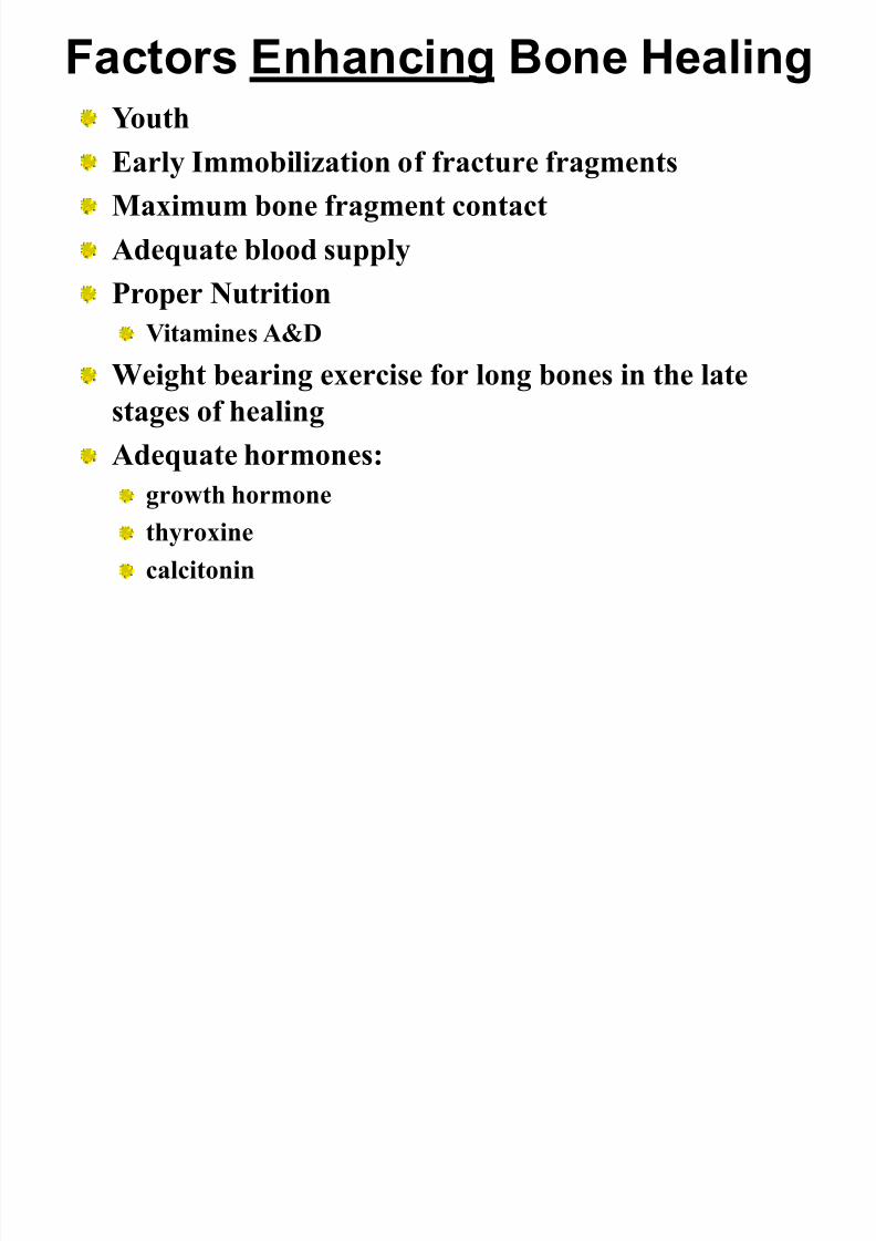
Factors Enhancing Bone HealingYouth
Early Immobilization of fracture fragments
Maximum bone fragment contact
Adequate blood supply
Proper NutritionVitamines A&D
Weight bearing exercise for long bones in the late
stages of healing
Adequate hormones:
growth hormone
thyroxine
calcitonin
8/3/2019 Sect 2.3B Tissue Healing
http://slidepdf.com/reader/full/sect-23b-tissue-healing 24/24
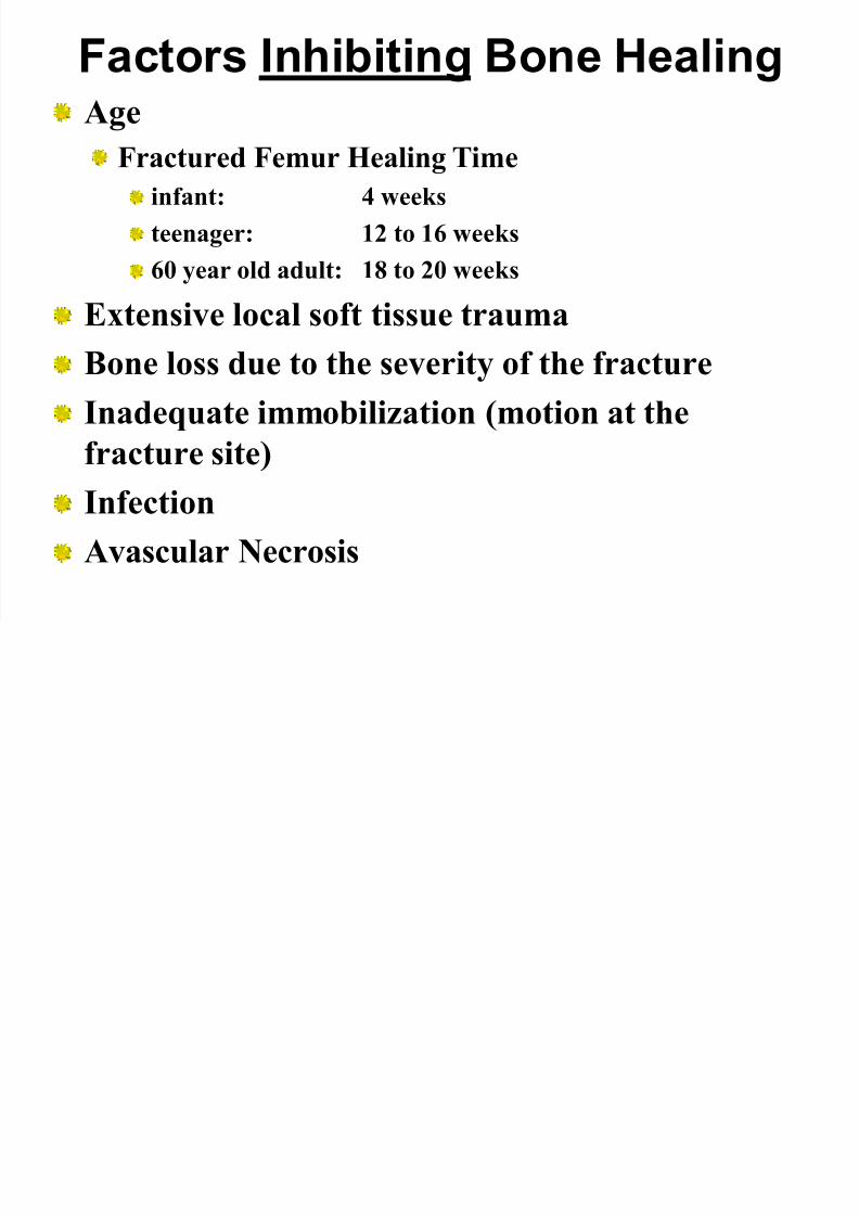
Factors Inhibiting Bone HealingAge
Fractured Femur Healing Time
infant: 4 weeks
teenager: 12 to 16 weeks
60 year old adult: 18 to 20 weeks
Extensive local soft tissue trauma
Bone loss due to the severity of the fracture
Inadequate immobilization (motion at thefracture site)
Infection
Avascular Necrosis