Embed Size (px)
Citation preview

Rev. Confirming Pages
257
9 M uscle was introduced in Chapter 1 as one of the four principal
tissue types that make up the human body. The ability to use
chemical energy to produce force and movement is present to a
limited extent in most cells, but in muscle cells it has become dominant. Muscles
generate force and movements used to regulate the internal environment, and
they also produce movements of the body in relation to the external environment.
In humans, the ability to communicate, whether by speech, writing, or artistic
expression, also depends on muscle contractions. Indeed, it is only by controlling
muscle activity that the human mind ultimately expresses itself.
Three types of muscle tissue can be identified on the basis of structure,
contractile properties, and control mechanisms—skeletal muscle, smooth
muscle, and cardiac muscle. Most skeletal muscle, as the name implies,
is attached to bone, and its contraction is responsible for supporting and
moving the skeleton. As described in Chapter 6, contraction of skeletal
muscle is initiated by action potentials in neurons of the somatic motor
division of the nervous system, and is usually under voluntary control.
Sheets of smooth muscle surround various hollow organs and tubes,
including the stomach, intestines, urinary bladder, uterus, blood vessels, and
airways in the lungs. Contraction of smooth muscle may propel the luminal
contents through the hollow organs, or it may regulate internal flow by changing
the tube diameter. In addition, contraction of smooth muscle cells makes the
hairs of the skin stand up and the pupil of the eye change diameter. In contrast
S E C T I O N A Skeletal Muscle
9.1 Structure 9.2 Molecular Mechanisms of
Skeletal Muscle Contraction Membrane Excitation: The
Neuromuscular Junction Excitation–Contraction Coupling Sliding-Filament Mechanism
9.3 Mechanics of Single-Fiber Contraction Twitch Contractions Load–Velocity Relation Frequency–Tension Relation Length–Tension Relation
9.4 Skeletal Muscle Energy Metabolism Muscle Fatigue
9.5 Types of Skeletal Muscle Fibers 9.6 Whole-Muscle Contraction
Control of Muscle Tension Control of Shortening Velocity Muscle Adaptation to Exercise Lever Action of Muscles and Bones
9.7 Skeletal Muscle Disorders Muscle Cramps Hypocalcemic Tetany Muscular Dystrophy Myasthenia Gravis
S E C T I O N B Smooth and Cardiac Muscle
9.8 Structure of Smooth Muscle 9.9 Smooth Muscle Contraction
and Its Control Cross-Bridge Activation Sources of Cytosolic Ca 2 1 Membrane Activation Types of Smooth Muscle
9.10 Cardiac Muscle Cellular Structure of Cardiac Muscle Excitation–Contraction Coupling in
Cardiac Muscle
Chapter 9 Clinical Case Study
Muscle
Colorized scanning electron micrograph (SEM) of freeze-fractured muscle fibers.
wid78305_ch09_257-299.indd 257 30/01/13 11:48 AM
Rev. Confirming Pages
258 Chapter 9
muscle shares this characteristic striped pattern, these two types are both referred to as striated muscle . The third basic muscle type, smooth muscle , derives its name from the fact that it lacks this striated appearance. Figure 9.1 compares the appearance of skeletal muscle cells to cardiac and smooth muscle cells.
9.1 Structure The most striking feature seen when viewing skeletal muscle through a microscope is a distinct series of alternating light and dark bands perpendicular to the long axis. Because cardiac
to skeletal muscle, smooth muscle contraction is not normally
under voluntary control. It occurs autonomously in some
cases, but frequently it occurs in response to signals from the
autonomic nervous system, hormones, autocrine or paracrine
signals, and other local chemical factors.
Cardiac muscle is the muscle of the heart. Its contraction
generates the pressure that propels blood through the
circulatory system. Like smooth muscle, it is regulated by
the autonomic nervous system, hormones, and autocrine or
paracrine signals; and it can undergo spontaneous contractions.
Several of the general principles of physiology described in
Chapter 1 are demonstrated in this chapter. One of these principles,
that structure is a determinant of—and has coevolved with—
function, is apparent in the elaborate specialization of muscle
cells and whole muscles that enable them to generate force and
movement. The principle that controlled exchange of materials
occurs between compartments and across cellular membranes is
exemplified by the movements of Ca 2 1 that underlie the regulation
of activation and relaxation of muscle. The laws of chemistry and
physics are fundamental to the molecular mechanism by which
muscle cells convert chemical energy into force, and also to the
mechanics governing bone–muscle lever systems. Finally, the
transfer and balance of matter and energy are demonstrated by
the ability of muscle cells to generate, store, and utilize energy via
multiple metabolic pathways.
This chapter will describe skeletal muscle first, followed
by smooth and cardiac muscle. Cardiac muscle, which
combines some of the properties of both skeletal and smooth
muscle, will be described in more depth in Chapter 12 in
association with its role in the circulatory system.
S E C T I O N A
Skeletal Muscle
Figure 9.1 Comparison of (a) skeletal muscle to (b) cardiac and (c) smooth muscle as seen with light microscopy (top panels) and in schematic form (bottom panels). Both skeletal and cardiac muscle have a striated appearance. Cardiac and smooth muscle cells generally have a single nucleus, but skeletal muscle fibers are multinucleated.
NucleiStriations
(a) Skeletal muscle
Muscle fiberConnectivetissue Branching
Intercalated disk
(b) Cardiac muscle (c) Smooth muscle
NucleusStriations Nuclei Muscle cells
wid78305_ch09_257-299.indd 258 30/01/13 11:48 AM
Rev. Confirming Pages
Figure 9.2 Structure of skeletal muscle.
Muscle fiber (single muscle cell)
Myofibril
Muscle
Tendons
Connectivetissue
Bloodvessel
Sarcomere
M line
Z line
Z line Z line
A band I band
Z line
H zone
Thick (myosin) filament Thin (actin) filament
Muscle 259
the length of muscle fibers. In response to strain or injury, they become active and undergo mitotic proliferation. Daughter cells then differentiate into myoblasts that can either fuse together to form new fibers or fuse with stressed or damaged muscle fibers to reinforce and repair them. The capacity for forming new skeletal muscle fibers is considerable but may not restore a severely dam-aged muscle to the original number of muscle fibers. Some of the compensation for a loss of muscle tissue also occurs through a satellite cell-mediated increase in the size ( hypertrophy ) of the remaining muscle fibers. Muscle hypertrophy also occurs in response to heavy exercise. Evidence suggests that this occurs through a combination of hypertrophy of existing fibers, split-ting of existing fibers, and satellite cell proliferation, differ-entiation, and fusion. Many hormones and growth factors are involved in regulating these processes, such as growth hormone, insulin-like growth factor, and sex hormones (see Chapter 11).
The term muscle refers to a number of muscle fibers bound together by connective tissue ( Figure 9.2 ). The relation-ship between a single muscle fiber and a muscle is analogous to that between a single neuron and a nerve, which is composed of
Due to its elongated shape and the presence of multiple nuclei, a skeletal muscle cell is also referred to as a muscle fiber . Each muscle fiber is formed during development by the fusion of a number of undifferentiated, mononucleated cells known as myoblasts into a single, cylindrical, multi-nucleated cell. Skeletal muscle differentiation is completed around the time of birth, and these differentiated fibers con-tinue to increase in size from infancy to adulthood. Compared to other cell types, skeletal muscle fibers are extremely large. Adult skeletal muscle fibers have diameters between 10 and 100 m m and lengths that may extend up to 20 cm. Key to the maintenance and function of such large cells is the retention of the nuclei from the original myoblasts. Spread throughout the length of the muscle fiber, each participates in regulation of gene expression and protein synthesis within its local domain.
If skeletal muscle fibers are damaged or destroyed after birth as a result of injury, they undergo a repair process involv-ing a population of undifferentiated stem cells known as satellite cells . Satellite cells are normally quiescent, located between the plasma membrane and surrounding basement membrane along
wid78305_ch09_257-299.indd 259 30/01/13 11:48 AM

Rev. Confirming Pages
260 Chapter 9
Figure 9.3 (a) The heavy chains of myosin molecules form the core of a thick filament. The myosin molecules are oriented in opposite directions in either half of a thick filament. (b) Structure of thin filament and myosin molecule. Cross-bridge binding sites on actin are covered by tropomyosin. The two globular heads of each myosin molecule extend from the sides of a thick filament, forming a cross-bridge.
Cross-bridge
ATP binding sites
Light chainsHeavy chains
Thin filament
Thick filament
Myosin
Actin binding sites
Troponin
Tropomyosin
Actin
Cross-bridge
(a)
(b)
the axons of many neurons. Skeletal muscles are usually attached to bones by bundles of collagen fibers known as tendons .
In some muscles, the individual fibers extend the entire length of the muscle, but in most, the fibers are shorter, often oriented at an angle to the longitudinal axis of the muscle. The transmission of force from muscle to bone is like a number of people pulling on a rope, each person corresponding to a single muscle fiber and the rope corresponding to the connec-tive tissue and tendons.
Some tendons are very long, with the site where the tendon attaches to the bone far removed from the end of the muscle. For example, some of the muscles that move the fin-gers are in the forearm (wiggle your fingers and feel the move-ment of the muscles just below your elbow). These muscles are connected to the fingers by long tendons.
The striated pattern in skeletal (and cardiac) muscle results from the arrangement of two types of filaments within the cyto-plasm, the larger referred to as thick filaments and the smaller as thin filaments . These filaments are part of cylindrical bundles called myofibrils , which are approximately 1 to 2 m m in diameter (see Figure 9.2 ). Most of the cytoplasm of a fiber is filled with myofibrils, each extending from one end of the fiber to the other and linked to the tendons at the ends of the fiber. One unit of this repeating pattern of thick and thin filaments is known as a sarco-mere (from the Greek sarco, “muscle,” and mer, “part”).
The molecular structure of thick and thin filaments is shown in Figure 9.3 . The thick filaments are composed almost entirely of the protein myosin . The myosin molecule is com-posed of two large polypeptide heavy chains and four smaller light chains . These polypeptides combine to form a molecule that consists of two globular heads (containing heavy and light chains) and a long tail formed by the two intertwined heavy chains. The tail of each myosin molecule lies along the axis of the thick filament, and the two globular heads extend out to the sides, forming cross-bridges , which make contact with the thin filament and exert force during muscle contraction. Each globu-lar head contains two binding sites, one for attaching to the thin filament and one for ATP. The ATP binding site also serves as an enzyme—an ATPase that hydrolyzes the bound ATP, harnessing its energy for contraction. The thin filaments (which are about half the diameter of the thick filaments) are principally composed
of the protein actin , as well as two other proteins— troponin and tropomyosin —that play important roles in regulating con-traction. An actin molecule is a globular protein composed of a single polypeptide (a monomer) that polymerizes with other actin monomers to form a polymer made up of two intertwined, helical chains. These chains make up the core of a thin filament. Each actin molecule contains a binding site for myosin.
The alternating dark and light bands produced by the orderly, parallel arrangement of thick and thin filaments are apparent in a microscopic view of skeletal muscle ( Figure 9.4 ). The thick filaments are located in the middle of each sar-comere, where they create a wide, dark band known as the A band (see the Figure 9.4 legend for an explanation of the naming of the bands and zones of the sarcomere).
Each sarcomere contains two sets of thin filaments, one at each end. One end of each thin filament is anchored to a network of interconnecting proteins known as the Z line , whereas the other end overlaps a portion of the thick filaments. Two succes-sive Z lines define the limits of one sarcomere. Thus, thin fila-ments from two adjacent sarcomeres are anchored to the two sides of each Z line. (The term line refers to the appearance of these structures in two dimensions. Because myofibrils are cylindrical, it is more realistic to think of them as Z disks. ) A light band known as the I band lies between the ends of the A bands of two adjacent sarcomeres and contains those portions of the thin filaments that do not overlap the thick filaments. The I band is bisected by the Z line. Two additional bands are present in the A-band region of each sarcomere. The H zone is a narrow, light band in the center of the A band. It corresponds to the space between the opposing ends of the two sets of thin filaments in each sarcomere. A narrow, dark band in the center of the H zone, known as the M line (also technically a disk), corresponds to proteins that link together the central region of adjacent thick filaments. In addition, filaments composed of the elastic protein titin extend from the Z line to the M line and are linked to both the M-line proteins and the thick filaments. Both the M-line linkage between thick filaments and the titin filaments act to maintain the alignment of thick filaments in the middle of each sarcomere. A cross section through the A bands ( Figure 9.5 ) shows the regular arrangement of overlapping thick and thin filaments. Each thick filament is surrounded by a hexagonal array of six thin filaments, and each thin filament is
wid78305_ch09_257-299.indd 260 30/01/13 11:48 AM

Rev. Confirming Pages
Muscle 261
Figure 9.4 (a) High magnification of a sarcomere within myofibrils. (b) Arrangement of the thick and thin filaments in the sarcomere shown in (a). The names of the I and A bands come from “isotropy” and “anisotropy,” terms from physics indicating that the I band has uniform appearance in all directions and the A band has a nonuniform appearance in different directions. The names for the Z line, M line, and H zone are from their initial descriptions in German: zwischen (“between”), mittel (“middle”), and heller (“light”).
Sarcomere
M lineZ line Z line
I band
H zone
A band
Titin Thin filament Thick filament
(a)
(b)
Myo
fibril
Thickfilament
Thinfilament
(a)
(b)
Figure 9.5 (a) Electron micrograph of a cross section through three myofibrils in a single skeletal muscle fiber. (b) Hexagonal arrangements of the thick and thin filaments in the overlap region in a single myofibril. Six thin filaments surround each thick filament, and three thick filaments surround each thin filament. Titin filaments and cross-bridges are not shown. From H. E.
Huxley, J. Mol. Biol., 37:507–520 (1968).
PH Y S I O L O G I C A L I N Q U I R Y
■ Draw a cross-section diagram like the one in part (b) for a slice taken (1) in the H zone, (2) in the I band, (3) at the M line, and (4) at the Z line (ignore titin).
Answer can be found at end of chapter.
surrounded by a triangular arrangement of three thick filaments. Altogether, there are twice as many thin as thick filaments in the region of filament overlap.
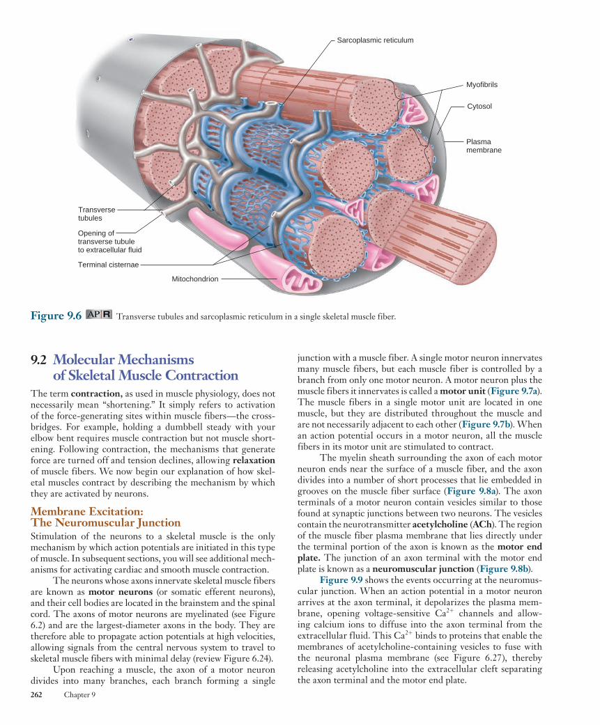
In addition to force-generating mechanisms, skeletal muscle fibers have an elaborate system of membranes that play important roles in the activation of contraction ( Figure 9.6 ). The sarcoplasmic reticulum in a muscle fiber is homologous
to the endoplasmic reticulum found in most cells. This struc-ture forms a series of sleevelike segments around each myofibril. At the end of each segment are two enlarged regions, known as terminal cisternae (sometimes also referred to as “lateral sacs”), that are connected to each other by a series of smaller tubular elements. Ca 2 1 is stored in the terminal cisternae and is released into the cytosol following membrane excitation.
A separate tubular structure, the transverse tubule ( T-tubule ) , lies directly between—and is intimately associ-ated with—the terminal cisternae of adjacent segments of the sarcoplasmic reticulum. The T-tubules and terminal cisternae surround the myofibrils at the region of the sarcomeres where the A bands and I bands meet. T-tubules are continuous with the plasma membrane (which in muscle cells is sometimes referred to as the sarcolemma ), and action potentials propa-gating along the surface membrane also travel throughout the interior of the muscle fiber by way of the T-tubules. The lumen of the T-tubule is continuous with the extracellular fluid surrounding the muscle fiber.
wid78305_ch09_257-299.indd 261 30/01/13 11:48 AM
Rev. Confirming Pages
262 Chapter 9
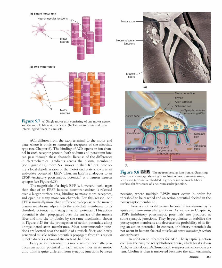
junction with a muscle fiber. A single motor neuron innervates many muscle fibers, but each muscle fiber is controlled by a branch from only one motor neuron. A motor neuron plus the muscle fibers it innervates is called a motor unit ( Figure 9.7a ). The muscle fibers in a single motor unit are located in one muscle, but they are distributed throughout the muscle and are not necessarily adjacent to each other ( Figure 9.7b ). When an action potential occurs in a motor neuron, all the muscle fibers in its motor unit are stimulated to contract.
The myelin sheath surrounding the axon of each motor neuron ends near the surface of a muscle fiber, and the axon divides into a number of short processes that lie embedded in grooves on the muscle fiber surface ( Figure 9.8a ). The axon terminals of a motor neuron contain vesicles similar to those found at synaptic junctions between two neurons. The vesicles contain the neurotransmitter acetylcholine ( ACh ) . The region of the muscle fiber plasma membrane that lies directly under the terminal portion of the axon is known as the motor end plate . The junction of an axon terminal with the motor end plate is known as a neuromuscular junction ( Figure 9.8b ).
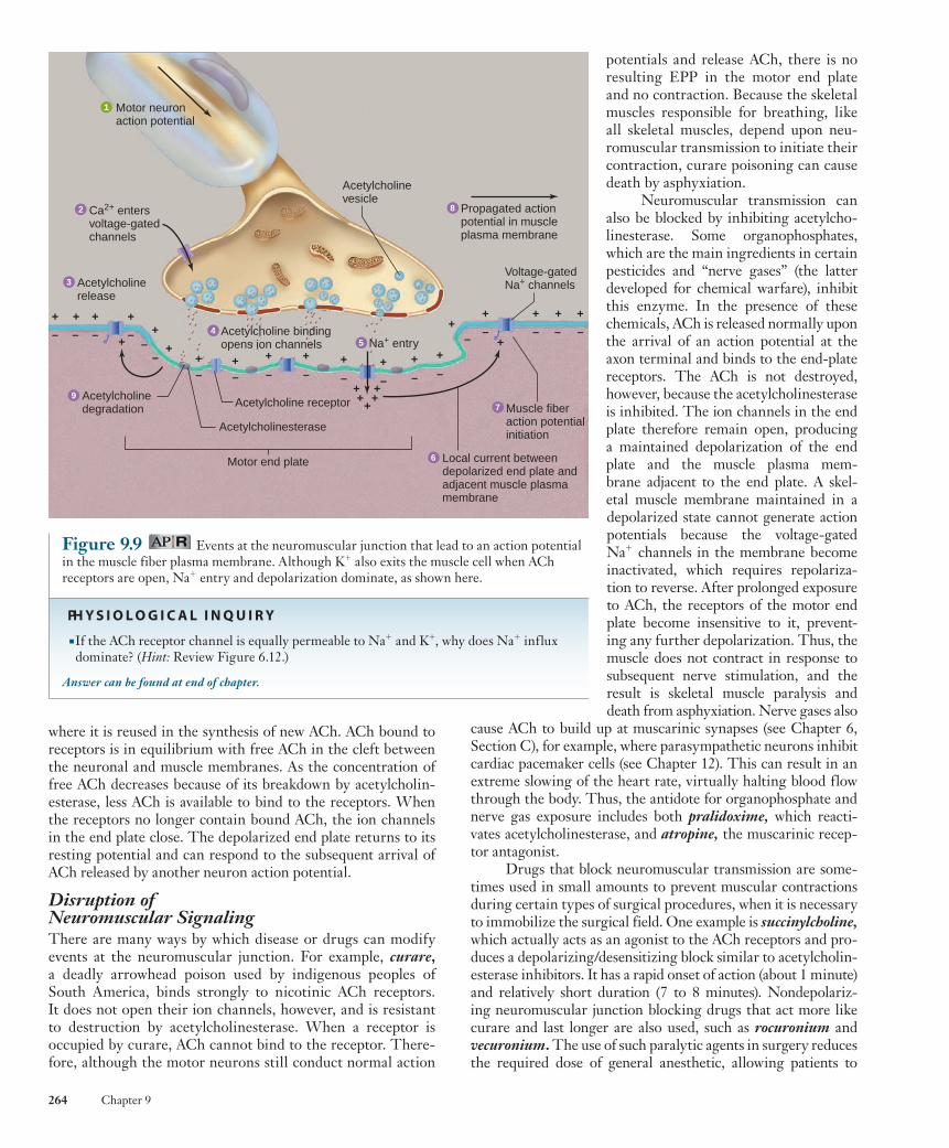
Figure 9.9 shows the events occurring at the neuromus-cular junction. When an action potential in a motor neuron arrives at the axon terminal, it depolarizes the plasma mem-brane, opening voltage-sensitive Ca 2 1 channels and allow-ing calcium ions to diffuse into the axon terminal from the extracellular fluid. This Ca 2 1 binds to proteins that enable the membranes of acetylcholine-containing vesicles to fuse with the neuronal plasma membrane (see Figure 6.27), thereby releasing acetylcholine into the extracellular cleft separating the axon terminal and the motor end plate.
9.2 Molecular Mechanisms of Skeletal Muscle Contraction
The term contraction , as used in muscle physiology, does not necessarily mean “shortening.” It simply refers to activation of the force-generating sites within muscle fibers—the cross-bridges. For example, holding a dumbbell steady with your elbow bent requires muscle contraction but not muscle short-ening. Following contraction, the mechanisms that generate force are turned off and tension declines, allowing relaxation of muscle fibers. We now begin our explanation of how skel-etal muscles contract by describing the mechanism by which they are activated by neurons.
Membrane Excitation: The Neuromuscular Junction Stimulation of the neurons to a skeletal muscle is the only mechanism by which action potentials are initiated in this type of muscle. In subsequent sections, you will see additional mech-anisms for activating cardiac and smooth muscle contraction.
The neurons whose axons innervate skeletal muscle fibers are known as motor neurons (or somatic efferent neurons), and their cell bodies are located in the brainstem and the spinal cord. The axons of motor neurons are myelinated (see Figure 6.2) and are the largest-diameter axons in the body. They are therefore able to propagate action potentials at high velocities, allowing signals from the central nervous system to travel to skeletal muscle fibers with minimal delay (review Figure 6.24).
Upon reaching a muscle, the axon of a motor neuron divides into many branches, each branch forming a single
Figure 9.6 Transverse tubules and sarcoplasmic reticulum in a single skeletal muscle fiber.
Myofibrils
Cytosol
Sarcoplasmic reticulum
Plasmamembrane
Transverse tubules
Opening oftransverse tubuleto extracellular fluid
Terminal cisternae
Mitochondrion
wid78305_ch09_257-299.indd 262 30/01/13 11:49 AM
Rev. Confirming Pages
Muscle 263
ACh diffuses from the axon terminal to the motor end plate where it binds to ionotropic receptors of the nicotinic type (see Chapter 6). The binding of ACh opens an ion chan-nel in each receptor protein; both sodium and potassium ions can pass through these channels. Because of the differences in electrochemical gradients across the plasma membrane (see Figure 6.12), more Na 1 moves in than K 1 out, produc-ing a local depolarization of the motor end plate known as an end-plate potential ( EPP ) . Thus, an EPP is analogous to an EPSP (excitatory postsynaptic potential) at a neuron–neuron synapse (see Figure 6.28).
The magnitude of a single EPP is, however, much larger than that of an EPSP because neurotransmitter is released over a larger surface area, binding to many more receptors, and opening many more ion channels. For this reason, one EPP is normally more than sufficient to depolarize the muscle plasma membrane adjacent to the end-plate membrane to its threshold potential, initiating an action potential. This action potential is then propagated over the surface of the muscle fiber and into the T-tubules by the same mechanism shown in Figure 6.23 for the propagation of action potentials along unmyelinated axon membranes. Most neuromuscular junc-tions are located near the middle of a muscle fiber, and newly generated muscle action potentials propagate from this region in both directions toward the ends of the fiber.
Every action potential in a motor neuron normally pro-duces an action potential in each muscle fiber in its motor unit. This is quite different from synaptic junctions between
Figure 9.7 (a) Single motor unit consisting of one motor neuron and the muscle fibers it innervates. (b) Two motor units and their intermingled fibers in a muscle.
(a) Single motor unit
Neuromuscular junctions
Motorneurons
(b) Two motor units
Motorneuron
(a)
Motor axon
Neuromuscularjunctions
Musclefibers
Figure 9.8 The neuromuscular junction. (a) Scanning electron micrograph showing branching of motor neuron axons, with axon terminals embedded in grooves in the muscle fiber’s surface. (b) Structure of a neuromuscular junction.
(b)
Myelin
Motor nerve fiber
Axon terminal
Schwann cell
Synaptic vesicles(containing ACh)
Active zone
Sarcolemma
Region ofsarcolemmawith ACh receptors
JunctionalfoldsNucleus of muscle fiber
Synapticcleft
neurons, where multiple EPSPs must occur in order for threshold to be reached and an action potential elicited in the postsynaptic membrane.
There is another difference between interneuronal syn-apses and neuromuscular junctions. As we saw in Chapter 6, IPSPs (inhibitory postsynaptic potentials) are produced at some synaptic junctions. They hyperpolarize or stabilize the postsynaptic membrane and decrease the probability of its fir-ing an action potential. In contrast, inhibitory potentials do not occur in human skeletal muscle; all neuromuscular junctions are excitatory.
In addition to receptors for ACh, the synaptic junction contains the enzyme acetylcholinesterase , which breaks down ACh, just as it does at ACh-mediated synapses in the nervous sys-tem. Choline is then transported back into the axon terminals,
wid78305_ch09_257-299.indd 263 30/01/13 11:49 AM
Rev. Confirming Pages
264 Chapter 9
potentials and release ACh, there is no resulting EPP in the motor end plate and no contraction. Because the skeletal muscles responsible for breathing, like all skeletal muscles, depend upon neu-romuscular transmission to initiate their contraction, curare poisoning can cause death by asphyxiation.
Neuromuscular transmission can also be blocked by inhibiting acetylcho-linesterase. Some organophosphates, which are the main ingredients in certain pesticides and “nerve gases” (the latter developed for chemical warfare), inhibit this enzyme. In the presence of these chemicals, ACh is released normally upon the arrival of an action potential at the axon terminal and binds to the end-plate receptors. The ACh is not destroyed, however, because the acetylcholinesterase is inhibited. The ion channels in the end plate therefore remain open, producing a maintained depolarization of the end plate and the muscle plasma mem-brane adjacent to the end plate. A skel-etal muscle membrane maintained in a depolarized state cannot generate action potentials because the voltage-gated Na 1 channels in the membrane become inactivated, which requires repolariza-tion to reverse. After prolonged exposure to ACh, the receptors of the motor end plate become insensitive to it, prevent-ing any further depolarization. Thus, the muscle does not contract in response to subsequent nerve stimulation, and the result is skeletal muscle paralysis and death from asphyxiation. Nerve gases also
cause ACh to build up at muscarinic synapses (see Chapter 6, Section C), for example, where parasympathetic neurons inhibit cardiac pacemaker cells (see Chapter 12). This can result in an extreme slowing of the heart rate, virtually halting blood flow through the body. Thus, the antidote for organophosphate and nerve gas exposure includes both pralidoxime , which reacti-vates acetylcholinesterase, and atropine , the muscarinic recep-tor antagonist.
Drugs that block neuromuscular transmission are some-times used in small amounts to prevent muscular contractions during certain types of surgical procedures, when it is necessary to immobilize the surgical field. One example is succinylcholine ,which actually acts as an agonist to the ACh receptors and pro-duces a depolarizing/desensitizing block similar to acetylcholin-esterase inhibitors. It has a rapid onset of action (about 1 minute) and relatively short duration (7 to 8 minutes). Nondepolariz-ing neuromuscular junction blocking drugs that act more like curare and last longer are also used, such as rocuronium and vecuronium . The use of such paralytic agents in surgery reduces the required dose of general anesthetic, allowing patients to
where it is reused in the synthesis of new ACh. ACh bound to receptors is in equilibrium with free ACh in the cleft between the neuronal and muscle membranes. As the concentration of free ACh decreases because of its breakdown by acetylcholin-esterase, less ACh is available to bind to the receptors. When the receptors no longer contain bound ACh, the ion channels in the end plate close. The depolarized end plate returns to its resting potential and can respond to the subsequent arrival of ACh released by another neuron action potential.
Disruption of Neuromuscular Signaling There are many ways by which disease or drugs can modify events at the neuromuscular junction. For example, curare, a deadly arrowhead poison used by indigenous peoples of South America, binds strongly to nicotinic ACh receptors. It does not open their ion channels, however, and is resistant to destruction by acetylcholinesterase. When a receptor is occupied by curare, ACh cannot bind to the receptor. There-fore, although the motor neurons still conduct normal action
3
9
2
54
6
7
8
1
+
+
Acetylcholinerelease
Motor neuronaction potential
Muscle fiberaction potentialinitiation
Local current betweendepolarized end plate andadjacent muscle plasmamembrane
Acetylcholine receptorAcetylcholinedegradation
Acetylcholinesterase
Motor end plate
Acetylcholinevesicle
Voltage-gatedNa+ channels
+ ++
– – –
+
+
++
–
+
– – –+
+–
– +–
–
+
–+
–+
+ +++
+
–
+
–
+
+
–
–
+–
+–
Na+ entryAcetylcholine bindingopens ion channels
Ca2+ entersvoltage-gatedchannels
Propagated actionpotential in muscleplasma membrane
Figure 9.9 Events at the neuromuscular junction that lead to an action potential in the muscle fiber plasma membrane. Although K 1 also exits the muscle cell when ACh receptors are open, Na 1 entry and depolarization dominate, as shown here.
PH Y S I O L O G I C A L I N Q U I R Y
■ If the ACh receptor channel is equally permeable to Na 1 and K 1 , why does Na 1 influx dominate? ( Hint: Review Figure 6.12.)
Answer can be found at end of chapter.
wid78305_ch09_257-299.indd 264 30/01/13 11:49 AM
Rev. Confirming Pages
Muscle 265
recover faster and with fewer complications. Patients must be artificially ventilated, however, to maintain respiration until the drugs have cleared from their bodies.
Another group of substances, including the toxin pro-duced by the bacterium Clostridium botulinum, blocks the release of acetylcholine from axon terminals. Botulinum toxin is an enzyme that breaks down proteins of the SNARE com-plex that are required for the binding and fusion of ACh vesicles with the plasma membrane of the axon terminal (review Figure 6.27). This toxin, which produces the food poisoning called botulism , is one of the most potent poisons known. Applica-tion of botulinum toxin to block ACh release is increasingly being used for clinical and cosmetic procedures, including the inhibition of overactive extraocular muscles, prevention of excessive sweat gland activity, treatment of migraine head-aches, and reduction of aging-related skin wrinkles.
Having described how action potentials in motor neu-rons initiate action potentials in skeletal muscle cells, we will now examine how that excitation results in muscle contraction.
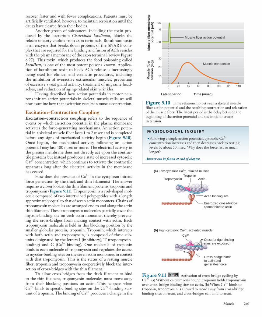
Excitation–Contraction Coupling Excitation–contraction coupling refers to the sequence of events by which an action potential in the plasma membrane activates the force-generating mechanisms. An action poten-tial in a skeletal muscle fiber lasts 1 to 2 msec and is completed before any signs of mechanical activity begin ( Figure 9.10 ). Once begun, the mechanical activity following an action potential may last 100 msec or more. The electrical activity in the plasma membrane does not directly act upon the contrac-tile proteins but instead produces a state of increased cytosolic Ca 2 1 concentration, which continues to activate the contractile apparatus long after the electrical activity in the membrane has ceased.
How does the presence of Ca 2 1 in the cytoplasm initiate force generation by the thick and thin filaments? The answer requires a closer look at the thin filament proteins, troponin and tropomyosin ( Figure 9.11 ). Tropomyosin is a rod-shaped mol-ecule composed of two intertwined polypeptides with a length approximately equal to that of seven actin monomers. Chains of tropomyosin molecules are arranged end to end along the actin thin filament. These tropomyosin molecules partially cover the myosin-binding site on each actin monomer, thereby prevent-ing the cross-bridges from making contact with actin. Each tropomyosin molecule is held in this blocking position by the smaller globular protein, troponin. Troponin, which interacts with both actin and tropomyosin, is composed of three sub-units designated by the letters I (inhibitory), T (tropomyosin- binding) and C (Ca 2 1 -binding). One molecule of troponin binds to each molecule of tropomyosin and regulates the access to myosin-binding sites on the seven actin monomers in contact with that tropomyosin. This is the status of a resting muscle fiber; troponin and tropomyosin cooperatively block the inter-action of cross-bridges with the thin filament.
To allow cross-bridges from the thick filament to bind to the thin filament, tropomyosin molecules must move away from their blocking positions on actin. This happens when Ca 2 1 binds to specific binding sites on the Ca 2 1 -binding sub-unit of troponin. The binding of Ca 2 1 produces a change in the
Muscle fiber action potential
0 20 40 60 80 100 120 140
Latent period Time (msec)
+30
0
–90
30
20
10
Mu
scle
fib
erte
nsi
on
(m
g)
Mu
scle
fib
er m
emb
ran
ep
ote
nti
al (
mV
)
Muscle contraction
Figure 9.10 Time relationship between a skeletal muscle fiber action potential and the resulting contraction and relaxation of the muscle fiber. The latent period is the delay between the beginning of the action potential and the initial increase in tension.
PH Y S I O L O G I C A L I N Q U I R Y
■ Following a single action potential, cytosolic Ca 2 1 concentration increases and then decreases back to resting levels by about 50 msec. Why does the force last so much longer?
Answer can be found at end of chapter.
Figure 9.11 Activation of cross-bridge cycling by Ca 2 1 . (a) Without calcium ions bound, troponin holds tropomyosin over cross-bridge binding sites on actin. (b) When Ca 2 1 binds to troponin, tropomyosin is allowed to move away from cross-bridge binding sites on actin, and cross-bridges can bind to actin.
(a) Low cytosolic Ca2+, relaxed muscle
(b) High cytosolic Ca2+, activated muscle
ActinTroponin
Tropomyosin
Energized cross-bridgecannot bind to actin
Actin binding site
Cross-bridge bindsto actin andgenerates force
Cross-bridge bindingsites are exposed
Ca2+
wid78305_ch09_257-299.indd 265 30/01/13 11:49 AM
Rev. Confirming Pages
266 Chapter 9
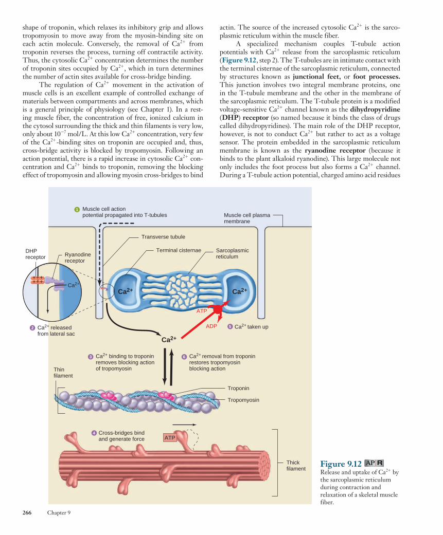
actin. The source of the increased cytosolic Ca 2 1 is the sarco-plasmic reticulum within the muscle fiber.
A specialized mechanism couples T-tubule action potentials with Ca 2 1 release from the sarcoplasmic reticulum ( Figure 9.12 , step 2). The T-tubules are in intimate contact with the terminal cisternae of the sarcoplasmic reticulum, connected by structures known as junctional feet , or foot processes .This junction involves two integral membrane proteins, one in the T-tubule membrane and the other in the membrane of the sarcoplasmic reticulum. The T-tubule protein is a modified voltage-sensitive Ca 2 1 channel known as the dihydropyridine ( DHP ) receptor (so named because it binds the class of drugs called dihydropyridines). The main role of the DHP receptor, however, is not to conduct Ca 2 1 but rather to act as a voltage sensor. The protein embedded in the sarcoplasmic reticulum membrane is known as the ryanodine receptor (because it binds to the plant alkaloid ryanodine). This large molecule not only includes the foot process but also forms a Ca 2 1 channel. During a T-tubule action potential, charged amino acid residues
shape of troponin, which relaxes its inhibitory grip and allows tropomyosin to move away from the myosin-binding site on each actin molecule. Conversely, the removal of Ca 2 1 from troponin reverses the process, turning off contractile activity. Thus, the cytosolic Ca 2 1 concentration determines the number of troponin sites occupied by Ca 2 1 , which in turn determines the number of actin sites available for cross-bridge binding.
The regulation of Ca 2 1 movement in the activation of muscle cells is an excellent example of controlled exchange of materials between compartments and across membranes, which is a general principle of physiology (see Chapter 1). In a rest-ing muscle fiber, the concentration of free, ionized calcium in the cytosol surrounding the thick and thin filaments is very low, only about 10 2 7 mol/L. At this low Ca 2 1 concentration, very few of the Ca 2 1 -binding sites on troponin are occupied and, thus, cross-bridge activity is blocked by tropomyosin. Following an action potential, there is a rapid increase in cytosolic Ca 2 1 con-centration and Ca 2 1 binds to troponin, removing the blocking effect of tropomyosin and allowing myosin cross-bridges to bind
Figure 9.12 Release and uptake of Ca 2 1 by the sarcoplasmic reticulum during contraction and relaxation of a skeletal muscle fiber.
Muscle cell actionpotential propagated into T-tubules Muscle cell plasma
membrane
Transverse tubule
Terminal cisternae Sarcoplasmicreticulum
Ca2+ binding to troponinremoves blocking action of tropomyosin
Ca2+ removal from troponinrestores tropomyosinblocking action
Ca2+
Ca2+ taken up
Troponin
Tropomyosin
Cross-bridges bindand generate force
Thickfilament
Thinfilament
Ca2+ Ca2+
ATP
ADP
ATP
Ca2+ releasedfrom lateral sac
++++++
++++++
Ca2+
DHPreceptor Ryanodine
receptor
3
2
4
5
6
1
wid78305_ch09_257-299.indd 266 30/01/13 11:49 AM

Rev. Confirming Pages
Muscle 267
within the DHP receptor protein induce a conformational change, which acts via the foot process to open the ryanodine receptor channel. Ca 2 1 is thus released from the terminal cister-nae of the sarcoplasmic reticulum into the cytosol, where it can bind to troponin. The increase in cytosolic Ca 2 1 in response to a single action potential is normally enough to briefly saturate all troponin-binding sites on the thin filaments.
A contraction is terminated by removal of Ca 2 1 from troponin, which is achieved by lowering the Ca 2 1 concentra-tion in the cytosol back to its prerelease level. The membranes of the sarcoplasmic reticulum contain primary active-transport proteins—Ca 2 1 -ATPases—that pump calcium ions from the cytosol back into the lumen of the reticulum. As we just saw, Ca 2 1 is released from the reticulum when an action potential begins in the T-tubule, but the pumping of the released Ca 2 1 back into the reticulum requires a much longer time. Therefore, the cytosolic Ca 2 1 concentration remains elevated, and the contraction contin-ues for some time after a single action potential.
To reiterate, just as contraction results from the release of Ca 2 1 stored in the sarcoplasmic reticulum, so contraction ends and relaxation begins as Ca 2 1 is pumped back into the reticulum (see Figure 9.12 ). ATP is required to provide the energy for the Ca 2 1 pump.
Sliding-Filament Mechanism When force generation produces shortening of a skeletal muscle fiber, the overlapping thick and thin filaments in each sarcomere move past each other, propelled by movements of the cross-bridges. During this shortening of the sarcomeres, there is no change in the lengths of either the thick or thin filaments. This is known as the sliding-filament mechanism of muscle contraction.
During shortening, each myosin cross-bridge attached to a thin filament actin molecule moves in an arc much like an oar on a boat. This swiveling motion of many cross-bridges forces the thin filaments attached to successive Z lines to move toward the center of the sarcomere, thereby shorten-ing the sarcomere ( Figure 9.13 ). One stroke of a cross-bridge produces only a very small movement of a thin filament rela-tive to a thick filament. As long as binding sites on actin remain exposed, however, each cross-bridge repeats its swivel-ing motion many times, resulting in large displacements of the filaments. It is worth noting that a common pattern of muscle shortening involves one end of the muscle remaining at a fixed position while the other end shortens toward it. In this case, as filaments slide and each sarcomere shortens internally, the center of each sarcomere also slides toward the fixed end of the muscle (this is depicted in Figure 9.14 ).
The sequence of events that occurs between the time a cross-bridge binds to a thin filament, moves, and then is set to repeat the process is known as a cross-bridge cycle . Each cycle consists of four steps: (1) attachment of the cross-bridge to a thin filament; (2) movement of the cross-bridge, producing tension in the thin filament; (3) detachment of the cross-bridge from the thin filament; and (4) energizing the cross-bridge so it can again attach to a thin filament and repeat the cycle. Each cross-bridge undergoes its own cycle of movement independently of other cross-bridges. At any instant during contraction, only some of the cross-bridges
Figure 9.13 Cross-bridges in the thick filaments bind to actin in the thin filaments and undergo a conformational change that propels the thin filaments toward the center of a sarcomere. (Only a few of the approximately 200 cross-bridges in each thick filament are shown.)
Z line Z lineCross-bridgeand thin filament
movement
Thin filamentThick filament
Relaxed(a)
Shortened(b)
A band
H zoneI band
H zonereduced
I bandreduced
Z lineZ line Z line
A bandunchanged
Figure 9.14 The sliding of thick filaments past overlapping thin filaments shortens the sarcomere with no change in thick or thin filament length. The I band and H zone are reduced.
PH Y S I O L O G I C A L I N Q U I R Y
■ Sphincter muscles are circular and generally not attached to bones. How would this diagram differ if the sarcomeres shown were part of a sphincter muscle?
Answer can be found at end of chapter.
are attached to the thin filaments, producing tension, while others are simultaneously in a detached portion of their cycle.
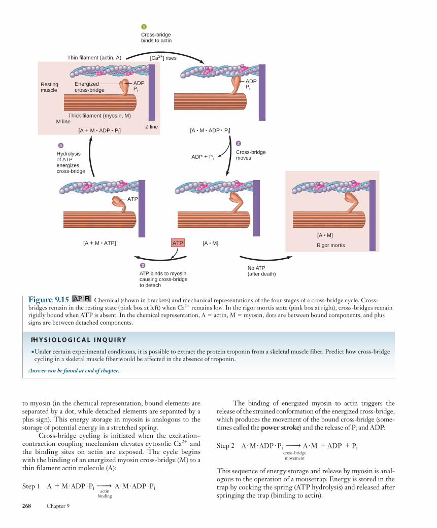
A general principle of physiology states that physiologi-cal processes are dictated by the laws of chemistry and physics (see Chapter 1), and the details of the cross-bridge mechanism are an excellent example. Figure 9.15 illustrates the chemical and physical events during the four steps of the cross-bridge cycle. The cross-bridges in a resting muscle fiber are in an ener-gized state resulting from the splitting of ATP, and the hydro-lysis products ADP and inorganic phosphate (P i ) are still bound
wid78305_ch09_257-299.indd 267 30/01/13 11:49 AM
Rev. Confirming Pages
268 Chapter 9
The binding of energized myosin to actin triggers the release of the strained conformation of the energized cross-bridge, which produces the movement of the bound cross-bridge (some-times called the power stroke ) and the release of P i and ADP:
Step 2 cross-bridgemovement
A .M.ADP .Pi A .M 1 ADP 1 Pi⎯ →⎯
This sequence of energy storage and release by myosin is anal-ogous to the operation of a mousetrap: Energy is stored in the trap by cocking the spring (ATP hydrolysis) and released after springing the trap (binding to actin).
to myosin (in the chemical representation, bound elements are separated by a dot, while detached elements are separated by a plus sign). This energy storage in myosin is analogous to the storage of potential energy in a stretched spring.
Cross-bridge cycling is initiated when the excitation–contraction coupling mechanism elevates cytosolic Ca 2 1 and the binding sites on actin are exposed. The cycle begins with the binding of an energized myosin cross-bridge (M) to a thin filament actin molecule (A):
Step 1 actin
binding
A 1 M.ADP.Pi A .M.ADP.Pi
3
24
1
Restingmuscle
Cross-bridgebinds to actin
Cross-bridgemovesADP + Pi
ATP binds to myosin, causing cross-bridge to detach
No ATP(after death)
Rigor mortis
Energizedcross-bridge
Thin filament (actin, A)
Thick filament (myosin, M)M line
Z line[A M ADP Pi]
[A M]
[A M]
Hydrolysisof ATPenergizescross-bridge
[A + M ATP] ATP
[Ca2+] rises
ADPPi
ADPPi
ATP
[A + M ADP Pi]
Figure 9.15 Chemical (shown in brackets) and mechanical representations of the four stages of a cross-bridge cycle. Cross-bridges remain in the resting state (pink box at left) when Ca 2 1 remains low. In the rigor mortis state (pink box at right), cross-bridges remain rigidly bound when ATP is absent. In the chemical representation, A 5 actin, M 5 myosin, dots are between bound components, and plus signs are between detached components.
PH Y S I O L O G I C A L I N Q U I R Y
■ Under certain experimental conditions, it is possible to extract the protein troponin from a skeletal muscle fiber. Predict how cross-bridge cycling in a skeletal muscle fiber would be affected in the absence of troponin.
Answer can be found at end of chapter.
wid78305_ch09_257-299.indd 268 30/01/13 11:49 AM

Rev. Confirming Pages
Muscle 269
The importance of ATP in dissociating actin and myosin during step 3 of a cross-bridge cycle is illustrated by rigor mortis , the gradual stiffening of skeletal muscles that begins several hours after death and reaches a maximum after about 12 hours. The ATP concentration in cells, including muscle cells, declines after death because the nutrients and oxygen the metabolic pathways require to form ATP are no longer supplied by the circulation. In the absence of ATP, the breakage of the link between actin and myosin does not occur (see Figure 9.15 ). The thick and thin filaments remain bound to each other by immobilized cross-bridges, producing a rigid condition in which the thick and thin filaments cannot be pulled past each other. The stiffness of rigor mortis disappears about 48 to 60 hours after death as the muscle tissue decomposes.
Table 9.2 summarizes the sequence of events that lead from an action potential in a motor neuron to the contraction and relaxation of a skeletal muscle fiber.
9.3 Mechanics of Single-Fiber Contraction
The force exerted on an object by a contracting muscle is known as muscle tension , and the force exerted on the muscle by an object (usually its weight) is the load . Muscle tension and load are opposing forces. Whether a fiber shortens depends on the relative magnitudes of the tension and the load. For mus-cle fibers to shorten and thereby move a load, muscle tension must be greater than the opposing load.
When a muscle develops tension but does not shorten or lengthen, the contraction is said to be an isometric (constant length) contraction . Such contractions occur when the mus-cle supports a load in a constant position or attempts to move an otherwise supported load that is greater than the tension developed by the muscle. A contraction in which the muscle changes length while the load on the muscle remains constant is an isotonic (constant tension) contraction .
Depending on the relative magnitudes of muscle tension and the opposing load, isotonic contractions can be associated with either shortening or lengthening of a muscle. When ten-sion exceeds the load, shortening occurs and it is referred to as concentric contraction . When an unsupported load is greater than the tension generated by cross-bridges, the result is an eccentric contraction (lengthening contraction). In this situ-ation, the load pulls the muscle to a longer length in spite of the opposing force produced by the cross-bridges. Such lengthen-ing contractions occur when an object being supported by mus-cle contraction is lowered, as when the knee extensors in your thighs are used to lower you to a seat from a standing position. It must be emphasized that in these situations the lengthening of muscle fibers is not an active process produced by the contractile proteins but a consequence of the external forces being applied to the muscle. In the absence of external lengthening forces, a fiber will only shorten when stimulated; it will never lengthen. All three types of contractions—isometric, concentric, and eccentric—occur in the natural course of everyday activities.
During each type of contraction, the cross-bridges repeat-edly go through the four steps of the cross-bridge cycle illustrated in Figure 9.15 . During step 2 of a concentric isotonic contraction,
During the cross-bridge movement, myosin is bound very firmly to actin, but this linkage must be broken to allow the cross-bridge to be reenergized and repeat the cycle. The binding of a new molecule of ATP to myosin breaks the link between actin and myosin:
Step 3 cross-bridge
dissociation from actin
A . M 1 ATP A 1 M.ATP⎯ →⎯
The dissociation of actin and myosin by ATP is an example of allosteric regulation of protein activity (see Figure 3.32a). The binding of ATP at one site on myosin decreases myosin’s affinity for actin bound at another site. Note that ATP is not split in this step; that is, it is not acting as an energy source but only as an allosteric modulator of the myosin head that weak-ens the binding of myosin to actin.
Following the dissociation of actin and myosin, the ATP bound to myosin is hydrolyzed, thereby re-forming the ener-gized state of myosin and returning the cross-bridge to its pre-power-stroke position:
Step 4 ATP hydrolysis
A 1 M .ATP A 1 M .ADP .Pi⎯ →⎯
Note that the hydrolysis of ATP (step 4) and the movement of the cross-bridge (step 2) are not simultaneous events. If bind-ing sites on actin are still exposed after a cross-bridge finishes its cycle, the cross-bridge can reattach to a new actin mono-mer in the thin filament and the cross-bridge cycle repeats. (In the event that the muscle is generating force without actually shortening, the cross-bridge will reattach to the same actin molecule as in the previous cycle.)
Thus, in addition to being used to maintain membrane excitability and regulate cytosolic Ca 2 1 , ATP performs two distinct roles in the cross-bridge cycle: (1) The energy released from ATP hydrolysis ultimately provides the energy for cross-bridge movement; and (2) ATP binding (not hydrolysis) to myosin breaks the link formed between actin and myosin dur-ing the cycle, allowing the next cycle to begin. Table 9.1 sum-marizes the functions of ATP in skeletal muscle contraction.
TABLE 9.1 Functions of ATP in Skeletal Muscle Contraction
Hydrolysis of ATP by the Na 1 /K 1 -ATPase in the plasma membrane maintains Na 1 and K 1 gradients, which allows the membrane to produce and propagate action potentials (review Figure 6.13).
Hydrolysis of ATP by the Ca 2 1 -ATPase in the sarcoplasmic reticulum provides the energy for the active transport of calcium ions into the reticulum, lowering cytosolic Ca 2 1 to prerelease concentrations, ending the contraction, and allowing the muscle fiber to relax.
Hydrolysis of ATP by myosin energizes the cross-bridges, providing the energy for force generation.
Binding of ATP to myosin dissociates cross-bridges bound to actin, allowing the bridges to repeat their cycle of activity.
wid78305_ch09_257-299.indd 269 30/01/13 11:49 AM

Rev. Confirming Pages
270 Chapter 9
TABLE 9.2 Sequence of Events Between a Motor Neuron Action Potential and Skeletal Muscle Fiber Contraction
1. Action potential is initiated and propagates to motor neuron axon terminals.
2. Ca21 enters axon terminals through voltage-gated Ca21 channels.
3. Ca21 entry triggers release of ACh from axon terminals.
4. ACh diffuses from axon terminals to motor end plate in muscle fiber.
5. ACh binds to nicotinic receptors on motor end plate, increasing their permeability to Na1 and K1.
6. More Na1 moves into the fiber at the motor end plate than K1 moves out, depolarizing the membrane and producing the end-plate potential (EPP).
7. Local currents depolarize the adjacent muscle cell plasma membrane to its threshold potential, generating an action potential that propagates over the muscle fiber surface and into the fiber along the T-tubules.
8. Action potential in T-tubules induces DHP receptors to pull open ryanodine receptor channels, allowing release of Ca21 from terminal cisternae of sarcoplasmic reticulum.
9. Ca21 binds to troponin on the thin filaments, causing tropomyosin to move away from its blocking position, thereby uncovering cross-bridge binding sites on actin.
10. Energized myosin cross-bridges on the thick filaments bind to actin:A 1 M · ADP · Pi → A · M · ADP · Pi
11. Cross-bridge binding triggers release of ATP hydrolysis products from myosin, producing an angular movement of each cross-bridge:
A · M · ADP · Pi → A · M 1 ADP 1 Pi
12. ATP binds to myosin, breaking linkage between actin and myosin and thereby allowing cross-bridges to dissociate from actin:
A · M 1 ATP → A 1 M · ATP
13. ATP bound to myosin is split, energizing the myosin cross-bridge:
M · ATP → M · ADP · Pi
14. Cross-bridges repeat steps 10 to 13, producing movement (sliding) of thin filaments past thick filaments. Cycles of cross-bridge movement continue as long as Ca21 remains bound to troponin.
15. Cytosolic Ca21 concentration decreases as Ca21-ATPase actively transports Ca21 into sarcoplasmic reticulum.
16. Removal of Ca21 from troponin restores blocking action of tropomyosin, the cross-bridge cycle ceases, and the muscle fiber relaxes.
the cross-bridges bound to actin rotate through their power stroke, causing shortening of the sarcomeres. In contrast, dur-ing an isometric contraction, the bound cross-bridges do exert a force on the thin filaments but they are unable to move it. Rather than the filaments sliding, the rotation during the power stroke is absorbed within the structure of the cross-bridge in this cir-cumstance. If isometric contraction is prolonged, cycling cross-bridges repeatedly rebind to the same actin molecule. During a lengthening contraction, the load pulls the cross-bridges in step 2 backward toward the Z lines while they are still bound to actin and exerting force. The events of steps 1, 3, and 4 are the same in all three types of contractions. Thus, the chemical changes in the contractile proteins during each type of contraction are the same. The end result (shortening, no length change, or lengthening) is determined by the magnitude of the load on the muscle.
Contraction terminology applies to both single fibers and whole muscles. In this section, we describe the mechan-ics of single-fiber contractions. Later, we will discuss the fac-tors controlling the mechanics of whole-muscle contraction.
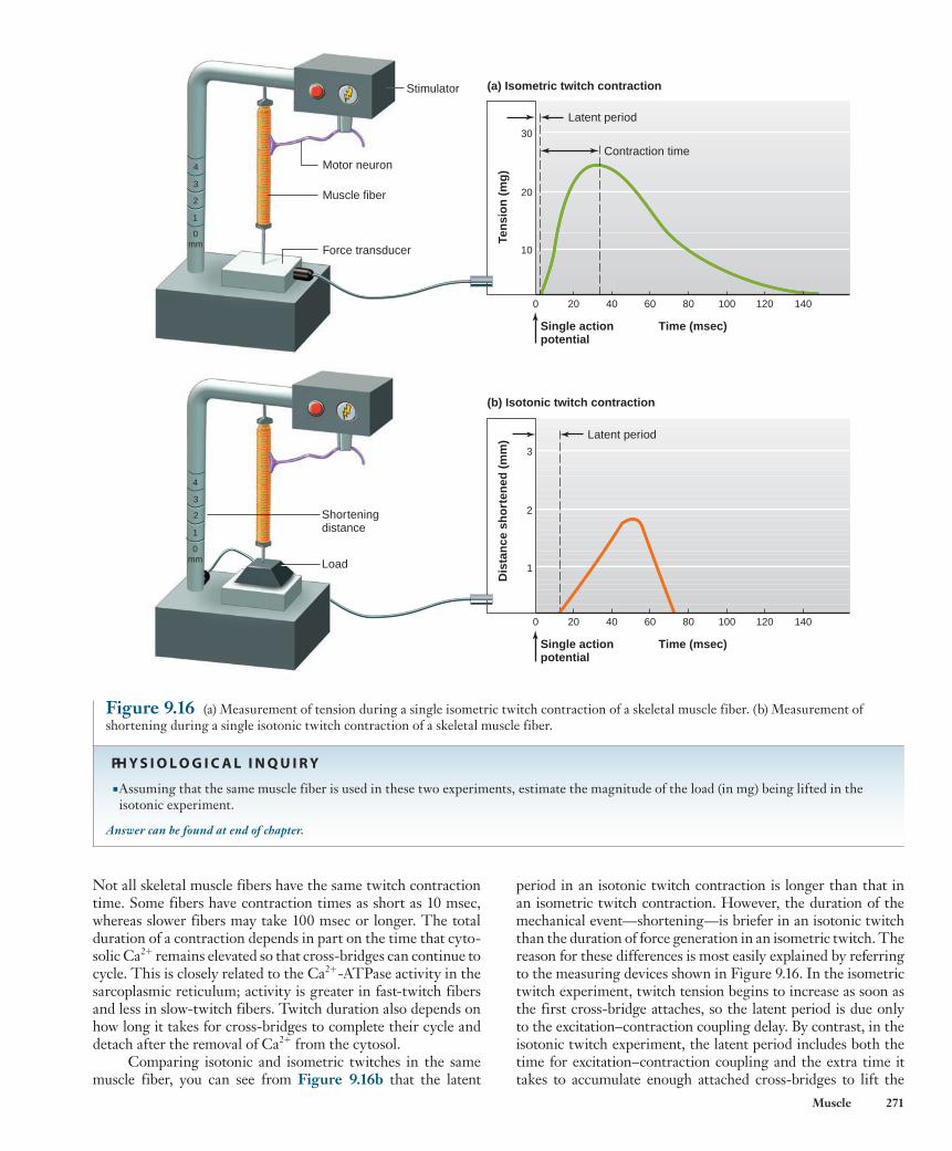
Twitch Contractions The mechanical response of a muscle fiber to a single action potential is known as a twitch . Figure 9.16a shows the main features of an isometric twitch. Following the action poten-tial, there is an interval of a few milliseconds known as the latent period before the tension in the muscle fiber begins to increase. During this latent period, the processes associated with excitation–contraction coupling are occurring. The time interval from the beginning of tension development at the end of the latent period to the peak tension is the contraction time .
wid78305_ch09_257-299.indd 270 30/01/13 11:49 AM
Rev. Confirming Pages
Muscle 271
period in an isotonic twitch contraction is longer than that in an isometric twitch contraction. However, the duration of the mechanical event—shortening—is briefer in an isotonic twitch than the duration of force generation in an isometric twitch. The reason for these differences is most easily explained by referring to the measuring devices shown in Figure 9.16 . In the isometric twitch experiment, twitch tension begins to increase as soon as the first cross-bridge attaches, so the latent period is due only to the excitation–contraction coupling delay. By contrast, in the isotonic twitch experiment, the latent period includes both the time for excitation–contraction coupling and the extra time it takes to accumulate enough attached cross-bridges to lift the
Not all skeletal muscle fibers have the same twitch contraction time. Some fibers have contraction times as short as 10 msec, whereas slower fibers may take 100 msec or longer. The total duration of a contraction depends in part on the time that cyto-solic Ca 2 1 remains elevated so that cross-bridges can continue to cycle. This is closely related to the Ca 2 1 -ATPase activity in the sarcoplasmic reticulum; activity is greater in fast-twitch fibers and less in slow-twitch fibers. Twitch duration also depends on how long it takes for cross-bridges to complete their cycle and detach after the removal of Ca 2 1 from the cytosol.
Comparing isotonic and isometric twitches in the same muscle fiber, you can see from Figure 9.16b that the latent
0 20 40 60 80 100 120 140
Time (msec)Single actionpotential
Ten
sio
n (
mg
)
30
20
10
Dis
tan
ce s
ho
rten
ed (
mm
)
3
2
1
0 20 40 60 80 100 120 140
Time (msec)Single actionpotential
(b) Isotonic twitch contraction
Latent period
Latent period
Stimulator
Contraction time
(a) Isometric twitch contraction
Motor neuron
4
3
2
1
0mm
4
3
2
1
0mm
Muscle fiber
Force transducer
Load
Shorteningdistance
Figure 9.16 (a) Measurement of tension during a single isometric twitch contraction of a skeletal muscle fiber. (b) Measurement of shortening during a single isotonic twitch contraction of a skeletal muscle fiber.
PH Y S I O L O G I C A L I N Q U I R Y
■ Assuming that the same muscle fiber is used in these two experiments, estimate the magnitude of the load (in mg) being lifted in the isotonic experiment.
Answer can be found at end of chapter.
wid78305_ch09_257-299.indd 271 30/01/13 11:49 AM
Rev. Confirming Pages
272 Chapter 9
Frequency–Tension Relation Because a single action potential in a skeletal muscle fiber lasts only 1 to 2 msec but the twitch may last for 100 msec, it is pos-sible for a second action potential to be initiated during the period of mechanical activity. Figure 9.19 illustrates the tension gener-ated during isometric contractions of a muscle fiber in response to multiple stimuli. The isometric twitch following the first stimulus, S 1 , lasts 150 msec. The second stimulus, S 2 , applied to the muscle fiber 200 msec after S 1 , when the fiber has completely relaxed, causes a second identical twitch. When a stimulus is applied before a fiber has completely relaxed from a twitch, it induces a contrac-tile response with a peak tension greater than that produced in a single twitch (S 3 and S 4 ). If the interval between stimuli is reduced further, the resulting peak tension is even greater (S 5 and S 6 ). Indeed, the mechanical response to S 6 is a smooth continuation of the mechanical response already induced by S 5 .
The increase in muscle tension from successive action potentials occurring during the phase of mechanical activity is known as summation . Do not confuse this with the summation of neuronal postsynaptic potentials described in Chapter 6. Post-synaptic potential summation involves additive voltage effects on the membrane, whereas here we are observing the effect of additional attached cross-bridges. A maintained contraction in
load off of the platform. Similarly, at the end of the twitch, the isotonic load comes back to rest on the platform well before all of the cross-bridges have detached in the isometric experiment.
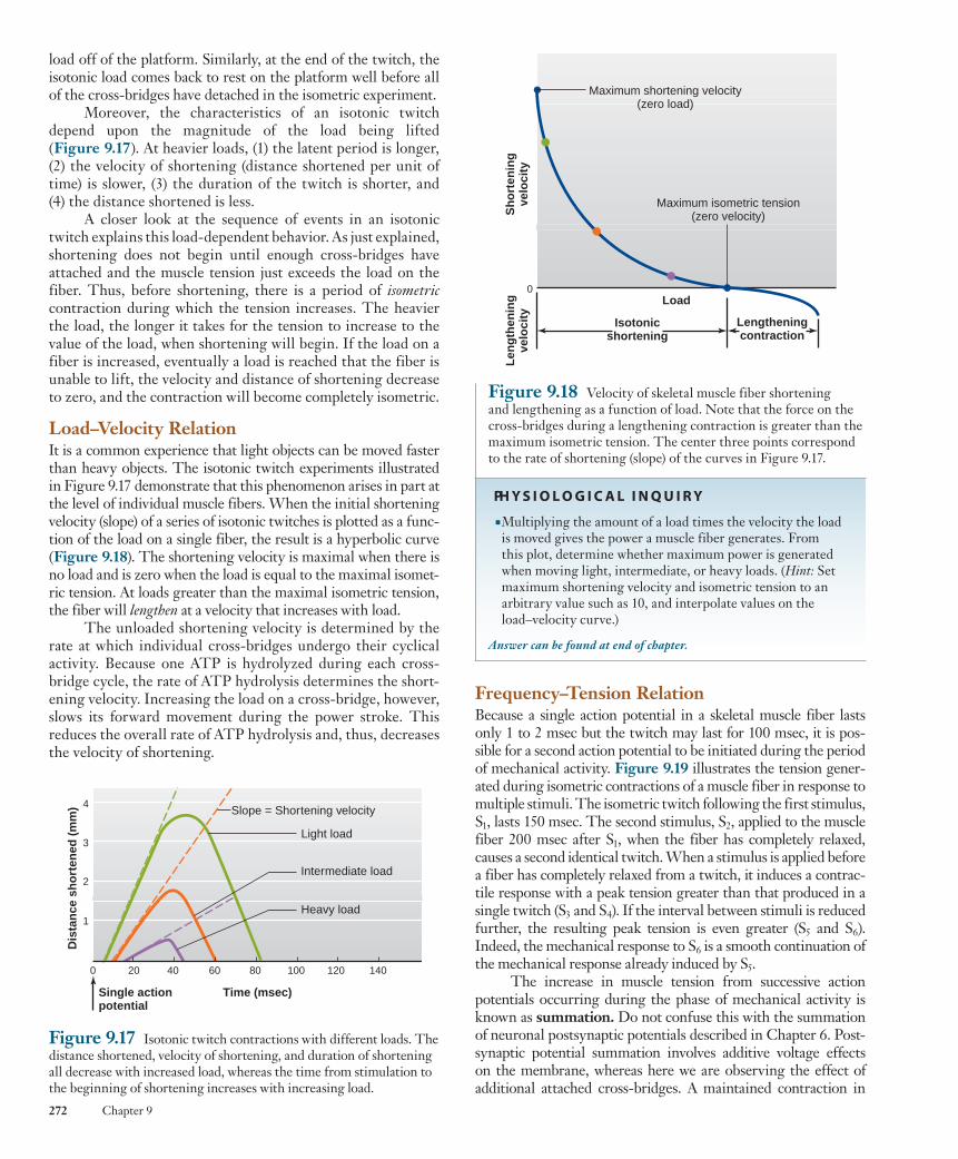
Moreover, the characteristics of an isotonic twitch depend upon the magnitude of the load being lifted ( Figure 9.17 ). At heavier loads, (1) the latent period is longer, (2) the velocity of shortening (distance shortened per unit of time) is slower, (3) the duration of the twitch is shorter, and (4) the distance shortened is less.
A closer look at the sequence of events in an isotonic twitch explains this load-dependent behavior. As just explained, shortening does not begin until enough cross-bridges have attached and the muscle tension just exceeds the load on the fiber. Thus, before shortening, there is a period of isometriccontraction during which the tension increases. The heavier the load, the longer it takes for the tension to increase to the value of the load, when shortening will begin. If the load on a fiber is increased, eventually a load is reached that the fiber is unable to lift, the velocity and distance of shortening decrease to zero, and the contraction will become completely isometric.
Load–Velocity Relation It is a common experience that light objects can be moved faster than heavy objects. The isotonic twitch experiments illustrated in Figure 9.17 demonstrate that this phenomenon arises in part at the level of individual muscle fibers. When the initial shortening velocity (slope) of a series of isotonic twitches is plotted as a func-tion of the load on a single fiber, the result is a hyperbolic curve ( Figure 9.18 ). The shortening velocity is maximal when there is no load and is zero when the load is equal to the maximal isomet-ric tension. At loads greater than the maximal isometric tension, the fiber will lengthen at a velocity that increases with load.
The unloaded shortening velocity is determined by the rate at which individual cross-bridges undergo their cyclical activity. Because one ATP is hydrolyzed during each cross-bridge cycle, the rate of ATP hydrolysis determines the short-ening velocity. Increasing the load on a cross-bridge, however, slows its forward movement during the power stroke. This reduces the overall rate of ATP hydrolysis and, thus, decreases the velocity of shortening.
Figure 9.17 Isotonic twitch contractions with different loads. The distance shortened, velocity of shortening, and duration of shortening all decrease with increased load, whereas the time from stimulation to the beginning of shortening increases with increasing load.
Heavy load
Slope = Shortening velocity
Light load
Intermediate load
Time (msec)Single actionpotential
Dis
tan
ce s
ho
rten
ed (
mm
) 4
3
2
1
0 20 40 60 80 100 120 140
Maximum shortening velocity(zero load)
Maximum isometric tension(zero velocity)
Load
Isotonicshortening
Lengtheningcontraction
Sh
ort
enin
gve
loci
tyL
eng
then
ing
velo
city
0
Figure 9.18 Velocity of skeletal muscle fiber shortening and lengthening as a function of load. Note that the force on the cross-bridges during a lengthening contraction is greater than the maximum isometric tension. The center three points correspond to the rate of shortening (slope) of the curves in Figure 9.17 .
PH Y S I O L O G I C A L I N Q U I R Y
■ Multiplying the amount of a load times the velocity the load is moved gives the power a muscle fiber generates. From this plot, determine whether maximum power is generated when moving light, intermediate, or heavy loads. ( Hint: Set maximum shortening velocity and isometric tension to an arbitrary value such as 10, and interpolate values on the load–velocity curve.)
Answer can be found at end of chapter.
wid78305_ch09_257-299.indd 272 30/01/13 11:49 AM

Rev. Confirming Pages
Muscle 273
Figure 9.19 Summation of isometric contractions produced by shortening the time between stimuli.
Ten
sio
n
100 200 300 4000
S1 S2
500 600 700
S3 S4
800 900 1000
S5 S6
Time (msec)
0
1
2
3
Rel
ativ
e te
nsi
on
S S S S S S100 200 300 400 500 600
S S S S S S S S S S S S S S S S700 800 900 1000
S S
Time (msec)
Twitch
Fused tetanus
Unfused tetanus
Figure 9.20 Isometric contractions produced by multiple stimuli (S) at 10 stimuli per second (unfused tetanus) and 100 stimuli per second (fused tetanus), as compared with a single twitch.
PH Y S I O L O G I C A L I N Q U I R Y
■ If the twitch contraction time is 35 msec and twitch duration is 150 msec, estimate the range of stimulation frequencies (stimuli per second) over which unfused tetanic contractions will occur.
Answer can be found at end of chapter.
response to repetitive stimulation is known as a tetanus (tetanic contraction). At low stimulation frequencies, the tension may oscillate as the muscle fiber partially relaxes between stimuli, pro-ducing an unfused tetanus . A fused tetanus , with no oscilla-tions, is produced at higher stimulation frequencies ( Figure 9.20 ).
As the frequency of action potentials increases, the level of tension increases by summation until a maximal fused tetanic tension is reached, beyond which tension no longer increases even with further increases in stimulation frequency. This maxi-mal tetanic tension is about three to five times greater than the isometric twitch tension. Different muscle fibers have different contraction times, so the stimulus frequency that will produce a maximal tetanic tension differs from fiber to fiber.
Why is tetanic tension so much greater than twitch tension? We can explain summation of tension in part by considering the relative timing of Ca 2 1 availability and cross-bridge binding. The isometric tension produced by a muscle fiber at any instant depends mainly on the total number of cross-bridges bound to actin and undergoing the power stroke of the cross-bridge cycle. Recall that a single action potential in a skeletal muscle fiber briefly releases enough Ca 2 1 to saturate troponin, and all the myosin-binding sites on the thin filaments are therefore initiallyavailable. However, the binding of energized cross-bridges to these sites (step 1 of the cross-bridge cycle) takes time, whereas the Ca 2 1 released into the cytosol begins to be pumped back into the sarcoplasmic reticulum almost immediately. Thus, after a
single action potential, the Ca 2 1 concentration begins to decrease and the troponin–tropomyosin complex reblocks many binding sites before cross-bridges have had time to attach to them.
In contrast, during a tetanic contraction, the succes-sive action potentials each release Ca 2 1 from the sarcoplasmic reticulum before all the Ca 2 1 from the previous action poten-tial has been pumped back into the sarcoplasmic reticulum. This results in a persistent elevation of cytosolic Ca 2 1 concen-tration, which prevents a decline in the number of available binding sites on the thin filaments. Under these conditions, more binding sites remain available and many more cross-bridges become bound to the thin filaments.
Other causes of the lower tension seen in a single twitch are elastic structures, such as muscle tendons and the protein titin, which delay the transmission of cross-bridge force to the ends of a fiber. Because a single twitch is so brief, cross-bridge activity is already declining before force has been fully trans-mitted through these structures. This is less of a factor dur-ing tetanic stimulation because of the much longer duration of cross-bridge activity and force generation.
Length–Tension Relation The springlike characteristic of the protein titin (see Figure 9.4 ), which is attached to the Z line at one end and the thick filaments at the other, is responsible for most of the passive elastic proper-ties of relaxed muscle fibers. With increased stretch, the passive
wid78305_ch09_257-299.indd 273 30/01/13 11:49 AM
Rev. Confirming Pages
274 Chapter 9
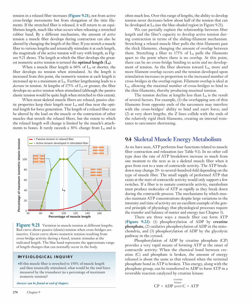
often much less. Over this range of lengths, the ability to develop tension never decreases below about half of the tension that can be developed at L 0 (see the blue-shaded region in Figure 9.21 ).
We can partially explain the relationship between fiber length and the fiber’s capacity to develop active tension dur-ing contraction in terms of the sliding-filament mechanism. Stretching a relaxed muscle fiber pulls the thin filaments past the thick filaments, changing the amount of overlap between them. Stretching a fiber to 175% of L 0 pulls the filaments apart to the point where there is no overlap. At this point, there can be no cross-bridge binding to actin and no develop-ment of tension. As the fiber shortens toward L 0 , more and more filament overlap occurs and the tension developed upon stimulation increases in proportion to the increased number of cross-bridges in the overlap region. Filament overlap is ideal at L 0 , allowing the maximal number of cross-bridges to bind to the thin filaments, thereby producing maximal tension.
The tension decline at lengths less than L 0 is the result of several factors. For example, (1) the overlapping sets of thin filaments from opposite ends of the sarcomere may interfere with the cross-bridges’ ability to bind and exert force; and (2) at very short lengths, the Z lines collide with the ends of the relatively rigid thick filaments, creating an internal resis-tance to sarcomere shortening.
9.4 Skeletal Muscle Energy Metabolism As we have seen, ATP performs four functions related to muscle fiber contraction and relaxation (see Table 9.1 ). In no other cell type does the rate of ATP breakdown increase so much from one moment to the next as in a skeletal muscle fiber when it goes from rest to a state of contractile activity. The ATP break-down may change 20- to several-hundred-fold depending on the type of muscle fiber. The small supply of preformed ATP that exists at the start of contractile activity would only support a few twitches. If a fiber is to sustain contractile activity, metabolism must produce molecules of ATP as rapidly as they break down during the contractile process. The mechanisms by which mus-cles maintain ATP concentrations despite large variations in the intensity and time of activity are an excellent example of the gen-eral principle of physiology that physiological processes require the transfer and balance of matter and energy (see Chapter 1).
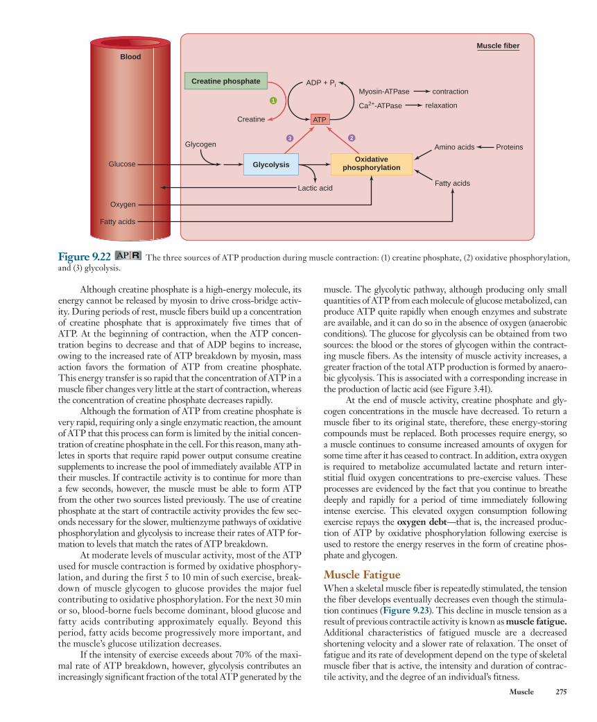
There are three ways a muscle fiber can form ATP ( Figure 9.22 ): (1) phosphorylation of ADP by creatine phosphate , (2) oxidative phosphorylation of ADP in the mito-chondria, and (3) phosphorylation of ADP by the glycolytic pathway in the cytosol.
Phosphorylation of ADP by creatine phosphate (CP) provides a very rapid means of forming ATP at the onset of contractile activity. When the chemical bond between cre-atine (C) and phosphate is broken, the amount of energy released is about the same as that released when the terminal phosphate bond in ATP is broken. This energy, along with the phosphate group, can be transferred to ADP to form ATP in a reversible reaction catalyzed by creatine kinase: creatine kinase
CP 1 ADP 34 C 1 ATP
tension in a relaxed fiber increases ( Figure 9.21 ), not from active cross-bridge movements but from elongation of the titin fila-ments. If the stretched fiber is released, it will return to an equi-librium length, much like what occurs when releasing a stretched rubber band. By a different mechanism, the amount of activetension a muscle fiber develops during contraction can also be altered by changing the length of the fiber. If you stretch a muscle fiber to various lengths and tetanically stimulate it at each length, the magnitude of the active tension will vary with length, as Fig-ure 9.21 shows. The length at which the fiber develops the great-est isometric active tension is termed the optimal length ( L 0 ).
When a muscle fiber length is 60% of L 0 or shorter, the fiber develops no tension when stimulated. As the length is increased from this point, the isometric tension at each length is increased up to a maximum at L 0 . Further lengthening leads to a decrease in tension. At lengths of 175% of L 0 or greater, the fiber develops no active tension when stimulated (although the passive elastic tension would be quite high when stretched to this extent).
When most skeletal muscle fibers are relaxed, passive elas-tic properties keep their length near L 0 and thus near the opti-mal length for force generation. The length of a relaxed fiber can be altered by the load on the muscle or the contraction of other muscles that stretch the relaxed fibers, but the extent to which the relaxed length will change is limited by the muscle’s attach-ments to bones. It rarely exceeds a 30% change from L 0 and is
40 60 80 100 120 140 160Percentage of muscle length
100
= Passive tension in relaxed fiber
= Active tension developed in stimulated fiber
80
60
40
20
0Per
cen
tag
e o
f m
axim
um
iso
met
ric
teta
nic
ten
sio
n
L0
Figure 9.21 Variation in muscle tension at different lengths. Red curve shows passive (elastic) tension when cross-bridges are inactive. Green curve shows isometric tension resulting from cross-bridge activity during a fused, tetanic stimulus at the indicated length. The blue band represents the approximate range of length changes that can normally occur in the body.
PH Y S I O L O G I C A L I N Q U I R Y
■ If this muscle fiber is stretched to 150% of muscle length and then tetanically stimulated, what would be the total force measured by the transducer (as a percentage of maximum isometric tension)?
Answer can be found at end of chapter.
wid78305_ch09_257-299.indd 274 30/01/13 11:49 AM
Rev. Confirming Pages
Muscle 275
muscle. The glycolytic pathway, although producing only small quantities of ATP from each molecule of glucose metabolized, can produce ATP quite rapidly when enough enzymes and substrate are available, and it can do so in the absence of oxygen (anaerobic conditions). The glucose for glycolysis can be obtained from two sources: the blood or the stores of glycogen within the contract-ing muscle fibers. As the intensity of muscle activity increases, a greater fraction of the total ATP production is formed by anaero-bic glycolysis. This is associated with a corresponding increase in the production of lactic acid (see Figure 3.41).
At the end of muscle activity, creatine phosphate and gly-cogen concentrations in the muscle have decreased. To return a muscle fiber to its original state, therefore, these energy-storing compounds must be replaced. Both processes require energy, so a muscle continues to consume increased amounts of oxygen for some time after it has ceased to contract. In addition, extra oxygen is required to metabolize accumulated lactate and return inter-stitial fluid oxygen concentrations to pre-exercise values. These processes are evidenced by the fact that you continue to breathe deeply and rapidly for a period of time immediately following intense exercise. This elevated oxygen consumption following exercise repays the oxygen debt —that is, the increased produc-tion of ATP by oxidative phosphorylation following exercise is used to restore the energy reserves in the form of creatine phos-phate and glycogen.
Muscle Fatigue When a skeletal muscle fiber is repeatedly stimulated, the tension the fiber develops eventually decreases even though the stimula-tion continues ( Figure 9.23 ). This decline in muscle tension as a result of previous contractile activity is known as muscle fatigue . Additional characteristics of fatigued muscle are a decreased shortening velocity and a slower rate of relaxation. The onset of fatigue and its rate of development depend on the type of skeletal muscle fiber that is active, the intensity and duration of contrac-tile activity, and the degree of an individual’s fitness.
Although creatine phosphate is a high-energy molecule, its energy cannot be released by myosin to drive cross-bridge activ-ity. During periods of rest, muscle fibers build up a concentration of creatine phosphate that is approximately five times that of ATP. At the beginning of contraction, when the ATP concen-tration begins to decrease and that of ADP begins to increase, owing to the increased rate of ATP breakdown by myosin, mass action favors the formation of ATP from creatine phosphate. This energy transfer is so rapid that the concentration of ATP in a muscle fiber changes very little at the start of contraction, whereas the concentration of creatine phosphate decreases rapidly.
Although the formation of ATP from creatine phosphate is very rapid, requiring only a single enzymatic reaction, the amount of ATP that this process can form is limited by the initial concen-tration of creatine phosphate in the cell. For this reason, many ath-letes in sports that require rapid power output consume creatine supplements to increase the pool of immediately available ATP in their muscles. If contractile activity is to continue for more than a few seconds, however, the muscle must be able to form ATP from the other two sources listed previously. The use of creatine phosphate at the start of contractile activity provides the few sec-onds necessary for the slower, multienzyme pathways of oxidative phosphorylation and glycolysis to increase their rates of ATP for-mation to levels that match the rates of ATP breakdown.
At moderate levels of muscular activity, most of the ATP used for muscle contraction is formed by oxidative phosphory-lation, and during the first 5 to 10 min of such exercise, break-down of muscle glycogen to glucose provides the major fuel contributing to oxidative phosphorylation. For the next 30 min or so, blood-borne fuels become dominant, blood glucose and fatty acids contributing approximately equally. Beyond this period, fatty acids become progressively more important, and the muscle’s glucose utilization decreases.
If the intensity of exercise exceeds about 70% of the maxi-mal rate of ATP breakdown, however, glycolysis contributes an increasingly significant fraction of the total ATP generated by the
Figure 9.22 The three sources of ATP production during muscle contraction: (1) creatine phosphate, (2) oxidative phosphorylation, and (3) glycolysis.
3 2
1
ATP
OxidativephosphorylationGlycolysis
Lactic acid
Glycogen
Creatine phosphate
Creatine
ADP + Pi
Fatty acids
Amino acids Proteins
Ca2+-ATPase
Myosin-ATPase contraction
relaxation
Muscle fiber
Glucose
Oxygen
Fatty acids
Blood
wid78305_ch09_257-299.indd 275 30/01/13 11:49 AM
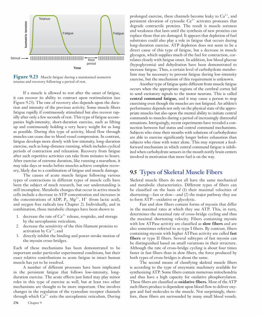
Rev. Confirming Pages
276 Chapter 9
prolonged exercise, these channels become leaky to Ca 2 1 , and persistent elevation of cytosolic Ca 2 1 activates proteases that degrade contractile proteins. The result is muscle soreness and weakness that lasts until the synthesis of new proteins can replace those that are damaged. It appears that depletion of fuel substrates could also play a role in fatigue that occurs during long-duration exercise. ATP depletion does not seem to be a direct cause of this type of fatigue, but a decrease in muscle glycogen, which supplies much of the fuel for contraction, cor-relates closely with fatigue onset. In addition, low blood glucose (hypoglycemia) and dehydration have been demonstrated to increase fatigue. Thus, a certain level of carbohydrate metabo-lism may be necessary to prevent fatigue during low-intensity exercise, but the mechanism of this requirement is unknown.
Another type of fatigue quite different from muscle fatigue occurs when the appropriate regions of the cerebral cortex fail to send excitatory signals to the motor neurons. This is called central command fatigue , and it may cause a person to stop exercising even though the muscles are not fatigued. An athlete’s performance depends not only on the physical state of the appro-priate muscles but also upon the mental ability to initiate central commands to muscles during a period of increasingly distressful sensations. Intriguingly, recent experiments have revealed a con-nection between fuel status and central command mechanisms. Subjects who rinse their mouths with solutions of carbohydrates are able to exercise significantly longer before exhaustion than subjects who rinse with water alone. This may represent a feed-forward mechanism in which central command fatigue is inhib-ited when carbohydrate sensors in the mouth notify brain centers involved in motivation that more fuel is on the way.
9.5 Types of Skeletal Muscle Fibers Skeletal muscle fibers do not all have the same mechanical and metabolic characteristics. Different types of fibers can be classified on the basis of (1) their maximal velocities of shortening—fast or slow—and (2) the major pathway they use to form ATP—oxidative or glycolytic.
Fast and slow fibers contain forms of myosin that differ in the maximal rates at which they use ATP. This, in turn, determines the maximal rate of cross-bridge cycling and thus the maximal shortening velocity. Fibers containing myosin with low ATPase activity are classified as slow fibers and are also sometimes referred to as type I fibers. By contrast, fibers containing myosin with higher ATPase activity are called fast fibers or type II fibers. Several subtypes of fast myosin can be distinguished based on small variations in their structure. Although the rate of cross-bridge cycling is about four times faster in fast fibers than in slow fibers, the force produced by both types of cross-bridges is about the same.
The second means of classifying skeletal muscle fibers is according to the type of enzymatic machinery available for synthesizing ATP. Some fibers contain numerous mitochondria and thus have a high capacity for oxidative phosphorylation. These fibers are classified as oxidative fibers . Most of the ATP such fibers produce is dependent upon blood flow to deliver oxy-gen and fuel molecules to the muscle. Not surprisingly, there-fore, these fibers are surrounded by many small blood vessels.
If a muscle is allowed to rest after the onset of fatigue, it can recover its ability to contract upon restimulation (see Figure 9.23 ). The rate of recovery also depends upon the dura-tion and intensity of the previous activity. Some muscle fibers fatigue rapidly if continuously stimulated but also recover rap-idly after only a few seconds of rest. This type of fatigue accom-panies high-intensity, short-duration exercise, such as lifting up and continuously holding a very heavy weight for as long as possible. During this type of activity, blood flow through muscles can cease due to blood vessel compression. In contrast, fatigue develops more slowly with low-intensity, long-duration exercise, such as long-distance running, which includes cyclical periods of contraction and relaxation. Recovery from fatigue after such repetitive activities can take from minutes to hours. After exercise of extreme duration, like running a marathon, it may take days or weeks before muscles achieve complete recov-ery, likely due to a combination of fatigue and muscle damage.
The causes of acute muscle fatigue following various types of contractions in different types of muscle cells have been the subject of much research, but our understanding is still incomplete. Metabolic changes that occur in active muscle cells include a decrease in ATP concentration and increases in the concentrations of ADP, P i , Mg 2 1 , H 1 (from lactic acid), and oxygen free radicals (see Chapter 2). Individually, and in combination, those metabolic changes have been shown to
1. decrease the rate of Ca 2 1 release, reuptake, and storage by the sarcoplasmic reticulum;
2. decrease the sensitivity of the thin filament proteins to activation by Ca 2 1 ; and
3. directly inhibit the binding and power-stroke motion of the myosin cross-bridges.
Each of these mechanisms has been demonstrated to be important under particular experimental conditions, but their exact relative contributions to acute fatigue in intact human muscle has yet to be resolved.
A number of different processes have been implicated in the persistent fatigue that follows low-intensity, long- duration exercise. The acute effects just listed may play minor roles in this type of exercise as well, but at least two other mechanisms are thought to be more important. One involves changes in the regulation of the ryanodine receptor channels through which Ca 2 1 exits the sarcoplasmic reticulum. During
Figure 9.23 Muscle fatigue during a maintained isometric tetanus and recovery following a period of rest.
Stimuli
Iso
met
ric
ten
sio
n
Tetanus
Fatigue Fatigue
Time
Restperiod
wid78305_ch09_257-299.indd 276 30/01/13 11:49 AM
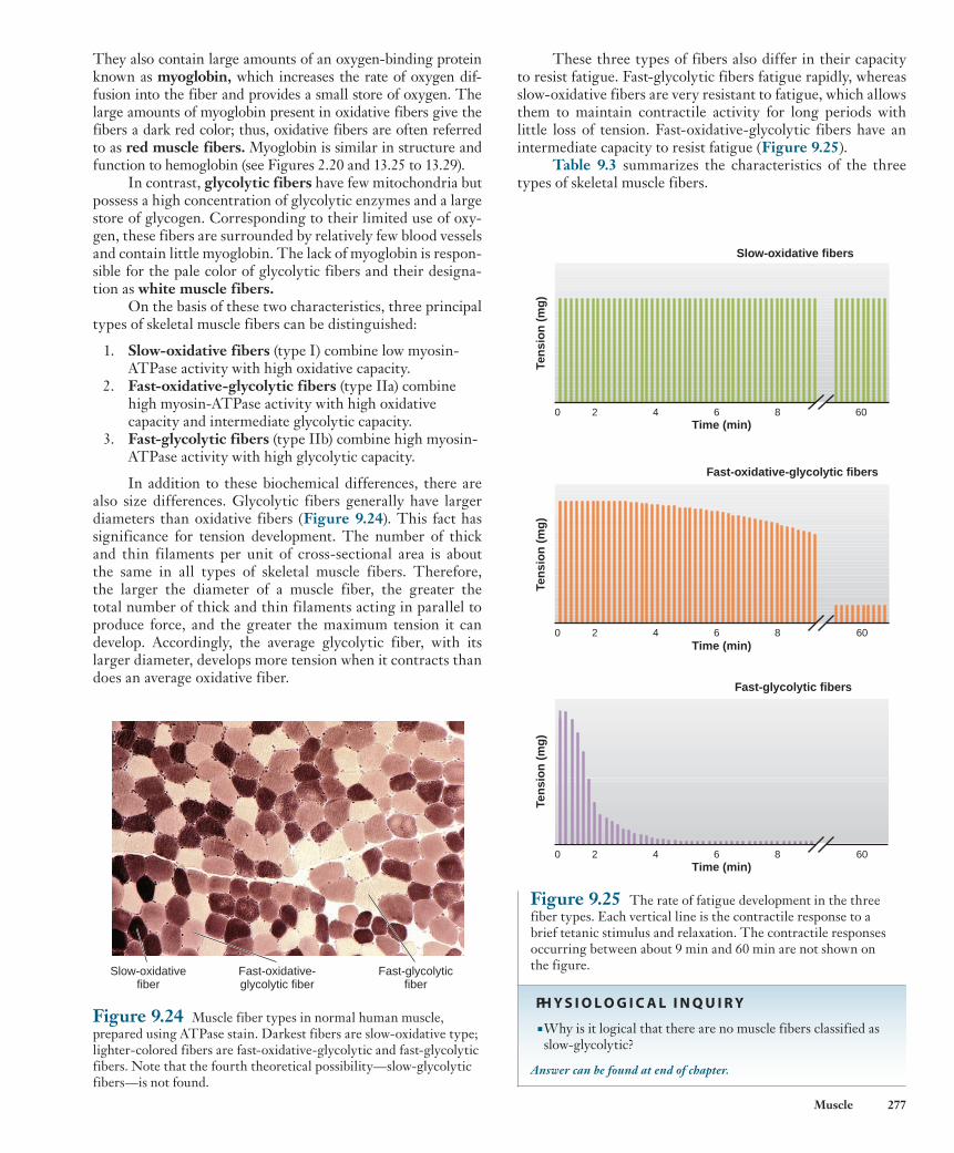
Rev. Confirming Pages
Muscle 277
They also contain large amounts of an oxygen-binding protein known as myoglobin , which increases the rate of oxygen dif-fusion into the fiber and provides a small store of oxygen. The large amounts of myoglobin present in oxidative fibers give the fibers a dark red color; thus, oxidative fibers are often referred to as red muscle fibers . Myoglobin is similar in structure and function to hemoglobin (see Figures 2.20 and 13.25 to 13.29).
In contrast, glycolytic fibers have few mitochondria but possess a high concentration of glycolytic enzymes and a large store of glycogen. Corresponding to their limited use of oxy-gen, these fibers are surrounded by relatively few blood vessels and contain little myoglobin. The lack of myoglobin is respon-sible for the pale color of glycolytic fibers and their designa-tion as white muscle fibers .
On the basis of these two characteristics, three principal types of skeletal muscle fibers can be distinguished:
1. Slow-oxidative fibers (type I) combine low myosin-ATPase activity with high oxidative capacity.
2. Fast-oxidative-glycolytic fibers (type IIa) combine high myosin-ATPase activity with high oxidative capacity and intermediate glycolytic capacity.
3. Fast-glycolytic fibers (type IIb) combine high myosin-ATPase activity with high glycolytic capacity.
In addition to these biochemical differences, there are also size differences. Glycolytic fibers generally have larger diameters than oxidative fibers ( Figure 9.24 ). This fact has significance for tension development. The number of thick and thin filaments per unit of cross-sectional area is about the same in all types of skeletal muscle fibers. Therefore, the larger the diameter of a muscle fiber, the greater the total number of thick and thin filaments acting in parallel to produce force, and the greater the maximum tension it can develop. Accordingly, the average glycolytic fiber, with its larger diameter, develops more tension when it contracts than does an average oxidative fiber.
Figure 9.24 Muscle fiber types in normal human muscle, prepared using ATPase stain. Darkest fibers are slow-oxidative type; lighter-colored fibers are fast-oxidative-glycolytic and fast-glycolytic fibers. Note that the fourth theoretical possibility—slow-glycolytic fibers—is not found.
Fast-glycolyticfiber
Slow-oxidativefiber
Fast-oxidative-glycolytic fiber
These three types of fibers also differ in their capacity to resist fatigue. Fast-glycolytic fibers fatigue rapidly, whereas slow-oxidative fibers are very resistant to fatigue, which allows them to maintain contractile activity for long periods with little loss of tension. Fast-oxidative-glycolytic fibers have an intermediate capacity to resist fatigue ( Figure 9.25 ).
Table 9.3 summarizes the characteristics of the three types of skeletal muscle fibers.
Ten
sio
n (
mg
)Te
nsi
on
(m
g)
Ten
sio
n (
mg
)
2 4 6 8
Fast-oxidative-glycolytic fibers
60Time (min)
0
2 4 6 8 60Time (min)
Fast-glycolytic fibers
0
0 2 4 6 8 60Time (min)
Slow-oxidative fibers
Figure 9.25 The rate of fatigue development in the three fiber types. Each vertical line is the contractile response to a brief tetanic stimulus and relaxation. The contractile responses occurring between about 9 min and 60 min are not shown on the figure.
PH Y S I O L O G I C A L I N Q U I R Y
■ Why is it logical that there are no muscle fibers classified as slow-glycolytic?
Answer can be found at end of chapter.
wid78305_ch09_257-299.indd 277 30/01/13 11:49 AM
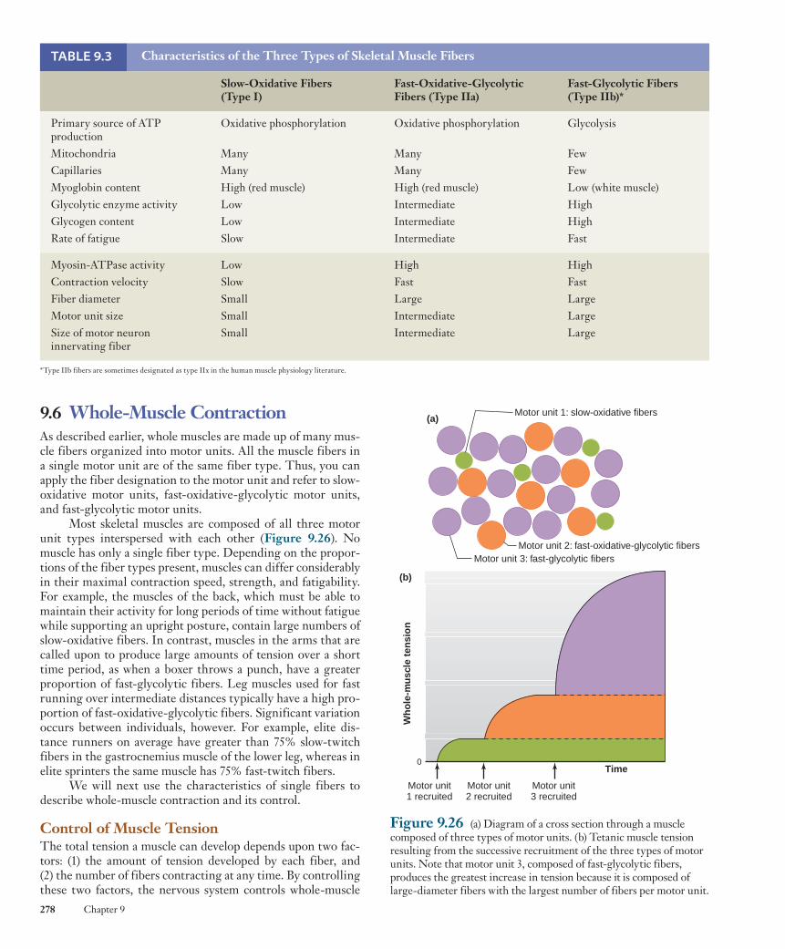
Rev. Confirming Pages
278 Chapter 9
9.6 Whole-Muscle Contraction As described earlier, whole muscles are made up of many mus-cle fibers organized into motor units. All the muscle fibers in a single motor unit are of the same fiber type. Thus, you can apply the fiber designation to the motor unit and refer to slow-oxidative motor units, fast-oxidative-glycolytic motor units, and fast-glycolytic motor units.
Most skeletal muscles are composed of all three motor unit types interspersed with each other ( Figure 9.26 ). No muscle has only a single fiber type. Depending on the propor-tions of the fiber types present, muscles can differ considerably in their maximal contraction speed, strength, and fatigability. For example, the muscles of the back, which must be able to maintain their activity for long periods of time without fatigue while supporting an upright posture, contain large numbers of slow-oxidative fibers. In contrast, muscles in the arms that are called upon to produce large amounts of tension over a short time period, as when a boxer throws a punch, have a greater proportion of fast-glycolytic fibers. Leg muscles used for fast running over intermediate distances typically have a high pro-portion of fast-oxidative-glycolytic fibers. Significant variation occurs between individuals, however. For example, elite dis-tance runners on average have greater than 75% slow-twitch fibers in the gastrocnemius muscle of the lower leg, whereas in elite sprinters the same muscle has 75% fast-twitch fibers.
We will next use the characteristics of single fibers to describe whole-muscle contraction and its control.
Control of Muscle Tension The total tension a muscle can develop depends upon two fac-tors: (1) the amount of tension developed by each fiber, and (2) the number of fibers contracting at any time. By controlling these two factors, the nervous system controls whole-muscle
TABLE 9.3 Characteristics of the Three Types of Skeletal Muscle Fibers
Slow-Oxidative Fibers (Type I)
Fast-Oxidative-Glycolytic Fibers (Type IIa)
Fast-Glycolytic Fibers (Type IIb) *
Primary source of ATP production Mitochondria Capillaries Myoglobin content Glycolytic enzyme activity Glycogen content Rate of fatigue
Oxidative phosphorylation
Many Many High (red muscle) Low Low Slow
Oxidative phosphorylation
Many Many High (red muscle) Intermediate Intermediate Intermediate
Glycolysis
Few Few Low (white muscle) High High Fast
Myosin-ATPase activity Contraction velocity Fiber diameter Motor unit size Size of motor neuron innervating fiber
Low Slow Small Small Small
High Fast Large Intermediate Intermediate
High Fast Large Large Large
*Type IIb fibers are sometimes designated as type IIx in the human muscle physiology literature.
Figure 9.26 (a) Diagram of a cross section through a muscle composed of three types of motor units. (b) Tetanic muscle tension resulting from the successive recruitment of the three types of motor units. Note that motor unit 3, composed of fast-glycolytic fibers, produces the greatest increase in tension because it is composed of large-diameter fibers with the largest number of fibers per motor unit.
Motor unit 2: fast-oxidative-glycolytic fibersMotor unit 3: fast-glycolytic fibers
Motor unit 1: slow-oxidative fibers
Motor unit1 recruited
Motor unit2 recruited
Motor unit3 recruited
Time0
Wh
ole
-mu
scle
ten
sio
n
(b)
(a)
wid78305_ch09_257-299.indd 278 30/01/13 11:49 AM
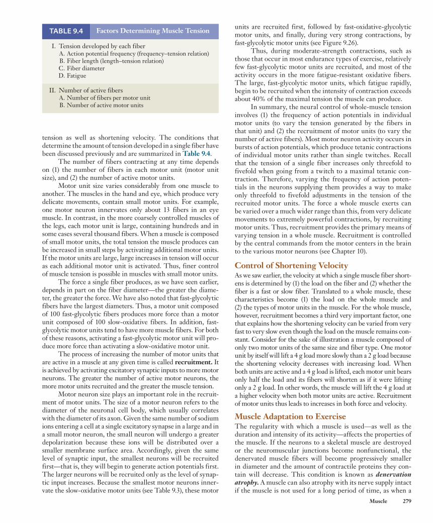
Rev. Confirming Pages
Muscle 279
units are recruited first, followed by fast-oxidative-glycolytic motor units, and finally, during very strong contractions, by fast-glycolytic motor units (see Figure 9.26 ).
Thus, during moderate-strength contractions, such as those that occur in most endurance types of exercise, relatively few fast-glycolytic motor units are recruited, and most of the activity occurs in the more fatigue-resistant oxidative fibers. The large, fast-glycolytic motor units, which fatigue rapidly, begin to be recruited when the intensity of contraction exceeds about 40% of the maximal tension the muscle can produce.
In summary, the neural control of whole-muscle tension involves (1) the frequency of action potentials in individual motor units (to vary the tension generated by the fibers in that unit) and (2) the recruitment of motor units (to vary the number of active fibers). Most motor neuron activity occurs in bursts of action potentials, which produce tetanic contractions of individual motor units rather than single twitches. Recall that the tension of a single fiber increases only threefold to fivefold when going from a twitch to a maximal tetanic con-traction. Therefore, varying the frequency of action poten-tials in the neurons supplying them provides a way to make only threefold to fivefold adjustments in the tension of the recruited motor units. The force a whole muscle exerts can be varied over a much wider range than this, from very delicate movements to extremely powerful contractions, by recruiting motor units. Thus, recruitment provides the primary means of varying tension in a whole muscle. Recruitment is controlled by the central commands from the motor centers in the brain to the various motor neurons (see Chapter 10).
Control of Shortening Velocity As we saw earlier, the velocity at which a single muscle fiber short-ens is determined by (1) the load on the fiber and (2) whether the fiber is a fast or slow fiber. Translated to a whole muscle, these characteristics become (1) the load on the whole muscle and (2) the types of motor units in the muscle. For the whole muscle, however, recruitment becomes a third very important factor, one that explains how the shortening velocity can be varied from very fast to very slow even though the load on the muscle remains con-stant. Consider for the sake of illustration a muscle composed of only two motor units of the same size and fiber type. One motor unit by itself will lift a 4 g load more slowly than a 2 g load because the shortening velocity decreases with increasing load. When both units are active and a 4 g load is lifted, each motor unit bears only half the load and its fibers will shorten as if it were lifting only a 2 g load. In other words, the muscle will lift the 4 g load at a higher velocity when both motor units are active. Recruitment of motor units thus leads to increases in both force and velocity.
Muscle Adaptation to Exercise The regularity with which a muscle is used—as well as the duration and intensity of its activity—affects the properties of the muscle. If the neurons to a skeletal muscle are destroyed or the neuromuscular junctions become nonfunctional, the denervated muscle fibers will become progressively smaller in diameter and the amount of contractile proteins they con-tain will decrease. This condition is known as denervation atrophy . A muscle can also atrophy with its nerve supply intact if the muscle is not used for a long period of time, as when a
tension as well as shortening velocity. The conditions that determine the amount of tension developed in a single fiber have been discussed previously and are summarized in Table 9.4 .
The number of fibers contracting at any time depends on (1) the number of fibers in each motor unit (motor unit size), and (2) the number of active motor units.
Motor unit size varies considerably from one muscle to another. The muscles in the hand and eye, which produce very delicate movements, contain small motor units. For example, one motor neuron innervates only about 13 fibers in an eye muscle. In contrast, in the more coarsely controlled muscles of the legs, each motor unit is large, containing hundreds and in some cases several thousand fibers. When a muscle is composed of small motor units, the total tension the muscle produces can be increased in small steps by activating additional motor units. If the motor units are large, large increases in tension will occur as each additional motor unit is activated. Thus, finer control of muscle tension is possible in muscles with small motor units.
The force a single fiber produces, as we have seen earlier, depends in part on the fiber diameter—the greater the diame-ter, the greater the force. We have also noted that fast-glycolytic fibers have the largest diameters. Thus, a motor unit composed of 100 fast-glycolytic fibers produces more force than a motor unit composed of 100 slow-oxidative fibers. In addition, fast-glycolytic motor units tend to have more muscle fibers. For both of these reasons, activating a fast-glycolytic motor unit will pro-duce more force than activating a slow-oxidative motor unit.
The process of increasing the number of motor units that are active in a muscle at any given time is called recruitment . It is achieved by activating excitatory synaptic inputs to more motor neurons. The greater the number of active motor neurons, the more motor units recruited and the greater the muscle tension.
Motor neuron size plays an important role in the recruit-ment of motor units. The size of a motor neuron refers to the diameter of the neuronal cell body, which usually correlates with the diameter of its axon. Given the same number of sodium ions entering a cell at a single excitatory synapse in a large and in a small motor neuron, the small neuron will undergo a greater depolarization because these ions will be distributed over a smaller membrane surface area. Accordingly, given the same level of synaptic input, the smallest neurons will be recruited first—that is, they will begin to generate action potentials first. The larger neurons will be recruited only as the level of synap-tic input increases. Because the smallest motor neurons inner-vate the slow-oxidative motor units (see Table 9.3 ), these motor
TABLE 9.4 Factors Determining Muscle Tension
I. Tension developed by each fiber A. Action potential frequency (frequency–tension relation) B. Fiber length (length–tension relation) C. Fiber diameter D. Fatigue
II. Number of active fibers A. Number of fibers per motor unit
B. Number of active motor units
wid78305_ch09_257-299.indd 279 30/01/13 11:49 AM
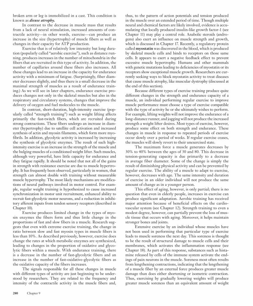
Rev. Confirming Pages
280 Chapter 9
thus, to the pattern of action potentials and tension produced in the muscle over an extended period of time. Though multiple neural and chemical factors are likely involved, evidence is accu-mulating that locally produced insulin-like growth factor-1 (see Chapter 11) may play a central role. Anabolic steroids (andro-gens) also exert an influence on muscle strength and growth, which is discussed in Chapter 17. Recently, a regulatory protein called myostatin was discovered in the blood, which is produced by skeletal muscle cells and binds to receptors on those same cells. It appears to exert a negative feedback effect to prevent excessive muscle hypertrophy. Humans and other mammals with genetic mutations leading to deficiencies of myostatin or its receptors show exceptional muscle growth. Researchers are cur-rently seeking ways to block myostatin activity to treat diseases that cause muscle atrophy, like muscular dystrophy (discussed at the end of this section).
Because different types of exercise training produce quite different changes in the strength and endurance capacity of a muscle, an individual performing regular exercise to improve muscle performance must choose a type of exercise compatible with the type of activity he or she ultimately wishes to perform. For example, lifting weights will not improve the endurance of a long-distance runner, and jogging will not produce the increased strength a weight lifter desires. Most types of exercise, however, produce some effect on both strength and endurance. These changes in muscle in response to repeated periods of exercise occur slowly over a period of weeks. If regular exercise ceases, the muscles will slowly revert to their unexercised state.
The maximum force a muscle generates decreases by 30% to 40% between the ages of 30 and 80. This decrease in tension-generating capacity is due primarily to a decrease in average fiber diameter. Some of the change is simply the result of diminishing physical activity and can be prevented by regular exercise. The ability of a muscle to adapt to exercise, however, decreases with age. The same intensity and duration of exercise in an older individual will not produce the same amount of change as in a younger person.
This effect of aging, however, is only partial; there is no question that even in elderly people, increases in exercise can produce significant adaptation. Aerobic training has received major attention because of beneficial effects on the cardio-vascular system (see Chapter 12). Strength training to even a modest degree, however, can partially prevent the loss of mus-cle tissue that occurs with aging. Moreover, it helps maintain stronger bones and joints.
Extensive exercise by an individual whose muscles have not been used in performing that particular type of exercise leads to muscle soreness the next day. This soreness is thought to be the result of structural damage to muscle cells and their membranes, which activates the inflammation response (see Chapter 18). As part of this response, substances such as hista-mine released by cells of the immune system activate the end-ings of pain neurons in the muscle. Soreness most often results from lengthening contractions, indicating that the lengthening of a muscle fiber by an external force produces greater muscle damage than does either shortening or isometric contraction. Thus, exercising by gradually lowering weights will produce greater muscle soreness than an equivalent amount of weight
broken arm or leg is immobilized in a cast. This condition is known as disuse atrophy .
In contrast to the decrease in muscle mass that results from a lack of neural stimulation, increased amounts of con-tractile activity—in other words, exercise—can produce an increase in the size (hypertrophy) of muscle fibers as well as changes in their capacity for ATP production.
Exercise that is of relatively low intensity but long dura-tion (popularly called “aerobic exercise”), such as distance run-ning, produces increases in the number of mitochondria in the fibers that are recruited in this type of activity. In addition, the number of capillaries around these fibers also increases. All these changes lead to an increase in the capacity for endurance activity with a minimum of fatigue. (Surprisingly, fiber diam-eter decreases slightly, and thus there is a small decrease in the maximal strength of muscles as a result of endurance train-ing.) As we will see in later chapters, endurance exercise pro-duces changes not only in the skeletal muscles but also in the respiratory and circulatory systems, changes that improve the delivery of oxygen and fuel molecules to the muscle.
In contrast, short-duration, high-intensity exercise (pop-ularly called “strength training”) such as weight lifting affects primarily the fast-twitch fibers, which are recruited during strong contractions. These fibers undergo an increase in diam-eter (hypertrophy) due to satellite cell activation and increased synthesis of actin and myosin filaments, which form more myo-fibrils. In addition, glycolytic activity is increased by increasing the synthesis of glycolytic enzymes. The result of such high-intensity exercise is an increase in the strength of the muscle and the bulging muscles of a conditioned weight lifter. Such muscles, although very powerful, have little capacity for endurance and they fatigue rapidly. It should be noted that not all of the gains in strength with resistance exercise are due to muscle hypertro-phy. It has frequently been observed, particularly in women, that strength can almost double with training without measurable muscle hypertrophy. The most likely mechanisms are modifica-tions of neural pathways involved in motor control. For exam-ple, regular weight training is hypothesized to cause increased synchronization in motor unit recruitment, enhanced ability to recruit fast-glycolytic motor neurons, and a reduction in inhibi-tory afferent inputs from tendon sensory receptors (described in Chapter 10).
Exercise produces limited change in the types of myo-sin enzymes the fibers form and thus little change in the proportions of fast and slow fibers in a muscle. Research sug-gests that even with extreme exercise training, the change in ratio between slow and fast myosin types in muscle fibers is less than 10%. As described previously, however, exercise does change the rates at which metabolic enzymes are synthesized, leading to changes in the proportion of oxidative and glyco-lytic fibers within a muscle. With endurance training, there is a decrease in the number of fast-glycolytic fibers and an increase in the number of fast-oxidative-glycolytic fibers as the oxidative capacity of the fibers increases.
The signals responsible for all these changes in muscle with different types of activity are just beginning to be under-stood by researchers. They are related to the frequency and intensity of the contractile activity in the muscle fibers and,
wid78305_ch09_257-299.indd 280 30/01/13 11:49 AM

Rev. Confirming Pages
Muscle 281
lifting. This explains a phenomenon well-known to athletic trainers: The shortening contractions of leg muscles used to run up flights of stairs result in far less soreness than the lengthening contractions used for running down. Interestingly, it has been demonstrated that most of the strength gains during weight lifting is due to the eccentric portion of the movement. It therefore seems that the mechanisms underlying muscle soreness and muscle adaptation to exercise are related.
Lever Action of Muscles and Bones A contracting muscle exerts a force on bones through its connect-ing tendons. When the force is great enough, the bone moves as the muscle shortens. A contracting muscle exerts only a pull-ing force, so that as the muscle shortens, the bones it is attached to are pulled toward each other. Flexion refers to the bendingof a limb at a joint, whereas extension is the straightening of a limb ( Figure 9.27 ). These opposing motions require at least two muscles, one to cause flexion and the other extension. Groups of muscles that produce oppositely directed movements at a joint are known as antagonists . For example, from Figure 9.27 we
Figure 9.27 Antagonistic muscles for flexion and extension of the forearm.
Tendon
Tendon
Tendon
Tendon
Triceps
Tricepscontracts
Biceps
Bicepscontracts
Extension Flexion
Figure 9.28 Contraction of the gastrocnemius muscle in the calf can lead either to flexion of the leg, if the quadriceps femoris muscle is relaxed, or to extension of the foot, if the quadriceps is contracting, preventing the knee joint from bending.
Quadricepsfemoris
Gastrocnemius
Gastrocnemiuscontracts
Quadricepsfemorisrelaxed
Quadricepsfemoriscontracts
Flexion of leg Extension of foot
can see that contraction of the biceps causes flexion of the arm at the elbow, whereas contraction of the antagonistic muscle, the triceps, causes the arm to extend. Both muscles exert only a pull-ing force upon the forearm when they contract.
Sets of antagonistic muscles are required not only for flexion–extension but also for side-to-side movements or rotation of a limb. The contraction of some muscles leads to two types of limb movement, depending on the contractile state of other mus-cles acting on the same limb. For example, contraction of the gas-trocnemius muscle in the calf causes a flexion of the leg at the knee, as in walking ( Figure 9.28 ). However, contraction of the gastroc-nemius muscle with the simultaneous contraction of the quadri-ceps femoris (which causes extension of the lower leg) prevents the knee joint from bending, leaving only the ankle joint capable of moving. The foot is extended, and the body rises on tiptoe.
wid78305_ch09_257-299.indd 281 30/01/13 11:49 AM
Rev. Confirming Pages
282 Chapter 9
9.7 Skeletal Muscle Disorders A number of conditions and diseases can affect the contrac-tion of skeletal muscle. Many of them are caused by defects in the parts of the nervous system that control contraction of the muscle fibers rather than by defects in the muscle fibers themselves. For example, poliomyelitis is a viral disease that destroys motor neurons, leading to the paralysis of skeletal muscle, and may result in death due to respiratory failure.
Muscle Cramps Involuntary tetanic contraction of skeletal muscles produces muscle cramps . During cramping, action potentials fire at abnor-mally high rates, a much greater rate than occurs during maximal voluntary contraction. The specific cause of this high activity is uncertain, but it is probably related to electrolyte imbalances in the extracellular fluid surrounding both the muscle and nerve fibers. These imbalances may arise from overexercise or persis-tent dehydration, and they can directly induce action potentials in motor neurons and muscle fibers. Another theory is that chemi-cal imbalances within the muscle stimulate sensory receptors in the muscle, and the motor neurons to the area are activated by reflex when those signals reach the spinal cord.
Hypocalcemic Tetany Hypocalcemic tetany is the involuntary tetanic contraction of skeletal muscles that occurs when the extracellular Ca 2 1 concen-tration decreases to about 40% of its normal value. This may
The muscles, bones, and joints in the body are arranged in lever systems—a good example of the general principle of physiology that physiological processes are dictated by the laws of chemistry and physics. The basic principle of a lever is illustrated by the flexion of the arm by the biceps muscle ( Figure 9.29 ), which exerts an upward pulling tension on the forearm about 5 cm away from the elbow joint. In this exam-ple, a 10 kg weight held in the hand exerts a downward load of 10 kg about 35 cm from the elbow. A law of physics tells us that the forearm is in mechanical equilibrium when the product of the downward load (10 kg) and its distance from the elbow (35 cm) is equal to the product of the isometric tension exerted by the muscle ( X ) and its distance from the elbow (5 cm); that is, 10 3 35 5 X 3 5. Thus, X 5 70 kg. The important point is that this system is working at a mechanical disadvantage because the tension exerted by the muscle (70 kg) is consider-ably greater than the load (10 kg) it is supporting.
However, the mechanical disadvantage that most muscle lever systems operate under is offset by increased maneuverabil-ity. As illustrated in Figure 9.30 , when the biceps shortens 1 cm, the hand moves through a distance of 7 cm. Because the muscle shortens 1 cm in the same amount of time that the hand moves 7 cm, the velocity at which the hand moves is seven times greater than the rate of muscle shortening. The lever system amplifies the velocity of muscle shortening so that short, relatively slow movements of the muscle produce faster movements of the hand. Thus, a pitcher can throw a baseball at 90 to 100 mph even though his muscles shorten at only a small fraction of this velocity.
Figure 9.30 The lever system of the arm amplifies the velocity of the biceps muscle, producing a greater velocity of the hand. The range of movement is also amplified (1 cm of shortening by the muscle produces 7 cm of movement by the hand).
PH Y S I O L O G I C A L I N Q U I R Y
■ If an individual’s biceps insertion was 5 cm from the elbow joint (as shown in Figure 9.29 ) and the center of the hand was 45 cm from the elbow joint, how fast would an object move if the biceps shortened at 2 cm/sec?
Answer can be found at end of chapter.
Force
1 cm
7 cm
Vm = Muscle contraction velocity
Vh = Hand velocity = 7 Vm
Figure 9.29 Mechanical equilibrium of forces acting on the forearm while supporting a 10 kg load. For simplicity, mass is used as a measure of the force here rather than newtons, which are the standard scientific units of force.
PH Y S I O L O G I C A L I N Q U I R Y
■ Describe what would happen if this weight was mounted on a rod that moved it 10 cm farther away from the elbow and the tension generated by the muscle was increased to 85 kg.
Answer can be found at end of chapter.
X = 70 kg
10 kg ë 35 cm = X ë 5 cmX = 70 kg
5 cm 30 cm 10 kg
10 kg
wid78305_ch09_257-299.indd 282 30/01/13 11:49 AM
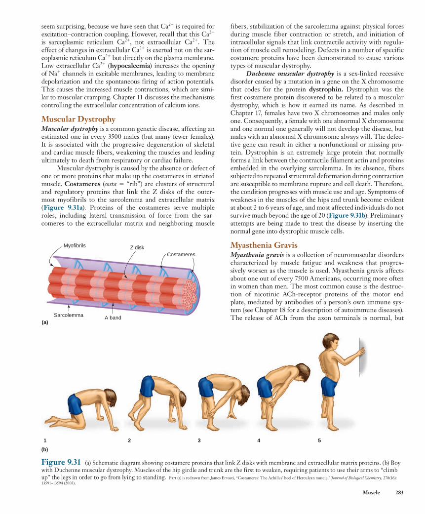
Rev. Confirming Pages
Muscle 283
fibers, stabilization of the sarcolemma against physical forces during muscle fiber contraction or stretch, and initiation of intracellular signals that link contractile activity with regula-tion of muscle cell remodeling. Defects in a number of specific costamere proteins have been demonstrated to cause various types of muscular dystrophy.
Duchenne muscular dystrophy is a sex-linked recessive disorder caused by a mutation in a gene on the X chromosome that codes for the protein dystrophin . Dystrophin was the first costamere protein discovered to be related to a muscular dystrophy, which is how it earned its name. As described in Chapter 17, females have two X chromosomes and males only one. Consequently, a female with one abnormal X chromosome and one normal one generally will not develop the disease, but males with an abnormal X chromosome always will. The defec-tive gene can result in either a nonfunctional or missing pro-tein. Dystrophin is an extremely large protein that normally forms a link between the contractile filament actin and proteins embedded in the overlying sarcolemma. In its absence, fibers subjected to repeated structural deformation during contraction are susceptible to membrane rupture and cell death. Therefore, the condition progresses with muscle use and age. Symptoms of weakness in the muscles of the hips and trunk become evident at about 2 to 6 years of age, and most affected individuals do not survive much beyond the age of 20 ( Figure 9.31b ). Preliminary attempts are being made to treat the disease by inserting the normal gene into dystrophic muscle cells.
Myasthenia Gravis Myasthenia gravis is a collection of neuromuscular disorders characterized by muscle fatigue and weakness that progres-sively worsen as the muscle is used. Myasthenia gravis affects about one out of every 7500 Americans, occurring more often in women than men. The most common cause is the destruc-tion of nicotinic ACh-receptor proteins of the motor end plate, mediated by antibodies of a person’s own immune sys-tem (see Chapter 18 for a description of autoimmune diseases). The release of ACh from the axon terminals is normal, but
seem surprising, because we have seen that Ca 2 1 is required for excitation–contraction coupling. However, recall that this Ca 2 1 is sarcoplasmic reticulum Ca 2 1 , not extracellular Ca 2 1 . The effect of changes in extracellular Ca 2 1 is exerted not on the sar-coplasmic reticulum Ca 2 1 but directly on the plasma membrane. Low extracellular Ca 2 1 ( hypocalcemia ) increases the opening of Na 1 channels in excitable membranes, leading to membrane depolarization and the spontaneous firing of action potentials. This causes the increased muscle contractions, which are simi-lar to muscular cramping. Chapter 11 discusses the mechanisms controlling the extracellular concentration of calcium ions.
Muscular Dystrophy Muscular dystrophy is a common genetic disease, affecting an estimated one in every 3500 males (but many fewer females). It is associated with the progressive degeneration of skeletal and cardiac muscle fibers, weakening the muscles and leading ultimately to death from respiratory or cardiac failure.
Muscular dystrophy is caused by the absence or defect of one or more proteins that make up the costameres in striated muscle. Costameres ( costa 5 “rib”) are clusters of structural and regulatory proteins that link the Z disks of the outer-most myofibrils to the sarcolemma and extracellular matrix ( Figure 9.31a ). Proteins of the costameres serve multiple roles, including lateral transmission of force from the sar-comeres to the extracellular matrix and neighboring muscle
Figure 9.31 (a) Schematic diagram showing costamere proteins that link Z disks with membrane and extracellular matrix proteins. (b) Boy with Duchenne muscular dystrophy. Muscles of the hip girdle and trunk are the first to weaken, requiring patients to use their arms to “climb up” the legs in order to go from lying to standing. Part (a) is redrawn from James Ervasti, “Costameres: The Achilles’ heel of Herculean muscle,” Journal of Biological Chemistry, 278(16): 13591–13594 (2003).
Myofibrils Z disk
A band
Costameres
Sarcolemma(a)
1 2 3 4 5
(b)
wid78305_ch09_257-299.indd 283 30/01/13 11:49 AM
Rev. Confirming Pages
284 Chapter 9
b. A single action potential in a motor neuron is sufficient to produce an action potential in a skeletal muscle fiber.
c. Figure 9.9 summarizes events at the neuromuscular junction. d. Signaling at the neuromuscular junction can be disrupted by
a number of different toxins, drugs, and disease processes. II. In a resting muscle, tropomyosin molecules that are in
contact with the actin subunits of the thin filaments block the attachment of cross-bridges to actin.
III. Contraction is initiated by an increase in cytosolic Ca 2 1 concentration. The calcium ions bind to troponin, producing a change in its shape that is transmitted via tropomyosin to uncover the binding sites on actin, allowing the cross-bridges to bind to the thin filaments. a. The increase in cytosolic Ca 2 1 concentration is triggered
by an action potential in the plasma membrane. The action potential is propagated into the interior of the fiber along the transverse tubules to the region of the sarcoplasmic reticulum, where dihydropyridine receptors sense the voltage change and pull open ryanodine receptors, releasing calcium ions from the reticulum.
b. Relaxation of a contracting muscle fiber occurs as a result of the active transport of cytosolic calcium ions back into the sarcoplasmic reticulum.
IV. When a skeletal muscle fiber actively shortens, the thin filaments are propelled toward the center of their sarcomere by movements of the myosin cross-bridges that bind to actin. a. The four steps occurring during each cross-bridge cycle
are summarized in Figure 9.15 . The cross-bridges undergo repeated cycles during a contraction, each cycle producing only a small increment of movement.
b. The functions of ATP in muscle contraction are summarized in Table 9.1 .
V. Table 9.2 summarizes the events leading to the contraction of a skeletal muscle fiber.
Mechanics of Single-Fiber Contraction I. Contraction refers to the turning on of the cross-bridge cycle.
Whether there is an accompanying change in muscle length depends upon the external forces acting on the muscle.
II. Three types of contractions can occur following activation of a muscle fiber: (1) an isometric contraction in which the muscle generates tension but does not change length; (2) an isotonic contraction in which the muscle shortens (concentric), moving a load; and (3) a lengthening (eccentric) contraction in which the external load on the muscle causes the muscle to lengthen during the period of contractile activity.
III. Increasing the frequency of action potentials in a muscle fiber increases the mechanical response (tension or shortening) up to the level of maximal tetanic tension.
IV. Maximum isometric tetanic tension is produced at the optimal sarcomere length L 0 . Stretching a fiber beyond its optimal length or decreasing the fiber length below L 0 decreases the tension generated.
V. The velocity of muscle fiber shortening decreases with increases in load. Maximum velocity occurs at zero load.
Skeletal Muscle Energy Metabolism I. Muscle fibers form ATP by the transfer of phosphate from
creatine phosphate to ADP, by oxidative phosphorylation of ADP in mitochondria, and by substrate-level phosphorylation of ADP in the glycolytic pathway.
II. At the beginning of exercise, muscle glycogen is the major fuel consumed. As the exercise proceeds, glucose and fatty acids from the blood provide most of the fuel, and fatty acids
the magnitude of the end-plate potential is markedly reduced because of the decreased availability of receptors. Even in normal muscle, the amount of ACh released with each action potential decreases with repetitive activity, and thus the mag-nitude of the resulting end-plate potential (EPP) decreases. In normal muscle, however, the EPP remains well above the threshold necessary to initiate a muscle action potential. In contrast, after a few motor nerve impulses in a myasthenia gravis patient, the magnitude of the EPP decreases below the threshold for initiating a muscle action potential.
A number of approaches are currently used to treat the dis-ease. One is to administer acetylcholinesterase inhibitors (e.g., pyridostigmine ). This can partially compensate for the reduc-tion in available ACh receptors by prolonging the time that acetylcholine is available at the synapse. Other therapies aim at blunting the immune response. Treatment with glucocorticoids is one way that immune function is suppressed (see Chapter 11). Removal of the thymus gland ( thymectomy ) reduces the pro-duction of antibodies and reverses symptoms in about 50% of patients. Plasmapheresis is a treatment that involves replacing the liquid fraction of blood (plasma) that contains the offend-ing antibodies. A combination of these treatments has greatly reduced the mortality rate for myasthenia gravis.
S E C T I O N A SUM M A RY There are three types of muscle—skeletal, smooth, and cardiac. Skeletal muscle is attached to bones and moves and supports the skeleton. Smooth muscle surrounds hollow cavities and tubes. Cardiac muscle is the muscle of the heart.
Structure I. Skeletal muscles, composed of cylindrical muscle fibers (cells),
are linked to bones by tendons at each end of the muscle. II. Skeletal muscle fibers have a repeating, striated pattern of light
and dark bands due to the arrangement of the thick and thin filaments within the myofibrils.
III. Actin-containing thin filaments are anchored to the Z lines at each end of a sarcomere. Their free ends partially overlap the myosin-containing thick filaments in the A band at the center of the sarcomere.
IV. Myosin molecules form the backbone of the thick filament and also have extensions called cross-bridges that span the gap between the thick and thin filaments. Each cross-bridge has two globular heads that contain a binding site for actin and an enzymatic site that splits ATP.
V. Skeletal muscle fibers have an elaborate membrane system in which the plasma membrane (sarcolemma) sends tubular extensions (T-tubules) throughout the cross section of the cell. T-tubules interact with terminal cisternae of the sarcoplasmic reticulum, in which Ca 2 1 is stored.
Molecular Mechanisms of Skeletal Muscle Contraction I. Branches of a motor neuron axon form neuromuscular
junctions with the muscle fibers in its motor unit. Each muscle fiber is innervated by a branch from only one motor neuron.a. Acetylcholine released by an action potential in a motor
neuron binds to receptors on the motor end plate of the muscle membrane, opening ion channels that allow the passage of sodium and potassium ions, which depolarize the end-plate membrane.
wid78305_ch09_257-299.indd 284 30/01/13 11:49 AM
Rev. Confirming Pages
Muscle 285
Skeletal Muscle Disorders I. Muscle cramps are involuntary tetanic contractions related to
heavy exercise and may be due to dehydration and electrolyte imbalances in the fluid surrounding muscle and nerve fibers.
II. When extracellular Ca 2 1 concentration decreases below normal, Na 1 channels of nerve and muscle open spontaneously, which causes the excessive muscle contractions of hypocalcemic tetany.
III. Muscular dystrophies are commonly occurring genetic disorders that result from defects of muscle-membrane-stabilizing proteins such as dystrophin. Muscles of individuals with Duchenne muscular dystrophy progressively degenerate with use.
IV. Myasthenia gravis is an autoimmune disorder in which destruction of ACh receptors of the motor end plate causes progressive loss of the ability to activate skeletal muscles.
S E C T I O N A REV IEW QUESTIONS 1. List the three types of muscle cells and their locations. 2. Diagram the arrangement of thick and thin filaments in a
striated muscle sarcomere, and label the major bands that give rise to the striated pattern.
3. Describe the organization of myosin, actin, tropomyosin, and troponin molecules in the thick and thin filaments.
4. Describe the location, structure, and function of the sarcoplasmic reticulum in skeletal muscle fibers.
5. Describe the structure and function of the transverse tubules. 6. Define motor unit and describe its structure. 7. Describe the sequence of events by which an action potential
in a motor neuron produces an action potential in the plasma membrane of a skeletal muscle fiber.
8. What is an end-plate potential, and what ions produce it? 9. Compare and contrast the transmission of electrical activity at
a neuromuscular junction with that at a synapse. 10. What prevents cross-bridges from attaching to sites on the thin
filaments in a resting skeletal muscle? 11. Describe the role and source of calcium ions in initiating
contraction in skeletal muscle. 12. Describe the four steps of one cross-bridge cycle. 13. Describe the physical state of a muscle fiber in rigor mortis and
the conditions that produce this state. 14. What three events in skeletal muscle contraction and relaxation
depend on ATP? 15. Describe the events that result in the relaxation of skeletal
muscle fibers. 16. Describe isometric, concentric, and eccentric contractions. 17. What factors determine the duration of an isotonic twitch in
skeletal muscle? An isometric twitch? 18. What effect does increasing the frequency of action potentials
in a skeletal muscle fiber have upon the force of contraction? Explain the mechanism responsible for this effect.
19. Describe the length–tension relationship in skeletal muscle fibers.
20. Describe the effect of increasing the load on a skeletal muscle fiber on the velocity of shortening.
21. What is the function of creatine phosphate in skeletal muscle contraction?
22. What fuel molecules are metabolized to produce ATP during skeletal muscle activity?
23. List the factors responsible for skeletal muscle fatigue. 24. What component of skeletal muscle fibers accounts for the
differences in the fibers’ maximal shortening velocities?
become progressively more important during prolonged exercise. When the intensity of exercise exceeds about 70% of maximum, glycolysis begins to contribute an increasing fraction of the total ATP generated.
III. A variety of factors may contribute to muscle fatigue, including a decrease in ATP concentration and increases in the concentrations of ADP, P i , Mg 2 1 , H 1 , and oxygen free radicals. Individually and in combination, those changes have effects such as decreasing Ca 2 1 uptake and storage by the sarcoplasmic reticulum, decreasing the sensitivity of the thin filaments to Ca 2 1 , and inhibiting the binding and power-stroke motion of the cross-bridges.
Types of Skeletal Muscle Fibers I. Three types of skeletal muscle fibers can be distinguished
by their maximal shortening velocities and the predominate pathway they use to form ATP: slow-oxidative, fast-oxidative-glycolytic, and fast-glycolytic fibers.a. Differences in maximal shortening velocities are due
to different myosin enzymes with high or low ATPase activities, giving rise to fast and slow fibers.
b. Fast-glycolytic fibers have a larger average diameter than oxidative fibers and therefore produce greater tension, but they also fatigue more rapidly.
II. All the muscle fibers in a single motor unit belong to the same fiber type, and most muscles contain all three types.
III. Table 9.3 summarizes the characteristics of the three types of skeletal muscle fibers.
Whole-Muscle Contraction I. The tension produced by whole-muscle contraction depends
on the amount of tension each fiber develops and the number of active fibers in the muscle ( Table 9.4 ).
II. Muscles that produce delicate movements have a small number of fibers per motor unit, whereas large powerful muscles have much larger motor units.
III. Fast-glycolytic motor units not only have large-diameter fibers but also tend to have large numbers of fibers per motor unit.
IV. Increases in muscle tension are controlled primarily by increasing the number of active motor units in a muscle, a process known as recruitment. Slow-oxidative motor units are recruited first; then fast-oxidative-glycolytic motor units are recruited; and finally, fast-glycolytic motor units are recruited only during very strong contractions.
V. Increasing motor-unit recruitment increases the velocity at which a muscle will move a given load.
VI. Exercise can alter a muscle’s strength and susceptibility to fatigue. a. Long-duration, low-intensity exercise increases a fiber’s
capacity for oxidative ATP production by increasing the number of mitochondria and blood vessels in the muscle, resulting in increased endurance.
b. Short-duration, high-intensity exercise increases fiber diameter as a result of increased synthesis of actin and myosin, resulting in increased strength.
VII. Movement around a joint generally involves groups of antagonistic muscles; some flex a limb at the joint and others extend the limb.
VIII. The lever system of muscles and bones generally requires muscle tension far greater than the load in order to sustain a load in an isometric contraction, but the lever system produces a shortening velocity at the end of the lever arm that is greater than the muscle-shortening velocity.
wid78305_ch09_257-299.indd 285 30/01/13 11:49 AM
Rev. Confirming Pages
286 Chapter 9
25. Summarize the characteristics of the three types of skeletal muscle fibers.
26. Upon what three factors does the amount of tension developed by a whole skeletal muscle depend?
27. Describe the process of motor-unit recruitment in controlling (a) whole-muscle tension and (b) velocity of whole-muscle shortening.
28. During increases in the force of skeletal muscle contraction, what is the order of recruitment of the different types of motor units?
29. What happens to skeletal muscle fibers when the motor neuron to the muscle is destroyed?
30. Describe the changes that occur in skeletal muscles following a period of (a) long-duration, low-intensity exercise training; and (b) short-duration, high-intensity exercise training.
31. How are skeletal muscles arranged around joints so that a limb can push or pull?
32. What are the advantages and disadvantages of the muscle-bone-joint lever system?
A band 260 acetylcholine (ACh) 262 acetylcholinesterase 263 actin 260 antagonist 281 cardiac muscle 258 central command fatigue 276 concentric contraction 269 contraction 262 contraction time 270 costameres 283 creatine phosphate 274 cross-bridge 261 cross-bridge cycle 267 dihydropyridine (DHP)
receptor 266 dystrophin 283 eccentric contraction 269
end-plate potential (EPP) 263 excitation–contraction
coupling 265 extension 281 fast fiber 276 fast-glycolytic fiber 277 fast-oxidative-glycolytic
fiber 277 flexion 281 foot process 266 fused tetanus 273 glycolytic fiber 277 H zone 260 heavy chains 260 hypertrophy 259 hypocalcemia 283 I band 260 isometric contraction 269
S E C T I O N A K EY TER MS
isotonic contraction 269 junctional feet 266 latent period 270 light chains 260 load 269 M line 260 motor end plate 262 motor neuron 262 motor unit 262 muscle 259 muscle fatigue 275 muscle fiber 259 myoblast 259 myofibril 260 myoglobin 277 myosin 260 myostatin 280 neuromuscular junction 262 optimal length (L 0 ) 274 oxidative fiber 276 oxygen debt 275 power stroke 268 recruitment 279 red muscle fiber 277 relaxation 262 rigor mortis 269 ryanodine receptor 266
sarcolemma 261 sarcomere 260 sarcoplasmic reticulum 261 satellite cell 259 skeletal muscle 258 sliding-filament
mechanism 267 slow fiber 276 slow-oxidative fiber 277 smooth muscle 258 striated muscle 258 summation 272 tendon 260 tension 269 terminal cisternae 261 tetanus 273 thick filament 260 thin filament 260 titin 260 transverse tubule
(T-tubule) 260 tropomyosin 260 troponin 261 twitch 270 unfused tetanus 273 white muscle fiber 277 Z line 260
atropine 264 botulism 265 curare 264 denervation atrophy 279 disuse atrophy 280 Duchenne muscular
dystrophy 283 hypocalcemic tetany 282 muscle cramp 282 muscular dystrophy 283
myasthenia gravis 283 plasmapheresis 284 poliomyelitis 282 pralidoxime 264 pyridostigmine 284 rocuronium 264 succinylcholine 264 thymectomy 284 vecuronium 264
S E C T I O N A CLINICA L TER MS
S E C T I O N B
Smooth and Cardiac Muscle
We now turn our attention to the other muscle types, begin-ning with smooth muscle. Two characteristics are common to all smooth muscles. They lack the cross-striated band-ing pattern found in skeletal and cardiac fibers (which makes them “smooth”), and the nerves to them are part of the auto-nomic division of the nervous system rather than the somatic division. Thus, smooth muscle is not normally under direct voluntary control.
Smooth muscle, like skeletal muscle, uses cross-bridge movements between actin and myosin filaments to gener-ate force, and calcium ions to control cross-bridge activity. However, the organization of the contractile filaments and the process of excitation–contraction coupling are quite dif-ferent in smooth muscle. Furthermore, there is considerable
diversity among smooth muscles with respect to the excitation– contraction coupling mechanism.
9.8 Structure of Smooth Muscle Each smooth muscle cell is spindle-shaped, with a diameter between 2 and 10 m m, and length ranging from 50 to 400 m m. They are much smaller than skeletal muscle fibers, which are 10 to 100 m m wide and can be tens of centimeters long (see Figure 9.1 ). Skeletal muscle fibers are sometimes large enough to run the entire length of the muscles in which they are found, whereas many individual smooth muscle cells are generally inter-connected to form sheetlike layers of cells ( Figure 9.32 ). Skel-etal muscle fibers are multinucleate cells with limited ability to
wid78305_ch09_257-299.indd 286 30/01/13 11:49 AM
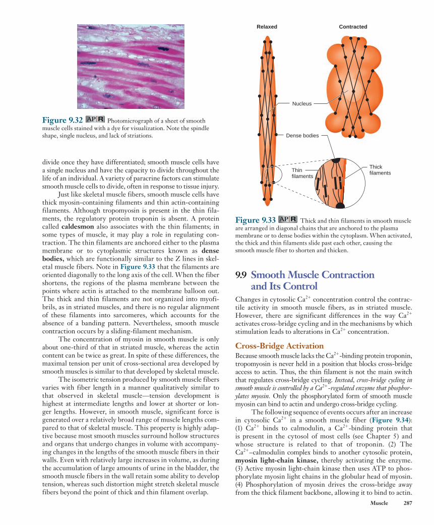
Rev. Confirming Pages
Muscle 287
9.9 Smooth Muscle Contraction and Its Control
Changes in cytosolic Ca 2 1 concentration control the contrac-tile activity in smooth muscle fibers, as in striated muscle. However, there are significant differences in the way Ca 2 1
activates cross-bridge cycling and in the mechanisms by which stimulation leads to alterations in Ca 2 1 concentration.
Cross-Bridge Activation Because smooth muscle lacks the Ca 2 1 -binding protein troponin, tropomyosin is never held in a position that blocks cross-bridge access to actin. Thus, the thin filament is not the main switch that regulates cross-bridge cycling. Instead, cross-bridge cycling in smooth muscle is controlled by a Ca 2 1 -regulated enzyme that phosphor-ylates myosin. Only the phosphorylated form of smooth muscle myosin can bind to actin and undergo cross-bridge cycling.
The following sequence of events occurs after an increase in cytosolic Ca 2 1 in a smooth muscle fiber ( Figure 9.34 ): (1) Ca 2 1 binds to calmodulin, a Ca 2 1 -binding protein that is present in the cytosol of most cells (see Chapter 5) and whose structure is related to that of troponin. (2) The Ca 2 1 –calmodulin complex binds to another cytosolic protein, myosin light-chain kinase , thereby activating the enzyme. (3) Active myosin light-chain kinase then uses ATP to phos-phorylate myosin light chains in the globular head of myosin. (4) Phosphorylation of myosin drives the cross-bridge away from the thick filament backbone, allowing it to bind to actin.
Figure 9.32 Photomicrograph of a sheet of smooth muscle cells stained with a dye for visualization. Note the spindle shape, single nucleus, and lack of striations.
Figure 9.33 Thick and thin filaments in smooth muscle are arranged in diagonal chains that are anchored to the plasma membrane or to dense bodies within the cytoplasm. When activated, the thick and thin filaments slide past each other, causing the smooth muscle fiber to shorten and thicken.
Relaxed Contracted
Dense bodies
Nucleus
ThickfilamentsThin
filaments
divide once they have differentiated; smooth muscle cells have a single nucleus and have the capacity to divide throughout the life of an individual. A variety of paracrine factors can stimulate smooth muscle cells to divide, often in response to tissue injury.
Just like skeletal muscle fibers, smooth muscle cells have thick myosin-containing filaments and thin actin- containing filaments. Although tropomyosin is present in the thin fila-ments, the regulatory protein troponin is absent. A protein called caldesmon also associates with the thin filaments; in some types of muscle, it may play a role in regulating con-traction. The thin filaments are anchored either to the plasma membrane or to cytoplasmic structures known as dense bodies , which are functionally similar to the Z lines in skel-etal muscle fibers. Note in Figure 9.33 that the filaments are oriented diagonally to the long axis of the cell. When the fiber shortens, the regions of the plasma membrane between the points where actin is attached to the membrane balloon out. The thick and thin filaments are not organized into myofi-brils, as in striated muscles, and there is no regular alignment of these filaments into sarcomeres, which accounts for the absence of a banding pattern. Nevertheless, smooth muscle contraction occurs by a sliding-filament mechanism.
The concentration of myosin in smooth muscle is only about one-third of that in striated muscle, whereas the actin content can be twice as great. In spite of these differences, the maximal tension per unit of cross-sectional area developed by smooth muscles is similar to that developed by skeletal muscle.
The isometric tension produced by smooth muscle fibers varies with fiber length in a manner qualitatively similar to that observed in skeletal muscle—tension development is highest at intermediate lengths and lower at shorter or lon-ger lengths. However, in smooth muscle, significant force is generated over a relatively broad range of muscle lengths com-pared to that of skeletal muscle. This property is highly adap-tive because most smooth muscles surround hollow structures and organs that undergo changes in volume with accompany-ing changes in the lengths of the smooth muscle fibers in their walls. Even with relatively large increases in volume, as during the accumulation of large amounts of urine in the bladder, the smooth muscle fibers in the wall retain some ability to develop tension, whereas such distortion might stretch skeletal muscle fibers beyond the point of thick and thin filament overlap.
wid78305_ch09_257-299.indd 287 30/01/13 11:49 AM
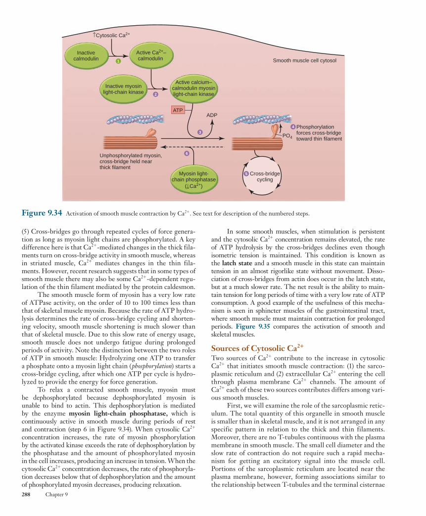
Rev. Confirming Pages
288 Chapter 9
In some smooth muscles, when stimulation is persistent and the cytosolic Ca 2 1 concentration remains elevated, the rate of ATP hydrolysis by the cross-bridges declines even though isometric tension is maintained. This condition is known as the latch state and a smooth muscle in this state can maintain tension in an almost rigorlike state without movement. Disso-ciation of cross-bridges from actin does occur in the latch state, but at a much slower rate. The net result is the ability to main-tain tension for long periods of time with a very low rate of ATP consumption. A good example of the usefulness of this mecha-nism is seen in sphincter muscles of the gastrointestinal tract, where smooth muscle must maintain contraction for prolonged periods. Figure 9.35 compares the activation of smooth and skeletal muscles.
Sources of Cytosolic Ca 2 1 Two sources of Ca 2 1 contribute to the increase in cytosolic Ca 2 1 that initiates smooth muscle contraction: (1) the sarco-plasmic reticulum and (2) extracellular Ca 2 1 entering the cell through plasma membrane Ca 2 1 channels. The amount of Ca 2 1 each of these two sources contributes differs among vari-ous smooth muscles.
First, we will examine the role of the sarcoplasmic retic-ulum. The total quantity of this organelle in smooth muscle is smaller than in skeletal muscle, and it is not arranged in any specific pattern in relation to the thick and thin filaments. Moreover, there are no T-tubules continuous with the plasma membrane in smooth muscle. The small cell diameter and the slow rate of contraction do not require such a rapid mecha-nism for getting an excitatory signal into the muscle cell. Portions of the sarcoplasmic reticulum are located near the plasma membrane, however, forming associations similar to the relationship between T-tubules and the terminal cisternae
(5) Cross-bridges go through repeated cycles of force genera-tion as long as myosin light chains are phosphorylated. A key difference here is that Ca 2 1 -mediated changes in the thick fila-ments turn on cross-bridge activity in smooth muscle, whereas in striated muscle, Ca 2 1 mediates changes in the thin fila-ments. However, recent research suggests that in some types of smooth muscle there may also be some Ca 2 1 -dependent regu-lation of the thin filament mediated by the protein caldesmon.
The smooth muscle form of myosin has a very low rate of ATPase activity, on the order of 10 to 100 times less than that of skeletal muscle myosin. Because the rate of ATP hydro-lysis determines the rate of cross-bridge cycling and shorten-ing velocity, smooth muscle shortening is much slower than that of skeletal muscle. Due to this slow rate of energy usage, smooth muscle does not undergo fatigue during prolonged periods of activity. Note the distinction between the two roles of ATP in smooth muscle: Hydrolyzing one ATP to transfer a phosphate onto a myosin light chain ( phosphorylation ) starts a cross-bridge cycling, after which one ATP per cycle is hydro-lyzed to provide the energy for force generation.
To relax a contracted smooth muscle, myosin must be dephosphorylated because dephosphorylated myosin is unable to bind to actin. This dephosphorylation is mediated by the enzyme myosin light-chain phosphatase , which is continuously active in smooth muscle during periods of rest and contraction (step 6 in Figure 9.34 ). When cytosolic Ca 2 1 concentration increases, the rate of myosin phosphorylation by the activated kinase exceeds the rate of dephosphorylation by the phosphatase and the amount of phosphorylated myosin in the cell increases, producing an increase in tension. When the cytosolic Ca 2 1 concentration decreases, the rate of phosphoryla-tion decreases below that of dephosphorylation and the amount of phosphorylated myosin decreases, producing relaxation.
Figure 9.34 Activation of smooth muscle contraction by Ca 2 1 . See text for description of the numbered steps.
Inactive myosinlight-chain kinase
Active calcium–calmodulin myosinlight-chain kinase
Myosin light-chain phosphatase
( Ca2+)
Inactivecalmodulin
Active Ca2+–calmodulin
Cytosolic Ca2+
ATPADP
2
34
5
6
1
Unphosphorylated myosin,cross-bridge held nearthick filament
Phosphorylationforces cross-bridgetoward thin filament
PO4
Cross-bridgecycling
Smooth muscle cell cytosol
wid78305_ch09_257-299.indd 288 30/01/13 11:49 AM

Rev. Confirming Pages
Muscle 289
a single twitch lasts several seconds in smooth muscle com-pared to a fraction of a second in skeletal muscle.
The degree of activation also differs between muscle types. In skeletal muscle, a single action potential releases suf-ficient Ca 2 1 to saturate all troponin sites on the thin filaments, whereas only a portion of the cross-bridges are activated in a smooth muscle fiber in response to most stimuli. There-fore, the tension generated by a smooth muscle cell can be graded by varying cytosolic Ca 2 1 concentration. The greater the increase in Ca 2 1 concentration, the greater the number of cross-bridges activated and the greater the tension.
In some smooth muscles, the cytosolic Ca 2 1 concentration is sufficient to maintain a low level of basal cross-bridge activ-ity in the absence of external stimuli. This activity is known as smooth muscle tone . Factors that alter the cytosolic Ca 2 1 con-centration also vary the intensity of smooth muscle tone.
Membrane Activation Many inputs to a smooth muscle plasma membrane can alter the contractile activity of the muscle ( Table 9.5 ). This con-trasts with skeletal muscle, in which membrane activation depends only upon synaptic inputs from somatic neurons. Some inputs to smooth muscle increase contraction, and oth-ers inhibit it. Moreover, at any one time, the smooth muscle plasma membrane may be receiving multiple inputs, with the contractile state of the muscle dependent on the relative inten-sity of the various inhibitory and excitatory stimuli. All these inputs influence contractile activity by altering cytosolic Ca 2 1
concentration as described in the previous section. Some smooth muscles contract in response to membrane
depolarization, whereas others can contract in the absence of any membrane potential change. Interestingly, in smooth mus-cles in which action potentials occur, calcium ions, rather than sodium ions, carry a positive charge into the cell during the rising phase of the action potential—that is, depolarization of the membrane opens voltage-gated Ca 2 1 channels, producing Ca 2 1 -mediated rather than Na 1 -mediated action potentials.
Smooth muscle is different from skeletal muscle in another important way with regard to electrical activity and cytosolic Ca 2 1 concentration. Smooth muscle cytosolic Ca 2 1 concentration can be increased (or decreased) by graded depo-larizations (or hyperpolarizations) in membrane potential, which increase or decrease the number of open Ca 2 1 channels.
Figure 9.35 Pathways leading from increased cytosolic Ca 2 1 to cross-bridge cycling in smooth and skeletal muscle fibers.
Cross-bridge cycleproduces tension and
shortening
Cross-bridge cycleproduces tension and
shortening
Smooth muscle Skeletal muscle
Phosphorylatedcross-bridges
bind to actin filaments
Myosin light-chain kinaseuses ATP to phosphorylate
myosin cross-bridges
Ca2+–calmodulin complexbinds to myosin
light-chain kinase
Ca2+ binds to calmodulinin cytosol
Myosin cross-bridgesbind to actin
Conformational changein troponin movestropomyosin out ofblocking position
Ca2+ binds to troponinon thin filaments
Cytosolic Ca2+ Cytosolic Ca2+
in skeletal muscle. Action potentials in the plasma membrane can be coupled to the release of sarcoplasmic reticulum Ca 2 1
at these sites. In some types of smooth muscles, action poten-tials are not necessary for Ca 2 1 release. Instead, second mes-sengers released from the plasma membrane, or generated in the cytosol in response to the binding of extracellular chemi-cal messengers to plasma membrane receptors, can trigger the release of Ca 2 1 from the more centrally located sarcoplasmic reticulum (review Figure 5.10 for a specific example).
What about extracellular Ca 2 1 in excitation–contraction coupling? There are voltage-sensitive Ca 2 1 channels in the plasma membranes of smooth muscle cells, as well as Ca 2 1 chan-nels controlled by extracellular chemical messengers. The Ca 2 1 concentration in the extracellular fluid is 10,000 times greater than in the cytosol; thus, the opening of Ca 2 1 channels in the plasma membrane results in an increased flow of Ca 2 1 into the cell. Because of the small cell size, the entering Ca 2 1 does not have far to diffuse to reach binding sites within the cell.
Removal of Ca 2 1 from the cytosol to bring about relax-ation is achieved by the active transport of Ca 2 1 back into the sarcoplasmic reticulum as well as out of the cell across the plasma membrane. The rate of Ca 2 1 removal in smooth mus-cle is much slower than in skeletal muscle, with the result that
TABLE 9.5 Inputs Influencing Smooth Muscle Contractile Activity
Spontaneous electrical activity in the plasma membrane of the muscle cell
Neurotransmitters released by autonomic neurons
Hormones
Locally induced changes in the chemical composition (paracrine factors, acidity, oxygen, osmolarity, and ion concentrations) of the extracellular fluid surrounding the cell
Stretch
wid78305_ch09_257-299.indd 289 30/01/13 11:49 AM
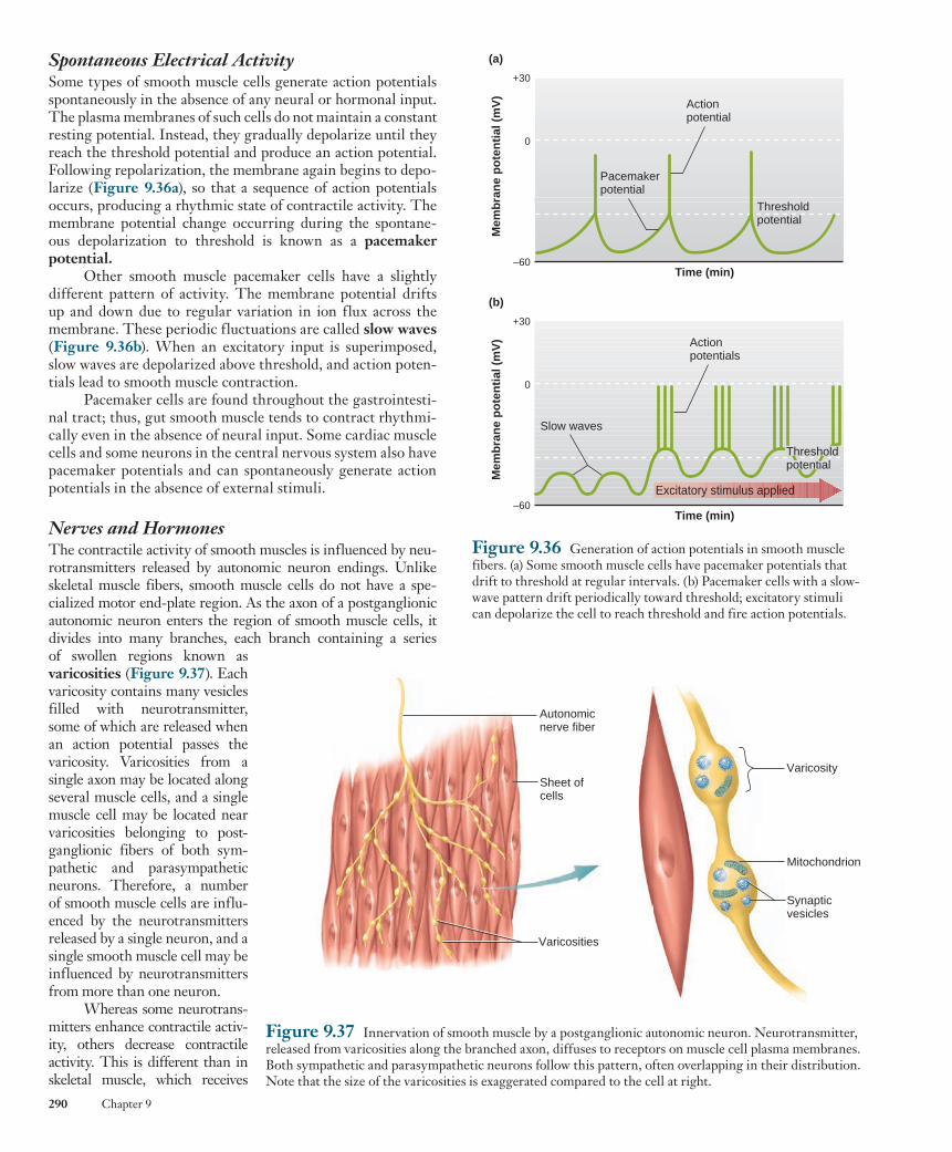
Rev. Confirming Pages
290 Chapter 9
Spontaneous Electrical Activity Some types of smooth muscle cells generate action potentials spontaneously in the absence of any neural or hormonal input. The plasma membranes of such cells do not maintain a constant resting potential. Instead, they gradually depolarize until they reach the threshold potential and produce an action potential. Following repolarization, the membrane again begins to depo-larize ( Figure 9.36a ), so that a sequence of action potentials occurs, producing a rhythmic state of contractile activity. The membrane potential change occurring during the spontane-ous depolarization to threshold is known as a pacemaker potential .
Other smooth muscle pacemaker cells have a slightly different pattern of activity. The membrane potential drifts up and down due to regular variation in ion flux across the membrane. These periodic fluctuations are called slow waves ( Figure 9.36b ). When an excitatory input is superimposed, slow waves are depolarized above threshold, and action poten-tials lead to smooth muscle contraction.
Pacemaker cells are found throughout the gastrointesti-nal tract; thus, gut smooth muscle tends to contract rhythmi-cally even in the absence of neural input. Some cardiac muscle cells and some neurons in the central nervous system also have pacemaker potentials and can spontaneously generate action potentials in the absence of external stimuli.
Nerves and Hormones The contractile activity of smooth muscles is influenced by neu-rotransmitters released by autonomic neuron endings. Unlike skeletal muscle fibers, smooth muscle cells do not have a spe-cialized motor end-plate region. As the axon of a postganglionic autonomic neuron enters the region of smooth muscle cells, it divides into many branches, each branch containing a series of swollen regions known as varicosities ( Figure 9.37 ). Each varicosity contains many vesicles filled with neurotransmitter, some of which are released when an action potential passes the varicosity. Varicosities from a single axon may be located along several muscle cells, and a single muscle cell may be located near varicosities belonging to post-ganglionic fibers of both sym-pathetic and parasympathetic neurons. Therefore, a number of smooth muscle cells are influ-enced by the neurotransmitters released by a single neuron, and a single smooth muscle cell may be influenced by neurotransmitters from more than one neuron.
Whereas some neurotrans-mitters enhance contractile activ-ity, others decrease contractile activity. This is different than in skeletal muscle, which receives
Figure 9.36 Generation of action potentials in smooth muscle fibers. (a) Some smooth muscle cells have pacemaker potentials that drift to threshold at regular intervals. (b) Pacemaker cells with a slow-wave pattern drift periodically toward threshold; excitatory stimuli can depolarize the cell to reach threshold and fire action potentials.
Actionpotential
Pacemakerpotential
Thresholdpotential
Time (min)
0
+30
–60
Mem
bra
ne
po
ten
tial
(m
V)
Actionpotentials
Slow waves
Time (min)
0
+30
–60M
emb
ran
e p
ote
nti
al (
mV
)
(b)
(a)
Thresholdpotential
Excitatory stimulus applied
Figure 9.37 Innervation of smooth muscle by a postganglionic autonomic neuron. Neurotransmitter, released from varicosities along the branched axon, diffuses to receptors on muscle cell plasma membranes. Both sympathetic and parasympathetic neurons follow this pattern, often overlapping in their distribution. Note that the size of the varicosities is exaggerated compared to the cell at right.
Varicosity
Synaptic vesicles
Mitochondrion
Autonomicnerve fiber
Varicosities
Sheet ofcells
wid78305_ch09_257-299.indd 290 30/01/13 11:49 AM

Rev. Confirming Pages
Muscle 291
on the electrical characteristics of their plasma membrane: single-unit smooth muscles and multiunit smooth muscles .
Single-Unit Smooth Muscle The muscle cells in a single-unit smooth muscle undergo syn-chronous activity, both electrical and mechanical; that is, the whole muscle responds to stimulation as a single unit. This occurs because each muscle cell is linked to adjacent fibers by gap junctions, which allow action potentials occurring in one cell to propagate to other cells by local currents. Therefore, electri-cal activity occurring anywhere within a group of single-unit smooth muscle cells can be conducted to all the other connected cells ( Figure 9.38 ).
Some of the cells in a single-unit muscle are pacemaker cells that spontaneously generate action potentials. These action potentials are conducted by way of gap junctions to the rest of the cells, most of which are not capable of pacemaker activity.
Nerves, hormones, and local factors can alter the contrac-tile activity of single-unit smooth muscles using the variety of mechanisms described previously for smooth muscles in gen-eral. The extent to which these muscles are innervated varies considerably in different organs. The axon terminals are often restricted to the regions of the muscle that contain pacemaker cells. The activity of the entire muscle can be controlled by reg-ulating the frequency of the pacemaker cells’ action potentials.
One additional characteristic of single-unit smooth muscles is that a contractile response can often be induced by stretching the muscle. In several hollow organs—the stomach, for example—stretching the smooth muscles in the walls of the organ as a result of increases in the volume of material in the lumen initiates a contractile response.
only excitatory input from its motor neurons; smooth muscle tension can be either increased or decreased by neural activity.
Moreover, a given neurotransmitter may produce oppo-site effects in different smooth muscle tissues. For example, norepinephrine, the neurotransmitter released from most postganglionic sympathetic neurons, enhances contraction of most vascular smooth muscle by acting on a -adrenergic receptors. By contrast, the same neurotransmitter produces relaxation of airway (bronchiolar) smooth muscle by acting on b 2 -adrenergic receptors. Thus, the type of response (excit-atory or inhibitory) depends not on the chemical messenger, per se, but on the receptors the chemical messenger binds to in the membrane and on the intracellular signaling mechanisms those receptors activate.
In addition to receptors for neurotransmitters, smooth muscle plasma membranes contain receptors for a variety of hormones. Binding of a hormone to its receptor may lead to either increased or decreased contractile activity.
Although most changes in smooth muscle contractile activity induced by chemical messengers are accompanied by a change in membrane potential, this is not always the case. Second messengers—for example, inositol trisphosphate—can cause the release of Ca 2 1 from the sarcoplasmic reticu-lum, producing a contraction without a change in membrane potential (review Figure 5.10).
Local Factors Local factors, including paracrine signals, acidity, oxygen and carbon dioxide concentration, osmolarity, and the ionic composition of the extracellular fluid, can also alter smooth muscle tension. Responses to local factors provide a means for altering smooth muscle contraction in response to changes in the muscle’s immediate internal environment, which can lead to regulation that is independent of long-distance signals from nerves and hormones.
Many of these local factors induce smooth muscle relax-ation. Nitric oxide (NO) is one of the most commonly encoun-tered paracrine compounds that produce smooth muscle relaxation. NO is released from some axon terminals as well as from a variety of epithelial and endothelial cells. Because of the short life span of this reactive molecule, it acts in a paracrine man-ner, influencing only those cells that are very near its release site.
Some smooth muscles can also respond by contracting when they are stretched. Stretching opens mechanically gated ion channels, leading to membrane depolarization. The result-ing contraction opposes the forces acting to stretch the muscle.
At any given moment, smooth muscle cells in the body receive many simultaneous signals. The state of contractile activity that results depends on the net magnitude of the sig-nals promoting contraction versus those promoting relaxation. This is a classic example of the general principle of physiology that most physiological functions are controlled by multiple regulatory systems, often working in opposition.
Types of Smooth Muscle The great diversity of the factors that can influence the con-tractile activity of smooth muscles in various organs has made it difficult to classify smooth muscle fibers. Many smooth muscles can be placed, however, into one of two groups, based
Figure 9.38 Innervation of a single-unit smooth muscle is often restricted to only a few cells in the muscle. Electrical activity is conducted from cell to cell throughout the muscle by way of the gap junctions between the cells.
Varicosities
Gap junctions
Autonomicnerve fiber
wid78305_ch09_257-299.indd 291 30/01/13 11:49 AM

Rev. Confirming Pages
292 Chapter 9
Excitation–Contraction Coupling in Cardiac Muscle As in skeletal muscle, contraction of cardiac muscle cells occurs in response to a membrane action potential that propagates through the T-tubules, but the mechanisms linking that exci-tation to the generation of force exhibit features of both skel-etal and smooth muscles ( Figure 9.40 ). Depolarization during cardiac muscle cell action potentials is in part due to an influx of Ca 2 1 through specialized voltage-gated channels. These Ca 2 1 channels are known as L-type Ca 2 1 channels (named for their L ong-lasting current) and are modified versions of the dihydropyridine (DHP) receptors that act as the voltage sensor in skeletal muscle cell excitation–contraction coupling. Not only does this entering Ca 2 1 participate in depolarization of the plasma membrane and cause a small increase in cyto-solic Ca 2 1 concentration, but it also serves as a trigger for the release of a much larger amount of Ca 2 1 from the sarcoplasmic reticulum. This occurs because ryanodine receptors in the cardiac sarcoplasmic reticulum terminal cisternae are Ca 2 1
channels; but rather than being opened directly by voltage as in skeletal muscle, they are opened by the binding of trig-ger Ca 2 1 in the cytosol. Once cytosolic Ca 2 1 is elevated, thin filament activation, cross-bridge cycling, and force generation occur by the same basic mechanisms described for skeletal muscle (review Figures 9.11 and 9.15 ).
Thus, even though most of the Ca 2 1 that initiates cardiac muscle contraction comes from the sarcoplasmic
The smooth muscles of the intestinal tract, uterus, and small-diameter blood vessels are examples of single-unit smooth muscles.
Multiunit Smooth Muscle Multiunit smooth muscles have no or few gap junctions. Each cell responds independently, and the muscle behaves as mul-tiple units. Multiunit smooth muscles are richly innervated by branches of the autonomic nervous system. The con-tractile response of the whole muscle depends on the num-ber of muscle cells that are activated and on the frequency of nerve stimulation. Although stimulation of the muscle by neurons leads to some degree of depolarization and a con-tractile response, action potentials do not occur in most mul-tiunit smooth muscles. Circulating hormones can increase or decrease contractile activity in multiunit smooth muscle, but stretching does not induce contraction in this type of mus-cle. The smooth muscles in the large airways to the lungs, in large arteries, and attached to the hairs in the skin are multi-unit smooth muscles.
It must be emphasized that most smooth muscles do not show all the characteristics of either single-unit or multiunit smooth muscles. These two prototypes represent the two extremes in smooth muscle characteristics, with many smooth muscles having overlapping characteristics.
9.10 Cardiac Muscle The third general type of muscle, cardiac muscle, is found only in the heart. Although many details about cardiac muscle will be discussed in the context of the cardiovascular system in Chapter 12, a brief explanation of its function and how it com-pares to skeletal and smooth muscle is presented here.
Cellular Structure of Cardiac Muscle Cardiac muscle combines properties of both skeletal and smooth muscle. Like skeletal muscle, it has a striated appear-ance due to regularly repeating sarcomeres composed of myosin-containing thick filaments interdigitating with thin filaments that contain actin. Troponin and tropomyosin are also present in the thin filament, and they have the same functions as in skeletal muscle. Cellular membranes include a T-tubule system and associated Ca 2 1 -loaded sarcoplasmic reticulum. The mechanism by which these membranes inter-act to release Ca 2 1 is different than in skeletal muscle, how-ever, as will be discussed shortly.
Like smooth muscle cells, individual cardiac muscle cells are relatively small (100 m m long and 20 m m in diam-eter) and generally contain a single nucleus. Adjacent cells are joined end to end at structures called intercalated disks , within which are desmosomes (see Figure 3.9) that hold the cells together and to which the myofibrils are attached ( Figure 9.39 ). Also found within the intercalated disks are gap junctions similar to those found in single-unit smooth muscle. Cardiac muscle cells also are arranged in layers and surround hollow cavities—in this case, the blood-filled chambers of the heart. When muscle in the walls of cardiac chambers con-tracts, it acts like a squeezing fist and exerts pressure on the blood inside.
Figure 9.39 Cardiac muscle. (a) Light micrograph. (b) Cardiac muscle cells and intercalated disks.
Striations
Nucleus
Intercalated disks
(a)
Gapjunction
Desmosome
(b)
Sarcolemma
Nucleus
Intercalateddisks
Cardiac musclecell
Mitochondrion
wid78305_ch09_257-299.indd 292 30/01/13 11:49 AM
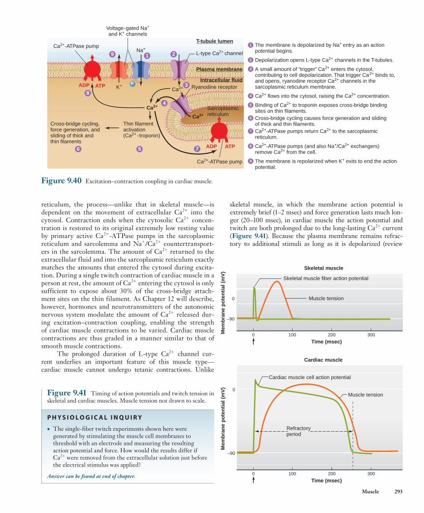
Rev. Confirming Pages
Muscle 293
skeletal muscle, in which the membrane action potential is extremely brief (1–2 msec) and force generation lasts much lon-ger (20–100 msec), in cardiac muscle the action potential and twitch are both prolonged due to the long-lasting Ca 2 1 current ( Figure 9.41 ). Because the plasma membrane remains refrac-tory to additional stimuli as long as it is depolarized (review
reticulum, the process—unlike that in skeletal muscle—is dependent on the movement of extracellular Ca 2 1 into the cytosol. Contraction ends when the cytosolic Ca 2 1 concen-tration is restored to its original extremely low resting value by primary active Ca 2 1 -ATPase pumps in the sarcoplasmic reticulum and sarcolemma and Na 1 /Ca 2 1 countertransport-ers in the sarcolemma. The amount of Ca 2 1 returned to the extracellular fluid and into the sarcoplasmic reticulum exactly matches the amounts that entered the cytosol during excita-tion. During a single twitch contraction of cardiac muscle in a person at rest, the amount of Ca 2 1 entering the cytosol is only sufficient to expose about 30% of the cross-bridge attach-ment sites on the thin filament. As Chapter 12 will describe, however, hormones and neurotransmitters of the autonomic nervous system modulate the amount of Ca 2 1 released dur-ing excitation–contraction coupling, enabling the strength of cardiac muscle contractions to be varied. Cardiac muscle contractions are thus graded in a manner similar to that of smooth muscle contractions.
The prolonged duration of L-type Ca 2 1 channel cur-rent underlies an important feature of this muscle type— cardiac muscle cannot undergo tetanic contractions. Unlike
Figure 9.40 Excitation–contraction coupling in cardiac muscle.
ADP ATP
ADP ATP
3
219
8
6 5
4
7
Ca2+-ATPase pump
Voltage-gated Na+
and K+ channels
L-type Ca2+ channel
Thin filamentactivation(Ca2+ -troponin)
Cross-bridge cycling,force generation, andsliding of thick andthin filaments
Sarcoplasmicreticulum
Ryanodine receptorIntracellular fluid
Plasma membrane
T-tubule lumen
K+
Na+
Ca2+
Ca2+
Ca2+
Ca2+-ATPase pump
The membrane is depolarized by Na+ entry as an actionpotential begins.
1
Depolarization opens L-type Ca2+ channels in the T-tubules.2
A small amount of “trigger” Ca2+ enters the cytosol,contributing to cell depolarization. That trigger Ca2+ binds to,and opens, ryanodine receptor Ca2+ channels in thesarcoplasmic reticulum membrane.
3
Ca2+ flows into the cytosol, raising the Ca2+ concentration.4
Binding of Ca2+ to troponin exposes cross-bridge bindingsites on thin filaments.
5
Cross-bridge cycling causes force generation and slidingof thick and thin filaments.
6
Ca2+-ATPase pumps (and also Na+/Ca2+ exchangers)remove Ca2+ from the cell.
8
Ca2+-ATPase pumps return Ca2+ to the sarcoplasmicreticulum.
7
The membrane is repolarized when K+ exits to end the actionpotential.
9
0
–90
Mem
bra
ne
po
ten
tial
(m
V)
0 100 200 300
Time (msec)
Time (msec)
Refractoryperiod
Cardiac muscle
Muscle tension
Cardiac muscle cell action potential
0 100 200 300
Skeletal muscle
Skeletal muscle fiber action potential
0
–90
Mem
bra
ne
po
ten
tial
(m
V)
Muscle tension
Figure 9.41 Timing of action potentials and twitch tension in skeletal and cardiac muscles. Muscle tension not drawn to scale.
P H Y S I O L O G I C A L I N Q U I R Y
■ The single-fiber twitch experiments shown here were generated by stimulating the muscle cell membranes to threshold with an electrode and measuring the resulting action potential and force. How would the results differ if Ca21 were removed from the extracellular solution just before the electrical stimulus was applied?
Answer can be found at end of chapter.
wid78305_ch09_257-299.indd 293 30/01/13 11:49 AM
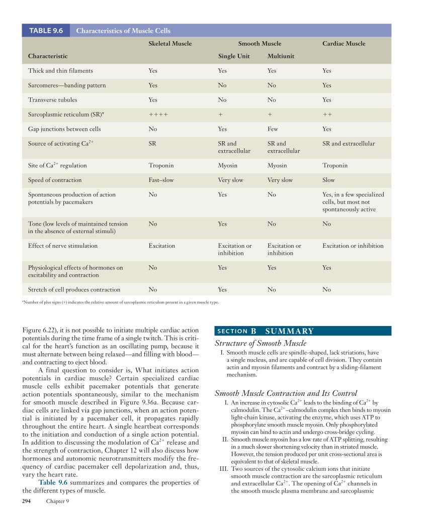
Rev. Confirming Pages
294 Chapter 9
TABLE 9.6 Characteristics of Muscle Cells
Skeletal Muscle Smooth Muscle Cardiac Muscle
Characteristic Single Unit Multiunit
Thick and thin filaments Yes Yes Yes Yes
Sarcomeres—banding pattern Yes No No Yes
Transverse tubules Yes No No Yes
Sarcoplasmic reticulum (SR) * 1 1 1 1 1 1 1 1
Gap junctions between cells No Yes Few Yes
Source of activating Ca 2 1 SR SR and extracellular
SR and extracellular
SR and extracellular
Site of Ca 2 1 regulation Troponin Myosin Myosin Troponin
Speed of contraction Fast–slow Very slow Very slow Slow
Spontaneous production of action potentials by pacemakers
No Yes No Yes, in a few specialized cells, but most not spontaneously active
Tone (low levels of maintained tension in the absence of external stimuli)
No Yes No No
Effect of nerve stimulation Excitation Excitation or inhibition
Excitation or inhibition
Excitation or inhibition
Physiological effects of hormones on excitability and contraction
No Yes Yes Yes
Stretch of cell produces contraction No Yes No No
*Number of plus signs ( 1 ) indicates the relative amount of sarcoplasmic reticulum present in a given muscle type.
Figure 6.22), it is not possible to initiate multiple cardiac action potentials during the time frame of a single twitch. This is criti-cal for the heart’s function as an oscillating pump, because it must alternate between being relaxed—and filling with blood—and contracting to eject blood.
A final question to consider is, What initiates action potentials in cardiac muscle? Certain specialized cardiac muscle cells exhibit pacemaker potentials that generate action potentials spontaneously, similar to the mechanism for smooth muscle described in Figure 9.36a . Because car-diac cells are linked via gap junctions, when an action poten-tial is initiated by a pacemaker cell, it propagates rapidly throughout the entire heart. A single heartbeat corresponds to the initiation and conduction of a single action potential. In addition to discussing the modulation of Ca 2 1 release and the strength of contraction, Chapter 12 will also discuss how hormones and autonomic neurotransmitters modify the fre-quency of cardiac pacemaker cell depolarization and, thus, vary the heart rate.
Table 9.6 summarizes and compares the properties of the different types of muscle.
S E C T I O N B SUM M A RY Structure of Smooth Muscle I. Smooth muscle cells are spindle-shaped, lack striations, have
a single nucleus, and are capable of cell division. They contain actin and myosin filaments and contract by a sliding-filament mechanism.
Smooth Muscle Contraction and Its Control I. An increase in cytosolic Ca 2 1 leads to the binding of Ca 2 1 by
calmodulin. The Ca 2 1 –calmodulin complex then binds to myosin light-chain kinase, activating the enzyme, which uses ATP to phosphorylate smooth muscle myosin. Only phosphorylated myosin can bind to actin and undergo cross-bridge cycling.
II. Smooth muscle myosin has a low rate of ATP splitting, resulting in a much slower shortening velocity than in striated muscle. However, the tension produced per unit cross-sectional area is equivalent to that of skeletal muscle.
III. Two sources of the cytosolic calcium ions that initiate smooth muscle contraction are the sarcoplasmic reticulum and extracellular Ca 2 1 . The opening of Ca 2 1 channels in the smooth muscle plasma membrane and sarcoplasmic
wid78305_ch09_257-299.indd 294 30/01/13 11:49 AM
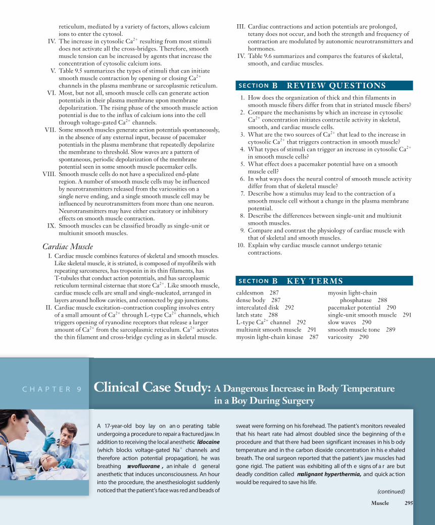
Rev. Confirming Pages
Muscle 295
III. Cardiac contractions and action potentials are prolonged, tetany does not occur, and both the strength and frequency of contraction are modulated by autonomic neurotransmitters and hormones.
IV. Table 9.6 summarizes and compares the features of skeletal, smooth, and cardiac muscles.
S E C T I O N B REV IEW QUESTIONS 1. How does the organization of thick and thin filaments in
smooth muscle fibers differ from that in striated muscle fibers? 2. Compare the mechanisms by which an increase in cytosolic
Ca 2 1 concentration initiates contractile activity in skeletal, smooth, and cardiac muscle cells.
3. What are the two sources of Ca 2 1 that lead to the increase in cytosolic Ca 2 1 that triggers contraction in smooth muscle?
4. What types of stimuli can trigger an increase in cytosolic Ca 2 1 in smooth muscle cells?
5. What effect does a pacemaker potential have on a smooth muscle cell?
6. In what ways does the neural control of smooth muscle activity differ from that of skeletal muscle?
7. Describe how a stimulus may lead to the contraction of a smooth muscle cell without a change in the plasma membrane potential.
8. Describe the differences between single-unit and multiunit smooth muscles.
9. Compare and contrast the physiology of cardiac muscle with that of skeletal and smooth muscles.
10. Explain why cardiac muscle cannot undergo tetanic contractions.
reticulum, mediated by a variety of factors, allows calcium ions to enter the cytosol.
IV. The increase in cytosolic Ca 2 1 resulting from most stimuli does not activate all the cross-bridges. Therefore, smooth muscle tension can be increased by agents that increase the concentration of cytosolic calcium ions.
V. Table 9.5 summarizes the types of stimuli that can initiate smooth muscle contraction by opening or closing Ca 2 1 channels in the plasma membrane or sarcoplasmic reticulum.
VI. Most, but not all, smooth muscle cells can generate action potentials in their plasma membrane upon membrane depolarization. The rising phase of the smooth muscle action potential is due to the influx of calcium ions into the cell through voltage-gated Ca 2 1 channels.
VII. Some smooth muscles generate action potentials spontaneously, in the absence of any external input, because of pacemaker potentials in the plasma membrane that repeatedly depolarize the membrane to threshold. Slow waves are a pattern of spontaneous, periodic depolarization of the membrane potential seen in some smooth muscle pacemaker cells.
VIII. Smooth muscle cells do not have a specialized end-plate region. A number of smooth muscle cells may be influenced by neurotransmitters released from the varicosities on a single nerve ending, and a single smooth muscle cell may be influenced by neurotransmitters from more than one neuron. Neurotransmitters may have either excitatory or inhibitory effects on smooth muscle contraction.
IX. Smooth muscles can be classified broadly as single-unit or multiunit smooth muscles.
Cardiac Muscle I. Cardiac muscle combines features of skeletal and smooth muscles.
Like skeletal muscle, it is striated, is composed of myofibrils with repeating sarcomeres, has troponin in its thin filaments, has T-tubules that conduct action potentials, and has sarcoplasmic reticulum terminal cisternae that store Ca 2 1 . Like smooth muscle, cardiac muscle cells are small and single-nucleated, arranged in layers around hollow cavities, and connected by gap junctions.
II. Cardiac muscle excitation–contraction coupling involves entry of a small amount of Ca 2 1 through L-type Ca 2 1 channels, which triggers opening of ryanodine receptors that release a larger amount of Ca 2 1 from the sarcoplasmic reticulum. Ca 2 1 activates the thin filament and cross-bridge cycling as in skeletal muscle.
S E C T I O N B K EY TER MS caldesmon 287 dense body 287 intercalated disk 292 latch state 288 L-type Ca 2 1 channel 292 multiunit smooth muscle 291 myosin light-chain kinase 287
myosin light-chain phosphatase 288
pacemaker potential 290 single-unit smooth muscle 291 slow waves 290 smooth muscle tone 289 varicosity 290
A 17-year-old boy lay on an o perating table undergoing a procedure to repair a fractured jaw. In addition to receiving the local anesthetic lidocaine (which blocks voltage-gated Na 1 channels and therefore action potential propagation), he was breathing sevofluorane , an inhale d general anesthetic that induces unconsciousness. An hour into the procedure, the anesthesiologist suddenly noticed that the patient’s face was red and beads of
sweat were forming on his forehead. The patient’s monitors revealed that his heart rate had almost doubled since the beginning of th e procedure and that there had been significant increases in his b ody temperature and in the carbon dioxide concentration in his e xhaled breath. The oral surgeon reported that the patient’s jaw muscles had gone rigid. The patient was exhibiting all of th e signs of a r are but deadly condition called malignant hyperthermia , and quick ac tion would be required to save his life.
C H A P T E R 9 Clinical Case Study : A Dangerous Increase in Body Temperature in a Boy During Surgery
(continued)
wid78305_ch09_257-299.indd 295 30/01/13 11:49 AM
Rev. Confirming PagesRev. Confirming Pages
296 Chapter 9
C H A P T E R 9 TEST QUESTIONS Answers found in Appendix A.
1. Which is a false statement about skeletal muscle structure? a. A myofibril is composed of multiple muscle fibers. b. Most skeletal muscles attach to bones by connective-tissue
tendons. c. Each end of a thick filament is surrounded by six thin
filaments. d. A cross-bridge is a portion of the myosin molecule. e. Thin filaments contain actin, tropomyosin, and troponin.
2. Which is correct regarding a skeletal muscle sarcomere? a. M lines are found in the center of the I band. b. The I band is the space between one Z line and the next. c. The H zone is the region where thick and thin filaments
overlap. d. Z lines are found in the center of the A band. e. The width of the A band is equal to the length of a thick
filament.
Most patients who suffer from malignant hyperthermia inherit an autosomal dominant mutation of a gene found on chromosome 19. This gene encodes the ryanodine receptors—the ion channels involved in releasing calcium ions from the sarcoplasmic reticulum in skeletal muscle. Although the channels function normally under most circumstances, they malfunction when exposed to some types of inhalant anesthetics or to dr ugs that d epolarize and block skeletal muscle neuromuscular junctions (like succinylcholine). In some cases, the malfunction does not occur until the second exposure to the triggering agent.
The mechanism of malignant h yperthermia involves an excessive opening of the ryanodine receptor channel, with massive release of Ca 2 1 from the sarcoplasmic reticulum into the cytosol of skeletal muscle cells. The rate of Ca 2 1 release is so great that sarcoplasmic reticulum Ca 2 1 -ATPase pumps are unable to work fast enough to re-sequester it. The excess Ca 2 1 results in persistent activation of cross-bridge cycling and muscle contraction and also stimulates Ca 2 1 -activated proteases that degrade muscle proteins. The metabolism of A TP by mus cle cells is in creased enormously during an episode, with a number of consequences, some of which will be discussed in greater detail in later chapters:
1. ATP is depleted, causing cross-bridges to enter the rigor state, and therefore muscle rigidity ensues.
2. Muscle cells must resort to anaerobic metabolism to produce ATP because oxygen cannot be delivered to muscles fast enough to maintain aerobic production of ATP, so patients develop lactic acidosis (acidified blood due to the buildup of lactic acid).
3. CO 2 production increases, generating carbonic acid that contributes to acidosis (see Chapter 13).
4. Muscles generate a tremendous amount of heat as a by-product of ATP breakdown and production, producing the hyperthermia characteristic of this condition.
5. The drive to maintain homeostasis of body temperature, pH, and oxygen and carbon dioxide levels triggers an increase in heart rate to support an increase in the rate of blood circulation (see Chapter 12).
6. Flushing of the skin and sweating occur to help dissipate excess heat (see Chapter 16).
The anesthesiologist immediately halted the surgical procedure, then substituted 100% oxygen for the sevofluorane in the boy’s breathing tube. Providing a high c oncentration of insp ired oxygen
See Chapter 19 for complete, integrative case studies.
increases the blood oxygen delivery to h elp muscles reestablish aerobic ATP production. The patient was then hyperventilated to help rid the body of excess CO 2 , and ice bags were placed on his body to keep his temperature from increasing further. He was also given multiple injections of dantrolene until his condition began to improve. Dantrolene , a drug originally developed as a muscle relaxant, blocks the flux of Ca 2 1 through the ryanodine receptor. Since its introduction as a treatment, the mortality rate from malignant hyperthermia has decreased from greater than 70% to approximately 5%.
The boy was transferred to th e intensive care unit, and his condition was monitored closely. Laboratory tests showed elevated blood H 1 , K 1 , Ca 2 1 , creatine kinase, and myoglobin concentrations, all of w hich are re leased during the rapid breakdown of mus cle tissue ( rhabdomyolysis ). Among the dangers faced by su ch patients are malfunction of cardiac and other e xcitable cells, from abnormal pH and electrolyte levels, and kidney failure resulting from the overwhelming load of waste products released from damaged muscle cells. Over the next several days, the boy’s condition improved and his blood chemistries returned to normal. Because the recognition and reaction by th e medical team ha d been swift, the boy only suffered from sore muscles for the next few weeks but had no lasting damage to vital organs.
Malignant hyperthermia has a r elatively low incidence, about one in 1 5,000 children and one in 5 0,000 adults. Because of its potentially lethal nature, however, it has become common practice to assess a given patient’s risk of developing the condition. Although definitive proof of malignant h yperthermia can be determined by t aking a mus cle biopsy and assessing its response to anesthetics, the test is invasive and only available in a few clinical laboratories, so it is not usually performed. Risk is more commonly assessed by taking a d etailed history that includes whether the patient or a g enetic relative has ev er had an a dverse reaction to anesthesia. Even if the family history is n egative, surgical teams need to have dantrolene on hand and be prepared. Advances in our understanding of the genetic basis of this disease make it likely that a reliable genetic screening test for malignant h yperthermia will someday be available.
Clinical terms: dantrolene, lidocaine, malignant hyperthermia, rhabdomyolysis, sevofluorane
(continued)
wid78305_ch09_257-299.indd 296 30/01/13 11:49 AM
Rev. Confirming Pages
Muscle 297
3. When a skeletal muscle fiber undergoes a concentric isotonic contraction, a. M lines remain the same distance apart. b. Z lines move closer to the ends of the A bands. c. A bands become shorter. d. I bands become wider. e. M lines move closer to the end of the A band.
4. During excitation–contraction coupling in a skeletal muscle fiber, a. the Ca21-ATPase pumps Ca21 into the T-tubule. b. action potentials propagate along the membrane of the
sarcoplasmic reticulum. c. Ca21 floods the cytosol through the dihydropyridine (DHP)
receptors. d. DHP receptors trigger the opening of terminal cisternae
ryanodine receptor Ca21 channels. e. acetylcholine opens the DHP receptor channel.
5. Why is the latent period longer during an isotonic twitch of a skeletal muscle fiber than it is during an isometric twitch? a. Excitation–contraction coupling is slower during an
isotonic twitch. b. Action potentials propagate more slowly when the fiber is
shortening, so extra time is required to activate the entire fiber. c. In addition to the time for excitation–contraction coupling,
it takes extra time for enough cross-bridges to attach to make the tension in the muscle fiber greater than the load.
d. Fatigue sets in much more quickly during isotonic contractions, and when muscles are fatigued the cross-bridges move much more slowly.
e. The latent period is longer because isotonic twitches only occur in slow (type I) muscle fibers.
6. What prevents a drop in muscle fiber ATP concentration during the first few seconds of intense contraction? a. Because cross-bridges are pre-energized, ATP is not needed
until several cross-bridge cycles have been completed. b. ADP is rapidly converted back to ATP by creatine
phosphate. c. Glucose is metabolized in glycolysis, producing large
quantities of ATP. d. The mitochondria immediately begin oxidative
phosphorylation. e. Fatty acids are rapidly converted to ATP by oxidative
glycolysis. 7. Which correctly characterizes a “fast-oxidative” type of skeletal
muscle fiber? a. few mitochondria and high glycogen content b. low myosin ATPase rate and few surrounding capillaries
c. low glycolytic enzyme activity and intermediate contraction velocity
d. high myoglobin content and intermediate glycolytic enzyme activity
e. small fiber diameter and fast onset of fatigue 8. Which is false regarding the structure of smooth muscle?
a. The thin filament does not include the regulatory protein troponin.
b. The thick and thin filaments are not organized in sarcomeres.
c. Thick filaments are anchored to dense bodies instead of Z lines.
d. The cells have a single nucleus. e. Single-unit smooth muscles have gap junctions connecting
individual cells. 9. The role of myosin light-chain kinase in smooth muscle is to
a. bind to calcium ions to initiate excitation–contraction coupling.
b. phosphorylate cross-bridges, thus driving them to bind with the thin filament.
c. split ATP to provide the energy for the power stroke of the cross-bridge cycle.
d. dephosphorylate myosin light chains of the cross-bridge, thus relaxing the muscle.
e. pump Ca21 from the cytosol back into the sarcoplasmic reticulum.
10. Single-unit smooth muscle differs from multiunit smooth muscle because a. single-unit muscle contraction speed is slow, and multiunit
is fast. b. single-unit muscle has T-tubules, and multiunit muscle
does not. c. single-unit muscles are not innervated by
autonomic nerves. d. single-unit muscle contracts when stretched, whereas
multiunit muscle does not. e. single-unit muscle does not produce action potentials
spontaneously, but multiunit muscle does. 11. Which of the following describes a similarity between cardiac
and smooth muscle cells? a. An action potential always precedes contraction. b. The majority of the Ca21 that activates contraction comes
from the extracellular fluid. c. Action potentials are generated by slow waves. d. An extensive system of T-tubules is present. e. Ca21 release and contraction strengths are graded.
C H A P T E R 9 GENERAL PRINCIPLES ASSESSMENT Answers found in Appendix A.
1. Some cardiac muscle cells are specialized to serve as pacemaker cells that generate action potentials at regular intervals. Stimulation by sympathetic neurotransmitters increases the frequency of action potentials generated, while parasympathetic stimulation reduces the frequency. Which of the general principles of physiology described in Chapter 1 does this best demonstrate?
2. A general principle of physiology states that physiological processes are dictated by the laws of chemistry and physics. The chemical
law of mass action tells us that the rate of a chemical reaction will slow down when there is a buildup in concentration of products of the reaction. How can this principle be applied as a contributing factor in muscle fatigue?
3. Explain how the process of skeletal muscle excitation–contraction coupling demonstrates the general principle of physiology that controlled exchange of materials occurs between compartments and across cellular membranes.
wid78305_ch09_257-299.indd 297 30/01/13 11:49 AM
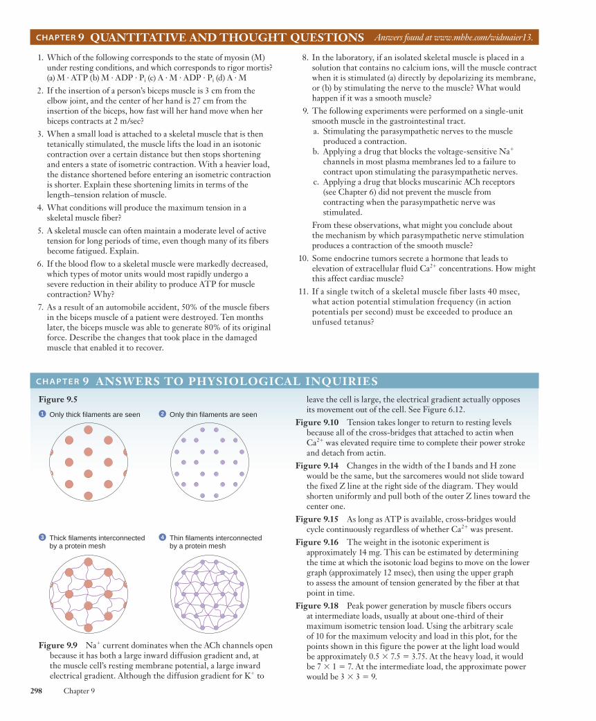
Rev. Confirming Pages
298 Chapter 9
Figure 9.5
1 Only thick filaments are seen 2 Only thin filaments are seen
3 Thick filaments interconnectedby a protein mesh
4 Thin filaments interconnectedby a protein mesh
Figure 9.9 Na1 current dominates when the ACh channels open because it has both a large inward diffusion gradient and, at the muscle cell’s resting membrane potential, a large inward electrical gradient. Although the diffusion gradient for K1 to
C H A P T E R 9 ANSWERS TO PHYSIOLOGICAL INQUIRIESleave the cell is large, the electrical gradient actually opposes its movement out of the cell. See Figure 6.12.
Figure 9.10 Tension takes longer to return to resting levels because all of the cross-bridges that attached to actin when Ca21 was elevated require time to complete their power stroke and detach from actin.
Figure 9.14 Changes in the width of the I bands and H zone would be the same, but the sarcomeres would not slide toward the fixed Z line at the right side of the diagram. They would shorten uniformly and pull both of the outer Z lines toward the center one.
Figure 9.15 As long as ATP is available, cross-bridges would cycle continuously regardless of whether Ca21 was present.
Figure 9.16 The weight in the isotonic experiment is approximately 14 mg. This can be estimated by determining the time at which the isotonic load begins to move on the lower graph (approximately 12 msec), then using the upper graph to assess the amount of tension generated by the fiber at that point in time.
Figure 9.18 Peak power generation by muscle fibers occurs at intermediate loads, usually at about one-third of their maximum isometric tension load. Using the arbitrary scale of 10 for the maximum velocity and load in this plot, for the points shown in this figure the power at the light load would be approximately 0.5 3 7.5 5 3.75. At the heavy load, it would be 7 3 1 5 7. At the intermediate load, the approximate power would be 3 3 3 5 9.
CHAPTER 9 QUANTITATIVE AND THOUGHT QUESTIONS Answers found at www.mhhe.com/widmaier13.
1. Which of the following corresponds to the state of myosin (M) under resting conditions, and which corresponds to rigor mortis? (a) M · ATP (b) M · ADP · Pi (c) A · M · ADP · Pi (d) A · M
2. If the insertion of a person’s biceps muscle is 3 cm from the elbow joint, and the center of her hand is 27 cm from the insertion of the biceps, how fast will her hand move when her biceps contracts at 2 m/sec?
3. When a small load is attached to a skeletal muscle that is then tetanically stimulated, the muscle lifts the load in an isotonic contraction over a certain distance but then stops shortening and enters a state of isometric contraction. With a heavier load, the distance shortened before entering an isometric contraction is shorter. Explain these shortening limits in terms of the length–tension relation of muscle.
4. What conditions will produce the maximum tension in a skeletal muscle fiber?
5. A skeletal muscle can often maintain a moderate level of active tension for long periods of time, even though many of its fibers become fatigued. Explain.
6. If the blood flow to a skeletal muscle were markedly decreased, which types of motor units would most rapidly undergo a severe reduction in their ability to produce ATP for muscle contraction? Why?
7. As a result of an automobile accident, 50% of the muscle fibers in the biceps muscle of a patient were destroyed. Ten months later, the biceps muscle was able to generate 80% of its original force. Describe the changes that took place in the damaged muscle that enabled it to recover.
8. In the laboratory, if an isolated skeletal muscle is placed in a solution that contains no calcium ions, will the muscle contract when it is stimulated (a) directly by depolarizing its membrane, or (b) by stimulating the nerve to the muscle? What would happen if it was a smooth muscle?
9. The following experiments were performed on a single-unit smooth muscle in the gastrointestinal tract. a. Stimulating the parasympathetic nerves to the muscle
produced a contraction. b. Applying a drug that blocks the voltage-sensitive Na1
channels in most plasma membranes led to a failure to contract upon stimulating the parasympathetic nerves.
c. Applying a drug that blocks muscarinic ACh receptors (see Chapter 6) did not prevent the muscle from contracting when the parasympathetic nerve was stimulated.
From these observations, what might you conclude about the mechanism by which parasympathetic nerve stimulation produces a contraction of the smooth muscle?
10. Some endocrine tumors secrete a hormone that leads to elevation of extracellular fluid Ca21 concentrations. How might this affect cardiac muscle?
11. If a single twitch of a skeletal muscle fiber lasts 40 msec, what action potential stimulation frequency (in action potentials per second) must be exceeded to produce an unfused tetanus?
wid78305_ch09_257-299.indd 298 30/01/13 11:49 AM
Rev. Confirming Pages
Muscle 299
Figure 9.20 Unfused tetanic contractions will occur at between 6.7 and 28.6 stimuli per second. In order for an unfused tetanus to occur, the interval between stimuli must be less than 150 msec but greater than 35 msec. (If the interval was greater than 150 msec, twitches would not summate, and if less than 35 msec, a fused tetanus would occur.) To calculate the corresponding frequencies:
1 stimulus/150 msec 3 1000 msec/sec 5 6.7 stimuli/sec 1 stimulus/35 msec 3 1000 msec/sec 5 28.6 stimuli/sec
Figure 9.21 The passive tension at 150% of muscle length would be about 35% of the maximum isometric tension (see the red curve). When stimulated at that length, the active tension developed would be an additional 35% (see the green curve). The total tension measured would therefore be approximately 70% of the maximum isometric tetanic tension.
Figure 9.25 Muscle fibers containing the slow isoform of myosin contract and hydrolyze ATP relatively slowly. Their requirement for ATP can thus be satisfied by aerobic/oxidative mechanisms that, although slow, are extremely efficient (a yield of 38 ATP per glucose molecule with water and carbon dioxide as waste products—see Chapter 3). It would not be efficient for a slow fiber to produce its ATP predominantly by glycolysis, a
process that is extremely rapid and relatively inefficient (only 2 ATP per glucose and lactic acid as a waste product).
Figure 9.29 The force acting upward on the forearm (85 3 5 5 425) would be less than the downward-acting force (10 3 45 5 450), so the muscle would undergo a lengthening (eccentric) contraction and the weight would move toward the ground.
Figure 9.30 The object would move nine times farther than the biceps in the same amount of time, or 18 cm/sec.
Figure 9.41 The skeletal muscle experiment would look the same. The calcium ions for contraction in skeletal muscle come from inside the sarcoplasmic reticulum. (Note: If the stimulus had been applied via a motor neuron, the lack of external Ca21 would have prevented exocytosis of ACh and there would have been no action potential or contraction in the skeletal muscle cell.) Removing extracellular Ca21 in the cardiac muscle experiment would eliminate both the prolonged plateau of the action potential and the contraction. Although the majority of the Ca21 that activates contraction also comes from the sarcoplasmic reticulum in cardiac muscle, its release is triggered by entry of Ca21 from the extracellular fluid through L-type channels during the action potential.
Visit this book’s website at www.mhhe.com/widmaier13 for chapter quizzes, interactive learning exercises, and other study tools.
human physiology
wid78305_ch09_257-299.indd 299 30/01/13 11:49 AM