Embed Size (px)
Citation preview
7/26/2019 SEGMENTATION AND CLASSIFICATION OF OPTICAL DISC IN RETINAL IMAGE
http://slidepdf.com/reader/full/segmentation-and-classification-of-optical-disc-in-retinal-image 1/5
International Journal of Innovative Research in Advanced Engineering (IJIRAE) ISSN: 2349-2763 Issue 04, Volume 3 (April 2016) SPECIAL ISSUE www.ijirae.com
________________________________________________________________________________________________________
IJIRAE: Impact Factor Value – SJIF: Innospace, Morocco (2015): 3.361 | PIF: 2.469 | Jour Info: 4.085 |
Index Copernicus 2014 = 6.57
© 2014- 16, IJIRAE- All Rights Reserved Page -41
EARLY STAGE DIAGNOSIS OF LUNG CANCER USINGCT-SCAN IMAGES BASED ON CELLULAR LEARNING
AUTOMATE
SAKTHI NEELA.P.K
Department of M.E (Medical electronics)
Sengunthar College of engineering
Namakkal, Tamilnadu, India
Mr.M.S.Muhammadu sathikraja, HOD & Assistant professor
Department of M.E (Medical electronics)
Sengunthar college of engineering
Namakkal, Tamilnadu,India
Abstract—The early detection of lung cancer is a challenging problem due to the structure of the cancer cells, where most of
the cells are overlapped with each other. This paper presents The project presents diagnosis of early stage lung cancer by making the image better and enhance it from noising, corruption or interference by using Gabor filtering and FIR filter to
remove Gaussian noise. The noise removed image is given to two level histogram stage then given to Image Segmentation stage: to divide and segment the enhanced images by using canny edge detection algorithm. Then segmented image is Feature Extracted to obtain the general features of the enhanced segmented image using Binarization and Masking Approach. The
proposed technique is efficient for segmentation principles to be a region of interest foundation for feature extraction obtaining. The proposed technique gives very promising results comparing with other used techniques. Relying on general features, a normality comparison is made. The main detected features for accurate images comparison are pixels percentage
and mask-labelling with high accuracy and robust operation.
Keywords—Image preprocessing, Gabor filtering, Thresholding Technique, Cany edge detection, Image Segmentation,
Feature extraction.
INTRODUCTION
Lung cancer is considered to be as the main cause of cancer death worldwide, and it is difficult to detect in its early stages because symptoms appear only at advanced stages causing the mortality rate to be the highest among all other types of cancer.More people die because of lung cancer than any other types of cancer such as: breast, colon, and prostate cancers. There issignificant evidence indicating that the early detection of lung cancer will decrease the mortality rate. The most recent estimatesaccording to the latest statistics provided by world health organization indicates that around 7.6 million deaths worldwide eachyear because of this type of cancer. Furthermore, mortality from cancer are expected to continue rising, to become around 17million worldwide in 2030[1].
There are many techniques to diagnosis lung cancer, such as Chest Radiograph (x-ray), Computed Tomography (CT), MagneticResonance Imaging (MRI scan) and Sputum Cytology[2]. However, most of these techniques are expensive and time consuming.In other words, most of these techniques are detecting the lung cancer in its advanced stages, where the patient’s chance ofsurvival is very low. Therefore, there is a great need for a new technology to diagnose the lung cancer in its early stages. Image
processing techniques provide a good quality tool for improving the manual analysis.For this reason we attempt to use automatic diagnostic system for detecting lung cancer in its early stages based on the analysis ofthe sputum color images. In order to formulate a rule we have developed a thresholding technique for unsupervised segmentationof the sputum color image to divide the images into several meaningful sub regions. Image segmentation has been used as the firststep in image classification and clustering. There are many algorithms which have been proposed in other articles for medicalimage segmentation, such as histogram analysis, regional growth, edge detection and Adaptive Thresholding[2]. A review of suchimage segmentation techniques can be found in[5]. Other authors have considered the use of color information as the keydiscriminating factor for cell segmentation for lung cancer diagnosis[7].The analysis of sputum images have been used in[8] fordetecting tuberculosis; it consists of analyzing sputum images for detecting bacilli. They used analysis techniques and featureextraction for the enhancement of the images, such as edge detection, heuristic knowledge, region labeling and removing.
7/26/2019 SEGMENTATION AND CLASSIFICATION OF OPTICAL DISC IN RETINAL IMAGE
http://slidepdf.com/reader/full/segmentation-and-classification-of-optical-disc-in-retinal-image 2/5
International Journal of Innovative Research in Advanced Engineering (IJIRAE) ISSN: 2349-2763 Issue 04, Volume 3 (April 2016) SPECIAL ISSUE www.ijirae.com
________________________________________________________________________________________________________
IJIRAE: Impact Factor Value – SJIF: Innospace, Morocco (2015): 3.361 | PIF: 2.469 | Jour Info: 4.085 |
Index Copernicus 2014 = 6.57
© 2014- 16, IJIRAE- All Rights Reserved Page -42
Thresholding has some benefits such as: fast processing, easy influence, resulting images do not keep weight space andanother method is region growing. This algorithm starts from a pixel of an image and checks other pixels that are around start point. The non assigned pixel that are neighbors with region checked are similar to the region then the region growing frequently but if differences between neighbor pix and region is more than the threshold the process is stopped. [5] The region growingsegmentation based thresholding is used on enhanced image.
I. IMAGE PREPROCESSING
GABOR FILTER
Image presentation based on Gabor function constitutes an excellent local and multi-scale decomposition in terms of logons thatare simultaneously (and optimally) localization in space and frequency domains. A Gabor filter is a linear filter whose impulseresponse is defined by a harmonic function multiplied by a Gaussian function. Because of the multiplication-convolution property(Convolution theorem), the Fourier transform of a Gabor filter's impulse response is the convolution of the Fourier transform ofthe harmonic function and the Fourier transform of the Gaussian function.
Its impulse response is defined by a sinusoidal wave (a plane wave for 2D Gabor filters) multiplied by a Gaussian filter . Becauseof the multiplication-convolution property (convolution), the of a Gabor filter's impulse response is the convolution of the Fouriertransform of the harmonic function and the Fourier transform of the Gaussian function. The filter has a real and an imaginarycomponent representing orthogonal directions. The two components may be formed into a complex number or used individually.
In this equation, represents the wavelength of the sinusoidal factor, represents the orientation of the normal to the parallel
stripes of a Gabor function, is the phase offset, is the sigma/standard deviation of the Gaussian envelope and is the spatialaspect ratio, and specifies the ellipticity of the support of the Gabor function.
Complex
Real
Imaginary
where
and
In this equation, represents the wavelength of the sinusoidal factor, represents the orientation of the normal to the parallel
stripes of a Gabor function, is the phase offset, is the sigma/standard deviation of the Gaussian envelope and is the spatialaspect ratio, and specifies the elasticity of the support of the Gabor function.
FAST FOURIER TRANSFORM
Fast Fourier Transform technique operates on Fourier transform of a given image. The frequency domain is a space inwhich each image value at image position F represents the amount that the intensity values in image “I” vary over a specificdistance related to F. Fast Fourier Transform is used here in image filtering (enhancement).
In particular, many of the existing techniques for image description and recognition depend highly on the segmentationresults. Segmentation divides the image into its constituent regions or objects. Segmentation of medical images in 2D, slice byslice has many useful applications for the medical professional such as: visualization and volume estimation of objects of interest,detection of abnormalities (e.g. tumors, polyps, etc.), tissue quantification and classification, and more . The goal of segmentationis to simplify and/or change the representation of the image into something that is more meaningful and easier to analyze. Imagesegmentation is typically used to locate objects and boundaries (lines, curves, etc.) in images. More precisely, image segmentationis the process of assigning a label to every pixel in an image such that pixels with the same label share certain visualcharacteristics.
7/26/2019 SEGMENTATION AND CLASSIFICATION OF OPTICAL DISC IN RETINAL IMAGE
http://slidepdf.com/reader/full/segmentation-and-classification-of-optical-disc-in-retinal-image 3/5
International Journal of Innovative Research in Advanced Engineering (IJIRAE) ISSN: 2349-2763 Issue 04, Volume 3 (April 2016) SPECIAL ISSUE www.ijirae.com
________________________________________________________________________________________________________
IJIRAE: Impact Factor Value – SJIF: Innospace, Morocco (2015): 3.361 | PIF: 2.469 | Jour Info: 4.085 |
Index Copernicus 2014 = 6.57
© 2014- 16, IJIRAE- All Rights Reserved Page -43
The result of image segmentation is a set of segments that collectively cover the entire image, or a set of contoursextracted from the image (edge detection). All pixels in a given region are similar with respect to some characteristic or computed property, such as color, intensity, or texture.
II.
SEGMENTATION
HISTOGRAM THRESHOLDING
Thresholding is one of the most powerful tools for image segmentation. The segmented image obtained fromthresholding has the advantages of smaller storage space, fast processing speed and ease in manipulation, compared with graylevel image which usually contains 256 levels. Therefore, thresholding techniques have drawn a lot of attention during the past 20years. Thresholding is a non-linear operation that converts a gray-scale image into a binary image where the two levels areassigned to pixels that are below or above the specified threshold value. In this research, Otsu’s method that uses (gray thresh)function to compute global image threshold is used. Otsu’s method is based on threshold selection by statistical criteria. Otsusuggested minimizing the weighted sum of within-class variances of the object and background pixels to establish an optimumthreshold. Recalling that minimization of within-class variances is equivalent to maximization of between-class variance. Thismethod gives satisfactory results for bimodal histogram images. Threshold values based on this method will be between 0 and 1,after achieving the threshold value; image will be segmented based on it
A. Segmentation
Marker-driven watershed segmentation technique extracts seeds that indicate the presence of objects or background at
specific image locations. Marker locations are then set to be regional minima within the topological surface (typically, thegradient of the original input image), and the watershed algorithm is applied. Separating touching objects in an image is one of themost difficult image processing operations, where the watershed transform is often applied to such problem. Marker-controlledwatershed approach has two types: External associated with the background and Internal associated with the objects of interest.Image Segmentation using the watershed transforms works well if we can identify or “mark” foreground objects and backgroundlocations, to find “catchment basins” and “watershed ridge lines” in an image by treating it as a surface where light pixels are highand dark pixels are low
B.Feature Extraction
Image features Extraction stage is an important stage that uses algorithms and techniques to detect and isolate variousdesired portions or shapes (features) of a given image. To predict the probability of lung cancer presence, the following twomethods are used: binarization and masking, both methods are based on facts that strongly related to lung anatomy andinformation of lung CT imaging.
C.Binarization ApproachBinarization approach depends on the fact that the number of black pixels is much greater than white pixels in normal
lung images, so we started to count the black pixels for normal and abnormal images to get an average that can be used later as athreshold, if the Leonardo Electronic Journal of Practices and Technologies number of the black pixels of a new image is greaterthat the threshold, then it indicates that the image is normal, otherwise, if the number of the black pixels is less than the threshold,it indicates that the image in abnormal.
D.Masking ApproachMasking approach depends on the fact that the masses are appeared as white connected areas inside ROI (lungs), as they
increase the percent of cancer presence increase. The appearance of solid blue color indicates normal case while appearance ofRGB masses indicates the presence of cancer
III.
RESULTS AND DISCUSSION
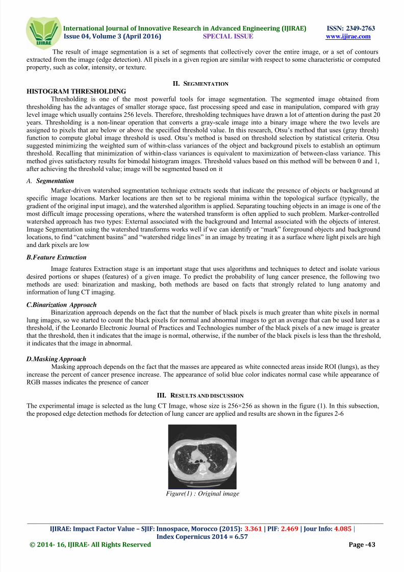
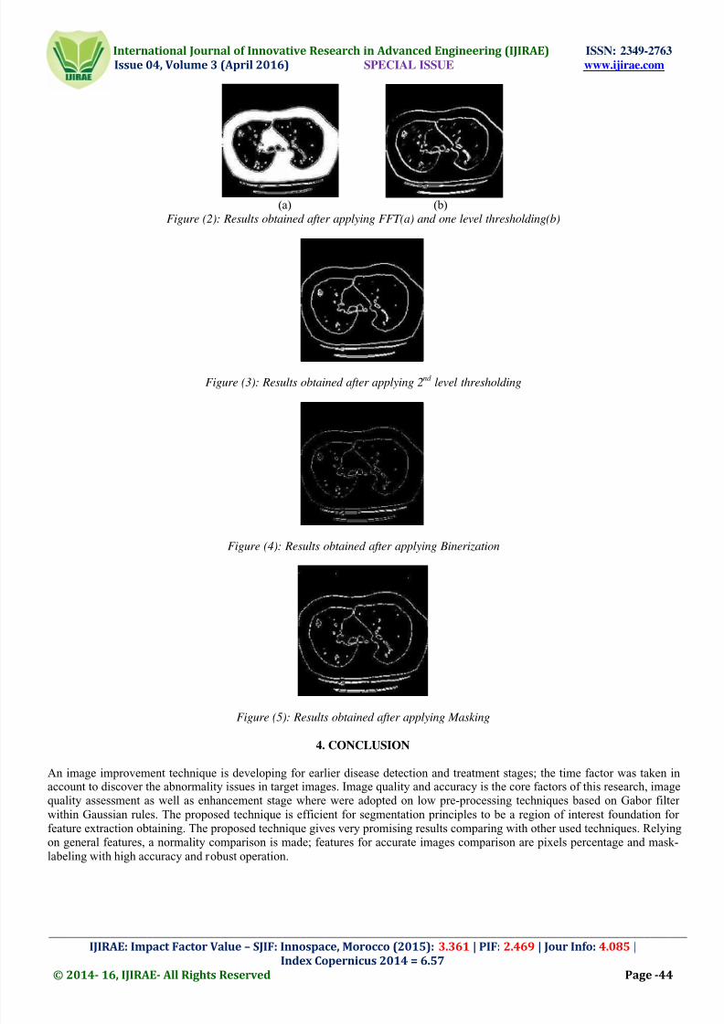
The experimental image is selected as the lung CT Image, whose size is 256×256 as shown in the figure (1). In this subsection,the proposed edge detection methods for detection of lung cancer are applied and results are shown in the figures 2-6
Figure(1) : Original image
7/26/2019 SEGMENTATION AND CLASSIFICATION OF OPTICAL DISC IN RETINAL IMAGE
http://slidepdf.com/reader/full/segmentation-and-classification-of-optical-disc-in-retinal-image 4/5
International Journal of Innovative Research in Advanced Engineering (IJIRAE) ISSN: 2349-2763 Issue 04, Volume 3 (April 2016) SPECIAL ISSUE www.ijirae.com
________________________________________________________________________________________________________
IJIRAE: Impact Factor Value – SJIF: Innospace, Morocco (2015): 3.361 | PIF: 2.469 | Jour Info: 4.085 |
Index Copernicus 2014 = 6.57
© 2014- 16, IJIRAE- All Rights Reserved Page -44
(a) (b)Figure (2): Results obtained after applying FFT(a) and one level thresholding(b)
Figure (3): Results obtained after applying 2nd
level thresholding
Figure (4): Results obtained after applying Binerization
Figure (5): Results obtained after applying Masking
4. CONCLUSION
An image improvement technique is developing for earlier disease detection and treatment stages; the time factor was taken inaccount to discover the abnormality issues in target images. Image quality and accuracy is the core factors of this research, imagequality assessment as well as enhancement stage where were adopted on low pre-processing techniques based on Gabor filterwithin Gaussian rules. The proposed technique is efficient for segmentation principles to be a region of interest foundation forfeature extraction obtaining. The proposed technique gives very promising results comparing with other used techniques. Relyingon general features, a normality comparison is made; features for accurate images comparison are pixels percentage and mask-labeling with high accuracy and robust operation.
7/26/2019 SEGMENTATION AND CLASSIFICATION OF OPTICAL DISC IN RETINAL IMAGE
http://slidepdf.com/reader/full/segmentation-and-classification-of-optical-disc-in-retinal-image 5/5
International Journal of Innovative Research in Advanced Engineering (IJIRAE) ISSN: 2349-2763 Issue 04, Volume 3 (April 2016) SPECIAL ISSUE www.ijirae.com
________________________________________________________________________________________________________
IJIRAE: Impact Factor Value – SJIF: Innospace, Morocco (2015): 3.361 | PIF: 2.469 | Jour Info: 4.085 |
Index Copernicus 2014 = 6.57
© 2014- 16, IJIRAE- All Rights Reserved Page -45
5. FUTURE SCOPE
The two level thresholding, canny edge detection and features extraction technique shows promising result. The systemdiagnosis capability can be enhanced by archiving images and patient records. The archives should be available for free to thetrained engineers and open source communities. The trained doctors and engineers working together need discover some newfeatures for better classification and prognosis of cancer in early stages. The above techniques can be applied on images from X-
ray or MRI for comparison of result. The images which give better result should be considered. The full automation of the systemcan be achieved by integrating diagnosis of cancer stage with best possible oncological treatment.
ACKNOWLEDGMENT
I wish to express my profound thanks to Medical Electronics engineering department, Sengunthar college of engineering,trichengode and faculty members for providing me all the facilities in making this project possible.. I also thank my parents forthe encouragement, support and attention. I am also grateful to my friend who supported me throughout this venture with valuablefeedback.
REFERENCES
[1].Sopore, J & K, 2014 “Efficient edge detection methods for diagnosis of lung cancer based on two-dimensional cellular
automata” ” in presented at 22nd International Conference on Systems, Signals and Image Processing.[2].Nunes É.D.O., Pérez M.G., 2013. “ Medical Image Segmentation by Multilevel Thresholding Based on HistogramDifference”, presented at 17th International Conference on Systems, Signals and Image Processing,
[3].Venkateshwarlu K., 2012 “Image Enhancement using Fuzzy Inference System” in presented at 19th International Conferenceon Systems, Signals and Image Processing,
[4].Mokhled S. AL-TARAWNEH,2012 “Lung Cancer Detection Using Image Processing Techniques” Leonardo ElectronicJournal of Practices and Technologies
[5].Rama .M ,2010 “Application of Neural Networks in Diagnosing Cancer Disease Using Demographic Data” in InternationalJournal of Computer Application.
[6].Sarika Tale,2015 “Lung Cancer Diagnosis using Computer Aided Diagnosis System “in International Journal of ComputerApplications.