Embed Size (px)
Citation preview

F. P Wedel, D.O.
Associate Adjunct Professor in Osteopathic Principles and Practice
A.T. Still University School of Osteopathic Medicine in Arizona, and private practice
in Family Medicine in Tucson, Arizona
Evaluation, Diagnosis and Treatment of
Selected Somatic Dysfunctions
related to the
SHORT LEG
Learning Objectives
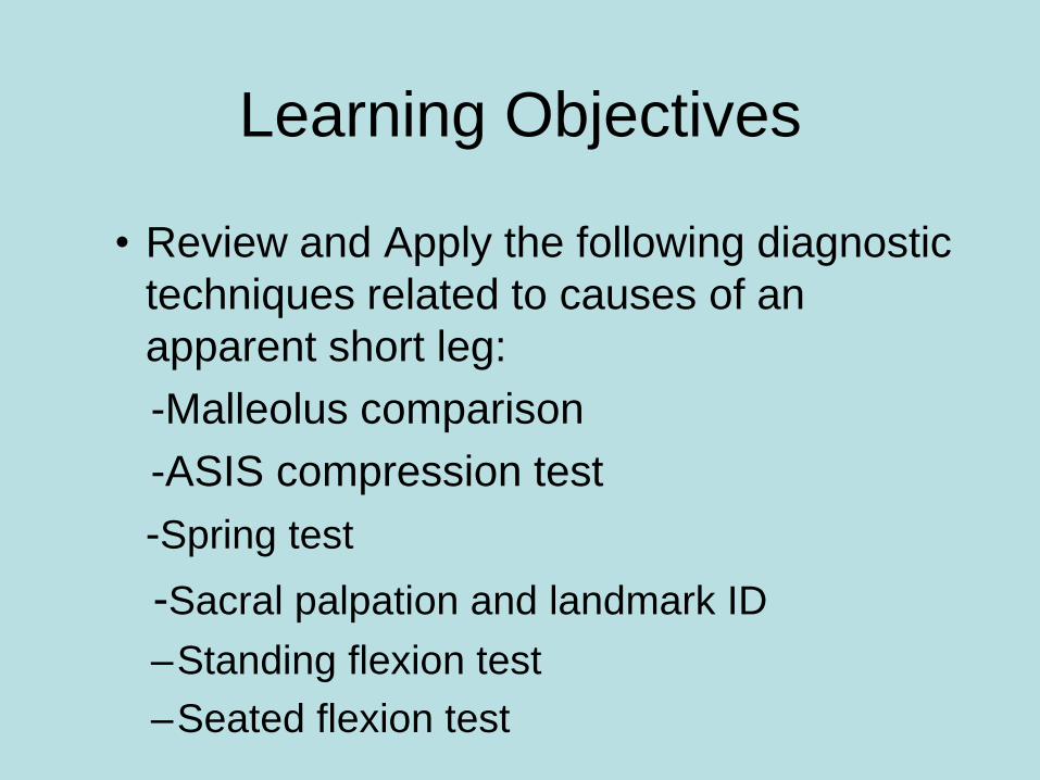
• Review and Apply the following diagnostic
techniques related to causes of an
apparent short leg:
-Malleolus comparison
-ASIS compression test
-Spring test
-Sacral palpation and landmark ID
–Standing flexion test
–Seated flexion test

Ilial-Sacral Techniques Covered:
1. Prone and Supine direct, muscle energy and HVLA for Innominate dysfunction: posterior rotation and upslip
2. Prone and Supine direct, muscle energy, and HVLA for sacral rotation on both same and opposite axes (torsions)
3. Prone, direct, muscle energy for sacral shears

ASIS SUPERIOR R___ L___
COMPRESSION + R___ L___
MALLEOLI SHORT R___ L___
PSIS SUPERIOR R___ L___
SULCUS DEEP R___ L___
ILA INF/ POST R___ L___
SPRING (IT DID) R___ L___
LUMBAR SPASM R___ L___
SEATED FLEXION + R___ L___
STANDING FLEXION+ R___ L___
DIAGNOSIS:
UPSLIP INNOMINATE
SACRAL SHEAR – UNILATERAL FLEXION OR EXTENSION
POSTERIOR/ ANTERIOR INNOMINATE
ANTERIOR / POSTERIOR SACRAL TORSION
TREATMENT CHOICE ME___ HVLA___
DID IT WORK YES___ NO___
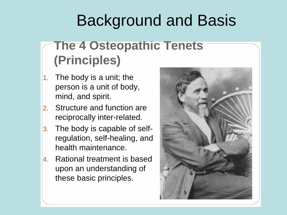
Background and Basis
The 4 Osteopathic Tenets
(Principles)
1. The body is a unit; the
person is a unit of body,
mind, and spirit.
2. Structure and function are
reciprocally inter-related.
3. The body is capable of self-
regulation, self-healing, and
health maintenance.
4. Rational treatment is based
upon an understanding of
these basic principles.

Somatic Dysfunction - Defined• “Impaired or altered function of related components
of the somatic (body framework) system:
• Skeletal, arthrodial, and myofascial structures,
• And…
• Related vascular, lymphatic, and neural elements”

Treatment Options for Somatic
Dysfunctions
• All somatic dysfunctions have a restrictive
barrier which are considered “pathologic”
• This restriction inhibits movement in one
direction which causes asymmetry within
the joint:
• The goal of osteopathic treament
is to eliminate the restrictive
barrier thus restoring symmetry….
Somatic Dysfunction:
CHARACTERISTICS
“The acronym TART is used to remember the
abnormal changes that accompany somatic
dysfunction. (Tenderness by itself is not always an
indication of somatic dysfunction):
•Tissue texture changes
•Asymmetry
•Restricted range of motion
•Tenderness
Kimberly Manual, chapter 3

Treatment Methodologies
• Indirect – movement away
from the barrier and more
functional than structural
–Cranial-sacral
–Counterstrain
–Balanced ligamentous tension
(BLT)
–Facilitated Positional Release
Treatment Methodologies
• Direct – engagement of the
restrictive barrier and movement
through it and to it by using the
body part as a lever
• Muscle Energy and MFR
• HVLA
• Chapman’s and Lymphatics

• ANATOMY
SACRAL
STRUCTURE,LIGAMENTS AND
MUSCLES

THE SACRUM
Means “sacred”
because of its density it is the last bone to decay and because it
protects
the reproductive system


Forces on the sacrum
• Angle of the sacroiliac joint “wedges” the sacrum in an anterior direction
– Prevents posterior movement
• Dorsal (posterior) sacroiliac ligaments much stronger than anterior sacroiliac ligaments
• Purpose: counteract significant pelvic forces pushing apex posteriorly.
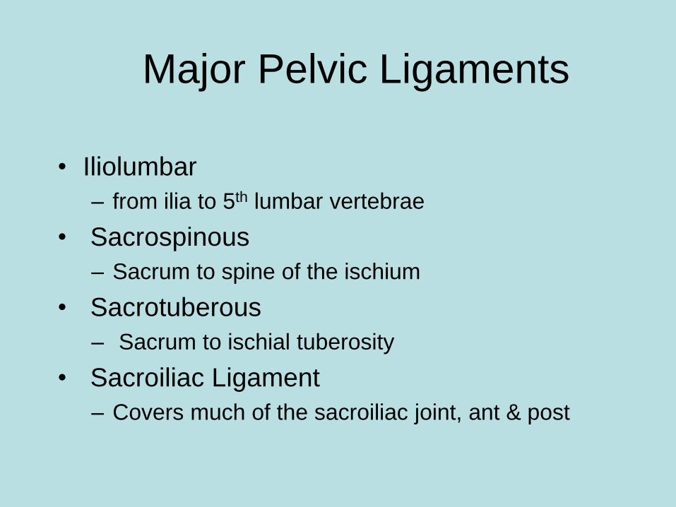
Major Pelvic Ligaments
• Iliolumbar
– from ilia to 5th lumbar vertebrae
• Sacrospinous
– Sacrum to spine of the ischium
• Sacrotuberous
– Sacrum to ischial tuberosity
• Sacroiliac Ligament
– Covers much of the sacroiliac joint, ant & post
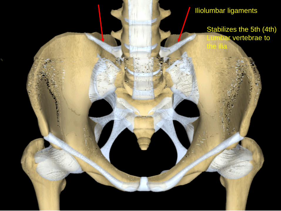
Iliolumbar ligaments
Stabilizes the 5th (4th)
Lumbar vertebrae to
the ilia
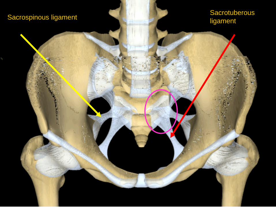
Sacrospinous ligamentSacrotuberous
ligament
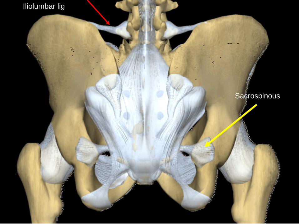
Iliolumbar lig
Sacrospinous

Sacrotuberous Ligament
• Runs from lower sacral tubercles to ischial
tuberosity
• Gluteus maximus attachment
• Tendon of the biceps femoris attachment
• Connects with fascia of the pelvis
– from sacrum to ischial tuberosity
– stabilizes anterior motion

Both Sacrospinous
& Sacrotuberousstabilize to prevent
posterior - superior
rotation of the sacral
apex around a
transverse axis

Sacroiliac Ligament
• Sacroiliac
– actually three ligaments
• Anterior or ventral sacroiliac
– from 3rd sacral segment to lateral preauricular sulcus
• interosseous sacroiliac
– massive bond between the upper parts of the joint
• dorsal sacroiliac
– Partly covers the interosseous, from lateral sacral
crest to PSIS and internal iliac crest.

Ventral/Anterior
Sacroiliac

interosseous
Posterior sacroiliac


Pelvic muscle attachments
from above.• Attach to Sacrum
– Erector Spinae• Iliocostalis
• Longissimus
• Spinalis
– Multifidus
• Attach to Innominates– Obliques (internal, external, transverse)
– Quadratus Lumborum
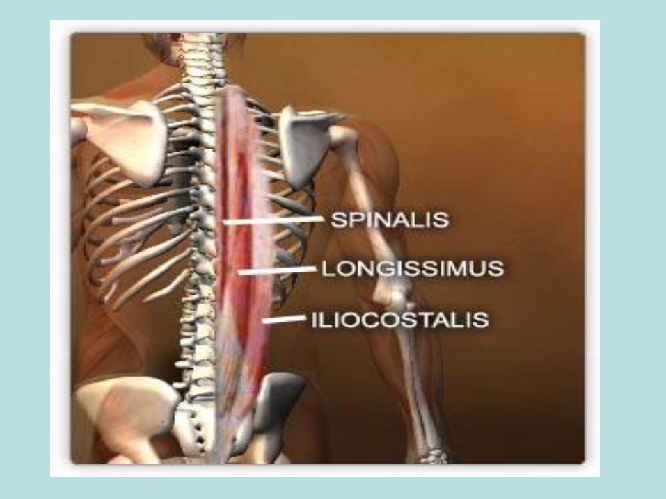

Posterior
Muscles
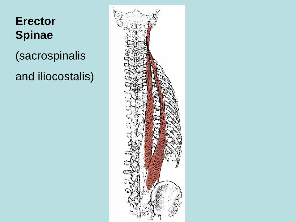
Erector
Spinae
(sacrospinalis
and iliocostalis)
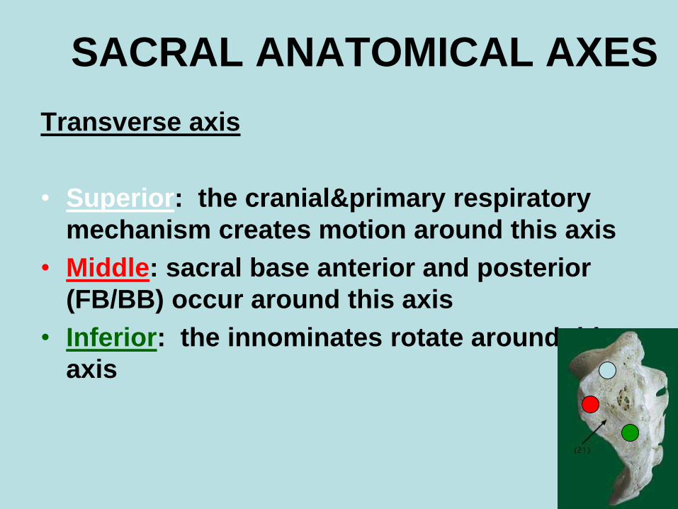
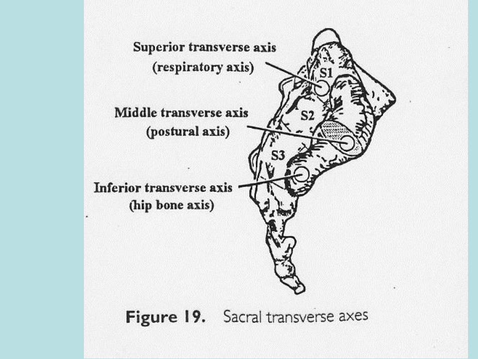
SACRAL ANATOMICAL AXES
Transverse axis
• Superior: the cranial&primary respiratory
mechanism creates motion around this axis
• Middle: sacral base anterior and posterior
(FB/BB) occur around this axis
• Inferior: the innominates rotate around this
axis
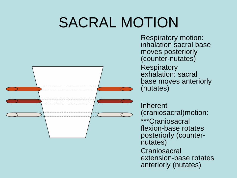
SACRAL MOTION Respiratory motion: inhalation sacral base moves posteriorly (counter-nutates)
Respiratory exhalation: sacral base moves anteriorly (nutates)
Inherent (craniosacral)motion:
***Craniosacral flexion-base rotates posteriorly (counter-nutates)
Craniosacral extension-base rotates anteriorly (nutates)
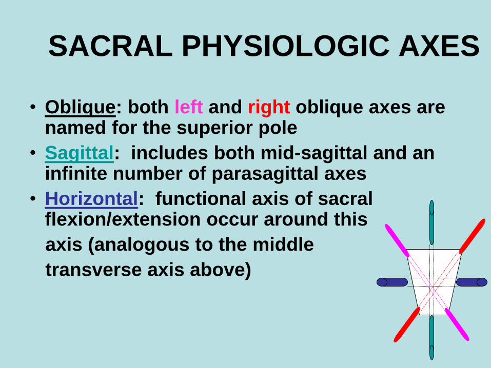
SACRAL PHYSIOLOGIC AXES
• Oblique: both left and right oblique axes are named for the superior pole
• Sagittal: includes both mid-sagittal and an infinite number of parasagittal axes
• Horizontal: functional axis of sacral flexion/extension occur around this
axis (analogous to the middle
transverse axis above)
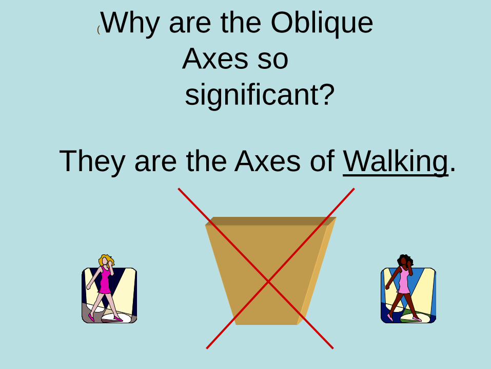
(Why are the Oblique
Axes so
significant?
They are the Axes of Walking.
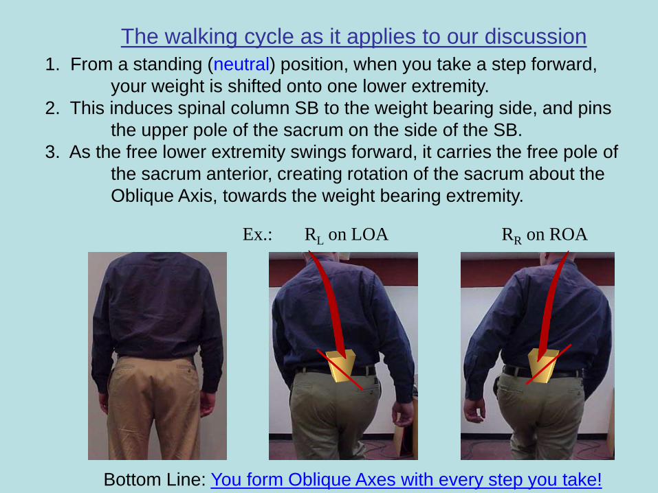
The walking cycle as it applies to our discussion
1. From a standing (neutral) position, when you take a step forward,
your weight is shifted onto one lower extremity.
2. This induces spinal column SB to the weight bearing side, and pins
the upper pole of the sacrum on the side of the SB.
3. As the free lower extremity swings forward, it carries the free pole of
the sacrum anterior, creating rotation of the sacrum about the
Oblique Axis, towards the weight bearing extremity.
Bottom Line: You form Oblique Axes with every step you take!
Ex.: RL on LOA RR on ROA
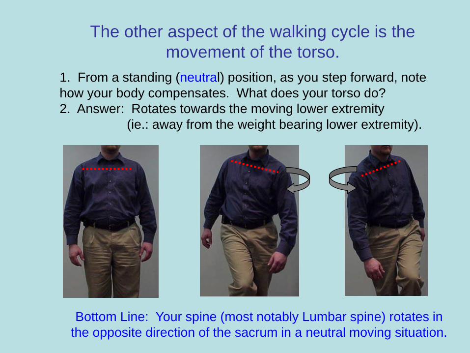
The other aspect of the walking cycle is the
movement of the torso.
1. From a standing (neutral) position, as you step forward, note
how your body compensates. What does your torso do?
2. Answer: Rotates towards the moving lower extremity
(ie.: away from the weight bearing lower extremity).
Bottom Line: Your spine (most notably Lumbar spine) rotates in
the opposite direction of the sacrum in a neutral moving situation.
Useful Tests
SUPINE
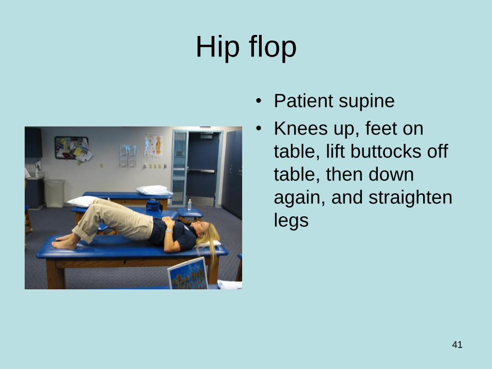
Hip flop
• Patient supine
• Knees up, feet on
table, lift buttocks off
table, then down
again, and straighten
legs
41
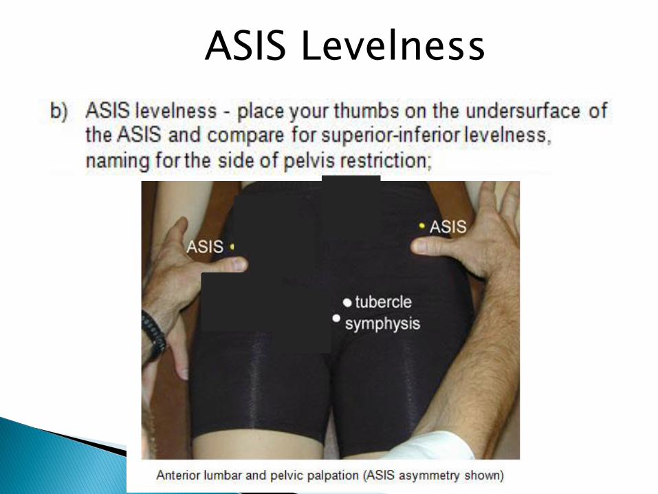
ASIS Levelness

Have the patient lie supine. The patient is then asked to raise his/her bottom up off the table and then set it back down again.
Doctor Stands with head and shoulders centered over the patient.
Contact the ASIS ◦ Stabilize one ASIS while
applying pressure at a 45 degree angle to the other ASIS
Positive test - restricted movement of the Sacroiliac joint -> rock like motion
Negative test - a sense of give or resilience => bounce or spring like motion
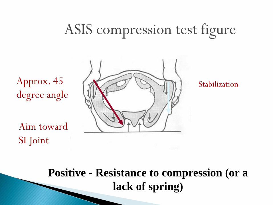
ASIS compression test figure
Positive - Resistance to compression (or a
lack of spring)
Approx. 45
degree angleStabilization
Aim toward
SI Joint
45
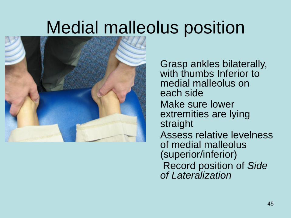
Medial malleolus position
Grasp ankles bilaterally, with thumbs Inferior to medial malleolus on each side
Make sure lower extremities are lying straight
Assess relative levelness of medial malleolus (superior/inferior)
Record position of Side of Lateralization
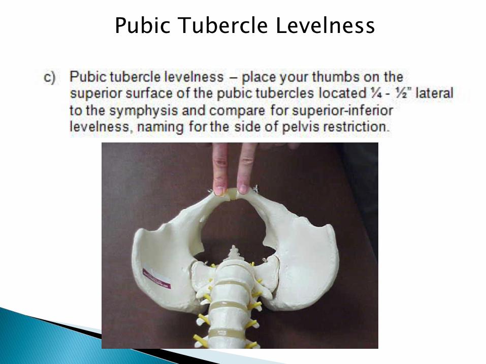
Pubic Tubercle Levelness


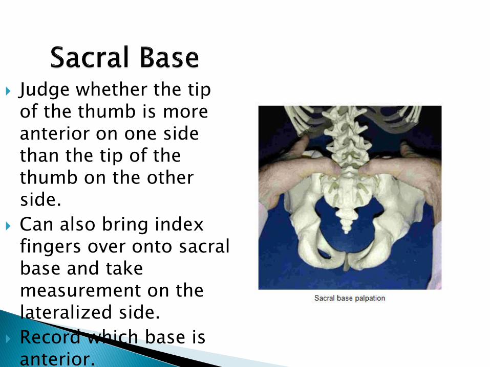
Judge whether the tip of the thumb is more anterior on one side than the tip of the thumb on the other side.
Can also bring index fingers over onto sacral base and take measurement on the lateralized side.
Record which base is anterior.
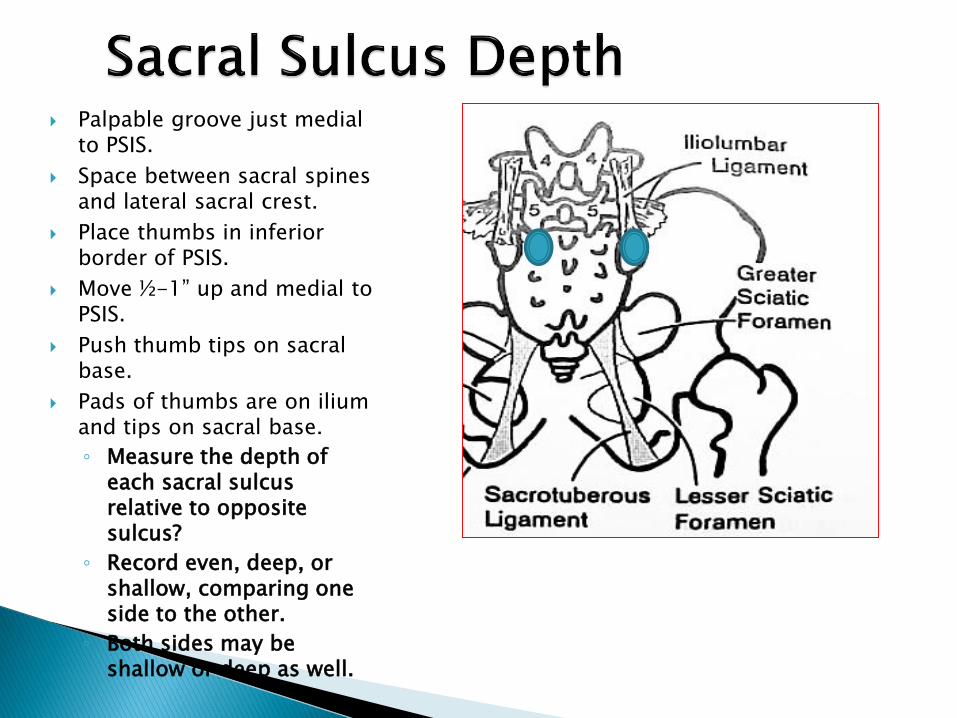
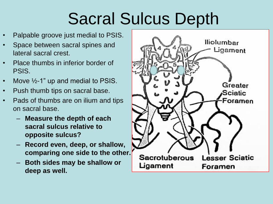
Palpable groove just medial to PSIS.
Space between sacral spines and lateral sacral crest.
Place thumbs in inferior border of PSIS.
Move ½-1” up and medial to PSIS.
Push thumb tips on sacral base.
Pads of thumbs are on ilium and tips on sacral base.
◦ Measure the depth of each sacral sulcus relative to opposite sulcus?
◦ Record even, deep, or shallow, comparing one side to the other.
◦ Both sides may be shallow or deep as well.
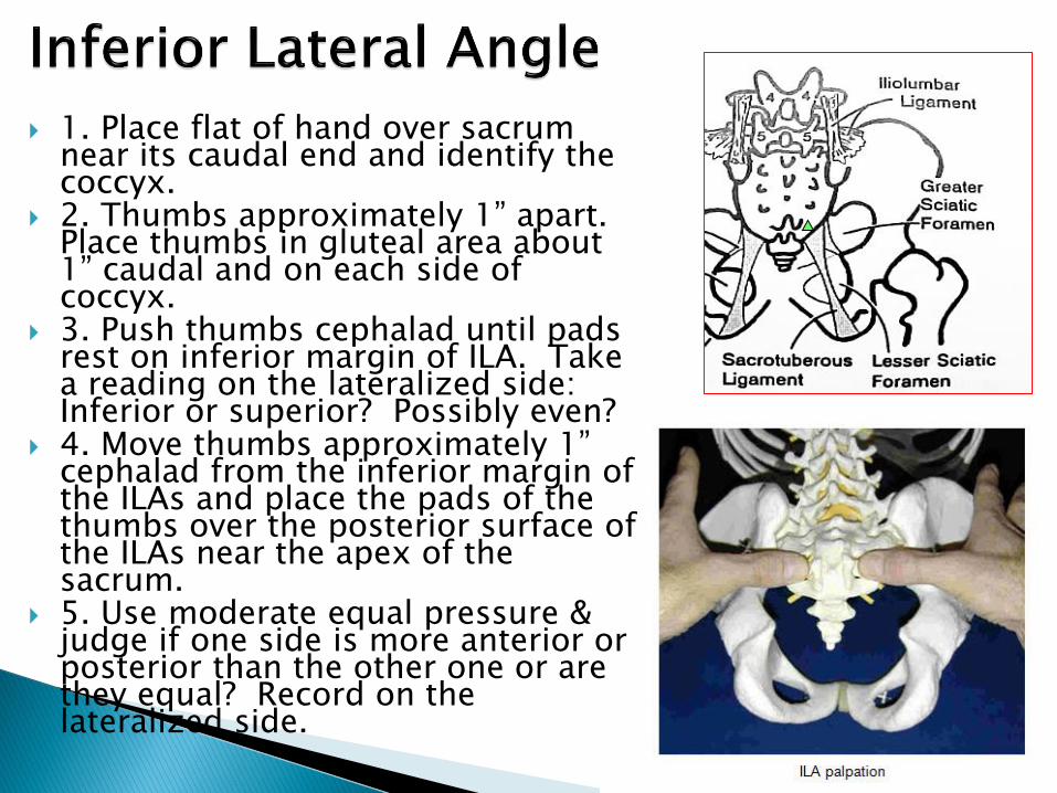
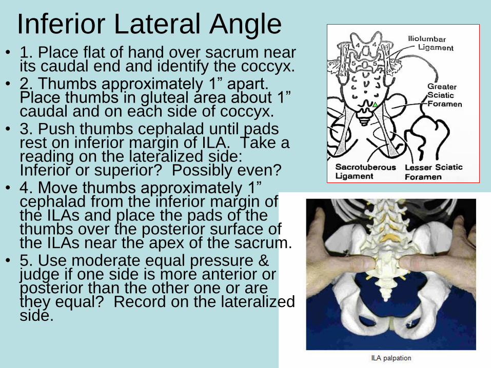
1. Place flat of hand over sacrum near its caudal end and identify the coccyx.
2. Thumbs approximately 1” apart. Place thumbs in gluteal area about 1” caudal and on each side of coccyx.
3. Push thumbs cephalad until pads rest on inferior margin of ILA. Take a reading on the lateralized side: Inferior or superior? Possibly even?
4. Move thumbs approximately 1” cephalad from the inferior margin of the ILAs and place the pads of the thumbs over the posterior surface of the ILAs near the apex of the sacrum.
5. Use moderate equal pressure & judge if one side is more anterior or posterior than the other one or are they equal? Record on the lateralized side.
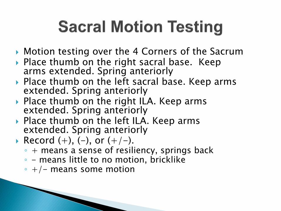
Motion testing over the 4 Corners of the Sacrum Place thumb on the right sacral base. Keep
arms extended. Spring anteriorly Place thumb on the left sacral base. Keep arms
extended. Spring anteriorly Place thumb on the right ILA. Keep arms
extended. Spring anteriorly Place thumb on the left ILA. Keep arms
extended. Spring anteriorly Record (+), (-), or (+/-).◦ + means a sense of resiliency, springs back◦ - means little to no motion, bricklike◦ +/- means some motion
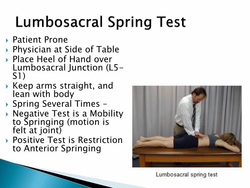
Patient Prone Physician at Side of Table Place Heel of Hand over
Lumbosacral Junction (L5-S1)
Keep arms straight, and lean with body
Spring Several Times – Negative Test is a Mobility
to Springing (motion is felt at joint)
Positive Test is Restriction to Anterior Springing
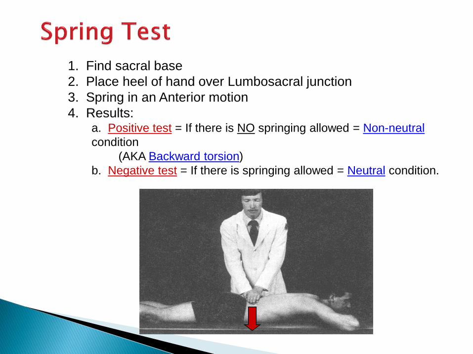
1. Find sacral base
2. Place heel of hand over Lumbosacral junction
3. Spring in an Anterior motion
4. Results:a. Positive test = If there is NO springing allowed = Non-neutral
condition
(AKA Backward torsion)
b. Negative test = If there is springing allowed = Neutral condition.

STAND
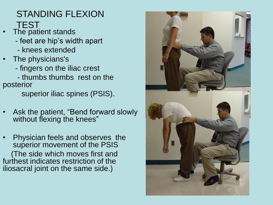
• The patient stands
- feet are hip’s width apart
- knees extended
• The physicians's
- fingers on the iliac crest
- thumbs thumbs rest on the posterior
superior iliac spines (PSIS).
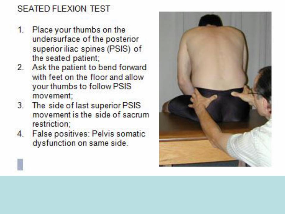
• Ask the patient, “Bend forward slowly without flexing the knees”
• Physician feels and observes the superior movement of the PSIS
(The side which moves first and furthest indicates restriction of the iliosacral joint on the same side.)
STANDING FLEXION
TEST

To make a Sacral Diagnosis you
will need to know the following:
• Static (Pure) Landmarks– Sacral base - Ant/ Post
– ILA -Ant/Post
– ASIS & PSIS -Sup./Inf.
– Pubes -Sup./Inf & Ant./Post
• Mixed Landmarks– Sacral Sulcus- Deep/Shallow
– STL - Tight/ Loose/ Equal
• Motion Testing
– Spring test
– L5
– Sacrum

HANDS ON SECTION 1
• TIME TO GO : TO THE TABLES
• WITH A PARTNER
• GET INSTRUCTIONS AND RESULTS
SHEET
• Ideally, you have now enough information
to make one of the following diagnoses:
• We will go through each and its treatment
options
• You will then go back to tables and choose
a method to fix your partner’s problem
SHORT LEG CONDITIONS
• Anatomic –congenital or acquired
• Sacral Shear
• Sacral Torsion
• Posterior Innominate
• Upslipped Innominate

Pelvis side shifts and rotates toward long leg
Innominate rotates anterior on side of short leg or posterior on side of long leg
Foot of long leg pronates, internally rotating lower leg
Lumbosacral angle increases by 2 to 3 degrees
Sacral Shears

To diagnose a sacral shear
• 1) need positive seated flexion test
• 2) need a deep sulcus
• 3) need an ILA that is posterior on same
side as deep sulcus
If deep sulcus and ILA are right sided but
seated flexion + on L = unilateral extension;
if deep sulcus and ILA are right sided and
seated flexion + on R = unilateral flexion
SHEARS
• So – a shear is either a
• Unilateral sacral flexion or a
• Unilateral sacral extension
• The extension shear will have a Short Leg
• The flexion shear will have a Long leg –
but the other will be Short ( relatively)
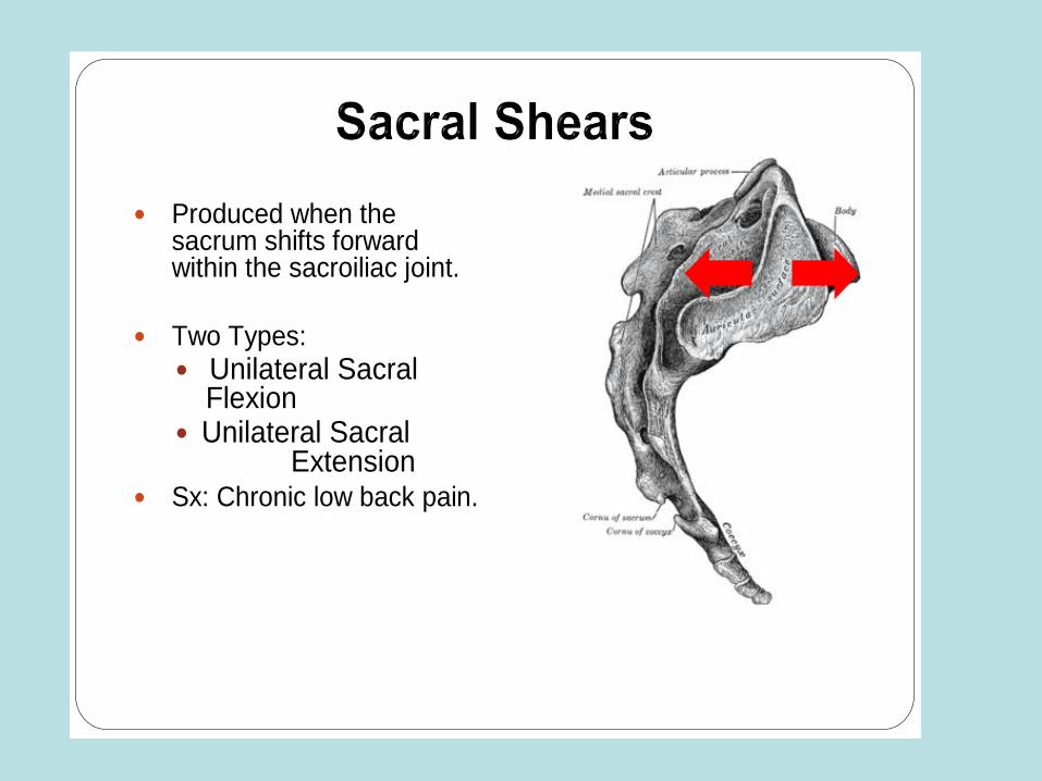
Produced when the sacrum shifts forward within the sacroiliac joint.
Two Types:
Unilateral Sacral Flexion
Unilateral Sacral Extension
Sx: Chronic low back pain.
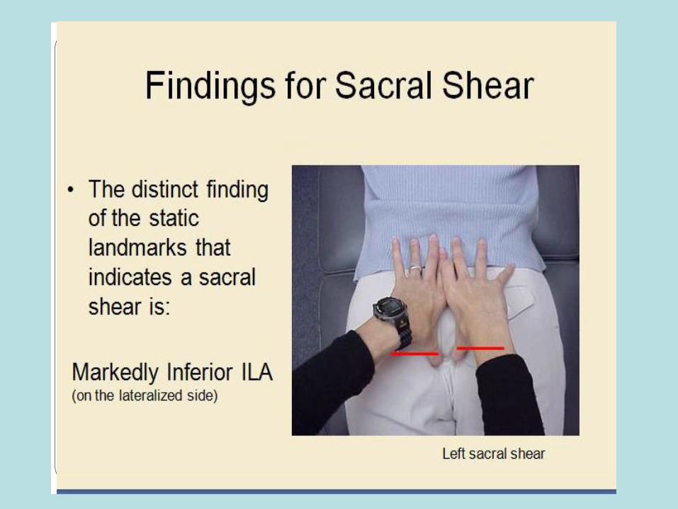

Naming the Shear
The shear is named for the side of the inferior ILA..
The sulcus is deep on same side- (which distinguishes this from a torsion)
The seated flexion positive side will tell you how to interpret whether it is a unilateral flexion or extension,
i.e.,sulcus deep and ILA on R with R seated flexion + =
R unilateral Flexion;
L unilateral extension if seated is + L with the same findings of: deep sulcus R and ILA post/infR

Sacral Shear Extension(unilateral)
TREATING A SHEAR
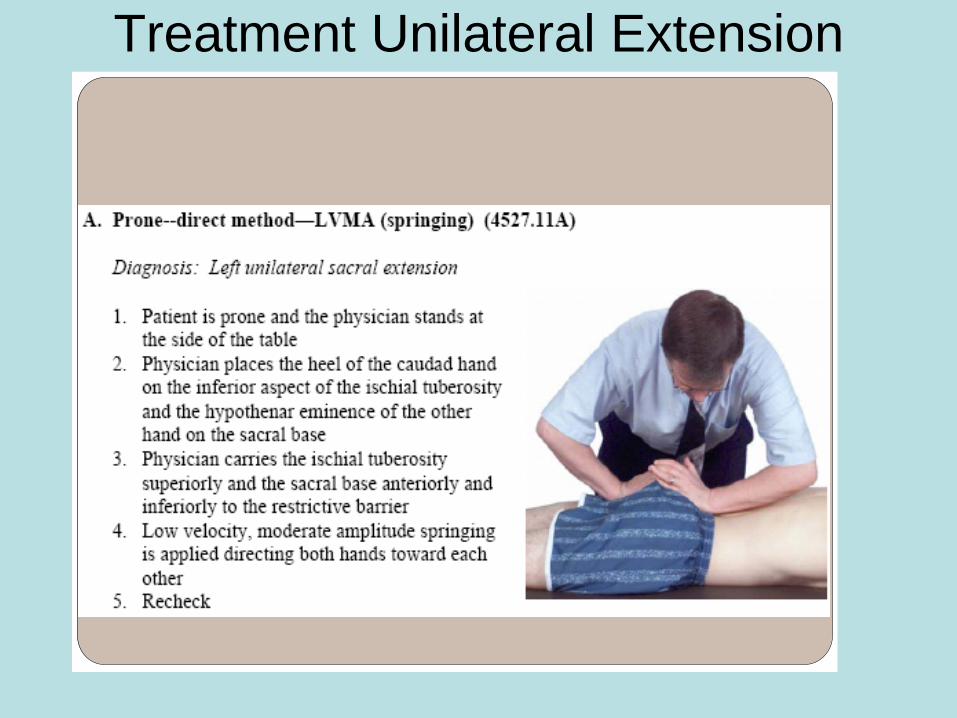
Treatment Unilateral Extension
• Sacral Shear – Unilateral sacral flexion
• The treatment side leg here will be long
but the other leg will be short


Shear Treatments-unilateral
flexions
Springing Respiratory ForceLeft unilateral sacral flexion
Springing Respiratory ForceLeft Unilateral Sacral Flexion A
Monitor at PSIS
Abduct and internally rotate LE until motion is felt.
Maintain that position by leaning of the patient’s femur/leg
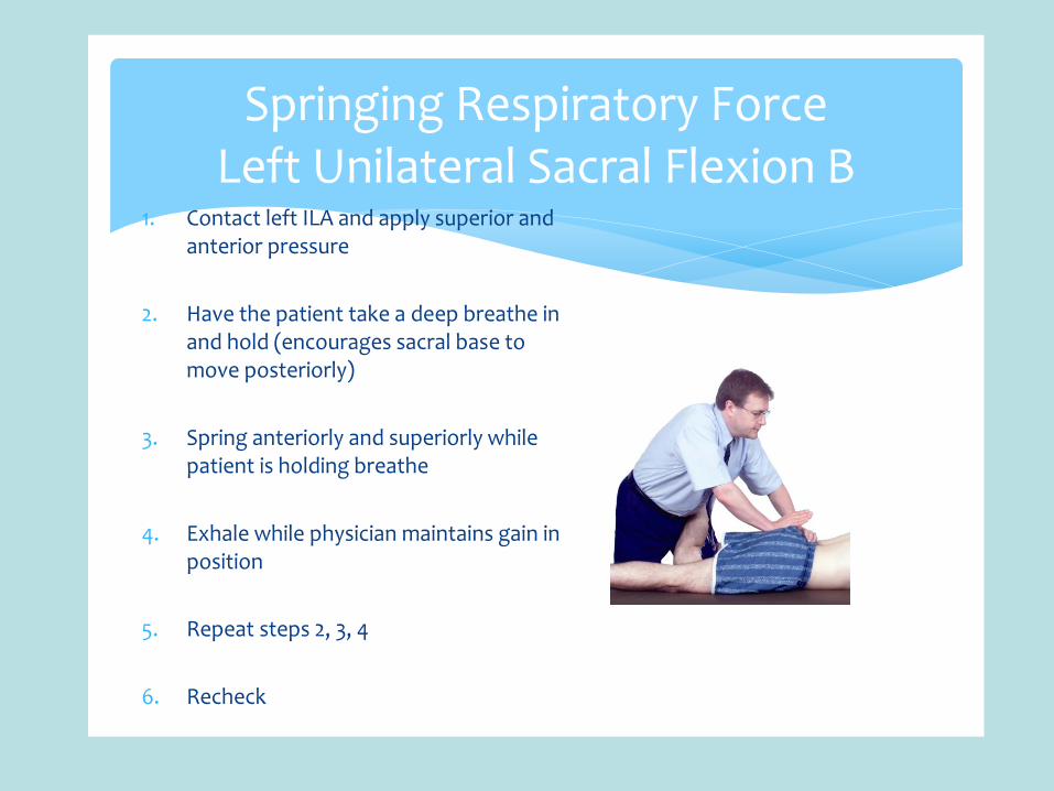
Springing Respiratory ForceLeft Unilateral Sacral Flexion B
1. Contact left ILA and apply superior and anterior pressure
2. Have the patient take a deep breathe in and hold (encourages sacral base to move posteriorly)
3. Spring anteriorly and superiorly while patient is holding breathe
4. Exhale while physician maintains gain in position
5. Repeat steps 2, 3, 4
6. Recheck

• A FEW DETAILS ON LUMBAR AND
SACRAL DIAGNOSES - sorry
FRYETTE’S LAWS • Law I: When the spine is in neutral, sidebending to one side will
be accompanied by horizontal rotation to the opposite side. In type I somatic dysfunction, this law can be seen when more than one vertebrae are out of alignment and cannot be returned to neutral by flexion or extension. The involved group of vertebrae demonstrates a coupled relationship between side bending and rotation. When the spine is neutral, side bending forces are applied to a group of typical vertebrae and the entire group will rotate toward the opposite side: the side of produced convexity. Extreme type I dysfunction is similar to scoliosis.
• Law II: When the spine is flexed or extended (non-neutral), sidebending to one side will be accompanied by rotation to the same side. In type II somatic dysfunction of the spine, this law can be seen when only one vertebrae is out of place and becomes much worse on flexion or extension. There will be rotation and sidebending in the same direction when this dysfunction is present
• Law III: When motion is introduced in one plane it will modify (reduce) motion in the other two planes. Type III sums up the other two laws by stating dysfunction in one plane will negatively affect all other planes of motion

Lumbosacral
Mechanics
• Example L rotation on LOA
• Lumbar spine neutral: SL RR
(note in all torsions, L5 will
rotate opposite of sacrum)
• Requires normal lordosis
• Occurs when (R) sacral base
rotates anterior (“forward”)
and does not rotate back
(feels “springy”)
• left ILA posterior, & inferior
(i.e., taking a step forward on r
A
P
SL RR
Lumbosacral motion
• Lumbar spine and sacrum rotate in
OPPOSITE directions
Neutral (type I) mechanics:
Example: L on LOA, the right sacral base
moves anteriorly while L5 is SLRR
In non-neutral (type 2) mechanics, the
sacral base rotates backwards…
Example: R on LOA, the right sacral base
moves posteriorly while L5 is RLSL
Sacral Motion
In Neutral (type I) mechanics:
the sacral base moves Anteriorly and
rotates opposite to the rotation of L5 ;
Its axis motion is the SAME as the SB
side of L5 so :
Example: L5 RrSl causes a L on LOA, ( L
on L) sacral torsion the R edge is
rotated L and anterior while the axis is
left so it = to the SB side of L5
Sacral Motion
In Non-Neutral (type 2) mechanics, the
sacral base rotates backwards.
It is still opposite to the L5 rotation side and
Its axis is the same as the L5 SB side
Example: L5 RlSl causes a R on LOA, the
right sacral base moves Posteriorly while
its axis side is Left and oblique
Sacral Torsion Rules
• A sacral torsion requires a deep sulcus
and a posterior and inferior ILA to be on
opposite sides
A deep right sulcus must have an posterior
and inferior ILA on the Left by the above
definition
Sacral Sulcus Depth• Palpable groove just medial to PSIS.
• Space between sacral spines and
lateral sacral crest.
• Place thumbs in inferior border of
PSIS.
• Move ½-1” up and medial to PSIS.
• Push thumb tips on sacral base.
• Pads of thumbs are on ilium and tips
on sacral base.
– Measure the depth of each
sacral sulcus relative to
opposite sulcus?
– Record even, deep, or shallow,
comparing one side to the other.
– Both sides may be shallow or
deep as well.
Inferior Lateral Angle• 1. Place flat of hand over sacrum near
its caudal end and identify the coccyx.• 2. Thumbs approximately 1” apart.
Place thumbs in gluteal area about 1” caudal and on each side of coccyx.
• 3. Push thumbs cephalad until pads rest on inferior margin of ILA. Take a reading on the lateralized side: Inferior or superior? Possibly even?
• 4. Move thumbs approximately 1” cephalad from the inferior margin of the ILAs and place the pads of the thumbs over the posterior surface of the ILAs near the apex of the sacrum.
• 5. Use moderate equal pressure & judge if one side is more anterior or posterior than the other one or are they equal? Record on the lateralized side.
Sacral Torsion Rules
• Name of lesion:
• Is for rotation and axis sides:
• R on R = right rotation on a right axis
R on L = right rotation on left axis
Sacral Torsion Rules
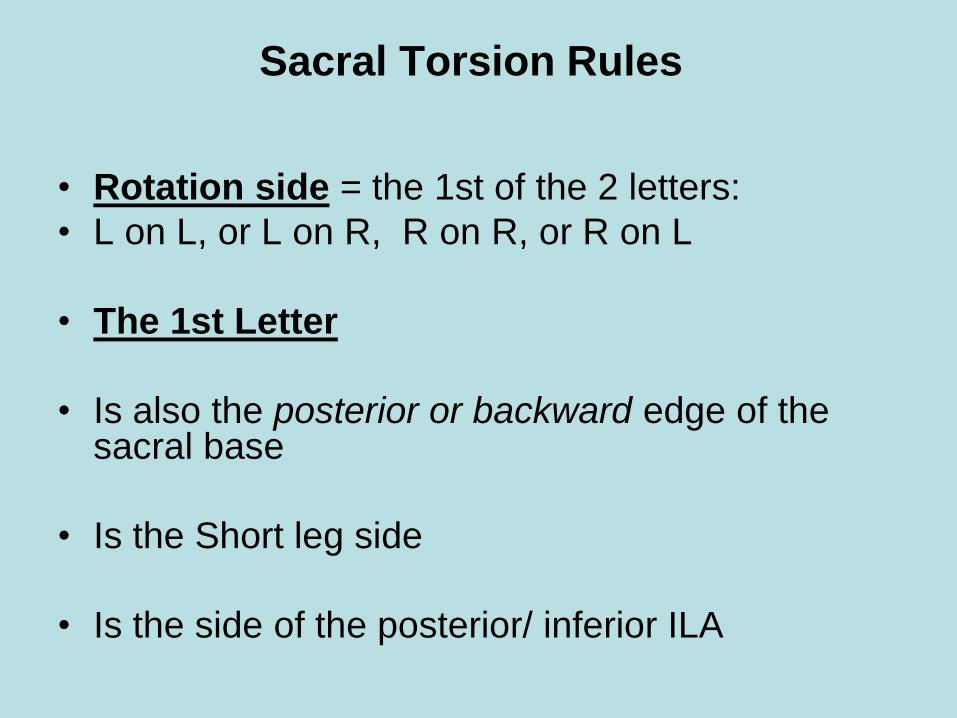
• Rotation side = the 1st of the 2 letters:
• L on L, or L on R, R on R, or R on L
• The 1st Letter
• Is also the posterior or backward edge of the sacral base
• Is the Short leg side
• Is the side of the posterior/ inferior ILA

Sacral Torsion Rules
The second letter ( R on R) refers to
the Axis side of the sacrum that is in
play
Is the same as the side bent side of L5
i.e., if L5 is Sidebent R the sacrum will
be on its R axis
Sacral Torsion Rules
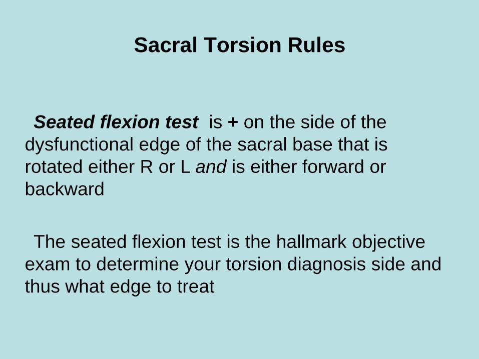
Seated flexion test is + on the side of the
dysfunctional edge of the sacral base that is
rotated either R or L and is either forward or
backward
The seated flexion test is the hallmark objective
exam to determine your torsion diagnosis side and
thus what edge to treat
Sacral Torsion Rules
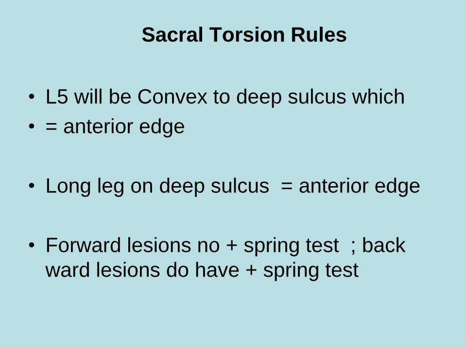
• L5 will be Convex to deep sulcus which
• = anterior edge
• Long leg on deep sulcus = anterior edge
• Forward lesions no + spring test ; back
ward lesions do have + spring test

• FORWARD TORSIONS

Neutral - Left Oblique Axis Findings
Name: L on LOA, RL on LOA,
Landmarks – Static:
Sacral Base: L posterior
Sacral Sulcus: L shallow
ILA: L Post/ Inf.
STL: L Tight
Motion Testing:
Spring: - (neg)
L5: SLRR
Sacral Base L - R +
ILA: L +/- R +/-Left Right
Midline
A +
P+/-
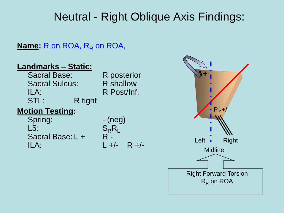
Neutral - Right Oblique Axis Findings:
Name: R on ROA, RR on ROA,
Landmarks – Static: Sacral Base: R posteriorSacral Sulcus: R shallowILA: R Post/Inf.STL: R tight
Motion Testing:Spring: - (neg)L5: SRRL
Sacral Base: L + R -ILA: L +/- R +/-
Right Forward Torsion
RR on ROA
Left Right
Midline
P+/-
A+
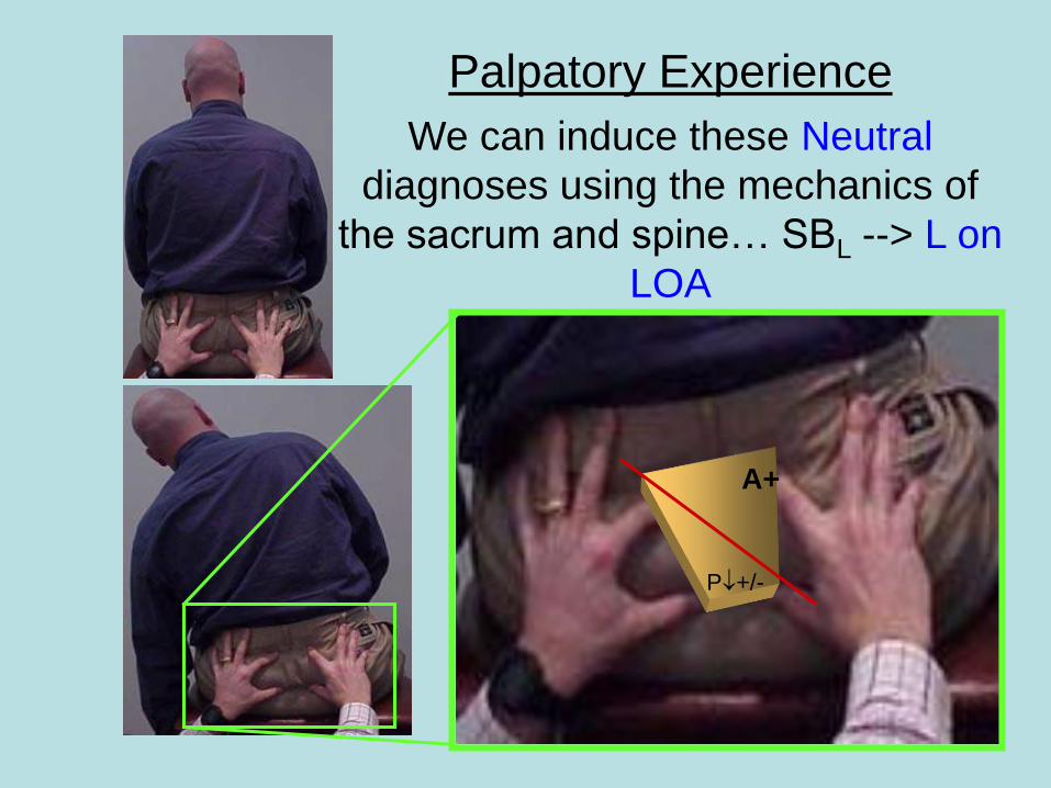
Palpatory Experience
We can induce these Neutral
diagnoses using the mechanics of
the sacrum and spine… SBL --> L on
LOA
A+
P+/-
• Treatment Options for
• Forward Sacral Torsions

Forward Sacral Torsion ME
(a right on right sacral torsion)1. Axis side down; chest on the table
2. Monitor at the lumbo-sacral junction
3. Flex the knees and hips until motion is felt at the lumbo-sacral junction
4. Support legs/knees with thigh or pillow
5. Apply pressure downward on lower legs/ankles
6. Ask patient to try to raise feet towards the ceiling while you resist
7. Rest
8. Re-engage barrier by repositioning ankles downward
9. Repeat 6, 7, 8
10. Recheck
Relative contra-indications-acute sacroiliac sprain, acute sacrum fracture, severe knee arthritis, deep venous thrombosis, or premature labor
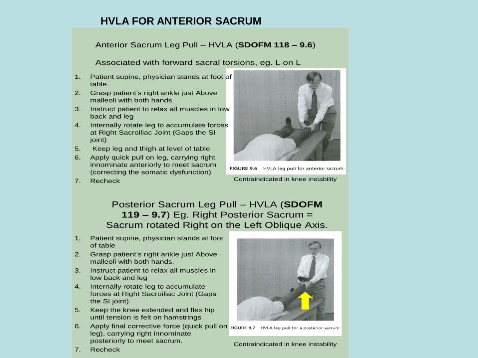
Anterior Sacrum Leg Pull – HVLA (SDOFM 118 – 9.6)
Associated with forward sacral torsions, eg. L on L
1. Patient supine, physician stands at foot of
table
2. Grasp patient’s right ankle just Above
malleoli with both hands.
3. Instruct patient to relax all muscles in low
back and leg
4. Internally rotate leg to accumulate forces
at Right Sacroiliac Joint (Gaps the SI
joint)
5. Keep leg and thigh at level of table
6. Apply quick pull on leg, carrying right
innominate anteriorly to meet sacrum
(correcting the somatic dysfunction)
7. Recheck Contraindicated in knee instability
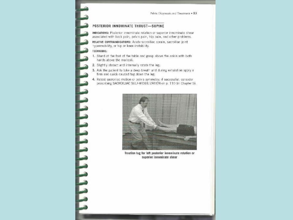
Posterior Sacrum Leg Pull – HVLA (SDOFM
119 – 9.7) Eg. Right Posterior Sacrum =
Sacrum rotated Right on the Left Oblique Axis.
1. Patient supine, physician stands at foot
of table
2. Grasp patient’s right ankle just Above
malleoli with both hands.
3. Instruct patient to relax all muscles in
low back and leg
4. Internally rotate leg to accumulate
forces at Right Sacroiliac Joint (Gaps
the SI joint)
5. Keep the knee extended and flex hip
until tension is felt on hamstrings
6. Apply final corrective force (quick pull on
leg), carrying right innominate
posteriorly to meet sacrum.
7. RecheckContraindicated in knee instability
HVLA FOR ANTERIOR SACRUM

POSTERIOR SACRAL
TORSIONS

Non-Neutral: Left Oblique Axis Findings
(right on left sacral torsion)
Name: R on LOA, RR on LOA,
Landmarks – Static:
Sacral Base: L Anterior
Sacral Sulcus: L Deep
ILA: L Ant/ Sup
STL: L Loose
Motion Testing:
Spring: + (positive)
L5: RLSL
Sacral Base L - R +/-
ILA: L + R +/-
Left Right
Midline
P+/-
A+
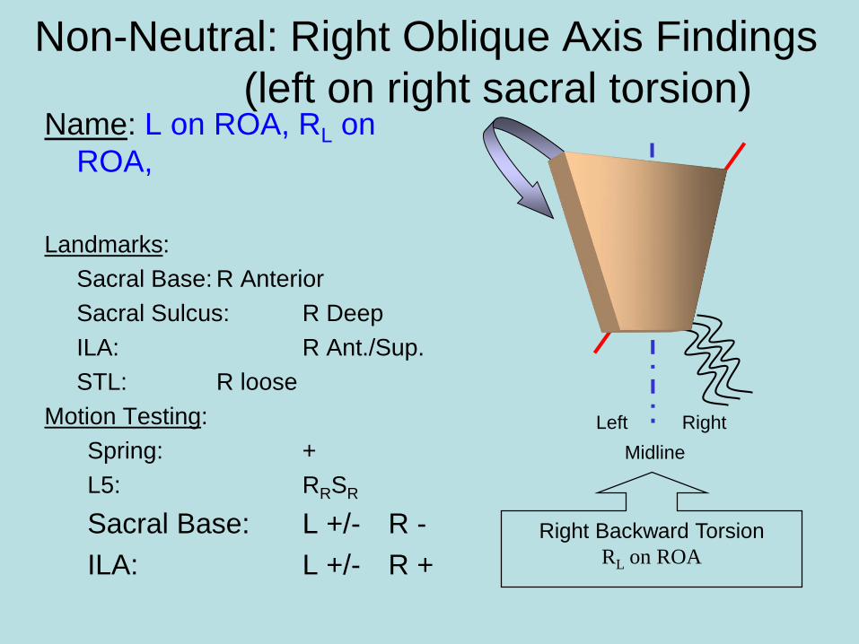
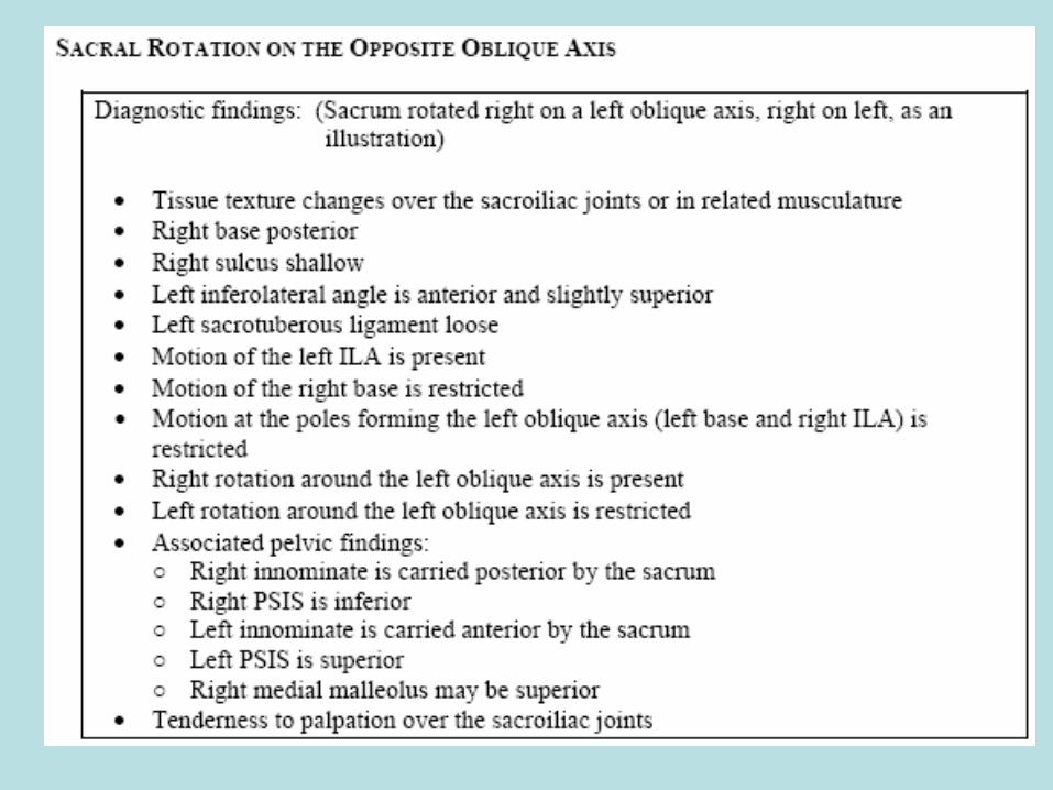
Non-Neutral: Right Oblique Axis Findings
(left on right sacral torsion)Name: L on ROA, RL on
ROA,
Landmarks:
Sacral Base: R Anterior
Sacral Sulcus: R Deep
ILA: R Ant./Sup.
STL: R loose
Motion Testing:
Spring: +
L5: RRSR
Sacral Base: L +/- R -
ILA: L +/- R +
A+
P+/-
Left Right
Midline
Right Backward Torsion
RL on ROA
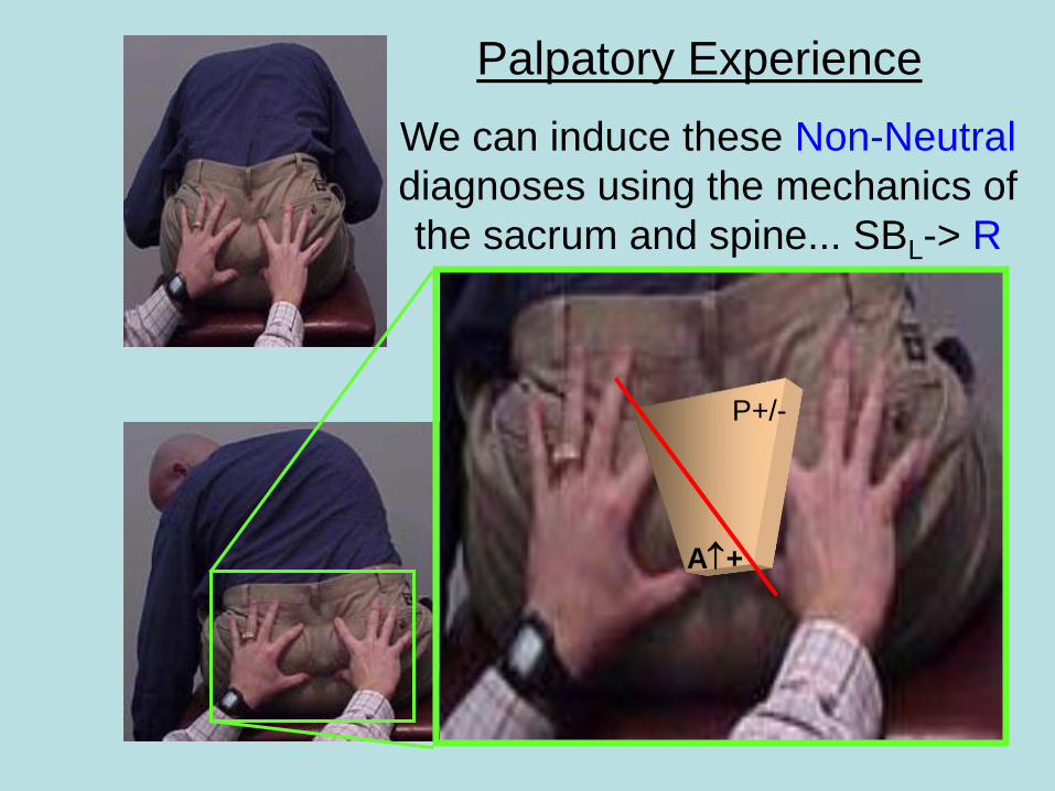
Palpatory Experience
We can induce these Non-Neutral
diagnoses using the mechanics of
the sacrum and spine... SBL-> R
on LOA
P+/-
A+
Posterior Sacral Torsion Treatment
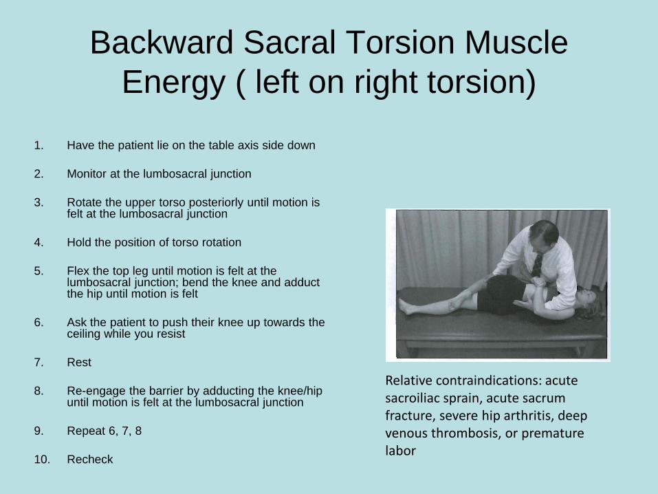
Backward Sacral Torsion Muscle
Energy ( left on right torsion)
1. Have the patient lie on the table axis side down
2. Monitor at the lumbosacral junction
3. Rotate the upper torso posteriorly until motion is felt at the lumbosacral junction
4. Hold the position of torso rotation
5. Flex the top leg until motion is felt at the lumbosacral junction; bend the knee and adduct the hip until motion is felt
6. Ask the patient to push their knee up towards the ceiling while you resist
7. Rest
8. Re-engage the barrier by adducting the knee/hip until motion is felt at the lumbosacral junction
9. Repeat 6, 7, 8
10. Recheck
Relative contraindications: acute sacroiliac sprain, acute sacrum fracture, severe hip arthritis, deep venous thrombosis, or premature labor
HVLA FOR POSTERIOR SACRUM
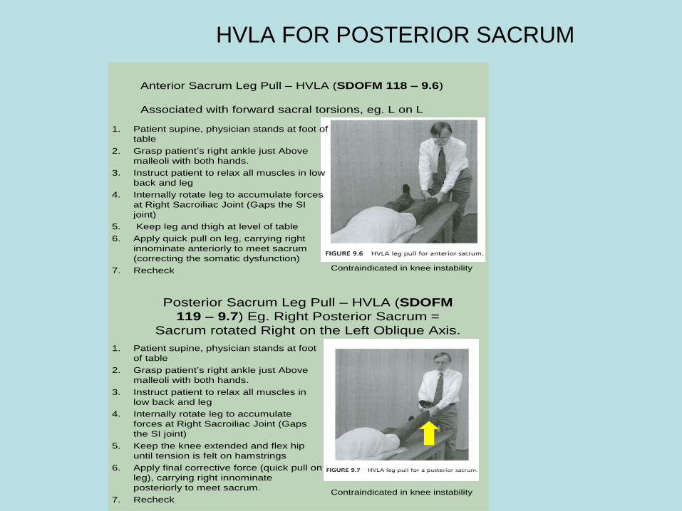
Anterior Sacrum Leg Pull – HVLA (SDOFM 118 – 9.6)
Associated with forward sacral torsions, eg. L on L
1. Patient supine, physician stands at foot of
table
2. Grasp patient’s right ankle just Above
malleoli with both hands.
3. Instruct patient to relax all muscles in low
back and leg
4. Internally rotate leg to accumulate forces
at Right Sacroiliac Joint (Gaps the SI
joint)
5. Keep leg and thigh at level of table
6. Apply quick pull on leg, carrying right
innominate anteriorly to meet sacrum
(correcting the somatic dysfunction)
7. Recheck Contraindicated in knee instability
Posterior Sacrum Leg Pull – HVLA (SDOFM
119 – 9.7) Eg. Right Posterior Sacrum =
Sacrum rotated Right on the Left Oblique Axis.
1. Patient supine, physician stands at foot
of table
2. Grasp patient’s right ankle just Above
malleoli with both hands.
3. Instruct patient to relax all muscles in
low back and leg
4. Internally rotate leg to accumulate
forces at Right Sacroiliac Joint (Gaps
the SI joint)
5. Keep the knee extended and flex hip
until tension is felt on hamstrings
6. Apply final corrective force (quick pull on
leg), carrying right innominate
posteriorly to meet sacrum.
7. RecheckContraindicated in knee instability


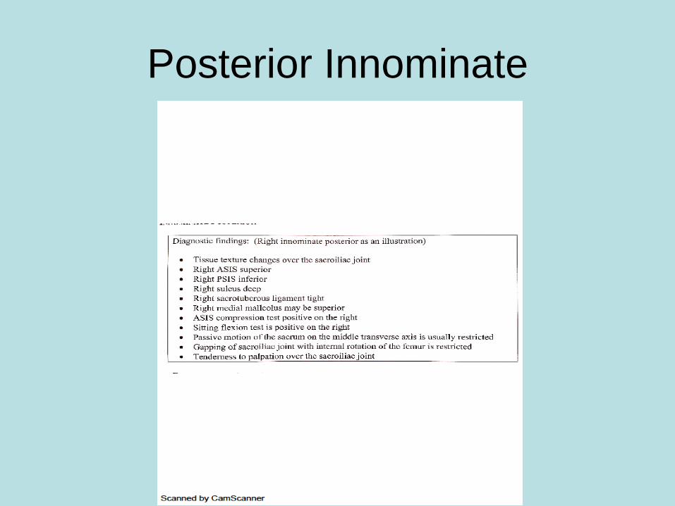
Posterior Innominate
• Muscle Energy and HVLA
Posterior Innominate

Treatment Options
Muscle Energy Posteriorly Rotated Ilium
1. Monitor at the PSIS and posterior iliac crest
2. Extend the leg (anteriorly rotating the ilium) until motion is felt at the PSIS
3. Have the patient attempt to pull leg down towards table against resistance
4. Relax
5. Reposition
6. Repeat 2, 3, 4, 5
7. Recheck


Anterior Innominate
• Standing Flexion will be +on the side of:
• ASIS will be inferior
• PSIS will be superior
• Leg will be Long - so opposite leg will
appear SHORT

Anterior Innominate
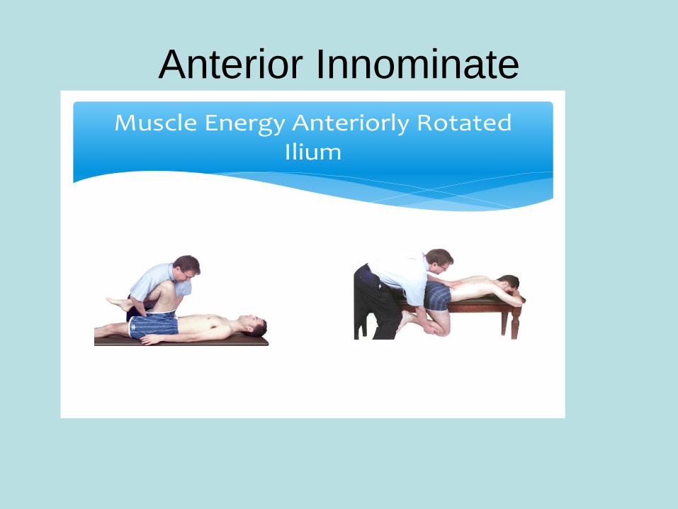
Anterior Innominate
Muscle Energy Anteriorly Rotated Ilium
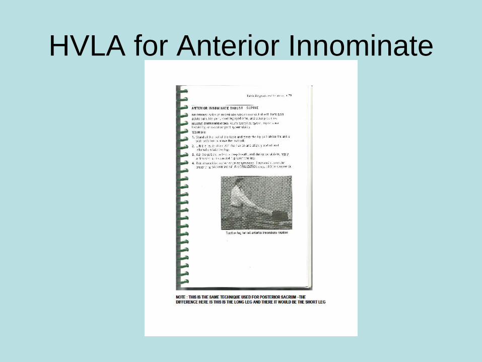
HVLA for Anterior Innominate

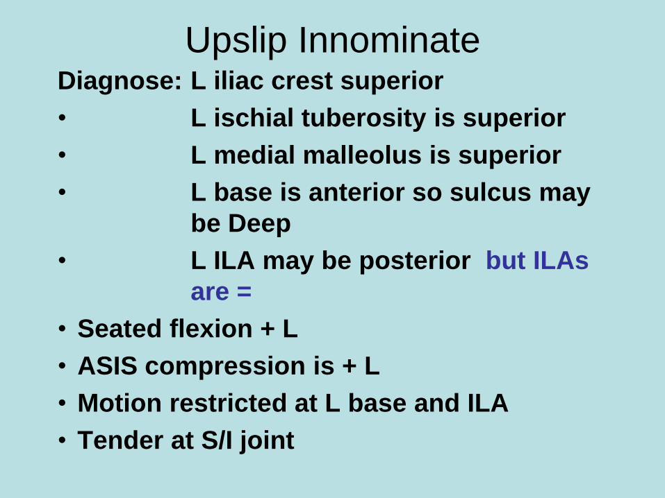
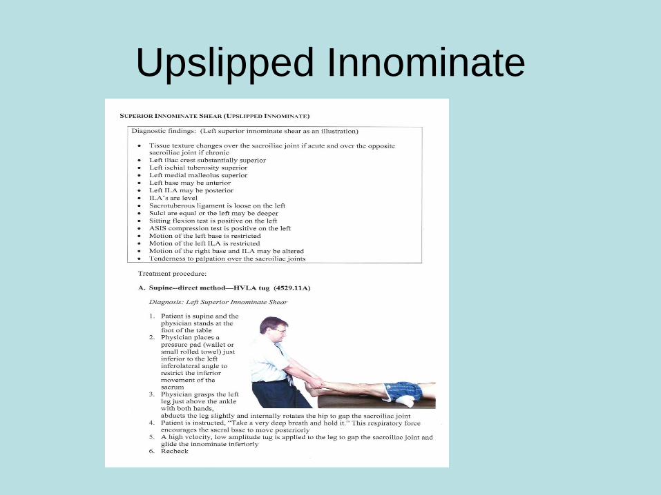
Upslip InnominateDiagnose: L iliac crest superior
• L ischial tuberosity is superior
• L medial malleolus is superior
• L base is anterior so sulcus may
be Deep
• L ILA may be posterior but ILAs
are =
• Seated flexion + L
• ASIS compression is + L
• Motion restricted at L base and ILA
• Tender at S/I joint
Upslipped Innominate

VERTICAL AXES
DYSFUNCTIONS
May or may not have short leg but need to know
122
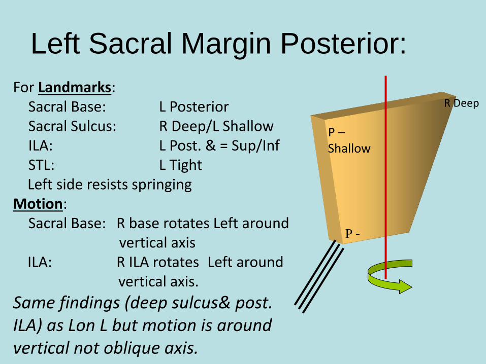
For Landmarks:Sacral Base: L PosteriorSacral Sulcus: R Deep/L ShallowILA: L Post. & = Sup/InfSTL: L TightLeft side resists springing
Motion: Sacral Base: R base rotates Left around
vertical axisILA: R ILA rotates Left around
vertical axis.
Same findings (deep sulcus& post. ILA) as Lon L but motion is around vertical not oblique axis.
P –Shallow
P -
Left Sacral Margin Posterior:
R Deep
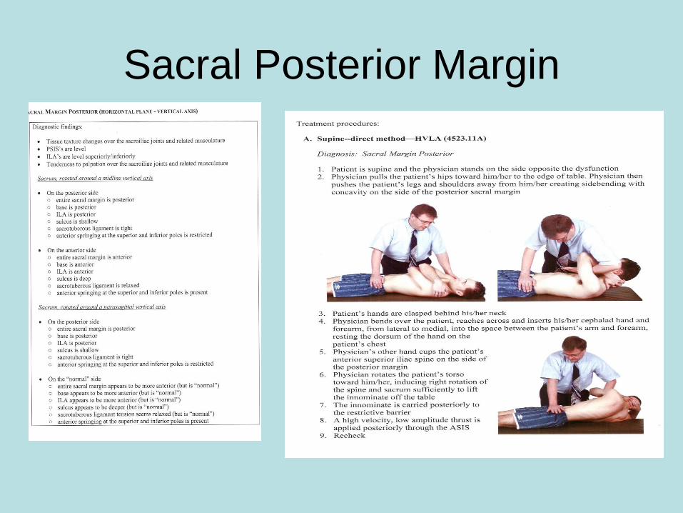
Sacral Posterior Margin


• Who has a shear?
• Who has a torsion?
• Who has a post/ant innominate
• Who has a upslip?
• Who is not sure?
• We will gather around as each diagnosis is
treated until ours is done
• Then
• We are done !

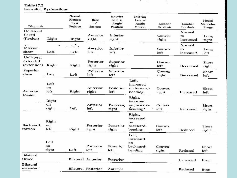
• Some tables for your enjoyment !!
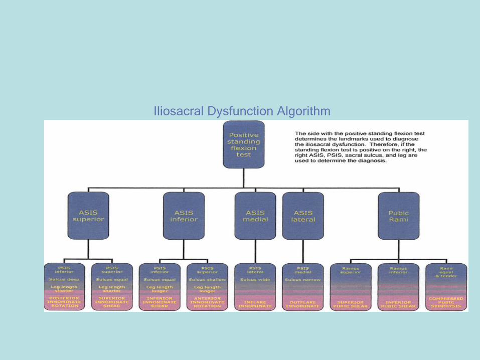
Iliosacral Dysfunction Algorithm

SACRAL DIAGNOSISDiagnosis Seated Flexion
Test
Sacral
Base/Sulci
ILA levelness L5
Rot
Spring
Test
LS Flexion
Asymmtry
Left on left Right Anterior right Posterior left Right Negative Decreased
Left on Right Left Anterior right Posterior left Right Positive Increased
Right on right Left Anterior left Posterior Right Left Negative Decreased
Right on Left Right Anterior Left Posterior Right Left Positive Increased
Left Unilat Fiex Left Anterior Left Posterior Left-
Negative Decreased
Left Unilat Ext Left Anterior Right Posterior Right-
Positive Increased
Right Unilat Flex Right Anterior Right Posterior right-
Negative Decreased
Right Unilat Ext Right Anterior Right Posterior left-
Positive Increased
Ant Margin - R Right Anterior Right Anterior Right Left Negative Decreased
Ant Margin-L Left Anterior Left Anterior Left Right Negative Decreased
Post Margin-R Right Shallow R Posterior Right Right Positive Increased
Post Margin-L Left Shallow L Posterior Left Left Positive Increased
Bilateral Flexion N/A Deep Bilateral Shallow Bilateral
-
Negative N/A
Bilateral Extnsn N/A Shallow
Bilateral
Deep Bilateral-
Positive N/A

SOURCES AND RESOURCES
• KIMBERLY MANUAL-2006 EDITION
• POCKET MANUAL OF OMT-2ND EDITION
• PRINCIPLES OF MANUAL MEDICINE-GREENMAN
• OMT REVIEW-SAVARESE-3RD EDITION
• LECTURES FROM OMM FACULTY – A.T.STILL UNIVERSITY-
PHOENIX AZ- WITH PERMISSION
• LECTURES FROM DR.HARMON MYERS
• JAOA Vol 91 No 3 March 1991
• CLINICAL APPLICATION OF COUNERSTRAIN – MYERS 2006