Embed Size (px)
Citation preview

Selective AKR1C3 inhibitors do notrecapitulate the anti-leukaemic activitiesof the pan-AKR1C inhibitormedroxyprogesterone acetateF Khanim1,8, N Davies2,8, P Velica3, R Hayden1, J Ride1, C Pararasa4, M G Chong2, U Gunther2, N Veerapen1,P Winn5, R Farmer5, E Trivier6, L Rigoreau6, M Drayson7 and C Bunce*,1
1School of Biosciences, University of Birmingham, Birmingham B15 2TT, UK; 2School of Cancer Sciences, University ofBirmingham, Birmingham B15 2TT, UK; 3Haematology Department, UCL Cancer Institute, London WC1E 6DD, UK; 4School of Lifeand Health Sciences, Aston University, Birmingham B4 7ET, UK; 5Centre for Systems Biology, University of Birmingham,Birmingham B15 2TT, UK; 6CRT Discovery Laboratories, Babraham, Cambridge CB22 3AT, UK and 7Immunity and Infection,University of Birmingham, Birmingham B15 2TT, UK
Background: We and others have identified the aldo-keto reductase AKR1C3 as a potential drug target in prostate cancer, breastcancer and leukaemia. As a consequence, significant effort is being invested in the development of AKR1C3-selective inhibitors.
Methods: We report the screening of an in-house drug library to identify known drugs that selectively inhibit AKR1C3 over theclosely related isoforms AKR1C1, 1C2 and 1C4. This screen initially identified tetracycline as a potential AKR1C3-selective inhibitor.However, mass spectrometry and nuclear magnetic resonance studies identified that the active agent was a novel breakdownproduct (4-methyl(de-dimethylamine)-tetracycline (4-MDDT)).
Results: We demonstrate that, although 4-MDDT enters AML cells and inhibits their AKR1C3 activity, it does not recapitulate theanti-leukaemic actions of the pan-AKR1C inhibitor medroxyprogesterone acetate (MPA). Screens of the NCI diversity set and anindependently curated small-molecule library identified several additional AKR1C3-selective inhibitors, none of which hadthe expected anti-leukaemic activity. However, a pan AKR1C, also identified in the NCI diversity set faithfully recapitulated theactions of MPA.
Conclusions: In summary, we have identified a novel tetracycline-derived product that provides an excellent lead structure withproven drug-like qualities for the development of AKR1C3 inhibitors. However, our findings suggest that, at least in leukaemia,selective inhibition of AKR1C3 is insufficient to elicit an anticancer effect and that multiple AKR1C inhibition may be required.
Members of the aldo/keto reductase (AKR) superfamily catalysethe conversion of aldehydes and ketones to their correspondingalcohols by utilising NADH and/or NADPH as cofactors. We andothers have identified AKR1C3 as a potentially novel therapeutictarget in prostate cancer, breast cancer and leukaemia (Desmond
et al, 2003; Byrns and Penning, 2009; Houghton et al, 2011; Byrnset al, 2012; Mitsiades et al, 2012). Our studies in acute myeloidleukaemia (AML) have demonstrated that enforced AKR1C3overexpression represses the all-trans retinoic acid (ATRA)-induced differentiation of HL-60 AML cells (Desmond et al, 2003),
*Correspondence: Professor C Bunce; E-mail: [email protected] first authors.
Revised 3 January 2014; accepted 13 January 2014; published online 25 February 2014
& 2014 Cancer Research UK. All rights reserved 0007 – 0920/14
FULL PAPER
Keywords: aldo-keto reductase 1C3; inhibitors; leukaemia
British Journal of Cancer (2014) 110, 1506–1516 | doi: 10.1038/bjc.2014.83
1506 www.bjcancer.com | DOI:10.1038/bjc.2014.83

whereas conversely knockdown of AKR1C3 in K562 cells results inerythroid differentiation (Birtwistle et al, 2009). We have alsoshown that the known AKR inhibitors, indomethacin andmedroxyprogesterone acetate (MPA), promote the differentiationand apoptosis of AML cells (Bunce et al, 1994; Khanim et al,2009a). More recently, we have demonstrated that MPA incombination with bezafibrate (BEZ) has in vivo clinical activityagainst AML (Murray et al, 2010). We have therefore proposedAKR1C3 as a novel regulator of myeloid cell differentiation and apotential new therapeutic target in leukaemia.
The role of AKR1C3 in prostate and breast cancer has beenpredominantly inferred by virtue of altered mRNA and/or proteinexpression levels in tumour tissues and in association with diseaseprogression (Agung et al, 2005; Suzuki et al, 2005; Hofland et al,2010; Beckmann et al, 2011; Aderibigbe et al, 2012; Jamieson et al,2012), and the enzyme’s recognised 3,alpha- and 17,beta-hydroxysteroid activities and 9,alpha-11,beta prostaglandin D2(9a, 11b-PGD2) dehydrogenase activity (Matsuura et al, 1998;Kuhne et al, 2001; Byrns and Penning, 2009; Byrns et al, 2012).Byrns et al (2012) demonstrated that overexpression of AKR1C3 inLNCaP prostate cancer cells resulted in increased testosteroneproduction and resistance to finasteride. Single-nucleotidepolymorphisms in AKR1C3 have been associated with diseaseprogression and aggressiveness in prostate carcinomas (Izumotoet al, 2005; Richards et al, 2006). AKR1C3 polymorphisms havealso been shown to modulate the risk of other cancers, includingbladder cancer, childhood leukaemias and diffuse large B-celllymphoma (Bauman et al, 2004; Morakinyo et al, 2011; Adegokeet al, 2012c). Separately, it has been shown that AKR1C3 caninactivate and induce resistance to the anticancer drugs doxo-rubicin, oracin and cisplatin (Adegoke and Nyokong, 2012;Adegoke et al, 2012a, b), whereas elevated AKR1C3 proteinlevels were associated with radioresistance in non-small cell lungcarcinomas (Jamieson et al, 2012).
AKR1C3 belongs to a highly conserved AKR subfamily(in humans AKR1C1, -1C2, -1C3 and -1C4), which displaysdifferential tissue expression and possess overlapping but differingpleiotropic activities (Kuhne et al, 2001; Gratacos-Cubarsi et al,2007; Birtwistle et al, 2009; Velica et al, 2009; Rizner and Penning,2013). MPA does not display strong AKR1C3 selectivity amongstthis family and only inhibits AKR1C3 at doses higher than thoseused for its respective primary indications as a contraceptivesteroid. These considerations have driven many research groups tosearch for lead compounds with a greater selectivity towardsAKR1C3 that could be developed as novel cancer therapies(Halling-Sorensen et al, 2002; Ghia et al, 2003; Qiu et al, 2007;Zargar et al, 2011; Burnett et al, 2012; Giebel et al, 2013; Hebertet al, 2013; Wasylishen et al, 2013). In recognition of this, severallaboratories have undertaken structural studies of AKR1C3 boundto inhibitors and substrates and embarked on the identification oflead compounds for drug development (Zargar et al, 2011; Burnettet al, 2012; Hebert et al, 2013; Wasylishen et al, 2013).
New drug development is challenging and as the pharmacologistand Nobel laureate James Black is quoted as saying, ‘the mostfruitful basis for the discovery of a new drug is to start with an olddrug’. In order to screen existing drugs for greater selectiveAKR1C3 inhibition, we developed a high-throughput assay ofAKR1C1, -1C2, -1C3 and -1C4 activity based on the consumptionof NADPH in the presence of the pan-AKR1C substratephenanthrenequinone (PQ) (AKR1C-diaphorase assay). An initialscreen of an in-house custom-built library of 100 off-patent drugs(FMC1) identified tetracycline as a potential AKR1C3-selectiveinhibitor. However, mass spectrometry and nuclear magneticresonance (NMR) studies identified that the active agent was infact a novel tetracycline breakdown product (4-methyl(de-dimethylamine)-tetracycline (4-MDDT). We demonstrate herethat this novel agent enters AML cells and inhibits the AKR1C3
activity within, but unexpectedly does not recapitulate the anti-leukaemic actions of the pan-AKR1C3 inhibitor MPA either aloneor in combination with BEZ. Further screens of the NCI diversityset (http://dtp.cancer.gov) and of a small-molecule library curatedat the Cancer Research Technology (CRT) identified severaladditional AKR1C3-selective inhibitors. Importantly, none of theseAKR1C3-selective compounds had anti-leukaemic activity. How-ever, a pan AKR1C1, -1C2, -1C3 inhibitor also identified in theNCI diversity set faithfully recapitulated the actions of MPA.
MATERIALS AND METHODS
FMC100 drug library. All drugs were purchased from Sigma-Aldrich (Poole, UK). Stocks were prepared at 10 000� reportedpeak serum concentrations in dimethylsulphoxide DMSO, ethanolor water and stored at � 20 1C. Tetracycline hydrate (Sigma-Aldrich) was prepared at 225 mM in DMSO.
National cancer institute diversity set. NCI compounds wereordered from the NCI/DTP Open Chemical Repository (http://www.dtp.nci.nih.gov). Stocks were prepared as 10 mM in DMSO in96-well plates and stored at � 20 1C.
Cancer research technologies (CRT). A1, A6 and A9 wereidentified as AKR1C3-selective inhibitors by CRT using afluorescence-based assay and purified recombinant AKR1C1-4proteins (Hebert et al, 2013). Compounds A1((CRT0036521)3-(3,4-dihydro-1H-isoquinoline-2-sulphonyl)-benzoic acid), A6((CRT0083914) 1-[4-(2-methyl-piperidine-1-sulphonyl)-phenyl]-pyrrolidin-2-one), and A9 ((CRT0093964) [4-(4-chloro-phenyl)-piperazin-1-yl]-morpholin-4-yl-methanone)(Figure 7A), were pro-vided as 12–15 mM stocks in anhydrous DMSO and stored at 4 1C.
Production and purification of recombinant AKR1C proteins.N-terminally Hisx6-tagged recombinant AKR1C1, 2, 3 and 4 werecloned into pET28b vectors (Novagen, Darmstadt, Germany) andexpressed and purified as previously described (Lovering et al,2004; Davies et al, 2009).
High-throughput recombinant AKR1C-diaphorase assay.Supplementary Figure 1 shows a schematic for the fluorometricAKR1C activity assay using PQ. Compounds were screened againstpurified recombinant AKR1C1, 2, 3 and 4 proteins in duplicatewells of 96-well plates. In the first step, reactions contained 4mM
PQ, 5 mM b-NADPH, 15 mg recombinant AKR1C (recAKR1C)protein and 100 mM test compound in 50 mM potassium phosphatebuffer (pH 6.5). Dimethylsulphoxide and acetonitrile were bothpresent at 2% (v/v) each. Negative, positive and no-inhibitorcontrol wells were set in duplicate on each plate; negative controlhad no PQ, while the positive controls used 5mM MPA or 20mM
indomethacin. The assay as used measured uncompetitive inhibi-tion owing to the formation of the E–NADPþ –I complex (Byrnset al, 2012). PQ was non-limiting in this assay as the productspontaneously oxidises back to PQ, as determined by therestoration of the initial rate of NADPH consumption whenNADPH was re-added to the assay (Supplementary Figure 1B).Plates were incubated at 37 1C for 15 min before resazurinand diaphorase were added to final concentrations of 26.8 mM
and 0.54 U/ml, respectively. The contents of wells were mixedby repeated pipetting and plates incubated for a further 10min at37 1C. Fluorescence was measured (Ex530 nm/Em595 nm) in afluorimeter (Bio-Tek Instruments, Winooski, VT, USA) using theKC4 software (BioTek, Potten, UK). Dose titration experiments tocalculate IC50 values were performed as above. Percentage enzymeactivity was calculated using 100� (100� (R�B1)/(B0�B1)),where R is A530/590 of well with test compound, B1 is average ofA530/590 of duplicate wells without compound and B0 is averageA530/590 reading of duplicate wells without substrate.
AKR1C3 inhibitors lack anti-leukaemic activity BRITISH JOURNAL OF CANCER
www.bjcancer.com | DOI:10.1038/bjc.2014.83 1507
NADPH assay for AKR1C enzymatic activity. Assays for all fourhuman recombinant AKR1C enzymes were performed, asdescribed previously (Davies et al, 2009).
HPLC purification of tetracycline breakdown product4-MDDT. 4-MDDT was purified at room temperature usinga Dionex DX-600 HPLC system (Thermo Scientific, HemelHempstead, UK), Prevail Select C18 5 m (250 mm� 4.6 mm i.d.)HPLC column. Elution at a flow of 1 ml/min was performed with alinear gradient between solvent A (50% methanol : 0.1% trifluor-oacetic acid v/v) and solvent B (98% methanol : 0.1% trifluoroaceticacid v/v). Peak identification was performed by comparing spectra
(collected between 220 and 500 nm). Fractions were collected,dried down under nitrogen stream and 4-MDDT resuspended inDMSO at 50 mM using its molecular mass as 413, as measured byGC-MS analysis (see below).
Mass spectrometry and NMR. Mass spectrometry to define masswas performed by Dr Peter Ashton (School of Chemistry,University of Birmingham) on both freshly prepared tetracyclineand HPLC-purified tetracycline derivative (4-MDDT) by electro-spray mass spectrometry analysis, scanning for molecules withRMM 200–2000.
0
20 000
40 000AKR1C3
0
20 000
40 000AKR1C1
0
20 000
40 000AKR1C2
0
20 000
40 000
FM
C1
FM
C3
FM
C5
FM
C7
FM
C9
FM
C11
FM
C13
FM
C15
FM
C17
FM
C19
FM
C21
FM
C23
FM
C25
FM
C27
FM
C29
FM
C31
FM
C33
FM
C35
FM
C37
FM
C39
FM
C41
FM
C43
FM
C45
FM
C47
FM
C49
FM
C51
FM
C53
FM
C55
FM
C57
FM
C59
FM
C61
FM
C63
FM
C65
FM
C67
FM
C69
FM
C71
FM
C73
FM
C75
FM
C77
FM
C79
FM
C81
FM
C83
FM
C85
FM
C87
FM
C89
FM
C91
FM
C93
FM
C95
FM
C97
MP
AC
ON
T
AKR1C4NA
DP
H r
emai
ning
(flu
ores
cenc
e un
its)
MPA
MPA
MPA
MPA
FMC1 drug library
Tetracycline
0
0.00
050.
005
0.05 0.
5 5 50
0.00
050.
005
0.05 0.
5 5 50
20
40
60
80
100
120
140
AKR1C1
AKR1C2
AKR1C3
AKR1C4
(5aS,6S,12aS)-3,6,10,12,12a-pentahydroxy-4,6-dimethyl-1,11-dioxo-1,5,5a,6,11,12a-hexahydrotetracene-2-carboxamide
0
20
40
60
80
100
(4S,4aS,5aS,6S,12aS)-4-(Dimethylamino)-3,6,10,12,12a-pentahydroxy-6-methyl-1,11-dioxo-1,4,4a,5,5a,6,11,12a-octahydro-2-tetracenecarboxamide
Enz
yme
activ
ity (
%)
Enz
yme
activ
ity (
%)
4-MDDT (�M) 4-MDDT (�M)
rec AKR1C3
IC50=0.51 �M
CH3
H3C CH3N
NH2 NH2
4a5a6a
10a109
8
7 65
11a 12a
3 8
910
10a 11a11
5a6a7 4
3
2112
4a
12a
5
CH3 CH3
6
211211
4
OOOHOH OHOH
HOOH
O O O
OH
HOOH
OH
O
Figure 1. Identification of a novel tetracycline breakdown product. (A) A panel of 100 clinically licensed drugs (FMC100) at peak serumconcentrations was screened for AKR1C-inhibitory activity against recombinant purified AKR1C proteins using the AKR1C-diaphorase assay.Data shown is mean fluorescence arbitrary units. Black bars indicate tetracycline and the dashed line represents level of inhibition by 5 mM MPA.GC-MS and NMR were used to elucidate the structure of the novel tetracycline breakdown product 4-methyl,(didemethyl)-tetracycline (4-MDDT).(B) Structures and names of the parent tetracycline and novel tetracycline breakdown product. (C) The isoform specificity of HPLC-purified 4-MDDTwas determined against recombinant AKR1C proteins using the AKR1C-daphorase assay (see Materials and Methods). (D) The IC50 of 4-MDDT forrecombinant AKR1C3 protein was determined using the PQ assay (see Materials and Methods).
BRITISH JOURNAL OF CANCER AKR1C3 inhibitors lack anti-leukaemic activity
1508 www.bjcancer.com | DOI:10.1038/bjc.2014.83
To elucidate the structure, 1D and 2D NMR experiments wereperformed on both 10 mM freshly prepared tetracycline and HPLC-purified tetracycline derivative (4-MDDT). Spectra were recordedon a Bruker 500 MHz spectrometer (Bruker, Coventry, UK) and aBruker 600 MHz spectrometer (Bruker), both equipped withcryogenically cooled probes. All spectra were recorded at atemperature of 300 K, in either d6-DMSO or d3-acetonitrile.One-dimensional 1H NMR spectra were acquired using a spectralwidth of 7.2 kHz and 32 K data points. One-dimensional 13C NMRspectra were obtained using a spectral width of 24 kHz with 64 Kdata points. One-dimensional 15N NMR spectra were obtainedusing a spectral width of 25 kHz with 32 K data points. For furtherassignments verification, 2D COSY, TOCSY (100 ms mixing time)and NOESY (200 ms mixing time) spectra were obtained, along with13C-HSQC and 15N-HSQC (with the INEPT delay adjusted forshort and for long-range couplings) in order to identify NH andN(CH3)2 groups.
In silico docking studies. Simulated docking of tetracycline and4-MDDT into AKR1C3 (PDB ID 1S2C with flufenamic acidremoved) was performed using Autodock 4.2 (Wu et al, 2010). Thecoordinates of tetracycline with Mg2þ were adapted from the
crystal structure PDB ID 2TCT. The coordinates of 4-MDDT werecalculated by editing the tetracycline coordinates using PyMol.UCSF Chimera 1.6.2 (Li et al, 2010) was used to calculate AM-1BCC charges for ligands in different proton configurations with/without Mg2þ . Polar hydrogen atoms and partial charges for theprotein and NADPH cofactor were calculated using the standardsettings in MGLTools. The input and output files for Autodockwere processed using the standard MGLTools scripts. The centre ofthe docking site was the average coordinate of the flufenamic acidbound in PDB 1S2C.
A number of calculations included some flexibility of theAKR1C3 structure by allowing torsional rotation of residueshaving the most contact with the ligand (ASN167, PHE306,PHE311, TYR216, TRP227, LEU54, LEU128, SER129, LEU122,TRP86, MET120). In addition, the two magnesium-bound 4-MDDT configurations were docked with the same flexible residuesas above but removing TYR216 and MET120 from the list andadding ARG226.
Cell culture and drug treatments. HL60, KG1a and K562(ATCC) cell lines were maintained in RPMI-1640 medium withL-glutamine supplemented with 10% v/v heat-inactivated
AKR1C3 1AKR1C2 1AKR1C1 1AKR1C4 1
AKR1C3 71AKR1C2 71AKR1C1 71AKR1C4 71
AKR1C3 141AKR1C2 141AKR1C1 141AKR1C4 141
AKR1C3 211AKR1C2 211AKR1C1 211AKR1C4 211
AKR1C3 281AKR1C2 281AKR1C1 281AKR1C4 281
ARG 226ARG 226
PHE 306
SER 129
SER 118
PHE 306
SER 129
SER 118
MMDSKHQCVKL L NDGHFMPVLG VLG FGTYAPPPPEVP P RSKALALEVTKL AIL AIEAGAGFRHRHID D SAHLYNNNNEEEEQ Q VGLAIVGLAIRSKIAIAMDSKYQCVKL L NDGHFMPVLG VLG FGTYAPAEVP P KSKALALEAVKL AIL AIEAGAGFHHID D SAHVYNNNNEEEEQ Q VGLAIVGLAIRSKIAIAMDSKYQCVKL L NDGHFMPVLG VLG FGTYAPAEVP P KSKALALEATKL AIL AIEAGAGFRHRHID D SAHLYNNNNEEEEQ Q VGLAIVGLAIRSKIAIAMDPKYQRVEL L NDGHFMPVLG VLG FGTYAPPPPEVP P RNRNRAVEVTKL AIL AIEAGAGFRHRHID D SAYLYNNNNEEEEQ Q VGLAIVGLAIRSKIAIA
DGSVKRKREDEDIF YF YTSTSKLWSTSTFH RH RPELVLVRPALALE E NSLKKKKAQLDY Y VDLYLILIHSPM M SLKPGEEEELSP P TDEDENGKVIVIFDDGSVKRKREDEDIF YF YTSTSKLWSNSH RH RPELVLVRPALALE E RSLKNLQLDY Y VDLYLILIHFPV V SVKPGEEEEVIVIP P KDEDENGKILILFDDGSVKRKREDEDIF YF YTSTSKLWCNSH RH RPELVLVRPALALE E RSLKNLQLDY Y VDLYLILIHFPV V SVKPGEEEEVIVIP P KDEDENGKILILFDDGSVKRKREDEDIF YF YTSTSKLWCTFF QF QPQMQMVQPALALE E SSLKKKKLQLDY Y VDLYLLHFPM M ALKPGETPLTPLP P KDEDENGKVIVIFD
IVDLCTTWEA MEKCKDAGLA AGLA KSIGVIGVSNFNR RR RQLEMILILNKP P GLGLKYKPVCNQ NQ VECHPYFYFNRS S KLLLLDFCKSKDTVDLCATWEA MEKCKDAGLA AGLA KSIGVIGVSNFNH H RLLEMILILNKP P GLGLKYKPVCNQ NQ VECHPYFYFNQNQR KR KLLLLDFCKSKDTVDLCATWEA VEKCKDAGLA AGLA KSIGVIGVSNFNR RR RQLEMILILNKP P GLGLKYKPVCNQ NQ VECHPYFYFNQNQR KR KLLLLDFCKSKDTVDLSATWEV MEKCKDAGLA AGLA KSIGVIGVSNFNC C RQLEMILILNKP P GLGLKYKPVCNQ NQ VECHPYLNQNQS S KLLLLDFCKSKD
IVLVAIVLVAYSALG ALG SQRDKRWVDP P NSPVLLVLLEDEDPV LV LCALAALAKKHKR KKHKR TPALIALALIALRYQ Q LQRGVVVLAGVVVLAK K SYNEQRIRQNQNIVLVAIVLVAYSALG ALG SHRHREEEEPWVDP P NSPVLLVLLEDEDPV LV LCALAALAKKHKR KKHKR TPALIALALIALRYQ Q LQRGVVVLAGVVVLAK K SYNEQRIRQNQNIVLVAIVLVAYSALG ALG SHRHREEEEPWVDP P NSPVLLVLLEDEDPV LV LCALAALAKKHKR KKHKR TPALIALALIALRYQ Q LQRGVVVLAGVVVLAK K SYNEQRIRQNQNIVLVAIVLVAHSALG ALG TQRHKLWVDP P NSPVLLVLLEDEDPV LV LCALAALAKKHKKKHKQ Q TPALIALALIALRYQ Q LQRGVVVLAGVVVLAK K SYNEQRIREN
VQVFEFQLTA A EDEDMKAIAIDGLGLD RNLHLHYFNSNSDS S FASHPNYPYS S DEDEYVQVFEFQLTS TS EEEEMKAIAIDGLGLN RNVRYLTLDI I FAGAGPPPPNYPFS S DEDEYVQVFEFQLTS TS EEEEMKAIAIDGLGLN RNVRYLTLDI I FAGAGPPPPNYPFS S DEDEYIQVFEFQLTS TS EDEDMKVLVLDGLGLN RNYRYVVMVVMDF LMDF LMDHPDYPFS S DEDEY
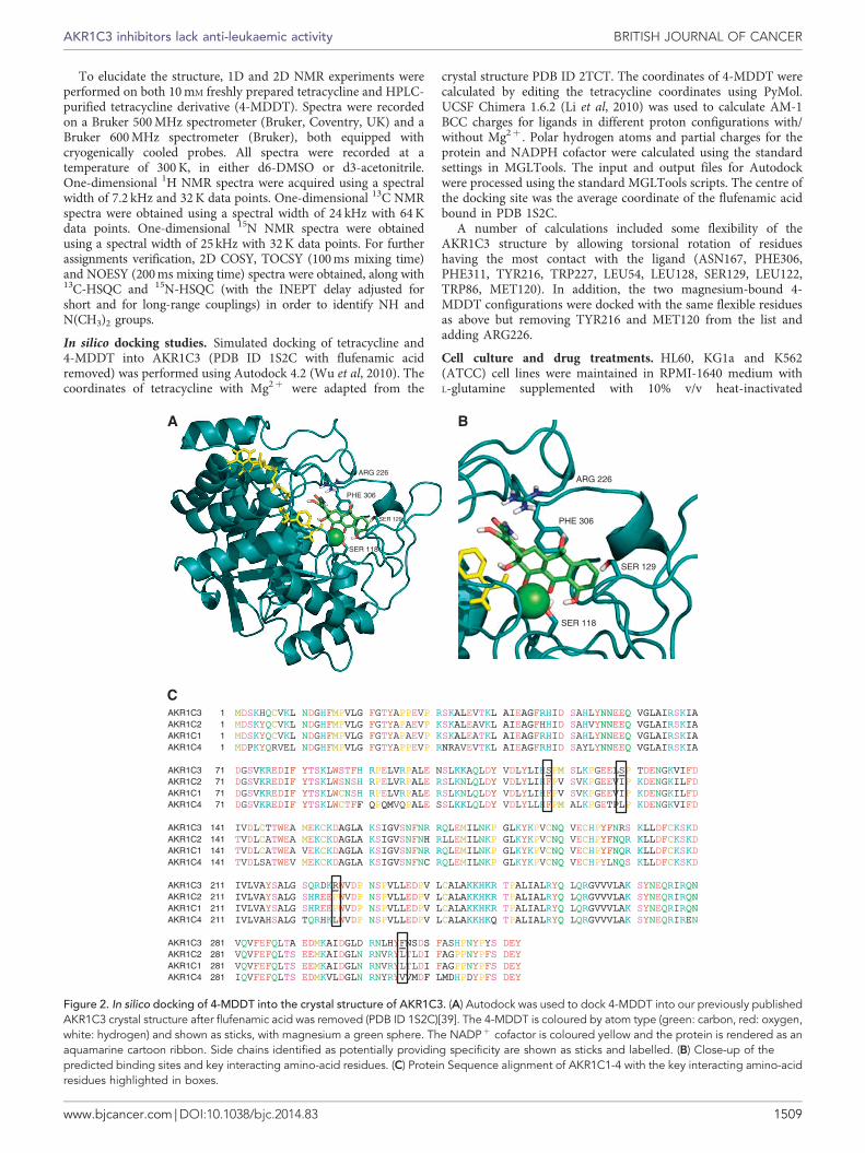
Figure 2. In silico docking of 4-MDDT into the crystal structure of AKR1C3. (A) Autodock was used to dock 4-MDDT into our previously publishedAKR1C3 crystal structure after flufenamic acid was removed (PDB ID 1S2C)[39]. The 4-MDDT is coloured by atom type (green: carbon, red: oxygen,white: hydrogen) and shown as sticks, with magnesium a green sphere. The NADPþ cofactor is coloured yellow and the protein is rendered as anaquamarine cartoon ribbon. Side chains identified as potentially providing specificity are shown as sticks and labelled. (B) Close-up of thepredicted binding sites and key interacting amino-acid residues. (C) Protein Sequence alignment of AKR1C1-4 with the key interacting amino-acidresidues highlighted in boxes.
AKR1C3 inhibitors lack anti-leukaemic activity BRITISH JOURNAL OF CANCER
www.bjcancer.com | DOI:10.1038/bjc.2014.83 1509

fetal bovine serum and penicillin (100 units/ml)/streptomycin(100 mg/ml) (all from Gibco, Invitrogen Ltd, Paisley, UK) at 37 1Cwith 5% CO2. Cells were passaged every 2 days maintaining celldensities between 0.25–1.5� 106 cells/ml. Drug treatments in 96-well plate assays were performed using 1� 104 cells/well in 200 ml,or in flasks at 0.25� 106 cells/ml in 4 ml media. Appropriatevolumes of DMSO and/or ethanol were added as solvent controlsin control treatments.
Chronic lymphocytic leukaemia (CLL) cells. Chronic lymphocyticleukaemia blood samples were obtained from unselected patients
diagnosed with CLL, according to standard morphologic,immunophenotypic and clinical criteria, attending the outpatientclinic at Birmingham Heartlands Hospital following informedwritten consent for the study, which had received local ethicalapproval. Primary mononuclear cells were prepared using FicollPaque-Plus (Anachem, Luton, UK) as previously described.Resultant cells were cultured and treated in a 96-well plateco-culture system, with L-control fibroblasts, as describedpreviously (Hayden et al, 2010). Viability was assessed at 24 husing annexin V and propidium iodide staining, and flowcytometry (Hayden et al, 2010).
0
10
20
30
40
50
60
No BEZ BEZ
% C
D11
b-po
sitiv
e ce
lls
HL60
CONT 1
MPA
*
*
*
0
10
20
30
40
50
60
70
80
0 0.05 0.5 5 50 MPA
Enz
yme
activ
ity (
%)
KG1a-Intracellular AKR1C3 activity
*
* *
0
50
100
150
200
250
300 HL60
0
50
100
150
200
250 K562
05
10152025303540 KG1a
0
10
20
30
40
50
0
5
10
15
20
25
0
2
4
6
8
10
No BEZ
0.5 mM BEZ
**
*
**
No BEZ BEZ
CONT
MPA
Cum
ulat
ive
cell
num
ber
(106 )
CONTM
PA
4-M
DDT
CONTM
PA
4-M
DDT
CONTM
PA
4-M
DDT
CONTM
PA
4-M
DDT
CONTM
PA
4-M
DDT
CONTM
PA
4-M
DDT
% C
D11
b-po
sito
ve c
ells
4-MDDT
0102030405060708090
No ATRA 10 nM ATRA
HL60
CONT
MPA
*4-MDDT
4-MDDT
4-MDDT (�M)
Figure 3. 4-MDDT does not recapitulate the actions of MPA against AML cell lines. HL-60, K562 and KG1a cells were treated for 7 days witheither solvent control, 50mM 4-MDDT or 5 mM MPA, alone or in combination with 0.5 mM BEZ. (A) Cumulative cell counts for HL60, K562 and KG1aafter 7 days treatment. Data shown is mean of a minimum of N¼3 experiments±s.e.m. (B and D) HL60 differentiation was assessed at day 7 bystaining for CD11b and flow cytometry following treatment with either solvent control, 50mM 4-MDDT or 5mM MPA with or without 0.5 mM BEZ or10 nM ATRA. Data shown is mean of a minimum of N¼ 3 experiments±s.e.m. (C) Cytospins of HL60 cells treated for 7 days as described abovewere stained with Jenner–Giemsa to demonstrate changes in cell morphology. (E) Intracellular AKR1C3 11b-prostaglandin reductase activity inKG1a cells was assessed in the presence of a dose titration of 4-MDDT or 5 mM MPA using 3H-PGD2 and thin layer chromatography. Data shown ismean of N¼ 4 experiments±s.e.m. *Po0.01.
BRITISH JOURNAL OF CANCER AKR1C3 inhibitors lack anti-leukaemic activity
1510 www.bjcancer.com | DOI:10.1038/bjc.2014.83

3H-PGD2 turnover analysis by thin layer chromatography.AKR1C3-mediated PGD2 turnover in intact cells was determinedby thin layer chromatography as described previously (Desmondet al, 2003; Davies et al, 2009).
Measurement of cell viability. HL60, KG1a and K562 cellviability was measured using manual counts, or by flow cytometryon a BD FACSCalibur utilising Cell Quest Pro software(Becton Dickinson, Plymouth, UK), viable gates and fluorescentCytocount beads (DakoCytomation, Ely, UK).
Assessment of cell differentiation. HL-60 cells were treated for7 days with re-feeding and re-treating every 2 days. Differentiationwas assessed by flow cytometry (Becton Dickinson FACS Caliburand Becton Dickinson Cell Quest software) using PE-CD11b(Becton Dickinson).
Jenner–Giemsa staining of slides. Cytospins were prepared from75–100 ml of culture. Slides were air dried, methanol fixed andstained as described previously (Khanim et al, 2009a).
RESULTS
The four human AKR1C enzymes share greater than 86% amino-acid sequence identity and, also share overlapping substratepromiscuities (Deyashiki et al, 1994; Dufort et al, 1996; Velicaet al, 2009). Thus identifying inhibitors selective for individualenzymes from this subfamily represents a considerable challenge.To this end we have developed a readily accessible indirect AKR1Cfluorescence-based assay that can be used for high-throughputscreening (Supplementary Figure 1). The assay utilises the pan-human AKR1C substrate PQ and NADPH as cofactors andmeasures AKR1C activity indirectly by determining levels ofNADPH remaining in the presence or absence of inhibitor(Supplementary Figure 1).
As part of our drug redeployment strategy in leukaemia andlymphoma, we have assembled a library (FMC1) of 100 off-patentcommonly used drugs of a known toxicological profile. This libraryhas been constructed to reflect each of the drug’s peak serumconcentration and differs from other libraries that normally
contain drugs at a common arbitrary concentration. The FMC1library was screened initially at 10� peak serum concentrationsagainst recombinant AKR1C1, -1C2, -1C3 and -1C4 usingthe AKR1C-diaphorase-based assay (Figure 1). Indomethacin andMPA were included as control inhibitors. Wells were includedcontaining enzyme and no inhibitor (þ ve control) and withoutenzyme or inhibitor (� ve control). This screen identified thebroad-spectrum polyketide antibiotic tetracycline as a potentialAKR1C3-selective inhibitor (Figure 1A).
Tetracycline is known to be unstable with factors such as light,temperature, and pH strongly affecting its chemical stability (Baggaet al, 2005; Patriciu et al, 2005; Feng et al, 2006). Under alkalineconditions, tautomerisation, demethylation, and the formation ofterranoic acid and isoforms have been described (Guan and Xiong,2011). Under acidic conditions, epimerisation reactions oftetracyclines take place (Patriciu et al, 2005; Wang et al, 2010) togive 4-epimeric, anhydrous, a/b apo, and ter forms (Guan andXiong, 2011). As the solution of tetracycline hydrate in DMSObeing tested was observed to change colour within a few days, thesolution was subjected to reverse-phase HPLC analysis thatrevealed the rapid conversion of the dissolved tetracycline to anunknown breakdown product. Freshly prepared tetracyclinesolutions demonstrated no AKR1C3-inhibitory activity; theAKR1C3-selective activity of the stored solution was shown to bedue to the breakdown product, the presence of which wasconfirmed by column chromatography. The purified tetracyclinebreakdown product was analysed by MS to give a suggested Mr of413, which differed by B31 Da from the actual mass of tetracycline(444.43 Da). This tetracycline breakdown moiety was subjected toNMR analysis of its structure, which identified a substitution atcarbon 4 replacing the dimethylamino group with a methyl group(Figure 1B; Supplementary Tables 1–3). Searches of severaldatabases (www.chemspider.com, http://pubchem.ncbi.nlm.nih.gov)did not identify any other tetracycline derivatives with asimilar structure. Hence, to our knowledge this is the firstdescription of this tetracycline derivative that we have termed4-methyl,(didemethyl)-tetracycline (4-MDDT) to distinguishfrom the 4-dimethylamino,6-methyl-tetracycline parent molecule.Analysis of the purified compound in the AKR1C-diaphorase assayconfirmed that the selective AKR1C3-inhibitory activity resided in
CONT
BEZ
MPA
Pro
pidi
um io
dide
0
CONTM
PA
4-M
DDT
MPA/4
-MDDT
20
40
60
80
100
% A
nnex
in V
-pos
itive
cel
ls
– BEZ
+ BEZ*
*
104
103
102
101
100
104
103
102
101
100
100 101 102 103 104 100 101 102 103 104 100 101 102 103 104
100 101 102 103 104 100 101 102 103 104 100 101 102 103 104
100 101 102 103 104
MPA/ 4-MDDT4-MDDT
BEZ /MPA (BaP) BEZ/ 4-MDDT
Annexin V
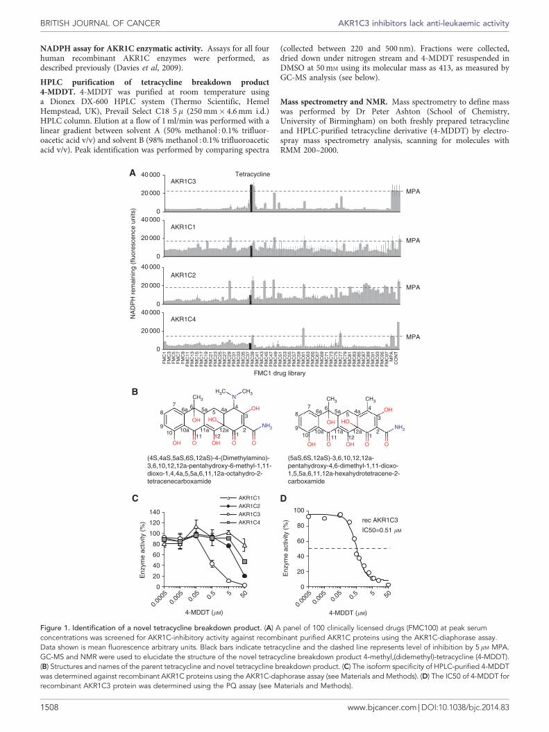
Figure 4. 4-MDDT does not recapitulate the actions of MPA against primary CLL cells. Peripheral blood mononuclear cells from CLL patientswere cultured on non-CD40L-expressing stroma in a ratio of 10 : 1 in 96-well plates. Treatments with either solvent control, 0.5 mM BEZ, 5 mM MPA,BEZ/MPA (BaP), 5.1mM 4-MDDT, MPA/4-MDDT or BEZ/4-MDDT were performed in triplicate for 24 h, before harvesting and staining with AnnexinV (AV) and propidium iodide (PI), and analysis by flow cytometry. Plot shown is for n¼ 4 samplesþ s.e.m.
AKR1C3 inhibitors lack anti-leukaemic activity BRITISH JOURNAL OF CANCER
www.bjcancer.com | DOI:10.1038/bjc.2014.83 1511

the 4-MDDT derivative (Figure 1C) and not the parent compoundand had an IC50 of 0.51 mM (Figure 1D). 4-MDDT is more stablecompared with the parent compound.
To interrogate the basis of the specificity of 4-MDDT forAKR1C3, we performed ligand-protein docking calculations usingour published AKR1C3 structure (Figure 2) (Lovering et al, 2004).Given the uncertain proton configuration and the flexibility of bothligand and protein, we did not anticipate finding the exact bindingorientation of 4-MDDT in the active site but anticipated someinsights into ligand specificity. Tetracycline is generally foundassociated with a magnesium ion, so we performed dockingexperiments both in the presence and absence of magnesium todetermine if this affected the analysis. Our calculations indicatethat magnesium-bound tetracycline is too large for the active site ofthe AKR1C3 crystal structure, whereas magnesium-bound4-MDDT fits (Figure 2). This discrimination was less obviousfor non-magnesium-bound ligands. Given the experimental data,this would seem to support the binding of 4-MDDT with a
bound magnesium. With regard to understanding AKR1C3selectivity, the docked 4-MDDT contacted residues of AKR1C3that differ greatly in physicochemical properties from those of theother AKR1C subfamily members, notably S118 (c.f. F), S129(c.f. I/L), R226 (c.f P/L) and F306 (c.f. L/V) (Figure 2B). Given theotherwise highly similar sequences between the AKR1C isoforms(Figure 2C), we might anticipate these residues in AKR1C3 tobe of significance. However, a detailed description of themechanism of 4-MDDT selectivity for AKR1C3 will requiredetailed crystallography studies.
We next turned to investigating the action of 4-MDDT againstHL-60, K562 and KG1a AML cell lines. As previously described(Khanim et al, 2009a), 5mM MPA alone had a moderate butstatistically anti-proliferative effect on all three AML cell lines andgreatly enhanced AML cell killing when combined with BEZ(Figure 3A). In contrast, 50 mM 4-MDDT did not display theseactivities (Figure 3A). We also previously demonstrated that theaction of BEZ and MPA (BaP) against K562 and KG1a cells was
0
20
40
60
80
100A
B
28086 30188 73100 75600 75747 82147 83436 85512 209931 211094%
Inhi
btio
n by
100
�M
com
poun
d
AKR1C1 AKR1C2 AKR1C3 AKR1C4
AKR1C1
AKR1C2
AKR1C3
49.92± 1.0
>100 >100 >100 >100 >100 >100 >100 >10070.87± 7.3
AKR1C4
IC50 (�M)
NCI compound
300
250
200
150
100
50
0
3035
25
4540
20151050
3035
25
4540
20151050
BEZ
CONT1
MPA
MPA
NSC 731
00
NSC 731
00
NSC 756
00
NSC 756
00
NSC 821
47
NSC 821
47
NSC 211
094
NSC 211
094
CON1M
PA
NSC 731
00
NSC 756
00
NSC 821
47
NSC 211
094
CON1M
PA
NSC 731
00
NSC 756
00
NSC 821
47
NSC 211
094
BEZM
PA
NSC 731
00
NSC 756
00
NSC 821
47
NSC 211
094
BEZM
PA
NSC 731
00
NSC 756
00
NSC 821
47
NSC 211
094
Cum
ulat
ive
cell
num
ber
(106 )
Cum
ulat
ive
cell
num
ber
(106 )
Cum
ulat
ive
cell
num
ber
(106 )
Cum
ulat
ive
cell
num
ber
(106 )
Cum
ulat
ive
cell
num
ber
(106 )
Cum
ulat
ive
cell
num
ber
(106 )
30
25
20
15
10
5
0 01234567
HL60
HL60
K562
**
* **
*
*
K562
KG1a250
200
150
100
50
0
KG1a
No BEZ
0.5 mM BEZ
>1004.86± 6.3
>100 >100 >100 >100 >100 >10080.48± 10.6
44.47± 3.0
3.88± 0.4
>100 1.95± 0.4
4.19± 2.3
3.12± 0.3
2.40± 0.3
6.52± 0.6
24.44± 1.3
0.56± 0.1
1.18± 0.3
32.54± 6.3
44.33± 4.3
89.67± 4.3
>100 >100 >100 >100 >1002.37± 0.3
30.06± 1.9
Figure 5. Identification of AKR1C3-selective inhibitors from the NCI Diversity Set. (A) NCI compounds were screened against recombinantAKR1C proteins using the AKR1C-diaphorase assay (see Materials and Methods) and % inhibition of activity measured. IC50s of selectedcompounds against all four AKR1C isoforms, shown below the bar chart, were calculated using dose titrations of each compound againstrecombinant AKR1C protein in the NADPH assay. (B) Cumulative cell counts for HL60, K562 and KG1a after 7 days treatment with either solventcontrol, 5 mM MPA, or the selected compounds at 10 times the IC50 concentration, without (top three panels) or with 0.5 mM BEZ (bottom threepanels). Data shown is mean of a minimum of N¼ 3 experiments±s.e.m. *Po0.01.
BRITISH JOURNAL OF CANCER AKR1C3 inhibitors lack anti-leukaemic activity
1512 www.bjcancer.com | DOI:10.1038/bjc.2014.83

mediated by direct cell killing but that in the case of HL-60 celldeath was preceded by the onset of cell differentiation(Khanim et al, 2009b). Here, we again observed that MPA, whencombined with BEZ, strongly promoted HL-60 cell differentiationas measured by expression of CD11b and phenotypic changes(Figure 3B and C). However, the effect of 4-MDDT was very muchreduced (Figure 3B and C). Finally, as previously observed,MPA promoted the differentiation of HL-60 cells in response toATRA but again 4-MDDT did not (Figure 3D). The failure of4-MDDT to exert MPA-like activities were not due to a failureto enter cells and inhibit AKR1C3 as demonstrated by theinhibition of intracellular 11b-PGD2-ketoreductase activity inKG1a cells (Figure 3E).
Our previous studies demonstrated that the anti-leukaemicactions of MPA, when combined with BEZ, extended to CLL cells(Hayden et al, 2009). We therefore tested 4-MDDT alone and incombination with BEZ or MPA against four primary CLL samples.As previously reported, neither BEZ nor MPA as single agentsmarkedly increased apoptosis of CLL cells, but the combination(BaP) markedly increased apoptosis. Similarly 4-MDDT had littleeffect against CLL cells but unlike MPA did not induce apoptosiswhen combined with BEZ (Figure 4).
These findings suggested that inhibition of AKR1C3 alone is notadequate to exert an anti-leukaemic effect. To further testthis hypothesis, we screened a large panel of small chemicalcompounds from the NCI diversity set for alternative AKR1C3-selective inhibitors using the AKR1C-diaphorase assay. The IC50values of compounds demonstrating AKR1C3 selectivity against allAKR1C isoforms was measured using the NADPH assay.As shown in Figure 5A, this screen identified 10 compounds thatshowed varying degrees of AKR1C3-selective inhibition. Four ofthese compounds were selected for analysis of their actions againstHL-60, K562 and KG1a cells when used alone and when incombination with BEZ (Figure 5B; Supplementary Figure 2). As inthe case of 4-MDDT, none of the NCI compounds matched theanti-proliferative actions of MPA and none accentuated cell killingin the presence of BEZ (Figure 5B). An independent screen,
using an alternative assay (Hebert et al, 2013), of the CRT small-molecule library identified three distinct chemical series (A1, A6,A9) (Supplementary Figure 3) also displaying AKR1C3 selectivity.Inhibition of intracellular AKR1C3 activity as measured using a11b-PGD2-ketoreductase TLC assay for representative inhibitors ineach of these classes identified low nM IC50 values (3–68 nM)clearly demonstrating the effective penetrance of these agentsinto cells (Figure 6A). Despite this, these agents again did notrecapitulate the combinatorial anti-leukaemic activity with BEZdisplayed by the pan-AKR inhibitor MPA (Figure 6B).
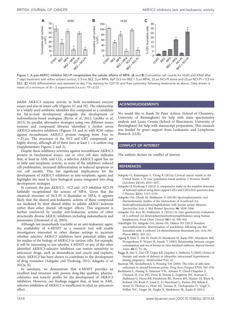
At first sight these new observations appear to contradict ourprevious observations that overexpression of AKR1C3 demotesHL-60 differentiation in response to ATRA and that indomethacinand MPA demonstrably inhibit AKR1C3 in AML cells and exertanti-leukaemic activity (Desmond et al, 2003; Khanim et al, 2009a).However, we have reported that AML cells variably co-expressAKR1-C1, -1C2 and -1C3 (Birtwistle et al, 2009). We have alsodemonstrated that both jasmonic acid and methyl jasmonate,which inhibited AKR1C2 and -1C3 predominantly but also hadhad some activity against AKR1C1 and -1C4, were sufficient topromote apoptosis and/or differentiation in leukaemia cell(Davies et al, 2009). A unifying explanation would be that theleukaemic properties of AML cells are reinforced by the combinedactivity of these enzymes and that pan-inhibitors are required tocircumvent this. In this regard our screens of the NCI diversity setidentified a compound, NCI-pan inhibitor (NCI-PI) that at 10mM
exhibited inhibitory activities against AKR1C1 -1C2 and -1C3.When used at 25 mM this compound faithfully recapitulatedthe anti-leukaemic actions of MPA against HL-60 and K562(Figure 7A and B).
DISCUSSION
These studies have identified 4-MDDT, a novel tetracyclinebreakdown product, as an excellent lead compound for thedevelopment of a specific AKR1C3 inhibitor. 4-MDDT was able to
0
20
40
60
80
100K562
0
20
40
60
80
100
120
140
Cum
ulat
ive
cell
num
ber
(×10
6 )
HL60
05
10152025303540
KG1a
0
20
40
60
80
100
120
Enz
yme
activ
ity (
%)
0
20
40
60
80
100
120
0
20
40
60
80
100
120A1
IC50= 3 nM
A6IC50= 39 nM
A9IC50= 68 nM
* **
**
0.00
010.
001
0.01 0.
1 1 10 100
0.00
010.
001
0.01 0.
1 1 10 100
0.00
010.
001
0.01 0.
1 1 10 100
BEZ
BEZ/A1
BEZ/A6
BEZ/A9
CONTBAP
BEZ
BEZ/A1
BEZ/A6
BEZ/A9
CONTBAP
BEZ
BEZ/A1
BEZ/A6
BEZ/A9
CONTBAP
A9 (�M)A6 (�M)A1 (�M)
Figure 6. AKR1C3-selective inhibitors from CRT also do not recapitulate the cellular effects of MPA. (A) Dose titrations of A1, A6 or A9 were usedto determine the IC50 of the selected CRT compounds, for recombinant AKR1C3 protein as determined using the PQ assay (see Materials andMethods). (B) Cumulative cell counts for HL60, K562 and KG1a after 7 days treatment with either solvent control, 0.5 mM BEZ, BAP (0.5 mM
BEZþ 5mM MPA), or 10mM A1/A6/A9 with 0.5 mM BEZ. Data shown is mean of a minimum of N¼3 experiments±s.e.m. *Po0.01.
AKR1C3 inhibitors lack anti-leukaemic activity BRITISH JOURNAL OF CANCER
www.bjcancer.com | DOI:10.1038/bjc.2014.83 1513
inhibit AKR1C3 enzyme activity in both recombinant enzymeassays and also in intact cells (Figures 1C and 3E). The relationshipto a widely used antibiotic identifies this compound as a candidatefor hit-to-lead development alongside the development ofindomethacin-based analogues (Byrns et al, 2012; Liedtke et al,2013). In parallel, alternative strategies using two different assayssystems and compound libraries identified a further sevenAKR1C3-selective inhibitors (Figures 5A and 6) with IC50 valuesagainst recombinant AKR1C3 protein ranging from 3 nM toB25 mM. The structures of the NCI and CRT compounds arehighly diverse, although all of them have at least 1� 6-carbon ring(Supplementary Figures 2 and 3).
Despite these inhibitory activities against recombinant AKR1C3protein in biochemical assays, our in vitro cell data indicatesthat, at least in AML and CLL, a selective AKR1C3 agent has noor little anti-neoplastic activity, as none of the inhibitors reducedcell proliferation, increased differentiation or induced apoptosis inour cell models. This has significant implications for thedevelopment of AKR1C3 inhibitors as anti-neoplastic agents andhighlights the need to have biological assays integrated into drugdevelopment strategies.
In contrast, the pan AKR1C1, -1C2 and -1C3 inhibitor NCI-PIfaithfully recapitulated the actions of MPA. Given that thechemical structure of NCI-PI is dissimilar to MPA, it appearslikely that the shared anti-leukaemic actions of these compoundare mediated by their shared ability to inhibit AKR1C isoformsrather than other shared ‘off-target’ effects. This argument isfurther reinforced by similar anti-leukaemic actions of otherstructurally diverse AKR1C inhibitors including indomethacin andjasmonates (Desmond et al, 2003).
Although not immediately tractable as an anti-leukaemic agent,the availability of 4-MDDT as a research tool will enableinvestigators interested in other disease settings to ascertainwhether selective AKR1C3 inhibitors have potential utility andfor studies of the biology of AKR1C3 in various cells. For example,it will be interesting to test whether 4-MDDT or any of the otheridentified AKR1C3-selective inhibitors can restore sensitivity toanticancer drugs, such as doxorubicin and oracin and cisplatin,where AKR1C3 has been shown to contribute to the developmentof drug resistance (Adegoke and Nyokong, 2012; Adegoke et al,2012a, b).
In summary, we demonstrate that 4-MDDT provides anexcellent lead structure with proven drug-like qualities, pharma-cokinetics and toxicity profile for the development of AKR1C3inhibitors. However, our findings suggest that, at least in AML,selective inhibition of AKR1C3 is insufficient to elicit an anticancereffect.
ACKNOWLEDGEMENTS
We would like to thank Dr Peter Ashton (School of Chemistry,University of Birmingham) for help with mass spectrometryanalysis and Laura Cronin (School of Biosciences, University ofBirmingham) for help with manuscript preparation. This researchwas funded by grant support from Leukaemia and LymphomaResearch (LLR).
CONFLICT OF INTEREST
The authors declare no conflict of interest.
REFERENCES
Adegoke O, Kulasingam S, Virnig B (2012a) Cervical cancer trends in theUnited States: a 35-year population-based analysis. J Womens Health(Larchmt) 21(10): 1031–1037.
Adegoke O, Nyokong T (2012) A comparative study on the sensitive detectionof hydroxyl radical using thiol-capped CdTe and CdTe/ZnS quantum dots.J Fluoresc 22(6): 1513–1519.
Adegoke OA, Ghosh M, Mukherjee A (2012b) Spectrophotometric andthermodynamic studies of the interactions of 4-carboxyl-2,6-dinitrophenylazohydroxynaphthalenes with bovine serum albumin.Spectrochim Acta A Mol Biomol Spectrosc 96: 1038–1046.
Adegoke OA, Kyu JK, Mukherjee A (2012c) In vitro genotoxicity evaluationof 4-carboxyl-2,6-dinitrophenylazohydroxynaphthalenes using humanlymphocytes. Food Chem Toxicol 50(3–4): 936–941.
Aderibigbe SA, Adegoke OA, Idowu OS, Olaleye SO (2012) Sensitivespectrophotometric determination of aceclofenac following azo dyeformation with 4-carboxyl-2,6-dinitrobenzene diazonium ion. Acta PolPharm 69(2): 203–211.
Agung B, Otoi T, Abe H, Hoshi H, Murakami M, Karja NW, Murakami MK,Wongsrikeao P, Watari H, Suzuki T (2005) Relationship between oxygenconsumption and sex of bovine in vitro fertilized embryos. Reprod DomestAnim 40(1): 51–56.
Bagga R, Jain V, Das CP, Gupta KR, Gopalan S, Malhotra S (2005) Choice oftherapy and mode of delivery in idiopathic intracranial hypertensionduring pregnancy. MedGenMed 7(4): 42.
Bauman DR, Steckelbroeck S, Penning TM (2004) The roles of aldo-ketoreductases in steroid hormone action. Drug News Perspect 17(9): 563–578.
Beckmann L, Husing A, Setiawan VW, Amiano P, Clavel-Chapelon F,Chanock SJ, Cox DG, Diver R, Dossus L, Feigelson HS, Haiman C,Hallmans G, Hayes RB, Henderson BE, Hoover RN, Hunter DJ, Khaw K,Kolonel LN, Kraft P, Lund E, Le Marchand L, Peeters PH, Riboli E,Stram D, Thomas G, Thun MJ, Tumino R, Trichopoulos D, Vogel U,Willett WC, Yeager M, Ziegler R, Hankinson SE, Kaaks R (2011)
0
20
40
60
80
100
120
Cum
ulat
ive
cell
num
ber
(×10
6 )
HL60
0
CONTBEZ
MPA
BEZ + M
PA
NCI-PI
BEZ + N
CI-PI
CONTBEZ
MPA
BEZ + M
PA
NCI-PI
BEZ + N
CI-PI
CONTBEZ
MPA
BEZ + M
PA
NCI-PI
BEZ + N
CI-PI
10
20
30
40
50
60
70
Cum
ulat
ive
cell
num
ber
(×10
6 )
K562
0
5
10
15
20
25
30
35
% C
D11
b-po
sitiv
e ce
lls
*
* *
*
* *
**
*
Figure 7. A pan-AKR1C inhibitor NCI-PI recapitulates the cellular effects of MPA. (A and B) Cumulative cell counts for HL60 and K562 after7 days treatment with either solvent control, 0.5 mM BEZ, 5 mM MPA, BaP (0.5 mM BEZþ 5mM MPA), 25mM NCI-PI alone and 25mM NCI-PIþ 0.5 mM
BEZ. (C) HL60 differentiation was assessed at day 7 by staining for CD11b and flow cytometry following treatments as above. Data shown ismean of a minimum of N¼3 experiments±s.e.m. *Po0.01.
BRITISH JOURNAL OF CANCER AKR1C3 inhibitors lack anti-leukaemic activity
1514 www.bjcancer.com | DOI:10.1038/bjc.2014.83

Comprehensive analysis of hormone and genetic variation in 36 genesrelated to steroid hormone metabolism in pre- and postmenopausalwomen from the breast and prostate cancer cohort consortium (BPC3).J Clin Endocrinol Metab 96(2): E360–E367.
Birtwistle J, Hayden RE, Khanim FL, Green RM, Pearce C, Davies NJ,Wake N, Schrewe H, Ride JP, Chipman JK, Bunce CM (2009) The aldo-keto reductase AKR1C3 contributes to 7,12-dimethylbenz(a)anthracene-3,4-dihydrodiol mediated oxidative DNA damage in myeloid cells:implications for leukemogenesis. Mutat Res 662(1–2): 67–74.
Bunce CM, French PJ, Durham J, Stockley RA, Michell RH, Brown G (1994)Indomethacin potentiates the induction of HL60 differentiation toneutrophils, by retinoic acid and granulocyte colony-stimulating factor,and to monocytes, by vitamin D3. Leukemia 8(4): 595–604.
Burnett AK, Russell NH, Hills RK, Kell J, Freeman S, Kjeldsen L, Hunter AE,Yin J, Craddock CF, Dufva IH, Wheatley K, Milligan D (2012) Addition ofgemtuzumab ozogamicin to induction chemotherapy improves survivalin older patients with acute myeloid leukemia. J Clin Oncol 30(32):3924–3931.
Byrns MC, Mindnich R, Duan L, Penning TM (2012) Overexpression ofaldo-keto reductase 1C3 (AKR1C3) in LNCaP cells diverts androgenmetabolism towards testosterone resulting in resistance to the 5alpha-reductase inhibitor finasteride. J Steroid Biochem Mol Biol 130(1-2):7–15.
Byrns MC, Penning TM (2009) Type 5 17beta-hydroxysteroid dehydrogenase/prostaglandin F synthase (AKR1C3): role in breast cancer and inhibitionby non-steroidal anti-inflammatory drug analogs. Chem Biol Interact178(1–3): 221–227.
Davies NJ, Hayden RE, Simpson PJ, Birtwistle J, Mayer K, Ride JP, Bunce CM(2009) AKR1C isoforms represent a novel cellular target for jasmonatesalongside their mitochondrial-mediated effects. Cancer Res 69(11):4769–4775.
Desmond JC, Mountford JC, Drayson MT, Walker EA, Hewison M, Ride JP,Luong QT, Hayden RE, Vanin EF, Bunce CM (2003) The aldo-ketoreductase AKR1C3 is a novel suppressor of cell differentiation thatprovides a plausible target for the non-cyclooxygenase-dependentantineoplastic actions of nonsteroidal anti-inflammatory drugs.Cancer Res 63(2): 505–512.
Deyashiki Y, Ogasawara A, Nakayama T, Nakanishi M, Miyabe Y, Sato K,Hara A (1994) Molecular cloning of two human liver 3 alpha-hydroxysteroid/dihydrodiol dehydrogenase isoenzymes that are identicalwith chlordecone reductase and bile-acid binder. Biochem J 299(Pt 2):545–552.
Dufort I, Soucy P, Labrie F, Luu-The V (1996) Molecular cloning ofhuman type 3 3 alpha-hydroxysteroid dehydrogenase that differs from20 alpha-hydroxysteroid dehydrogenase by seven amino acids. BiochemBiophys Res Commun 228(2): 474–479.
Feng C, Beller EM, Bagga S, Boyce JA (2006) Human mast cells expressmultiple EP receptors for prostaglandin E2 that differentially modulateactivation responses. Blood 107(8): 3243–3250.
Ghia P, Guida G, Stella S, Gottardi D, Geuna M, Strola G, Scielzo C,Caligaris-Cappio F (2003) The pattern of CD38 expression defines adistinct subset of chronic lymphocytic leukemia (CLL) patients atrisk of disease progression. Blood 101(4): 1262–1269.
Giebel S, Labopin M, Mohty M, Mufti GJ, Niederwieser D, Cornelissen JJ,Janssen JJ, Milpied N, Vindelov L, Petersen E, Arnold R, Bacigalupo A,Blaise D, Craddock C, Nagler A, Frassoni F, Sadus-Wojciechowska M,Rocha V (2013) The impact of center experience on results of reducedintensity: allogeneic hematopoietic SCT for AML. An analysis from theAcute Leukemia Working Party of the EBMT. Bone Marrow Transplant48(2): 238–242.
Gratacos-Cubarsi M, Fernandez-Garcia A, Picouet P, Valero-Pamplona A,Garcia-Regueiro JA, Castellari M (2007) Formation of tetracyclinedegradation products in chicken and pig meat under different thermalprocessing conditions. J Agr Food Chem 55(11): 4610–4616.
Guan KL, Xiong Y (2011) Regulation of intermediary metabolism by proteinacetylation. Trends Biochem Sci 36(2): 108–116.
Halling-Sorensen B, Sengelov G, Tjornelund J (2002) Toxicity of tetracyclinesand tetracycline degradation products to environmentally relevantbacteria, including selected tetracycline-resistant bacteria. Arch EnvironCon Tox 42(3): 263–271.
Hayden RE, Pratt G, Davies NJ, Khanim FL, Birtwistle J, Delgado J, Pearce C,Sant T, Drayson MT, Bunce CM (2009) Treatment of primary CLL cellswith bezafibrate and medroxyprogesterone acetate induces apoptosis and
represses the pro-proliferative signal of CD40-ligand, in part throughincreased 15dDelta12,14,PGJ2. Leukemia 23(2): 292–304.
Hayden RE, Pratt G, Drayson MT, Bunce CM (2010) Lycorine sensitizesCD40 ligand-protected chronic lymphocytic leukemia cells to bezafibrate-and medroxyprogesterone acetate-induced apoptosis but dasatanib doesnot overcome reported CD40-mediated drug resistance. Haematologica95(11): 1889–1896.
Hebert AS, Dittenhafer-Reed KE, Yu W, Bailey DJ, Selen ES, Boersma MD,Carson JJ, Tonelli M, Balloon AJ, Higbee AJ, Westphall MS, Pagliarini DJ,Prolla TA, Assadi-Porter F, Roy S, Denu JM, Coon JJ (2013) Calorierestriction and SIRT3 trigger global reprogramming of the mitochondrialprotein acetylome. Mol Cell 49(1): 186–199.
Hofland J, van Weerden WM, Dits NF, Steenbergen J, van Leenders GJ,Jenster G, Schroder FH, de Jong FH (2010) Evidence of limitedcontributions for intratumoral steroidogenesis in prostate cancer.Cancer Res 70(3): 1256–1264.
Houghton PJ, Lock R, Carol H, Morton CL, Phelps D, Gorlick R, Kolb EA,Keir ST, Reynolds CP, Kang MH, Maris JM, Wozniak AW, Gu Y,Wilson WR, Smith MA (2011) Initial testing of the hypoxia-activatedprodrug PR-104 by the pediatric preclinical testing program.Pediatr Blood Cancer 57(3): 443–453.
Izumoto S, Suzuki T, Kinoshita M, Hashiba T, Kagawa N, Wada K,Fujimoto Y, Hashimoto N, Saitoh Y, Maruno M, Yoshimine T (2005)Immunohistochemical detection of female sex hormone receptors incraniopharyngiomas: correlation with clinical and histologic features.Surg Neurol 63(6): 520–525.
Jamieson SM, Brooke DG, Heinrich D, Atwell GJ, Silva S, Hamilton EJ,Turnbull AP, Rigoreau LJ, Trivier E, Soudy C, Samlal SS, Owen PJ,Schroeder E, Raynham T, Flanagan JU, Denny WA (2012) 3-(3,4-Dihydroisoquinolin-2(1H)-ylsulfonyl)benzoic Acids: highly potent andselective inhibitors of the type 5 17-beta-hydroxysteroid dehydrogenaseAKR1C3. J Med Chem 55(17): 7746–7758.
Khanim FL, Hayden RE, Birtwistle J, Lodi A, Tiziani S, Davies NJ, Ride JP,Viant MR, Gunther UL, Mountford JC, Schrewe H, Green RM,Murray JA, Drayson MT, Bunce CM (2009a) Combined bezafibrate andmedroxyprogesterone acetate: potential novel therapy for acute myeloidleukaemia. PLoS One 4(12): e8147.
Khanim FL, Hayden RE, Birtwistle J, Lodi A, Tiziani S, Davies NJ, Ride JP,Viant MR, Gunther UL, Mountford JC, Schrewe H, Green RM,Murray JA, Drayson MT, Bunce CM (2009b) Combined bezafibrate andmedroxyprogesterone acetate: potential novel therapy for acute myeloidleukaemia. PLoS One 4(12): e8147.
Kuhne M, Korner U, Wenzel S (2001) Tetracycline residues in meat andbone meals. Part 2: The effect of heat treatments on bound tetracyclineresidues. Food Addit Contam 18(7): 593–600.
Li ZS, Wang W, Liao Z, Zou DW, Jin ZD, Chen J, Wu RP, Liu F, Wang LW,Shi XG, Yang Z, Wang L (2010) A long-term follow-up study onendoscopic management of children and adolescents with chronicpancreatitis. Am J Gastroenterol 105(8): 1884–1892.
Liedtke AJ, Adeniji AO, Chen M, Byrns MC, Jin Y, Christianson DW, Marnett LJ,Penning TM (2013) Development of potent and selective indomethacinanalogues for the inhibition of AKR1C3 (Type 5 17beta-hydroxysteroiddehydrogenase/prostaglandin F synthase) in castrate-resistant prostatecancer. J Med Chem 56(6): 2429–2446.
Lovering AL, Ride JP, Bunce CM, Desmond JC, Cummings SM, White SA(2004) Crystal structures of prostaglandin D(2) 11-ketoreductase(AKR1C3) in complex with the nonsteroidal anti-inflammatory drugsflufenamic acid and indomethacin. Cancer Res 64(5): 1802–1810.
Matsuura K, Shiraishi H, Hara A, Sato K, Deyashiki Y, Ninomiya M, Sakai S(1998) Identification of a principal mRNA species for human 3alpha-hydroxysteroid dehydrogenase isoform (AKR1C3) that exhibits highprostaglandin D2 11-ketoreductase activity. J Biochem (Tokyo) 124(5):940–946.
Mitsiades N, Sung CC, Schultz N, Danila DC, He B, Eedunuri VK, Fleisher M,Sander C, Sawyers CL, Scher HI (2012) Distinct patterns of dysregulatedexpression of enzymes involved in androgen synthesis and metabolism inmetastatic prostate cancer tumors. Cancer Res 72(23): 6142–6152.
Morakinyo AO, Iranloye BO, Daramola AO, Adegoke OA (2011) Antifertilityeffect of calcium channel blockers on male rats: association with oxidativestress. Adv Med Sci 56(1): 95–105.
Murray JA, Khanim FL, Hayden RE, Craddock CF, Holyoake TL, Jackson N,Lumley M, Bunce CM, Drayson MT (2010) Combined bezafibrate andmedroxyprogesterone acetate have efficacy without haematological
AKR1C3 inhibitors lack anti-leukaemic activity BRITISH JOURNAL OF CANCER
www.bjcancer.com | DOI:10.1038/bjc.2014.83 1515

toxicity in elderly and relapsed acute myeloid leukaemia (AML).Br J Haematol 149(1): 65–69.
Patriciu A, Mazilu D, Petrisor D, Bagga H, Kavoussi L, Stoianovici D (2005)A computer assisted method for guide-wore and catheter evaluation.Conf Proc IEEE Eng Med Biol Soc 1: 436–439.
Qiu W, Zhou M, Mazumdar M, Azzi A, Ghanmi D, Luu-The V, Labrie F,Lin SX (2007) Structure-based inhibitor design for an enzymethat binds different steroids: a potent inhibitor for human type 517beta-hydroxysteroid dehydrogenase. J Biol Chem 282(11):8368–8379.
Richards SM, Jensen RV, Liu M, Sullivan BD, Lombardi MJ, Rowley P,Schirra F, Treister NS, Suzuki T, Steagall RJ, Yamagami H, Sullivan DA(2006) Influence of sex on gene expression in the mouse lacrimal gland.Exp Eye Res 82(1): 13–23.
Rizner TL, Penning TM (2013) Role of aldo-keto reductase family 1 (AKR1)enzymes in human steroid metabolism. Steroids 79C: 49–63.
Suzuki T, Miki Y, Nakamura Y, Moriya T, Ito K, Ohuchi N, Sasano H (2005)Sex steroid-producing enzymes in human breast cancer. Endocr RelatCancer 12(4): 701–720.
Velica P, Davies NJ, Rocha PP, Schrewe H, Ride JP, Bunce CM (2009) Lack offunctional and expression homology between human and mouse aldo-ketoreductase 1C enzymes: implications for modelling human cancers.Mol Cancer 8: 121.
Wang Q, Zhang Y, Yang C, Xiong H, Lin Y, Yao J, Li H, Xie L, Zhao W,Yao Y, Ning ZB, Zeng R, Xiong Y, Guan KL, Zhao S, Zhao GP (2010)Acetylation of metabolic enzymes coordinates carbon source utilizationand metabolic flux. Science 327(5968): 1004–1007.
Wasylishen AR, Kalkat M, Kim SS, Pandyra A, Chan PK, Oliveri S, Sedivy E,Konforte D, Bros C, Raught B, Penn LZ (2013) MYC activity is negativelyregulated by a C-terminal lysine cluster. Oncogene 33: 1066–1072.
Wu RP, Hayashi T, Cottam HB, Jin G, Yao S, Wu CC, Rosenbach MD,Corr M, Schwab RB, Carson DA (2010) Nrf2 responses and thetherapeutic selectivity of electrophilic compounds in chronic lymphocyticleukemia. Proc Natl Acad Sci USA 107(16): 7479–7484.
Zargar S, Moreira TS, Samimi-Seisan H, Jeganathan S, Kakade D, Islam N,Campbell J, Adegoke OA (2011) Skeletal muscle protein synthesis and theabundance of the mRNA translation initiation repressor PDCD4 areinversely regulated by fasting and refeeding in rats. Am J PhysiolEndocrinol Metab 300(6): E986–E992.
This work is published under the standard license to publish agree-ment. After 12 months the work will become freely available andthe license terms will switch to a Creative Commons Attribution-NonCommercial-Share Alike 3.0 Unported License.
Supplementary Information accompanies this paper on British Journal of Cancer website (http://www.nature.com/bjc)
BRITISH JOURNAL OF CANCER AKR1C3 inhibitors lack anti-leukaemic activity
1516 www.bjcancer.com | DOI:10.1038/bjc.2014.83