Embed Size (px)
Citation preview

Selective Expression of Dopamine D3 Receptor mRNA inProliferative Zones during Embryonic Development of the Rat Brain
Jorge Diaz,1 Sophie Ridray,1 Virginie Mignon,2 Nathalie Griffon,2 Jean-Charles Schwartz,2 andPierre Sokoloff2
1Laboratoire de Physiologie, Universite Rene Descartes, 75006 Paris, France, and 2Unite de Neurobiologie etPharmacologie de l’Institut National de la Sante et de la Recherche Medicale, Centre Paul Broca, 75014 Paris, France
We studied by in situ hybridization histochemistry the expres-sion of D3 receptor (D3R) mRNA at various stages of rat braindevelopment. The first expression of D3R mRNA was detectedat embryonic day 14 (E14) in the striatal and rhinencephalicneuroepithelia and throughout the tectal neuroepithelium. FromE16 to E19 D3R mRNA expression extended along a rostrocau-dal axis to additional proliferative ventricular zones of the basalforebrain, including the neuroepithelia of the olfactory bulb,nucleus accumbens, septum, and amygdala, whereas D1 andD2 receptor (D1R and D2R) mRNAs were expressed predomi-nantly by migrating neuroblasts and/or differentiating striatalneurons. Only a few neuroblasts, migrating in the lateral corticalstream or developing as cerebellar Purkinje cells, expressedD3R mRNA from E18. At birth D3R expression mRNA appearedin differentiating neuronal fields of the nucleus accumbens andmedial mamillary body primordia and on P5 reached a distri-
bution similar to that found in adult. In addition, a transientupregulation was detected on P5 in the medial mamillary bod-ies, parietofrontal cortex, and olfactory tubercle. In the adultbrain D3R gene expression continued in the striatal proliferativesubventricular zone. The late expression D3R mRNA in neu-rons, after achievement of dopamine innervation, supports theexistence of a regulating factor released from dopamine neu-rons, as suggested by denervation studies in the adult. Thesustained and abundant D3R gene expression, predominantlyin germinative neuroepithelial zones actively involved in neuro-genesis of most basal forebrain structures, supports the hy-pothesis of a neurogenetic but minor morphogenetic modula-tory role for the D3R during CNS development.
Key words: in situ hybridization; neuroepithelium; neurogen-esis; forebrain development; dopamines; D3R mRNA
Catecholamines seem to affect a number of developmental pro-cesses in primitive organisms, including growth, regeneration, andmorphogenesis (Lauder, 1993). The dopaminergic system appearsearly in brain development of higher species (Voorn et al., 1988),and a neurodevelopmental role for dopamine has been suggested(Rosengarten and Friedhoff, 1979; Miller and Friedhoff, 1986).
Dopamine interacts with five dopamine receptor subtypes(D1R–D5R) having distinct localization and intracellular signal-ing, allowing this neurotransmitter to exert pleiotropic influenceson target cells (Sibley and Monsma, 1992; Gingrich and Caron,1993; Sokoloff and Schwartz, 1995). The D3R (Sokoloff et al.,1990) has a density in the adult rat brain two orders of magnitudelower than that of the D2R and is expressed in discrete brainregions, including the ventral striatum, mamillary bodies, andarchicerebellum (Bouthenet et al., 1991; Levesque et al., 1992;Diaz et al., 1995).
D1R and D3R both occur in the islands of Calleja and nucleusaccumbens (Le Moine and Bloch, 1996), whereas colocalization ofD2R and D3R seems exceptional (Bouthenet et al., 1991). It is notknown whether these respective coexpression and segregationoriginate early in development. Previous works have detailedontogeny of D1R and D2R in the rat brain (Guennoun and Bloch,1992; Schambra et al., 1994). D1R and D2R messages are present
from gestational day 14 in low amounts and increase slowly duringdevelopment. These receptors are expressed mainly by migratingand maturing neurons, similar in location to those found in theadult brain, suggesting that respective receptor expression is anintrinsic property of these neurons (Schambra et al., 1994).
The regulation of D3R gene expression in adult is highly de-pendent on dopamine innervation. Thus, a lesion of dopamineneurons, impairment of axonal transport, or reduction of dopa-mine neuron firing, but not removal of dopamine or its knowncotransmitter transmission, decreases D3R gene expression(Levesque et al., 1995). This has led to the proposal that D3Rgene regulation is under the positive influence of a yet unidenti-fied anterograde factor released from dopamine neurons.
The prenatal development of D3R expression is documentedvery poorly. With the use of the highly sensitive reverse PCR, D3Rtranscripts were detected faintly in rodent embryos from gesta-tional days 10–11 and clearly on day 14 (Cadoret et al., 1993;Fishburn et al., 1996). These studies, however, did not reveal thelocalization of D3R transcripts in embryonic brain. Autoradio-graphic analysis of rat brain during prenatal ontogeny with the useof the D2R/D3R ligand [125I]iodosulpride (Sales et al., 1988)revealed transient expressions of binding sites in the ventral partof the spinal cord and in the lateral ventricle lining that did notmatch D2R mRNA distribution and, therefore, possibly that wererelated to the D3R. In addition, a transient expression of D3Rprotein was observed in the parietal cortex during postnatal de-velopment of mouse brain (Demotes-Mainard et al., 1996).
Using in situ hybridization, we mainly show here that D3RmRNA expression is restricted almost entirely to the ventricular
Received Oct. 28, 1996; revised March 14, 1997; accepted March 21, 1997.This work was supported by a Biomed 2 Grant from the European Commission
(BMH4-CT96-0203). We are grateful to C. Sotelo for critical reading and helpfuldiscussion of this manuscript.
Correspondence should be addressed to Dr. Pierre Sokoloff, Unite de Neurobi-ologie et Pharmacologie, 2ter Rue d’Alesia, 75014 Paris, France.Copyright © 1997 Society for Neuroscience 0270-6474/97/174282-11$05.00/0
The Journal of Neuroscience, June 1, 1997, 17(11):4282–4292

neuropithelium during the whole prenatal ontogeny and that theneuronal expression of the D3R appears later, after the settling ofdopamine innervation.
MATERIALS AND METHODSAnimals and dissection. Timed pregnant female Wistar rats (Iffa-Credo) wereused. Gestational age was determined from the mating time, and embryonicday 1 (E1) was designated as the day after insemination. After a 21 dgestation the day of birth was designed as postnatal day 0 (P0), and subse-quent age was defined relative to this. For studies involving prenatal series,pregnant rats were decapitated, and embryos and fetuses were removed bycesarean section on E12, E14, E16, E17, E18, and E19. Postnatal studieswere made with animals at ages P0 and P5.
Tissue preparation. Embryos and fetuses (E12–E19) removed free fromthe amniotic membranes were whole-frozen in liquid monochlorodifluo-romethane (230°C) and stored at 270°C. The postnatal animals (P0 andP5 pups) were decapitated, and their heads were collected and frozen.Tissue sections (8 and 10 mm for pre- and postnatal, respectively) madein coronal and sagittal planes on a cryostat were thaw-mounted at roomtemperature on SuperFrost Plus slides (Menzel-Glasser), fixed for 40 minat 4°C in 4% paraformaldehyde made up in 0.1 M PBS, and rinsed twice(5 min each) in PBS at 4°C and then for 5 min in PBS at roomtemperature. The sections were rinsed briefly in distilled water, dehy-drated via graded alcohols, dried under a stream of cold air, and storedat 270°C until they were processed for in situ hybridization.
Probes. The probes used were 33P-labeled riboprobes. Probes for ratD2R and D3R mRNAs corresponded to respective sequences of the thirdintracellular loop of the receptor and are described elsewhere (Sokoloffet al., 1990; Bouthenet et al., 1991). The probe for rat D1R correspondedto a C-terminal fragment (nucleotides 1382–1708) obtained by PCR andsubcloned into pGEM-4Z. Riboprobes were synthesized with the Ribo-
probe Gemini System (Promega, Madison, WI), treated by RNase-freeDNase (Boehringer Mannheim, Mannheim, Germany), and recoveredfrom Chroma Spin 230 columns (Clontech, Cambridge, UK).
Figure 1. Expression of D3R mRNA in the brain of the 14 d embryo. A,Film autoradiographic hybridization signals from a parasagittal sectionhybridized with a D3R mRNA antisense probe. Hybridization signals arepresent only in neuroepithelia of striatum (St), rhinencephalon (indicatedby asterisk), and tectum (TNe). Ct, Cortex. B, Structure locations areshown on the same section counterstained with Mayer’s hemalum solutionand photographed with bright-field illumination. C, Photomicrograph ofan emulsion-dipped section with bright-field illumination at the level ofstriatal neuroepithelium lining the lateral ventricle (LV ), showing groupsof labeled cells identified by clusters of dark silver grains overlying indi-vidual nuclei in the neuroepithelium (Ne).
Table 1. Abbreviations used in figures
4V Fourth ventricleIX Cerebellum, lobule 9X Cerebellum, lobule 10Acc Nucleus accumbensAccSh Nucleus accumbens, shell partAccC Nucleus accumbens, core partAmy AmygdalaCC Corpus callosumCi Cingulate cortexCx CortexFr Frontal cortexICj Islands of CallejaICjM Island of Calleja, majorIn Insulate cortexLcs Lateral cortical streamLV Lateral ventricleM Mitotic zone of the neuroepitheliumMB Medial mamillary bodiesNe NeuroepitheliumOB Olfactory bulbPa PallidumPFCx Parietofrontal cortexPu Purkinje cell layerRe Reservoir of lateral cortical streamRh RhinencephalonS Synthetic zone of the neuroepitheliumSp SeptumSt StriatumSVZ Subventricular zoneTNe Tectal neuroepitheliumTu Pyramidal layer of olfactory tubercle
Diaz et al. • Ontogeny of D3 Receptor mRNA J. Neurosci., June 1, 1997, 17(11):4282–4292 4283
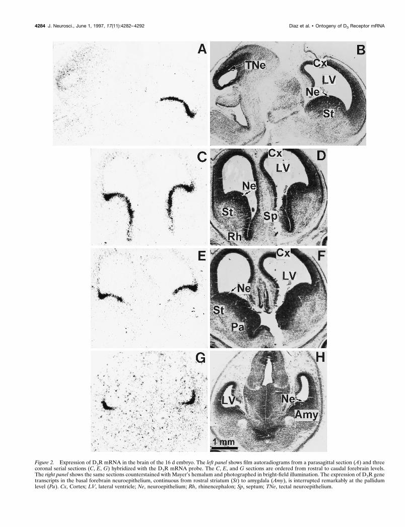
Figure 2. Expression of D3R mRNA in the brain of the 16 d embryo. The left panel shows film autoradiograms from a parasagittal section (A) and threecoronal serial sections (C, E, G) hybridized with the D3R mRNA probe. The C, E, and G sections are ordered from rostral to caudal forebrain levels.The right panel shows the same sections counterstained with Mayer’s hemalum and photographed in bright-field illumination. The expression of D3R genetranscripts in the basal forebrain neuroepithelium, continuous from rostral striatum (St) to amygdala (Amy), is interrupted remarkably at the pallidumlevel (Pa). Cx, Cortex; LV, lateral ventricle; Ne, neuroepithelium; Rh, rhinencephalon; Sp, septum; TNe, tectal neuroepithelium.
4284 J. Neurosci., June 1, 1997, 17(11):4282–4292 Diaz et al. • Ontogeny of D3 Receptor mRNA

In situ hybridization. Thawed sections were treated with proteinase K (1mg/ml) for 10 min at 37°C, acetylated in 0.1 M triethanolamine, pH 8, and0.25% acetic anhydride for 10 min, rinsed in 23 SSC (203 SSC is 0.3 MTris-citrate and 3 M NaCl), dehydrated in increased concentrations ofgraded ethanol, and air-dried. Sections were covered with 50 ml of ahybridization buffer containing 70% formamide (for D2R and D3R) or50% formamide (for D1R), 10% dextran sulfate, 13 Denhardt’s solution,43 SSC (for D2R and D3R) or 23 SSC (for D1R), 0.1% Na-pyrophosphate, 100 mg/ml yeast-tRNA, 100 mg/ml denatured salmonsperm-DNA, 50 mM Tris-HCl, pH 7.4, 1 mM EDTA, and 2 3 106 dpm ofa 33P-labeled antisense or sense probe. Slides were covered with squaresof Nescofilm (Roth) and incubated overnight (12–14 hr) at 55°C. Thensections were cooled at room temperature in 23 SSC, treated with RNaseA (200 mg/ml) for 1 hr at 37°C, and rinsed twice in 23 SSC for 15 min atroom temperature, in 0.53 SSC for 30 min at 55°C, and in 0.13 SSC for30 min at 60°C. Sections were dehydrated in graded alcohols containing300 mM ammonium acetate and were dried under a stream of cold air.Autoradiograms were generated by apposing the radiolabeled tissuesections to b-Max Hyperfilms (Amersham, Braunschweig, Germany) for1–2 weeks for D1R and D2R and 3–4 weeks for D3R.
Some sections from animals at each stage of development studied weredipped in a photographic emulsion (LM-1, Amersham) and exposed for30–40 d. Dipped sections were counterstained with Mayer’s hemalumsolution and examined with a photomicroscope Axiophot (Zeiss,Oberkochen, Germany).
RESULTSWe performed In situ hybridization using a 33P-labeled D3RcRNA probe on brain sections of embryos, heads of fetuses andpups, from the 12th gestation day (13 d after insemination, E12)until the 5th day after birth (P5). Two to four animals from at leasttwo different offspring were used at each stage. Structure identi-fication, name, and abbreviations (Table 1) are in agreement withthe recommendations of the Boulder Committee (1970) and theatlas of the rat developing brain by Bayer and Altman (1995). Theregional and cellular distribution of D3R mRNA expression wasdetermined with both film autoradiograms and emulsion-coatedmicroautoradiographies on coronal and sagittal sections taken atvarious levels throughout the whole developing brain, but onlyrelevant levels are illustrated on Figures 1–9. In addition, cRNAprobes for D1R and D2R subtypes were used in some experimentsfor comparison. Control hybridization experiments with senseprobes resulted in autoradiograms devoid of signal (data notshown).
Midgestational development (E12–E16)No hybridization signal was seen in any part of the developingbrain at E12 (data not shown). Specific hybridization signal forD3R message was detected on both autoradiograms andemulsion-coated sections from E14 (Fig. 1). At this stage discretegerminative ventricular zones of the basal forebrain and dorsalmidbrain exhibited distinct autoradiographic signals. In the fore-brain, hybridization labeling was detected at high levels in theneuroepithelium of striatum (also referred to as lateral ganglioniceminence) and at moderate levels in rhinencephalic and septalneuroepithelia (Fig. 1A,B). D3R mRNA expression in the fore-brain neuroepithelium abruptly ended at the rostral tip of thelateral ventricle, approximately corresponding to the limit be-tween striatal and cortical neuroepithelia or dorsal and basalforebrain (Fig. 1A,B). In the midbrain, hybridization signals weredetected at moderate levels in the tectal neuroepithelium. Exam-ination of microautoradiographic hybridization labeling showedthat D3R mRNA-labeled cells appeared as clusters of neuroepi-thelial cells at the vicinity of the lateral ventricle (Fig. 1C). Withinthe clusters a large majority of cells were labeled.
At E16 high levels of hybridization signals appeared as a narrowband all along the basal forebrain neuroepithelium from thestriatum up to the amygdala. However, this continuity of D3RmRNA expression was interrupted remarkably at the level of theneuroepithelium of pallidum, which did not express transcripts atall (Fig. 2E). The hybridization signals were more pronounced atthe limit between the striatal and cortical neuroepithelia (Figs.2A,C,E, 3A). Labeling at a moderate level was still apparent in thetectal neuroepithelium, whereas faint signals were present in thecortical neuroepithelium.
Photomicrographs (Fig. 3) showed that at E16 D3R mRNA wasexpressed by cells lining the lateral ventricle at the level ofventricular zones of striatum, rhinencephalon, septum, and amyg-dala and appeared mostly in clustering cells rather than in ahomogenous layer.
Late gestational development (E17–E19)The distribution of D3R mRNA in brain embryo at E17 did notdiffer greatly from that at E16 (data not shown). At E18 most of
Figure 3. High magnifications of D3R mRNA hybridization signals inbasal forebrain neuroepithelium of the 16 d embryo. The top and bottomrectangles in a bright-field photomicrograph from a coronal section coun-terstained with Mayer’s hemalum (A) delimit the fields shown in B and C,respectively. B, C, Dark-field photomicrographs taken from an emulsion-dipped section through the rostral striatal anlage that show the labeling inthe striatal (St), rhinencephalic (Rh), and septal (Sp) neuroepithelia bor-dering the ventral horn of the lateral ventricle (LV ), indicated by thinarrows in C, whereas the subventricular zone (SVZ) and the differentiatingfield of striatum are labeled only scarcely. The strongest signal is presentapproximately at the limit between striatal and cortical neuroepithelia inA, as determined by the dorsolateral corner of the lateral ventricle (indi-cated by thick arrows in A and B). Cx, Cortex; Ne, neuroepithelium.
Diaz et al. • Ontogeny of D3 Receptor mRNA J. Neurosci., June 1, 1997, 17(11):4282–4292 4285

the D3R mRNA still was expressed in the basal forebrain neuro-epithelium, spanning the entire striatal border of the lateralventricle and extending ventrally up to the ventral tip surroundedby primordia of nucleus accumbens and septum. In addition,hybridization signals were present in tectal neuroepithelium but atmuch lower levels, as compared with previous stages of gestationand in cortical neuroepithelium (Figs. 4A,C, 5A). Labeled cellsappeared as clusters in a 100-mm-thick band lining the ventricle(Fig. 4E), corresponding to the synthetic and mitotic zones of theneuroepithelium, as evidenced by Bayer and Altman (1991) withthe use of [3H]thymidine labeling, whereas the differentiating fieldwas labeled only very scarcely. This shows that D3R-expressingcells are proliferative and not postmitotic differentiating cells.
The first D3R mRNA-expressing neurons appeared at E18.Scattered labeled migrating neuronblasts were found to leave thejunction between striatal and cortical ventricular zones, join thelateral cortical stream, and accumulate in the reservoir (Fig.5A,E), a region in which differentiated neurons sojourn beforemigrating to piriform and primary olfactory cortice and as yetunidentified areas of the basal telencephalon (Bayer and Altman,1991, 1995). A labeled cluster of settling Purkinje cells also was
seen in the caudal part of the cerebellar anlage (Fig. 4A). Thesemigrating Purkinje cells probably correspond to those destined tosettle in cerebellar cortex in lobule 10 rather than in lobule 9, inagreement with the caudorostral cytogenetic gradient observed inthe neurogenesis of Purkinje cells (Altman and Bayer, 1985).
The distribution of D3R mRNA was compared with that of D1Rand D2R mRNAs on adjacent sections of the forebrain. At E18(Fig. 5) the distributions of the three receptor subtypes wereoverlapping partially, but distinct. The three receptor mRNAswere found in the reservoir of the lateral cortical stream; this isthe unique area in which we found coexpression of D1R, D2R, andD3R mRNAs. D1R and, to a lesser, extent D3R, but not D2R,mRNAs were expressed in the cortical neuroepithelium. In thestriatal primordium D1R, D2R, and D3R mRNAs were expressedin distinct compartments: the neuroepithelium expressed exclu-sively D3R mRNA; the medial subventricular zone expressedalmost exclusively the D2R mRNA (very scarce D3R-expressingcells were, however, found in this area); differentiating fields ofstriatum, nucleus accumbens, and rhinencephalon expressed D1Rand D2R, but not D3R, mRNAs (Fig. 5).
At E19 (Fig. 6) the D3R mRNA was still prominent in the
Figure 4. Expression of D3R mRNA in the basal forebrain and cerebellar primordia of the 18 d embryo. Autoradiograms of parasagittal (A) and coronal(C) sections hybridized with the D3R mRNA probe show signals in neuroepithelia (Ne) of striatum (St), nucleus accumbens (Acc), rhinencephalon (Rh),septum (Sp), and in the more caudal Purkinje cell layer (Pu) of cerebellar primordium. B, D, Corresponding sections counterstained with Mayer’shemalum. High magnification of the striatal ventricular zone under bright-field illumination (E) illustrates that labeling is restricted to clusters of labeledcells present through the synthetic (S) and mitotic (M ) neuroepithelial zones, delimiting a continuous periventricular band 100 mm thick. Cx, Cortex; LV,lateral ventricle; SVZ, subventricular zone.
4286 J. Neurosci., June 1, 1997, 17(11):4282–4292 Diaz et al. • Ontogeny of D3 Receptor mRNA

Figure 5. Compared expressions of D1R, D2R, and D3R mRNAs in the forebrain of the 18 d embryo. Shown are film autoradiograms of adjacent coronalsections at a rostral level of the developing striatum, hybridized with probes specific for either D3R (A), D2R (C), or D1R (D) mRNAs. B, Bright-fieldphotomicrograph of section A, counterstained with the Mayer’s hemalum. Cells in the reservoir of lateral cortical stream (indicated by arrows) expressthe three receptor subtype mRNAs (A, C, D). The cortical neuroepithelium and cortical plate (Cx) are labeled by the D1R and D3R, but not D2R, mRNAprobes. D1R, D2R, and D3R mRNAs are expressed in different striatal compartments, as shown in E, which represents a schematic coronal section at thesame level and developmental stage, taken from the atlas by Bayer and Altman (1995) in which the right cerebral hemisphere displays the structures (Acc,nucleus accumbens; Ci, cingulate cortex; Fr, frontal cortex; In, insulate cortex; Lcs, lateral cortical stream; Re, reservoir; Rh, rhinencephalon; Sp, septum;St, striatum) and the left hemisphere displays the respective distributions of D1R, D2R, and D3R mRNAs. In the striatum D1R mRNA is present in thedifferentiation field (St), D2R mRNA in the subventricular zone (SVZ) and in the differentiation field, and D3R mRNA in the neuroepithelium (Ne).
Diaz et al. • Ontogeny of D3 Receptor mRNA J. Neurosci., June 1, 1997, 17(11):4282–4292 4287

neuroepithelium, extending from rostral striatum to amygdala.The neuroepithelium of olfactory bulb also was labeled. At thisstage labeling appeared as a band becoming thinner than previ-ously, because the cortical ventricular zone of the neuroepithe-lium also had become thinner (Bayer and Altman, 1991). Labelingin Purkinje cell plate also appeared stronger and more extended.
Postnatal development (P0–P5)At birth the D3R mRNA distribution still resembled that oflate embryos, with only a few distinct characteristics found inadults (Fig. 7). The striatal neuroepithelium still was labeledheavily, although to a lesser level than in embryos. Labeling ofsubventricular zones of striatum and nucleus accumbens be-came stronger but did not appear in differentiating fields (Fig.7A,C). Strong labeling also appeared in medial mamillary body(Fig. 7A), a region of high D3R expression in adults (Bouthenetet al., 1991). Also as in adults, labeled Purkinje cells werefound within the cerebellum, in the entire lobule 10, and aspatches in lobule 9 (Fig. 7A,E–H ).
More prominent changes occurred between P0 and P5. At thislatter stage the distribution was already almost identical to that inthe adult brain, with a few exceptions. Labeling along the striatal
subventricular zone got thinner and was distributed as patches(Figs. 8D, 9A). In adults D3R-expressing cells also were found inthe striatal subventricular (also referred to as subependymal) zone(Fig. 9C,D); a labeling of ependymal cells could not be excluded.At P5 major localizations found in adults were settled, as inlobules 9 and 10 of cerebellum, medial mamillary body, andventral striatum (Fig. 8A). In the ventral striatum, although la-beling was identical at P5 and in adults in the shell part of nucleusaccumbens and in the islands of Calleja, there was D3R mRNA inthe core of nucleus accumbens and in the olfactory tubercle (Fig.8C,D), which were strongly reduced in adults (Diaz et al., 1995).A moderate labeling was detected at P5 in the parietofrontalcortex (Fig. 8A,C,D), which remained in adults as a laminatedband of scarce positive cells in layer IV (data not shown).
DISCUSSIONWe detected the first appearance of D3R mRNA at E14, inagreement with results obtained by PCR (Cadoret et al., 1993),and autoradiography by using the highly sensitive D2R/D3R ra-dioligand [125I]iodosulpride (Sales et al., 1988). Our results sug-gest that D3R mRNA is more abundant during pre- and earlypostnatal ontogeny than in adult. The striatal neuroepitheliumexpresses strong signals from E14, culminating at E18, and pro-gressively declines after birth. These signals seem to be of similaramplitude to those generated by a D2R mRNA probe, althoughthe D2R is one to two orders of magnitude more abundant thanthe D3R in adult brain (Levesque et al., 1992). The high abun-dance of D3R protein in the striatal neuroepithelium also isindicated by the strongest [125I]iodosulpride binding in this re-gion, which exceeds that of D2R binding in differentiating striatum(Sales et al., 1988). Moreover, labeled neuroepithelium extendsthrough almost the whole basal forebrain, whereas the D3RmRNA is expressed in the derived structures at a very low leveland /or in discrete areas. In addition, transient expressions inneocortical and paleocortical regions, medial mamillary body, andthe core part of nucleus accumbens were found in newborns.
Comparisons among the expression patterns of the D1R, D2R,and D3R mRNAs revealed marked differences, suggesting distinctexpression regulation and roles (Table 2). Whereas the D3RmRNA is expressed almost exclusively in the proliferative neuro-epithelium during prenatal ontogeny, D1R and D2R mRNAs areexpressed only transiently and sparingly in this structure andappear mostly in differentiating neurons, before dopamine inner-vation (Table 2). In addition, there is no overlap of D1R, D2R,and D3R mRNAs, except in the reservoir of the lateral corticalstream. In many instances the distribution of D3R mRNA mark-edly differs during prenatal and postnatal developments. Accord-ing to the fate of D3R mRNA-expressing cells, three distinctpatterns can be distinguished.
The first and most documented pattern corresponds to D3RmRNA-expressing neuroepithelial cells, generating neurons inwhich this marker thereafter becomes undetectable. Thus, prena-tal D3R mRNA is confined almost entirely in neuroepithelial cellsof striatum, amygdala, olfactory bulb, and tectum, most of theprogeny of which rapidly lose the capacity to transcribe the D3Rgene during migration and differentiation. D3R mRNA labeling inthe striatal neuroepithelium overlaps the synthetic and mitoticzones, which are restricted to a thin layer close to the ventricle atearly stages (E14–E16) but which spread in the subventricularzone at later stages (Bayer and Altman, 1995). At birth and duringpostnatal development the synthetic zone gets thinner along thelateral ventricle, just as the D3R mRNA labeling does. Addition-
Figure 6. Expression of D3R mRNA in the brain of the 19 d embryo. Theleft panel shows film autoradiograms from parasagittal (A, E) and coronal(C) sections hybridized with the D3R mRNA probe. The right panels showcorresponding sections counterstained with Mayer’s hemalum and photo-graphed in bright-field illumination. Hybridization signals are detected athigh level in several periventricular neuroepithelial regions (Ne) of thebasal forebrain, including the striatum (St in A), amygdala (Amy in C), andin a group of Purkinje cells (Pu in E). Low expression of D3R is seen in theventricular zone of the cortex and cortical plate (Cx), olfactory bulb (OB),and subventricular zone (SVZ) of the striatum in A. LV, Lateral ventricle.
4288 J. Neurosci., June 1, 1997, 17(11):4282–4292 Diaz et al. • Ontogeny of D3 Receptor mRNA

ally, cells continue to proliferate in the subependymal zone, cor-responding to a remnant of neuroepithelium (Privat and Leblond,1972; Jacobson, 1991), in which D3R mRNA occurs in adults.
The restricted expression of D3R mRNA in the neuroepithe-lium may be linked to the cell proliferative activity via othernegative regulation by factors arresting cell division and triggeringcell differentiation or may be linked to positive regulation bytranscription factors (Arenander and De Vellis, 1994). In thislatter respect, it is noteworthy that expressions of Dlx-2 andMash-1 homeobox genes (Porteus et al., 1991, 1994; Robinson etal., 1991), candidates for regulating patterning and differentiationof the forebrain, match, to some extent, D3R mRNA distribution.Alternatively, D3R gene expression may be activated by unknownfactors, either released from neuroepithelium or circulating in thelateral ventricle and poorly diffusing through the parenchyma.
Both microheterogeneity and compartmentation are observedin the distribution of D3R gene transcripts within the neuroepi-thelium. Instead of forming a continuous sheet of neuroepithelialcells, D3R mRNA-expressing cells, rather, are organized in clus-ters evident throughout the entire development. Similar alterna-tion of [3H]thymidine-labeled neuroepithelial cells has suggesteddifferent migration kinetic properties among these cells (Altmanand Bayer, 1990). Distinct properties among neuroepithelial cellsalso may be related to their distinct fate. Lineage studies withretroviral markers suggest that certain germinal cells becomerestricted only to generating neurons (Luskin et al., 1988). Mostadult medium-sized striatal neurons are distributed among dynor-
phin /substance P and D1R-expressing neurons and enkephalinand D2R-expressing neurons (Gerfen et al., 1990; Curran andWatson, 1995; Le Moine and Bloch, 1995). Adult striatal neuronsalso belong to either striosomes or matrix, making different out-puts (Graybiel, 1984). Birth-dating studies have shown that strio-some cells become postmitotic at a distinctly earlier time thanmatrix cells (Van der Kooy and Fishell, 1987). In adults, whereascolocalization of D2R and D3R seems exceptional if it exists(Bouthenet et al., 1991; Diaz et al., 1994), there are examples ofD1R and D3R colocalization (Le Moine and Bloch, 1996; Bordetet al., 1997) (S. Ridray, N. Griffon, E. Souil, V. Mignon, S.Carboni, J. Diaz, J.-C. Schwartz, P. Sokoloff, unpublished obser-vation) suggesting that D3R mRNA-expressing cells in the neu-roepithelium may correspond to a distinct subpopulation.
In addition, the continuous distribution of D3R mRNA withinthe basal forebrain neuroepithelium is interrupted remarkably atthe level of the pallidal neuroepithelium while persisting at moreventral or caudal levels. It is noteworthy that cell fate is distinct inthe striatal and pallidal neuroepithelium, the latter presumablygiving rise mostly to pallidal cholinergic magnocellular cells in noor only very low levels of D3R mRNA expressed in adults (Diaz etal., 1995).
The second and less documented pattern corresponds to cellsthat express D3R mRNA in both the neuroepithelium and itsprogeny. D3R mRNA is abundant in the neuroepithelium ofnucleus accumbens at its appearance on E18 and in the shellsubdivision after the appearance of the secondary germinal matrix
Table 2. Comparison among appearances of D1R, D2R, and D3R mRNAs, [125I]iodosulpride binding, and dopamine immunoreactivity
Structures Figure numbersa
Dopamine receptor mRNA[125I]ISbindingb DA-IRcD1Rd D2Rd D3R
Olfactory bulb (Ne) 6A E18 0 E163P0 E17 ndRhinencephalon (Ne) 2C, 3C, 4C 0 0 E14 E15 0Islands of Calleja 8A,C,D nd nd [P0–P5] nd E19Septum (Ne) 2C, 3C, 5A, 7C 0 0 E16 E15 0Septum (DF) 8C E18 nd [P0–P5]h [P0–P7] [P0–P5]Nucleus accumbens
Ne 4A, 5A, 7C 0 0 E18 E20 0SVZ 7C, 8D, 9D 0 0 E19 nd 0DF (shell) 8C,D E18e E18e Pg5 f P0 P2DF (core) 8C,D E18 E18 [P0–P5]h E19 E19e
StriatumNe 1–7 E143E17g E143E17g E14 E14 E18g
SVZ 6A, 7A,B, 8D, 9A E14 E14 E19g E14 E16g
DF 8C,9D E17 E16 [P0–P5] f E15 E14Pallidum (Ne) 2E 0 0 0 0 0Amygdala (Ne) 2G, 6C nd nd E16 0 0Olfactory tubercle 8C,D E18 E18 [P0–P5] E18 E21Lateral cortical stream (Re) 5 nd nd E18 nd ndCortex (Ne) 2E, 6A,C, 7C E143E17 0 E16g E13 0Cortical plate 6A,C E16 E18 E16g E16 E17Fronto-parietal cortex (DF) 8C,D E18 E18 [P0–P5]h P7 P17Median mammillary bodies 7A, 8A nd nd P0 P14 ndTectum (Ne) 1A, 2A E14 0 E143E18 P5 0Cerebellum lobule 9 4A, 6E, 7A,E, 8A 0 0 E18 E20 0
lobule 10 7A, 8A 0 0 [E19–P0] E20 0
DF, Differentiation field; Ne, neuroepithelium; Re, reservoir; SVZ, subventricular zone; E3E or E3P, transient expression; [E-E or P-P], the exact time of appearance couldnot be determined; 0, undetectable; nd, not determined from available data. aFigure number in the present study illustrating the presence or absence of D3R mRNA;b[125I]iodosulpride binding, from Sales et al. (1988); cDopamine immunoreactivity, from Voorn et al. (1988) and Kalsbeek et al. (1992); dfrom Guennoun and Bloch (1992),Schambra et al. (1994), and present study; elateral part; fmedial part; gvery sparse labeling; hlabeling very sparse in adult—the exact date of disappearance is not known.
Diaz et al. • Ontogeny of D3 Receptor mRNA J. Neurosci., June 1, 1997, 17(11):4282–4292 4289

(at P5; see Table 2). Our data indicate that the expression of D3RmRNA is not continuous during cell differentiation and migrationfrom the neuroepithelium but, rather, is triggered after the set-tling of dopamine innervation, which might exert a dual trophicinfluence. This is consistent with our hypothesis that regulation ofD3R gene expression in the shell of nucleus accumbens is underthe positive influence of (1) a trophic factor, synthesized bydopamine neurons, distinct from dopamine and its cotransmittersneurotensin and cholecystokinin, and conveyed by axonal trans-port, the release of which seems dependent on dopamine neuronactivity (Levesque et al., 1995); (2) activation of the D1R bydopamine or agonists (Bordet et al., 1997).
Nevertheless, a continuous expression of D3R mRNA occurs incells originating approximately from the junction between striataland cortical neuroepithelia, migrating through the lateral corticalstream, and sojourning in the reservoir (Bayer and Altman, 1995).Some cells arising from the cortical neuroepithelium differentiatein the parietofrontal cortex, which transiently expresses high levelsof D3R mRNA (present study) and binding (Demotes-Mainard etal., 1996), and these cells remain in adult rats in layer IV. D3RmRNA also persists in human parietal cortex, where it could bedetected by PCR in brain from normal subjects but not frompsychiatric patients who may express an abnormal shortenedtranscript (Schmauss et al., 1993).
The third pattern corresponds to D3R mRNA-expressing neu-rons that differentiate from a neuroepithelium in which this
marker is undetectable. D3R mRNA-expressing Purkinje cells,destined to settle in the archicerebellum (lobules 9 and 10) afterbirth (Bouthenet et al., 1991; Diaz et al., 1995), occur from theirdifferentiation at E18, but not in Purkinje cell progenitors. Astrong labeling also appears at P0 in the medial mamillary bodies,which derive from unlabeled hypothalamic neuroepithelium.Thus, in these structures, expression of the D3R seems to followa more classical pattern, being triggered after the neuronal dif-ferentiation, possibly in relation to a functional specialization.
A specific role of the D3R in neurogenesis, but not in morpho-genesis, can be inferred from its highest level of expression inprenatal ontogeny, as compared with adulthood and its selectivelocalization in the proliferative zone of the neuroepithelium dur-ing ontogeny. Such a role is supported by the mitogenic responseinduced by recombinant D3R stimulation in transfected cells(Chio et al., 1994; Pilon et al., 1994). However, the origin ofdopamine available for D3R stimulation is unclear in the neuro-epithelium, where no dopamine fibers are present. One study(Specht et al., 1978) reported a few cells positive for tyrosinehydroxylase immunoreactivity at E12.5–E15 at the surface ofstriatal neuroepithelium. Nevertheless, there is no indication thatthese cells indeed produce dopamine. Another possibility is thatthe D3R receives dopamine diffusing through the cerebrospinalfluid from dopamine neurons in diencephalon and mesencepha-lon. Dopamine has a higher affinity at the D3R than at any otherreceptor subtype, allowing this receptor to respond to nanomolar
Figure 7. In situ hybridization of D3R mRNA in the brain of a newborn pup. A, C, E, and G are autoradiograms of parasagittal (A) and coronal (C, E,G) sections hybridized with the D3R probe; B, D, F, and H are corresponding sections counterstained with Mayer’s hemalum. D3R mRNA is seen in theresidual neuroepithelium (Ne) and the adjacent subventricular zone (SVZ) of the striatum (St), nucleus accumbens (Acc), and septum (Sp) as well as inthe medial mamillary body (MB) and as patches in the Purkinje cell layer (Pu) of lobules 9 (IX ) and 10 (X ) in the cerebellar cortex. Cx, Cerebral cortex;LV, lateral ventricle; 4V, fourth ventricle.
4290 J. Neurosci., June 1, 1997, 17(11):4282–4292 Diaz et al. • Ontogeny of D3 Receptor mRNA

dopamine concentrations. In adult rat brain examples of D3Rlocalizations at some distance from dopaminergic fibers havesuggested nonsynaptic actions of diffusing dopamine through thisreceptor (Diaz et al., 1995). Finally, a constitutive, i.e., agonist-independent, activity of the D3R has been suggested from studiesthat use transfected cells (Griffon et al., 1996b).
Schizophrenia, sometimes assumed to result from a neurode-velopmental disorder, is marked namely by neuroanatomical ab-normalities such as ventricle enlargement (Weinberger, 1987;Waddington, 1993), possibly related to a defect in neuroepithe-lium proliferation. A role for the D3R in this pathological processis supported by some (but not all) genetic studies (Crocq et al.,
1992; Nimgaonkar et al., 1993; Mant et al., 1994; Griffon et al.,1996a).
REFERENCESAltman J, Bayer SA (1985) Embryonic development of the rat cerebel-
lum. III. Regional differences in the time of origin, migration, andsettling of Purkinje cells. J Comp Neurol 231:42–65.
Altman J, Bayer SA (1990) Vertical compartmentation and cellulartransformations in the germinal matrices of the embryonic rat cerebralcortex. Exp Neurol 107:23–35.
Arenander AT, De Vellis J (1994) Development of the nervous system.In: Basic neurochemistry: molecular, cellular, and medical aspects (Sie-gel GJ, ed), pp 573–606. New York: Raven.
Bayer SA, Altman J (1991) Cell migration in the developing neocortex.In: Neocortical development (Bayer SA, Altman J, eds), pp 116–127.New York: Raven.
Bayer SA, Altman J (1995) Principles of neurogenesis, neuronal migra-tion, and neuronal circuit formation. In: The rat nervous system (Paxi-nos G, ed), pp 1079–1098. New York: Academic.
Bordet R, Ridray S, Carboni S, Diaz J, Sokoloff P, Schwartz J-C (1997)Induction of dopamine D3 receptor expression as a mechanism ofbehavioral sensitization to levodopa. Proc Natl Acad Sci USA94:3363–3367.
Figure 8. Expression of D3R mRNA in the brain of the 5 d pup. Shownare autoradiograms of parasagittal (A) and coronal [rostral (C) and caudal(D) level of nucleus accumbens] sections hybridized with the D3R probe.B corresponds to section A, counterstained with Mayer’s hemalum. Highexpression of transcripts is observed in the major (ICjM ) and minor (ICj)islands of Calleja, nucleus accumbens shell (AccSh), medial mamillarybody (MB), patches in the subventricular zone (SVZ) of striatum (St), andPurkinje cell layer of the cerebellar lobules 9 (IX ) and 10 (X ), whereasmoderate signals are found in the core of nucleus accumbens (AccC),ventromedial region of the striatum (arrowhead), septum (Sp), parieto-frontal cortex (PFCx), and olfactory tubercle (Tu).
Figure 9. Expression of D3R gene transcripts in the subventricular zoneof striatum in brains from a 5 d pup (A, B) or adult (C, D). Photomicro-graphs under dark-field illumination (A, C) show labeling in the striatalsubventricular zone (SVZ) lining the lateral ventricle (LV ), as indicated byarrowheads. B, D, Corresponding sections counterstained with Mayer’shemalum. Acc, Nucleus accumbens; CC, corpus callosum; Sp, septum; St,striatum.
Diaz et al. • Ontogeny of D3 Receptor mRNA J. Neurosci., June 1, 1997, 17(11):4282–4292 4291

Boulder Committee (1970) Embryonic vertebrate central nervous sys-tem: revised terminology. Anat Rec 166:257–262.
Bouthenet M-L, Souil E, Martres M-P, Sokoloff P, Giros B, Schwartz J-C(1991) Localization of dopamine D3 receptor mRNA in the rat brainusing in situ hybridization histochemistry: comparison with D2 receptormRNA. Brain Res 564:203–219.
Cadoret MA, Jaber M, Bloch B (1993) Prenatal D1, D1b, and D3 dopa-mine receptor gene expression in the rat forebrain: detection by reversepolymerase chain reaction. Neurosci Lett 155:92–95.
Chio CL, Lajiness ME, Huff RM (1994) Activation of heterologouslyexpressed D3 dopamine receptors: comparison with D2 dopamine re-ceptors. Mol Pharmacol 45:51–60.
Crocq MA, Mant R, Asherson P, Williams J, Hode Y, Mayerova A,Collier D, Lannfelt L, Sokoloff P, Schwartz J-C, Gill M, Nacher J-P,McGuffin P, Owen NJ (1992) Association between schizophrenia andhomozygosity at the dopamine D3 receptor gene. J Med Genet29:858–860.
Curran EJ, Watson Jr SJ (1995) Dopamine receptor mRNA expressionpatterns by opioid peptide cells in the nucleus accumbens of the rat: adouble in situ hybridization study. J Comp Neurol 361:57–76.
Demotes-Mainard J, Henry C, Jeantet Y, Arsaut J, Arnauld E (1996)Postnatal ontogeny of dopamine D3 receptors in the mouse brain:autoradiographic evidence for a transient cortical expression. Dev BrainRes 94:166–174.
Diaz J, Levesque D, Griffon N, Lammers C, Martres M-P, Sokoloff P,Schwartz J-C (1994) Opposing roles for dopamine D2 and D3 recep-tors on neurotensin mRNA expression in nucleus accumbens. EurJ Neurosci 6:1384–1387.
Diaz J, Levesque D, Lammers CH, Griffon N, Martres M-P, Schwartz J-C,Sokoloff P (1995) Phenotypical characterization of neurons expressingthe dopamine D3 receptor. Neuroscience 65:731–745.
Fishburn CS, Bedford M, Lonai P, Fuchs S (1996) Early expression of D3dopamine receptors in murine embryonic development. FEBS Lett381:257–261.
Gerfen CR, Engber TM, Mahan LC, Susel Z, Chase TN, Monsma FJ,Sibley DR (1990) D1and D2 dopamine receptor-regulated gene ex-pression of striatonigral and striatopallidal neurons. Science250:1429–1432.
Gingrich JA, Caron MG (1993) Recent advances in the molecular biol-ogy of dopamine receptors. Annu Rev Neurosci 16:299–321.
Graybiel AM (1984) Correspondence between the dopamine islands andstriosomes of the mammalian striatum. Neuroscience 13:1157–1187.
Griffon N, Crocq MA, Pilon C, Martres M-P, Mayerova A, Uyanik G,Burgert E, Duval F, Nacher J-P, Javoy-Agid F, Tamminga CA, SchwartzJ-C, Sokoloff P (1996a) Dopamine D3 receptor gene: organization,transcript variants, and polymorphism associated with schizophrenia.Am J Med Genet Neuropsychiatr Genet 67:58–70.
Griffon N, Pilon C, Sautel F, Schwartz J-C, Sokoloff P (1996b) Antipsy-chotics with inverse agonist activity at the dopamine D3 receptor.J Neural Transm 103:1166–1175.
Guennoun R, Bloch B (1992) Ontogeny of D1 and DARPP-32 geneexpression in the rat striatum: an in situ hybridization study. Mol BrainRes 12:131–139.
Jacobson M (1991) The germinal cell, histogenesis, and lineage of nervecells. In: Developmental neurobiology, 3rd Ed, pp 41–93. New York:Plenum.
Kalsbeek A, Voorn P, Buijs M (1992) Development of dopamine-containing systems in the CNS. In: Handbook of chemical neuroanat-omy: ontogeny of transmitters and peptides in the CNS (Bjorklund A,Hokfelt T, Tohyama M, eds), pp 63–112. New York: Elsevier.
Lauder JM (1993) Neurotransmitters as growth regulatory signals: roleof receptors and second messengers. Trends Neurosci 16:233–240.
Le Moine C, Bloch B (1995) D1 and D2 dopamine receptor gene expres-sion in the striatum: sensitive cRNA probes demonstrate prominentsegregation of D1 and D2 mRNAs in distinct neuronal populations ofthe dorsal and ventral striatum. J Comp Neurol 355:418–426.
Le Moine C, Bloch B (1996) Expression of D3 dopamine receptor inpeptidergic neurons of the nucleus accumbens: comparison with D1 andD2 dopamine receptors. Neuroscience 73:131–143.
Levesque D, Diaz J, Pilon C, Martres M-P, Giros B, Souil E, Schott D,Morgat J-L, Schwartz J-C, Sokoloff P (1992) Identification, character-
ization, and localization of the dopamine D3 receptor in rat brain using7-[3H]-hydroxy-N,N di-N-propyl-2-aminotetralin. Proc Natl Acad SciUSA 89:8155–8159.
Levesque D, Martres M-P, Diaz J, Griffon N, Lammers CH, Sokoloff P,Schwartz J-C (1995) A paradoxical regulation of the dopamine D3receptor expression suggests the involvement of an anterograde factorfrom dopamine neurons. Proc Natl Acad Sci USA 92:1719–1723.
Luskin MB, Pearlman AL, Sanes JR (1988) Cell lineage in the cerebralcortex of the mouse studied in vivo and in vitro with a recombinantretrovirus. Neuron 1:635–647.
Mant R, Williams J, Asherson P, Parfitt E, McGuffin P, Owen MJ (1994)The relationship between homozygosity at the dopamine D3 receptorgene and schizophrenia. Am J Med Genet 54:21–26.
Miller JC, Friedhoff AJ (1986) Prenatal neuroleptic exposure alters post-natal striatal cholinergic activity in the rat. Dev Neurosci 8:111–116.
Nimgaonkar VL, Zhang XR, Caldwell JG, Ganguli R, Chakravarti A(1993) Association study of schizophrenia with dopamine D3 receptorgene polymorphisms: probable effects of family history of schizophre-nia? Am J Med Genet Neuropsychiatr Genet 48:214–217.
Pilon C, Levesque D, Dimitriadou V, Griffon N, Martres MP, SchwartzJ-C, Sokoloff P (1994) Functional coupling of the human dopamine D3receptor in a transfected NG 108-15 neuroblastoma–glioma hybrid cellline. Eur J Pharmacol Mol Pharmacol Sect 268:129–139.
Porteus MH, Bulfone A, Ciaranello RD, Rubenstein JLR (1991) Isola-tion and characterization of a novel cDNA clone encoding a homeodo-main that is developmentally regulated in the ventral forebrain. Neuron7:221–229.
Porteus MH, Bulfone A, Liu J-K, Puelles L, Lo L-C, Rubenstein JLR(1994) DLX-2, MASH-1, and MAP-2 expression and bromodeoxyuri-dine incorporation define molecularly distinct cell populations in theembryonic mouse forebrain. J Neurosci 14:6370–6383.
Privat A, Leblond CP (1972) The subependymal layer and neighboringregion in the brain of the young rat. J Comp Neurol 146:277–302.
Robinson GW, Wray S, Mahon KA (1991) Spatially restricted expressionof a member of a new family of murine Distal-less homeobox genes inthe developing forebrain. New Biol 3:1183–1194.
Rosengarten H, Friedhoff AJ (1979) Enduring changes in dopamine re-ceptor cells of pups from drug administration to pregnant and nursingrats. Science 203:1133–1135.
Sales N, Martres MP, Bouthenet ML, Schwartz JC (1988) Ontogeny ofdopaminergic D2 receptors in the rat nervous system: characterizationand detailed autoradiographic mapping with [125I]iodosulpride. Neuro-science 28:673–700.
Schambra UB, Duncan GE, Breese GR, Fornaretto MG, Caron MG,Fremeau JR (1994) Ontogeny of D1A and D2 dopamine receptor sub-types in rat brain using in situ hybridization and receptor binding.Neuroscience 62:65–85.
Schmauss C, Haroutunian V, Davis KL, Davidson M (1993) Selectiveloss of dopamine D3-type receptor mRNA expression in parietal andmotor cortices of patients with chronic schizophrenia. Proc Natl AcadSci USA 90:8942–8946.
Sibley DR, Monsma Jr FJ (1992) Molecular biology of dopamine recep-tors. Trends Pharmacol Sci 13:61–65.
Sokoloff P, Schwartz J-C (1995) The novel dopamine receptors, half adecade later. Trends Pharmacol 16:270–275.
Sokoloff P, Giros B, Martres M-P, Bouthenet M-L, Schwartz J-C (1990)Molecular cloning and characterization of a novel dopamine receptor(D3) as a target for neuroleptics. Nature 347:146–151.
Specht LA, Pickel VM, Joh TH, Reis DJ (1978) Immunocytochemicallocalization of tyrosine hydroxylase in processes within the ventricularzone of prenatal rat brain. Brain Res 156:315–321.
Van der Kooy D, Fishell G (1987) Neuronal birthdate underlies thedevelopment of striatal compartments. Brain Res 401:155–161.
Voorn P, Kalsbeek A, Jorritsma-Byham B, Groenewegen HJ (1988) Thepre- and postnatal development of the dopaminergic cell group inthe developmental mesencephalon and the dopaminergic innervation ofthe striatum of the rat. Neuroscience 25:857–887.
Waddington J (1993) Schizophrenia: developmental neuroscience andpathobiology. Lancet 341:531–536.
Weinberger DR (1987) Implications of normal brain development forthe pathogenesis of schizophrenia. Arch Gen Psychiatry 44:660–669.
4292 J. Neurosci., June 1, 1997, 17(11):4282–4292 Diaz et al. • Ontogeny of D3 Receptor mRNA