Embed Size (px)
Citation preview
Selective targeting of perivascular macrophagesfor clearance of �-amyloid in cerebralamyloid angiopathyCheryl A. Hawkesa and JoAnne McLaurina,b,1
aCentre for Research in Neurodegenerative Diseases and bDepartment of Laboratory Medicine and Pathobiology, University of Toronto, Toronto, Ontario,Canada M5S 3H2
Edited by Don W. Cleveland, University of California at San Diego, La Jolla, CA, and approved November 26, 2008 (received for review June 6, 2008)
Cerebral amyloid angiopathy (CAA), the deposition of �-amyloid (A�)peptides in leptomeningeal and cortical blood vessels, affects themajority of patients with Alzheimer’s disease (AD). Evidence suggeststhat vascular amyloid deposits may result from impaired clearance ofneuronal A� along perivascular spaces. We investigated the role ofperivascular macrophages in regulating CAA severity in the TgCRND8mouse model of AD. Depletion of perivascular macrophages signifi-cantly increased the number of thioflavin S-positive cortical bloodvessels. ELISA confirmed that this increase was underscored byelevations in total vascular A�42 levels. Conversely, stimulation ofperivascular macrophage turnover reduced cerebral CAA load, aneffect that was not mediated through clearance by microglia orastrocytes. These results highlight a function for the physiological roleof perivascular macrophages in the regulation of CAA and suggestthat selective targeting of perivascular macrophage activation mightconstitute a therapeutic strategy to clear vascular amyloid.
As many as 90% of all Alzheimer’s disease (AD) cases presentwith cerebral amyloid angiopathy (CAA), the deposition of
�-amyloid (A�) in cortical and leptomeningeal blood vessels (1).The vascular A� deposits observed in AD have been shown in vitroto induce degeneration of human and murine cerebrovascularsmooth muscle and endothelial cells and in vivo to inhibit angio-genesis, impair vascular tone, and decrease total cerebral bloodflow (2, 3). Pathological examination of AD brains positive forCAA has revealed capillary fragmentation, thickening, and redu-plication of vascular basement membranes and disruption of blood-brain barrier (BBB) permeability (4). More recently it has beendemonstrated that external vessel diameter, vessel wall thickness,and luminal area were decreased by more than 50% in patients withAD with disease duration exceeding 10 years compared withindividuals diagnosed 5 years before autopsy (5). Clinically, thedegree of CAA severity correlates with intracerebral hemorrhage,ischemic necrosis, and degree of dementia (6).
Impaired clearance of A� from the brain is thought to be one ofthe main causes of amyloid accumulation in sporadic AD. Severalendogenous mechanisms exist for the removal of soluble A� fromthe central nervous system (CNS) to the periphery, includingreceptor-mediated clearance at the BBB and via bulk movement ofinterstitial fluid.
In addition to putative problems with receptor-mediated A�transport across the BBB, it has been hypothesized that CAA mightarise as a result of impaired clearance of cerebral A� alongperivascular spaces (7). This suggestion is supported by histologicalstudies of AD brains that have identified A� deposits in dilatedperivascular spaces and within small intracortical vessels and ar-teries, a pattern consistent with drainage pathways nearest to thebrain (7). Further, dextran and ovalbumin tracers injected into theinterstitial fluid of the brain parenchyma, which distribute inpatterns identical to those of vascular amyloid deposited in CAA,are taken up by perivascular macrophages within 3 to 24 h afterinjection (8). Interestingly, reports from both human and animalanti-A� immunization studies have demonstrated increased CAAload to be associated with cortical A� plaque removal (9).
The perivascular spaces are an extension of the subpial space andare bordered peripherally by the basement membrane of the glialimitans and centrally by the outer surfaces of cerebral blood vessels(10). A heterogeneous population of cells reside within the perivas-cular spaces, including leptomeningeal mesothelial cells and mac-rophages, which, in combination with pericytes and astrocytic footprocesses, contribute to the formation of the immune BBB (10).Perivascular macrophages are a group of innate immune cells thatare distinguished from parenchymal microglia by their possession ofacid phosphatase, nonspecific esterase activity, and expression ofthe hemoglobin-haptoglobin scavenger receptor CD163 and themannose receptor CD206 (11, 12). Unlike parenchymal microglia,which exhibit very little turnover, perivascular macrophages areregularly replaced from the bone marrow at a rate of �30% over3 months (13). Although the full extent of the physiological role isnot known, perivascular macrophages have been shown to act asantigen presenting cells, perform phagocytosis, and respond totransient CNS and peripheral inflammation (14, 15).
Recently, Fiala et al. (16) reported that blood-derived macro-phages from patients with AD were less effective at phagocytosingA�42 than those from nondemented individuals. Given that perivas-cular macrophages are constitutively phagocytic, and in light oftheir localization within perivascular spaces and proximity to vas-cular amyloid, we hypothesized that perivascular macrophages playa role in regulating the deposition of vascular A�. To test thishypothesis, we examined the effect of perivascular macrophagedepletion and turnover on CAA severity in the TgCRND8 mousemodel of AD (17). The pattern of vascular amyloid deposition inleptomeningeal and small cortical blood vessels observed in thesemice mirrors that typically found in human CAA.
ResultsDepletion of Perivascular Macrophages Increases CAA Severity. Theuse of liposome-encapsulated clodronate, an intracellular toxin, hasbeen well characterized to study the effects of peripheral macro-phage depletion and has more recently been adapted to examine theinnate immune response to CNS injury (18, 19). To assess the effectof selective perivascular macrophage depletion on CAA severity,we injected clodronate- or vehicle-encapsulated liposomes into theleft lateral ventricle of 4-month-old TgCRND8 mice expressing amild degree of CAA (clodronate, n � 32; vehicle, n � 30).Clodronate administration significantly reduced the number ofperivascular macrophages throughout both ipsi- and contralateralbrain regions, including cortical and hippocampal areas, as dem-
Author contributions: C.A.H. and J.M. designed research; C.A.H. performed research; C.A.H.and J.M. analyzed data; and C.A.H. and J.M. wrote the paper.
The authors declare no conflict of interest.
This article is a PNAS Direct Submission.
1To whom correspondence should be addressed. E-mail: [email protected].
This article contains supporting information online at www.pnas.org/cgi/content/full/0805453106/DCSupplemental.
© 2009 by The National Academy of Sciences of the USA
www.pnas.org�cgi�doi�10.1073�pnas.0805453106 PNAS � January 27, 2009 � vol. 106 � no. 4 � 1261–1266
NEU
ROSC
IEN
CE
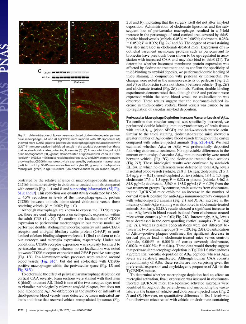
onstrated by the relative absence of macrophage-specific markerCD163 immunoreactivity in clodronate-treated animals comparedwith controls [Fig. 1 A and B and supporting information (SI) Fig.S1 A and B]. This reduction was quantitatively confirmed by a 56%� 4.5% reduction in levels of the macrophage-specific proteinCD206 between animals administered clodronate versus thosereceiving vehicle (P � 0.002; Fig. 1C).
Although macrophages are known to express the CD206 recep-tor, there are conflicting reports on cell-specific expression withinthe adult CNS (11, 20). To confirm the localization of CD206expression to perivascular macrophages in our mouse model, weperformed double labeling immunocytochemistry with anti-CD206receptor and anti-glial fibrillary acidic protein (GFAP) or anti-ionized calcium-binding adaptor molecule-1 (Iba1) antisera to ruleout astrocyte and microglia expression, respectively. Under ourconditions, CD206 receptor expression was expressly localized toperivascular macrophages, whereas no co-localization was notedbetween CD206 receptor expression and GFAP-positive astrocytes(Fig. 1D). Iba-1-immunoreactive processes were stained aroundblood vessels (Fig. S1C), but did not co-localize with CD206-positive macrophages within the perivascular space (Fig. 1E andFig. S1D).
To determine the effect of perivascular macrophage depletion oncortical CAA severity, brain sections were stained with thioflavinS (thioS) to detect A�. ThioS is one of the two accepted dyes usedto visualize pathologically relevant amyloid plaques, but does notbind monomeric A�. No differences in the number or intensity ofthioS-positive blood vessels were detected between untreated an-imals and those that received vehicle-encapsulated liposomes (Fig.
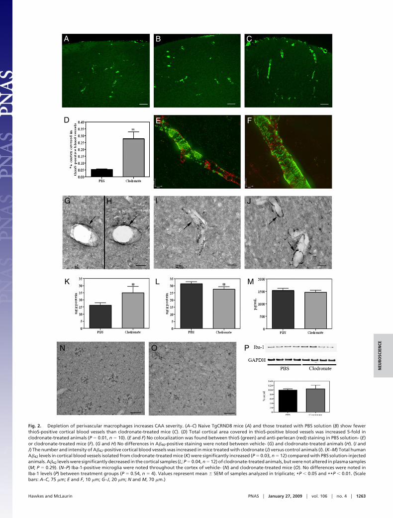
2 A and B), indicating that the surgery itself did not alter amyloiddeposition. Administration of clodronate liposomes and the sub-sequent loss of perivascular macrophages resulted in a 5-foldincrease in the percentage of total cortical area covered by thioS-positive blood vessels (vehicle, 0.05% � 0.005%; clodronate, 0.28%� 0.05%; P � 0.009; Fig. 2 C and D). The degree of vessel stainingwas also increased in clodronate-treated mice. Expression of en-dothelial basement membrane proteins such as perlecan and fi-bronectin have previously been shown to be up-regulated in asso-ciation with increased CAA and may also bind to thioS (21). Todetermine whether basement membrane protein expression wasaffected by clodronate treatment and to confirm the specificity ofthioS binding to amyloid deposits, we performed double labeling ofthioS staining in conjunction with perlecan or fibronectin. Nochanges were noted in the immunoreactivity of perlecan (Fig. 2 Eand F) or fibronectin (data not shown) between vehicle- (Fig. 2E)and clodronate-treated (Fig. 2F) animals. Further, double labelingexperiments demonstrated that, although thioS and perlecan wereexpressed within the same blood vessel, no co-localization wasobserved. These results suggest that the clodronate-induced in-crease in thioS-positive cortical blood vessels was caused by anup-regulation of vascular amyloid deposition.
Perivascular Macrophage Depletion Increases Vascular Levels of A�42.To confirm that vascular amyloid was specifically increased, weperformed double labeling immunocytochemistry of brain tissueswith anti-A�8–17 (clone 6F/3D) and anti-�-smooth muscle actin.Similar to the thioS staining, clodronate-treated mice showed agreater number of A�-positive blood vessels throughout the cortexcompared with vehicle-injected animals (Fig. S2 A–D). We nextexamined whether A�40 or A�42 was preferentially depositedfollowing clodronate treatment. No appreciable differences in theamount or intensity of vascular A�40 immunoreactivity were notedbetween vehicle- (Fig. 2G) and clodronate-treated tissue sections(Fig. 2H). These histological results were confirmed by sandwichELISA, in which no differences were detected in total A�40 levelsin isolated blood vessels (vehicle, 23.8 � 1.6 ng/g; clodronate, 21.5 �2.4 ng/g; P � 0.21), vessel-depleted cortex (vehicle, 18.4 � 1.0 ng/g;clodronate 17.6 � 1.5 ng/g; P � 0.32) or plasma (vehicle, 3,462 �84.8 pg/mL; clodronate, 3,569 � 185.8 pg/mL; P � 0.29) from thetwo treatment groups. By contrast, brain sections from clodronate-treated TgCRND8 mice exhibited an increase in the number ofcortical vessels positive for anti-A�42 immunoreactivity comparedwith vehicle-injected animals (Fig. 2 I and J). An increase in theintensity of anti-A�42 staining was also noted in clodronate-treatedanimals. Similarly, ELISA results showed a significant increase intotal A�42 levels in blood vessels isolated from clodronate-treatedmice versus controls (P � 0.03; Fig. 2K). Interestingly, A�42 levelswere decreased in the corresponding cortical samples (P � 0.04;Fig. 2L), whereas plasma concentrations were not different be-tween the two treatment groups (P � 0.29; Fig. 2M). Quantificationof A�8–17-positive plaques confirmed the significant decrease incortical plaque load in clodronate-treated mice versus controls(vehicle, 0.004% � 0.001% of cortex covered; clodronate,0.002% � 0.0003%; P � 0.04). These data would thereby suggestthat perivascular macrophage depletion in TgCRND8 mice inducesa preferential vascular deposition of A�42 peptides, whereas A�40levels are relatively unaffected. Although human CAA consistspredominantly of A�40, these results are not surprising given thepreferential expression and amyloidogenic properties of A�42 in theTgCRND8 mouse.
To determine whether macrophage depletion had an effect onmicroglial activation, Iba-1 expression was assessed in clodronate-injected TgCRND8 mice. Iba-1-positive activated microglia wereidentified throughout the parenchyma and surrounding the vascu-lature in the brains of vehicle- and clodronate-injected mice (Fig. 2N and O). However, no quantitative difference in Iba-1 levels wasfound between mice treated with vehicle- or clodronate-containing
A B
C
D E
Fig. 1. Administration of liposome-encapsulated clodronate depletes perivas-cular macrophages. (A and B) TgCRND8 mice injected with PBS liposomes (A)showed more CD163-positive perivascular macrophages (green) associated withGLUT-1- immunoreactive (red) blood vessels in the caudate putamen than thosethat received clodronate-containing liposomes (B). (C) Immunoblotting of rightbrain homogenates (30 �g/lane) demonstrated a significant reduction in CD206levels (P � 0.002, n � 5) in mice receiving clodronate. (D and E) Photomicrographsshowing that CD206 immunoreactivity is expressed by perivascular macrophages(red) but not by GFAP-immunoreactive astrocytes (D, green) or Iba1-positivemicroglia (E,green) inTgCRND8mice. (Scalebars:AandB, 10�m;DandE, 20�m.)
1262 � www.pnas.org�cgi�doi�10.1073�pnas.0805453106 Hawkes and McLaurin
A B C
D E F
G I J
K L M
N O P
H
Fig. 2. Depletion of perivascular macrophages increases CAA severity. (A–C) Naive TgCRND8 mice (A) and those treated with PBS solution (B) show fewerthioS-positive cortical blood vessels than clodronate-treated mice (C). (D) Total cortical area covered in thioS-positive blood vessels was increased 5-fold inclodronate-treated animals (P � 0.01, n � 10). (E and F) No colocalization was found between thioS (green) and anti-perlecan (red) staining in PBS solution- (E)or clodronate-treated mice (F). (G and H) No differences in A�40-positive staining were noted between vehicle- (G) and clodronate-treated animals (H). (I andJ) The number and intensity of A�42-positive cortical blood vessels was increased in mice treated with clodronate (J) versus control animals (I). (K–M) Total humanA�42 levels in cortical blood vessels isolated from clodronate-treated mice (K) were significantly increased (P � 0.03, n � 12) compared with PBS solution-injectedanimals. A�42 levels were significantly decreased in the cortical samples (L; P � 0.04, n � 12) of clodronate-treated animals, but were not altered in plasma samples(M; P � 0.29). (N–P) Iba-1-positive microglia were noted throughout the cortex of vehicle- (N) and clodronate-treated mice (O). No differences were noted inIba-1 levels (P) between treatment groups (P � 0.54, n � 4). Values represent mean � SEM of samples analyzed in triplicate; *P � 0.05 and **P � 0.01. (Scalebars: A–C, 75 �m; E and F, 10 �m; G–J, 20 �m; N and M, 70 �m.)
Hawkes and McLaurin PNAS � January 27, 2009 � vol. 106 � no. 4 � 1263
NEU
ROSC
IEN
CE
liposomes (Fig. 2P; P � 0.54). These data indicate that perivascularmacrophage depletion did not significantly alter microglial activity.
Increases in CAA severity have been associated with increases inmicro-hemorrhages in human studies and animal models of AD (9,22). To investigate the impact of increased A�42 deposition oncerebral ‘‘microbleeds,’’ we examined brain tissues for the hemo-globin breakdown product hemosiderin by Prussian blue staining(9). Despite the significant increase in vascular amyloid followingclodronate treatment, we were unable to detect Prussian bluelabeling of blood vessels in vehicle- or clodronate-treated animals.Staining was evident only along the tract mark at the site of injectionand could be distinctly identified within microglial cells (Fig. S2 Eand F). As such, no differences in Prussian blue staining were notedbetween clodronate- and vehicle-injected animals, suggesting thatincreased CAA did not compromise vascular integrity.
Chitin Administration Stimulates Turnover of Perivascular Macro-phages. Given our findings that perivascular macrophage depletionincreased CAA severity, we examined the effects of perivascularmacrophage turnover on vascular amyloid load. At 5 months of age,TgCRND8 mice deposit significant CAA and thus allowed fordetection of treatment-induced changes. To stimulate perivascularmacrophage turnover, we used chitin, a naturally occurring biopoly-mer of N-acetyl-�-D-glucosamine expressed in the cell walls offungi, crustaceans, insects, and worms (chitin, n � 21; vehicle, n �20). Chitin uptake in peripheral macrophages is believed to occurvia binding to the CD206 receptor (23).
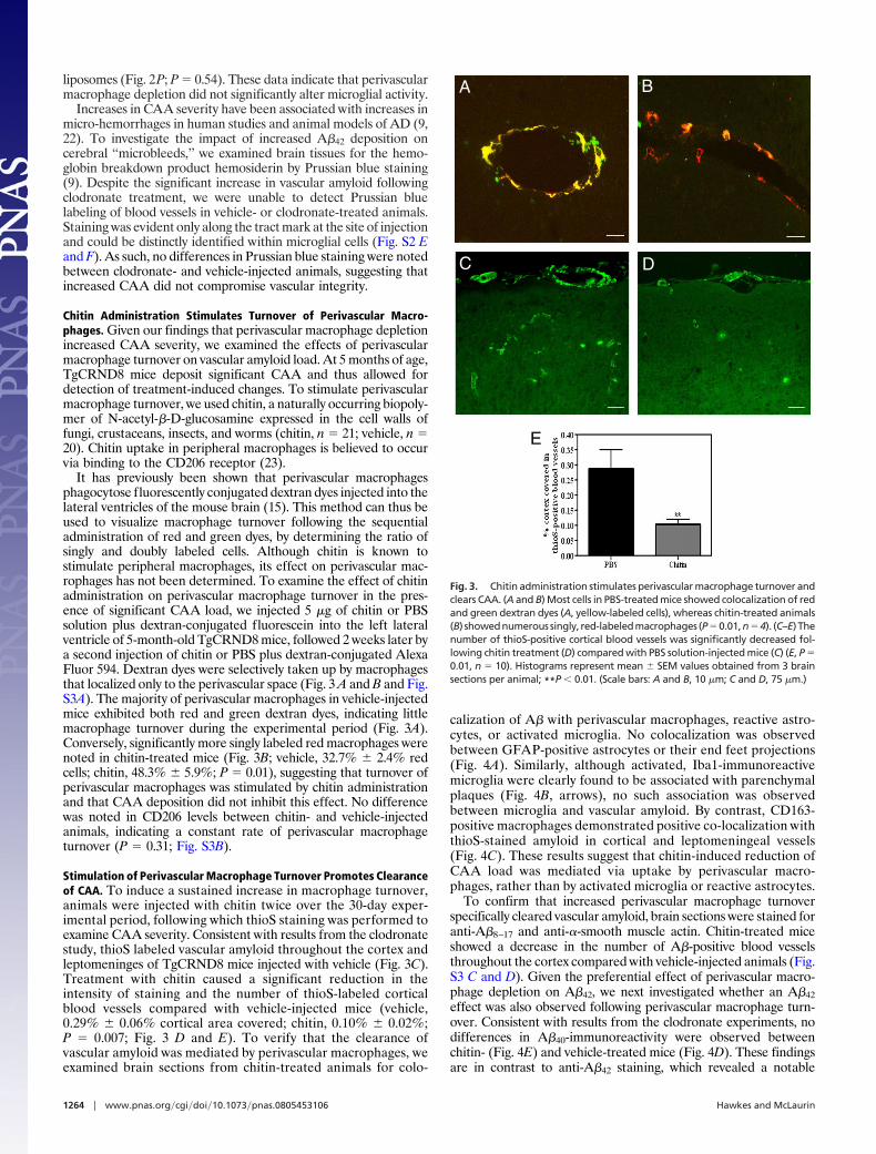
It has previously been shown that perivascular macrophagesphagocytose fluorescently conjugated dextran dyes injected into thelateral ventricles of the mouse brain (15). This method can thus beused to visualize macrophage turnover following the sequentialadministration of red and green dyes, by determining the ratio ofsingly and doubly labeled cells. Although chitin is known tostimulate peripheral macrophages, its effect on perivascular mac-rophages has not been determined. To examine the effect of chitinadministration on perivascular macrophage turnover in the pres-ence of significant CAA load, we injected 5 �g of chitin or PBSsolution plus dextran-conjugated fluorescein into the left lateralventricle of 5-month-old TgCRND8 mice, followed 2 weeks later bya second injection of chitin or PBS plus dextran-conjugated AlexaFluor 594. Dextran dyes were selectively taken up by macrophagesthat localized only to the perivascular space (Fig. 3 A and B and Fig.S3A). The majority of perivascular macrophages in vehicle-injectedmice exhibited both red and green dextran dyes, indicating littlemacrophage turnover during the experimental period (Fig. 3A).Conversely, significantly more singly labeled red macrophages werenoted in chitin-treated mice (Fig. 3B; vehicle, 32.7% � 2.4% redcells; chitin, 48.3% � 5.9%; P � 0.01), suggesting that turnover ofperivascular macrophages was stimulated by chitin administrationand that CAA deposition did not inhibit this effect. No differencewas noted in CD206 levels between chitin- and vehicle-injectedanimals, indicating a constant rate of perivascular macrophageturnover (P � 0.31; Fig. S3B).
Stimulation of Perivascular Macrophage Turnover Promotes Clearanceof CAA. To induce a sustained increase in macrophage turnover,animals were injected with chitin twice over the 30-day exper-imental period, following which thioS staining was performed toexamine CAA severity. Consistent with results from the clodronatestudy, thioS labeled vascular amyloid throughout the cortex andleptomeninges of TgCRND8 mice injected with vehicle (Fig. 3C).Treatment with chitin caused a significant reduction in theintensity of staining and the number of thioS-labeled corticalblood vessels compared with vehicle-injected mice (vehicle,0.29% � 0.06% cortical area covered; chitin, 0.10% � 0.02%;P � 0.007; Fig. 3 D and E). To verify that the clearance ofvascular amyloid was mediated by perivascular macrophages, weexamined brain sections from chitin-treated animals for colo-
calization of A� with perivascular macrophages, reactive astro-cytes, or activated microglia. No colocalization was observedbetween GFAP-positive astrocytes or their end feet projections(Fig. 4A). Similarly, although activated, Iba1-immunoreactivemicroglia were clearly found to be associated with parenchymalplaques (Fig. 4B, arrows), no such association was observedbetween microglia and vascular amyloid. By contrast, CD163-positive macrophages demonstrated positive co-localization withthioS-stained amyloid in cortical and leptomeningeal vessels(Fig. 4C). These results suggest that chitin-induced reduction ofCAA load was mediated via uptake by perivascular macro-phages, rather than by activated microglia or reactive astrocytes.
To confirm that increased perivascular macrophage turnoverspecifically cleared vascular amyloid, brain sections were stained foranti-A�8–17 and anti-�-smooth muscle actin. Chitin-treated miceshowed a decrease in the number of A�-positive blood vesselsthroughout the cortex compared with vehicle-injected animals (Fig.S3 C and D). Given the preferential effect of perivascular macro-phage depletion on A�42, we next investigated whether an A�42effect was also observed following perivascular macrophage turn-over. Consistent with results from the clodronate experiments, nodifferences in A�40-immunoreactivity were observed betweenchitin- (Fig. 4E) and vehicle-treated mice (Fig. 4D). These findingsare in contrast to anti-A�42 staining, which revealed a notable
A B
C D
E
Fig. 3. Chitin administration stimulates perivascular macrophage turnover andclears CAA. (A and B) Most cells in PBS-treated mice showed colocalization of redand green dextran dyes (A, yellow-labeled cells), whereas chitin-treated animals(B) showednumerous singly, red-labeledmacrophages (P�0.01,n�4). (C–E)Thenumber of thioS-positive cortical blood vessels was significantly decreased fol-lowing chitin treatment (D) compared with PBS solution-injected mice (C) (E, P �0.01, n � 10). Histograms represent mean � SEM values obtained from 3 brainsections per animal; **P � 0.01. (Scale bars: A and B, 10 �m; C and D, 75 �m.)
1264 � www.pnas.org�cgi�doi�10.1073�pnas.0805453106 Hawkes and McLaurin
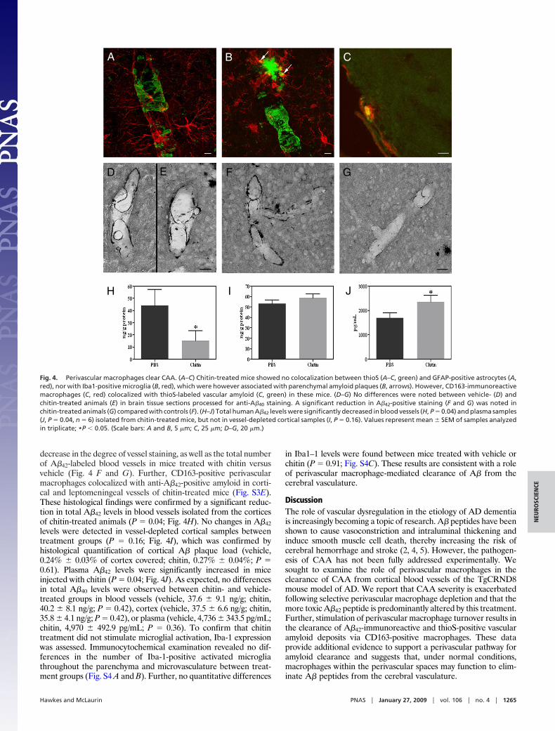
decrease in the degree of vessel staining, as well as the total numberof A�42-labeled blood vessels in mice treated with chitin versusvehicle (Fig. 4 F and G). Further, CD163-positive perivascularmacrophages colocalized with anti-A�42-positive amyloid in corti-cal and leptomeningeal vessels of chitin-treated mice (Fig. S3E).These histological findings were confirmed by a significant reduc-tion in total A�42 levels in blood vessels isolated from the corticesof chitin-treated animals (P � 0.04; Fig. 4H). No changes in A�42levels were detected in vessel-depleted cortical samples betweentreatment groups (P � 0.16; Fig. 4I), which was confirmed byhistological quantification of cortical A� plaque load (vehicle,0.24% � 0.03% of cortex covered; chitin, 0.27% � 0.04%; P �0.61). Plasma A�42 levels were significantly increased in miceinjected with chitin (P � 0.04; Fig. 4J). As expected, no differencesin total A�40 levels were observed between chitin- and vehicle-treated groups in blood vessels (vehicle, 37.6 � 9.1 ng/g; chitin,40.2 � 8.1 ng/g; P � 0.42), cortex (vehicle, 37.5 � 6.6 ng/g; chitin,35.8 � 4.1 ng/g; P � 0.42), or plasma (vehicle, 4,736 � 343.5 pg/mL;chitin, 4,970 � 492.9 pg/mL; P � 0.36). To confirm that chitintreatment did not stimulate microglial activation, Iba-1 expressionwas assessed. Immunocytochemical examination revealed no dif-ferences in the number of Iba-1-positive activated microgliathroughout the parenchyma and microvasculature between treat-ment groups (Fig. S4 A and B). Further, no quantitative differences
in Iba1–1 levels were found between mice treated with vehicle orchitin (P � 0.91; Fig. S4C). These results are consistent with a roleof perivascular macrophage-mediated clearance of A� from thecerebral vasculature.
DiscussionThe role of vascular dysregulation in the etiology of AD dementiais increasingly becoming a topic of research. A� peptides have beenshown to cause vasoconstriction and intraluminal thickening andinduce smooth muscle cell death, thereby increasing the risk ofcerebral hemorrhage and stroke (2, 4, 5). However, the pathogen-esis of CAA has not been fully addressed experimentally. Wesought to examine the role of perivascular macrophages in theclearance of CAA from cortical blood vessels of the TgCRND8mouse model of AD. We report that CAA severity is exacerbatedfollowing selective perivascular macrophage depletion and that themore toxic A�42 peptide is predominantly altered by this treatment.Further, stimulation of perivascular macrophage turnover results inthe clearance of A�42-immunoreactive and thioS-positive vascularamyloid deposits via CD163-positive macrophages. These dataprovide additional evidence to support a perivascular pathway foramyloid clearance and suggests that, under normal conditions,macrophages within the perivascular spaces may function to elim-inate A� peptides from the cerebral vasculature.
A B C
D F G
H I J
E
Fig. 4. Perivascular macrophages clear CAA. (A–C) Chitin-treated mice showed no colocalization between thioS (A–C, green) and GFAP-positive astrocytes (A,red), nor with Iba1-positive microglia (B, red), which were however associated with parenchymal amyloid plaques (B, arrows). However, CD163-immunoreactivemacrophages (C, red) colocalized with thioS-labeled vascular amyloid (C, green) in these mice. (D–G) No differences were noted between vehicle- (D) andchitin-treated animals (E) in brain tissue sections processed for anti-A�40 staining. A significant reduction in A�42-positive staining (F and G) was noted inchitin-treated animals (G) compared with controls (F). (H–J) Total human A�42 levels were significantly decreased in blood vessels (H, P � 0.04) and plasma samples(J, P � 0.04, n � 6) isolated from chitin-treated mice, but not in vessel-depleted cortical samples (I, P � 0.16). Values represent mean � SEM of samples analyzedin triplicate; *P � 0.05. (Scale bars: A and B, 5 �m; C, 25 �m; D–G, 20 �m.)
Hawkes and McLaurin PNAS � January 27, 2009 � vol. 106 � no. 4 � 1265
NEU
ROSC
IEN
CE
Our data suggests that uptake and removal of vascular amyloidby perivascular macrophages is ongoing under physiologic condi-tions in TgCRND8 mice and that disruption of this process exac-erbates CAA severity. This is supported by the findings that (i)dextran dye-positive cells were identified in only cells with amacrophage morphology within the perivascular space and not inthe parenchyma, indicating that macrophages did not migrate intothe neuropil; (ii) CAA levels were reduced/exacerbated throughoutall cortical areas, both ipsilateral and contralateral to the site ofinjection, suggesting that CAA severity was not mediated byactivation or inhibition of local resident microglia; (iii) chitinadministration had no effect on parenchymal plaque load or totalA� levels in vessel-depleted cortical samples; and (iv) thioS- andA�42-positive vascular amyloid co-localized with CD163-positivemacrophages, but not with Iba1-positive microglia or GFAP-positive astrocytes. However, given the complex interplay among allimmune cells, a possible cooperative action between perivascularmacrophages and microglia or astrocytes in the regulation of CAAcannot be completely discounted.
At present, there are no therapeutic strategies for the manage-ment of CAA. However, the importance of CAA and its interre-lationship with parenchymal amyloid was underscored recently byreports from animal and human anti-A� immunization studies, inwhich reductions in parenchymal plaque load were associated withsignificant increases in CAA severity and brain micro-hemorrhagesin amyloid-laden vessels (9). We did not find an increase in cerebralbleeding incidence associated with clodronate treatment in thepresent experiment, as a result of the age of our animals and therelatively short duration of treatment; however, CAA-associatedmicro-hemorrhages remain a major therapeutic concern. Althoughmuch more work is needed to characterize the inflammatoryresponse of perivascular macrophage stimulation in the AD brain,in the present study, chitin administration was well tolerated byTgCRND8 mice expressing moderate CAA pathology and nodeleterious side effects were noted following stimulation of perivas-cular macrophage turnover. Thus, the current findings suggest thatperivascular macrophages might be a potential target for therapeu-tic intervention in the clearance of CAA.
MethodsAnimals. TgCRND8 mice overexpressing the human Swedish (KM670/671NL) andIndianan (V717F) APP mutations under a hamster prion protein promoter weremaintained on an outbred C3H/C57BL6 background (17). Mice were age- andsex-matched and allowed food and water ad libitum. All experiments wereperformed in accordance with the guidelines stipulated by the University ofToronto and Canadian Council for Animal Care.
Depletion of Perivascular Macrophages. Perivascular macrophage depletion wascarried out according to the protocol adapted by Polfliet et al. (19) (see SIMethods). Four-month-old TgCRND8 mice were anesthetized with isofluraneand stereotaxically injected with 10 �L of PBS solution- or clodronate-containingliposomes into the left lateral ventricle (coordinates from Bregma: anteroposte-rior, �0.2 mm; mediolateral, 1.2 mm; dorsoventral, 2.3 mm; n � 30 per group).Animals were killed 1 month later and brains were processed for further analysis.
Stimulation of Perivascular Macrophage Turnover. Five-month-old TgCRND8mice received a 5-�L injection of either PBS solution or chitin (1 mg/mL) into theleft lateralventricle,whichwasrepeated14days later (n�20pergroup).Asubsetof animals received a combination of chitin or PBS solution plus 5 �L dextranfluorescein dye, followed 2 weeks later by an injection of chitin or PBS solutionplus 5 �L dextran Alexa Fluor 594 dye (n � 4 per group). Animals were killed 14days after the last injection and brains were processed for further analysis.
Immunocytochemistry. Mice were deeply anesthetized with sodium pentobar-bital and perfused with 0.1 M PBS solution (pH 7.4) and 10% formalin or snap-frozen. Sections (20 �m) were incubated overnight with anti-CD163 (1:500),anti-Iba-1 (1:1,000), anti-A�8–17 (1:500), or anti-A�40/42 (1:70) and developed byusing the glucose oxidase-DAB-nickel enhancement method (see SI Methods forantibody sources). For thioS staining, sections were treated with 1% thioS for 5min, differentiated twice in 70% EtOH, and washed in PBS solution. For thioS plusGFAP/Iba1/CD163/perlecan double labeling, sections were incubated overnightwith anti-GFAP (1:250), anti-Iba1 (1:250), anti-CD163 (1:100), or anti-perlecan(1:100); exposed to Alex Fluor 594-conjugated anti-rabbit/mouse (1:200); andthen processed for thioS staining (see SI Methods for additional staining proce-dures). Photomicrographs of visualized sections were captured by using a ZeissAxioscope 2 Plus microscope and exported to Adobe Photoshop CS.
Immunoblotting. Right brain hemisphere homogenates were separated by 4%–20% polyacrylamide gel electrophoresis (30 �g/lane), transferred to nitrocellu-lose membrane, and incubated overnight at 4°C with anti-CD163 (1:1,000), anti-CD206 (1:1,000), or anti-Iba1 (1:750) antibodies. Membranes were stripped, andre-probed with anti-GAPDH (1:10,000) to ensure equal protein loading.
Cortical Blood Vessel Isolation and A� ELISA Sample Preparation. Isolation ofcortical blood vessels was adapted from Patton et al. (22) (see SI Methods). TotalA�wasextractedfromcorticalfiltratesbysonicatinghomogenates in70%formicacid, followed by centrifugation (100,000 � g, 1 h, 4 °C) and neutralization of thesamples. Isolated blood vessels were washed, centrifuged (6,000 �g, 10 min, 4 °C)to remove supernatant, and treated similarly as cortical samples to extract totalA�40/42. Neutralized samples were diluted and analyzed by using commerciallyavailable sandwich ELISA kits, as per the manufacturer’s instructions (BioSource,Burlington, ON, Canada). Plasma samples were also assessed for A�40/42 levels.Samples were measured in triplicate, with mean values � SEM. reported fortreatment groups.
ACKNOWLEDGMENTS. The authors thank Mary Brown, Kevin DaSilva, DanielaFenili, and Dr. Teresa DeLuca for their technical assistance. This work was sup-portedbygrants fromtheCanadian Institutes forHealthResearch (C.H., J.M.), theNaturalSciencesandEngineeringResearchCouncil (J.M.), theAlzheimer’sSocietyof Canada (C.H.), and the Ontario Alzheimer’s Society (J.M.).
1. Attems J, Lauda F, Jellinger KA (2008) Unexpectedly low prevalence of intracerebral hemor-rhages in sporadic cerebral amyloid angiopathy: an autopsy study. J Neurol 255:70–76.
2. Miao J, et al. (2005) Cerebral microvascular amyloid beta protein deposition inducesvascular degeneration and neuroinflammation in transgenic mice expressing humanvasculotropic mutant amyloid beta precursor protein. Am J Pathol 167:505–515.
3. Shin HK, et al. (2007) Age-dependent cerebrovascular dysfunction in a transgenic mousemodel of cerebral amyloid angiopathy. Brain 130:2310–2319.
4. Perlmutter LS (1994) Microvascular pathology and vascular basement membrane compo-nents in Alzheimer’s disease. Mol Neurobiol 9:33–40.
5. Tian J, et al. (2006) Relationships in Alzheimer’s disease between the extent of Abeta depo-sition in cerebral blood vessel walls, as cerebral amyloid angiopathy, and the amount ofcerebrovascular smooth muscle cells and collagen. Neuropathol Appl Neurobiol 32:332–340.
6. Attems J, Quass M, Jellinger KA, Lintner F (2007) Topographical distribution of cerebralamyloid angiopathy and its effect on cognitive decline are influenced by Alzheimerdisease pathology. J Neurol Sci 257:49–55.
7. Nicoll JA, et al. (2004) Cerebral amyloid angiopathy plays a direct role in the pathogenesisof Alzheimer’s disease. Pro-CAA position statement. Neurobiol Aging 25:589–597.
8. Carare RO, et al. (2008) Solutes, but not cells, drain from the brain parenchyma alongbasement membranes of capillaries and arteries: Significance for cerebral amyloid angi-opathy and neuroimmunology. Neuropathol Appl Neurobiol 34:131–144.
9. Pfeifer M, et al. (2002) Cerebral hemorrhage after passive anti-A� immunotherapy.Science 298:1379.
10. Bechmann I, Galea I, Perry VH (2007) What is the blood-brain barrier (not)? TrendsImmunol 28:5–11.
11. Galea I, et al. (2005) Mannose receptor expression specifically reveals perivascular macro-phages in normal, injured, and diseased mouse brain. Glia 49:375–384.
12. Kim WK, et al. (2006) CD163 identifies perivascular macrophages in normal and viralencephalitic brains and potential precursors to perivascular macrophages in blood. Am JPathol 168:822–834.
13. Bechmann I, et al. (2001) Turnover of rat brain perivascular cells. Exp Neurol 168:242–249.
14. Williams K, Alvarez X, Lackner AA (2001) Central nervous system perivascular cells areimmunoregulatory cells that connect the CNS with the peripheral immune system. Glia36:156–164.
15. Bechmann I, et al. (2001) Immune surveillance of mouse brain perivascular spaces byblood-borne macrophages. Eur J Neurosci 14:1651–1658.
16. Fiala M, et al. (2005) Ineffective phagocytosis of amyloid-beta by macrophages of Alzhei-mer’s disease patients. J Alzheimers Dis 7:221–232.
17. ChishtiMA,etal. (2001)Early-onsetamyloiddepositionandcognitivedeficits in transgenicmice expressing a double mutant form of amyloid precursor protein 695. J Biol Chem276:21562–21570.
18. Van Rooijen N, Sanders A (1994) Liposome mediated depletion of macrophages:Mechanism of action, preparation of liposomes and applications. J Immunol Methods174:83–93.
19. Polfliet MM, et al. (2001) A method for the selective depletion of perivascular andmeningeal macrophages in the central nervous system. J Neuroimmunol 116:188–195.
20. Burudi EM, Regnier-Vigouroux A (2001) Regional and cellular expression of the mannosereceptor in the post-natal developing mouse brain. Cell Tissue Res 303:307–317.
21. Wyss-Coray T, et al. (2000) Chronic overproduction of transforming growth factor-beta1by astrocytes promotes Alzheimer’s disease-like microvascular degeneration in transgenicmice. Am J Pathol 156:139–150.
22. Patton RL, et al. (2006) Amyloid-beta peptide remnants in AN-1792-immunized Alzhei-mer’s disease patients: A biochemical analysis. Am J Pathol 169:1048–1063.
1266 � www.pnas.org�cgi�doi�10.1073�pnas.0805453106 Hawkes and McLaurin


![Thomas Haider JC Lab 20141020.ppt [Kompatibilitätsmodus] · Vienna, 2014 Blood-Brain Barrier Background Muldoon et al. (2013) Neurons Pericytes Astrocytes Perivascular Macrophages](https://img.pdfslide.net/doc/110x75/5b0543897f8b9a0a548e9fd9/thomas-haider-jc-lab-kompatibilittsmodus-2014-blood-brain-barrier-background-muldoon.jpg)