Embed Size (px)
Citation preview
Biological Report 85(1.5) Contaminant Hazard Reviews October 1985 Report No. 5
SELENIUM HAZARDS TO FISH, WILDLIFE, AND INVERTEBRATES: A SYNOPTIC REVIEW
By
Ronald Eisler
Patuxent Wildlife Research Center U.S. Fish and Wildlife Service
Laurel, MD 20708
SUMMARY
Ecological and toxicological aspects of selenium (Se) in the environment are reviewed, including its chemistry, background residues in biological and other materials, and toxic, sublethal, and latent effects (including the effects of Se deficiency). Recommendations are presented, including proposed Se criteria for protection of sensitive species of fish and wildlife.
Most authorities agree on five points. First, Se deficiency is not as well documented as Se poisoning, but may be equally significant. Second, Se released as a result of anthropogenic activities (including fossil fuel combustion and metal smelting), as well as that in naturally seleniferous areas, poses the greatest threat of poisoning to fish and wildlife. Third, additional research is required on chemical and biological transformations among valence states, allotropic forms, and isomers of Se. Fourth, Se metabolism and degradation are both significantly modified by interaction with various heavy metals, agricultural chemicals, microorganisms, and numerous physicochemical factors, and until these interactions are resolved it will be difficult to meaningfully interpret Se residues in various tissues. And fifth, documented biological responses to Se deficiency or to selenosis vary widely, even among closely related taxonomic groups.
It is generally agreed that Se deficiency may be prevented in fish, small laboratory mammals, and livestock by feeding diets containing 50 to 100 ppb of Se. The concentration range of total inorganic selenite currently recommended for aquatic life protection--35 ppb in freshwater to 54 ppb in marine waters--is below the range of 60 to 600 ppb that is fatal to sensitive aquatic species. In freshwater, it is also below the range of 47 to 53 ppb associated with growth inhibition of freshwater algae, anemia and reduced hatching in trout, and shifts in species composition of freshwater algae communities. Accordingly, current recommendations for Se with respect to aquatic life appear to afford an adequate measure of protection. However, some studies have shown that Se water concentrations of 9 to 12 ppb are associated with inhibited reproduction of certain freshwater teleosts, suggesting that the current Se criterion for protection of freshwater life should be revised downward. Also, high bioconcentration and accumulation of Se from water by numerous species of algae, fish, and invertebrates is well documented at levels of 0.015 to 3.3 ppb, which are substantially below the recommended range of 35 to 54 ppb. The significance of Se residues in aquatic biota is still unclear, and more research appears to be needed on Se pharmacokinetics in aquatic environments.
Aerosol concentrations exceeding 4.0 ug Se/m3 are considered potentially harmful to human health; however, no comparable data base for birds and other wildlife species is available at this time.
Selenium in natural foods is less toxic than are purified forms of Se. The consensus is that Se toxicity is prevented in livestock if dietary Se concentrations do not exceed 5,000 ppb in natural forage, or 2,000 ppb in feeds supplemented with purified Se. Minimum toxic concentrations of Se in the rat (a sensitive species), fed diets containing natural Se were 1,400 ppb of Se as judged by evidence of liver changes, and 3,000 ppb as estimated from longevity and histopathology; these values were only 350 and 750 ppb, respectively, when diets low in natural Se were fortified with purified Se. The evidence is incomplete for migratory waterfowl and other birds, but diets containing more than 5,000 ppb of Se are demonstrably harmful.
This report should be cited as:
Eisler R. 1985. Selenium hazards to fish, wildlife and invertebrates: a synoptic review. U.S. Fish and Wildlife Service Biological Report 85 (1.5).
2
SUMMARY INTRODUCTION ENVIRONMENTAL CHEMISTRY BACKGROUND CONCENTRATIONS DEFICIENCY AND PROTECTIVE EFFECTS TOXICITY MAMMALS AND BIRDS AQUATIC ORGANISMS SUBLETHAL AND LATENT EFFECTS AQUATIC ORGANISMS TERRESTRIAL INVERTEBRATES BIRDS MAMMALS CURRENT RECOMMENDATIONS LITERATURE CITED
TABLES Number
1 Selenium concentrations in nonbiological materials. 2 Selenium concentrations in field populations of selected species of flora and fauna. Values shown
are in total Se (mg/kg, or ppm) fresh weight (FW), dry weight (DW), or ash weight (AW). Hyphenated numbers show range and single numbers the mean; where both appear, the range is in parentheses.
3 Toxicity of selenium salts to aquatic biota. Values shown are in ug/l (ppb) in medium fatal to 50% of the organisms during exposure for various intervals.
4 Proposed selenium criteria for prevention of Se deficiency and for protection against selenosis.
ACKNOWLEDGMENTS I thank L. Garrett, M. Shawe and B. Graff for library services; I. Moore for secretarial help; J. Jacknow, D.J. Hoffman, H.M. Ohlendorf, and T.P. Lowe for reviewing the manuscript; and P.H. Eschmeyer and J.R. Zuboy for editorial services.
3
INTRODUCTION Selenium (Se) was first identified as an element in 1817 by the Swedish chemist Berzelius. It is now firmly
established that Se is beneficial or essential in amounts from trace to part-per-billion concentrations for humans and some plants and animals, but toxic at some concentrations present in the environment. Selenium deficiency was recently reported among cattle grazing in the Florida Everglades, which showed evidence of anemia, slow growth, and reduced fertility (Morris et al. 1984). Conversely, calves of Indian buffaloes died of Se poisoning after eating rice husks grown in naturally seleniferous soils (Prasad et al. 1982); severe reproductive and developmental abnormalities were observed in aquatic birds nesting at Se-contaminated irrigation drainwater ponds in the San Joaquin Valley, California (Ohlendorf et al. 1986); and fish contained high body burdens of Se and failed to reproduce in a lake receiving dissolved Se by way of a power plant fly ash sluice water return (Cumbie and Van Horn 1978). Selenium poisoning is an ancient and well-documented disease (Rosenfeld and Beath 1964). Signs of it were reported among domestic livestock by Marco Polo in western China near the borders of Turkestan and Tibet in about the year 1295; among livestock, chickens, and children in Columbia, South America, by Father Pedro Simon in 1560; among human adults in Irapuato, Mexico, in about 1764; and among horses of the U.S. Cavalry in South Dakota in 1857 and again in 1893 (Rosenfeld and Beath 1964). In 1907-08, more than 15,000 sheep died in a region north of Medicine Bow, Wyoming, after grazing on seleniferous plants. The incidents have continued, and the recent technical literature abounds with isolated examples of selenosis among domestic animals.
Selenium has been the subject of many reviews (Rosenfeld and Beath 1964; Frost 1972; Sandholm 1973; Zingaro and Cooper 1974; Frost and Ingvoldstad 1975; Anon. 1975; NAS 1976; Harr 1978; EPA 1980; Lo and Sandi 1980; Shamberger 1981; Wilber 1980, 1983; Fishbein 1977, 1983; NRC 1983; Reddy and Massaro 1983). These authorities agree that Se is widely distributed in nature, being especially abundant with sulfide minerals of various metals, such as iron, lead, and copper. The major source of environmental Se is the weathering of natural rock. The amount of Se entering the atmosphere as a result of anthropogenic activities is estimated to be 3,500 metric tons annually, of which most is attributed to combustion of coal; however, aside from highly localized contamination, the contribution of Se by man's activities is small in comparison with that attributable to natural sources. Collectively, all authorities agree that Se may favorably or adversely affect growth, survival, and reproduction of algae and higher plants, bacteria and yeasts, crustaceans, molluscs, insects, fish, birds, and mammals (including humans). Most acknowledge that sensitivity to Se and its compounds is extremely variable in all classes of organisms and, except for some instances of Se deficiency or of selenosis, metabolic pathways and modes of action are imperfectly understood. For example, selenium “indicator” plants can accumulate Se to concentrations of thousands of parts per million without ill effects; in these plants Se promotes growth, whereas in crop plants accumulations as low as 25 to 50 ppm may be toxic. Thus, plants and waters high in Se are considered potentially hazardous to livestock and to aquatic life and other natural resources in seleniferous zones.
In this account I briefly review the technical literature on ecological and toxicological aspects of selenium (with emphasis on fish and wildlife and their predators and prey), and provide current recommendations for the protection of sensitive species of concern to the U.S. Fish and Wildlife Service. This is part of a continuing series of synoptic reviews prepared in response to informational requests from environmental specialists of the Service.
ENVIRONMENTAL CHEMISTRY Selenium is characterized by an atomic weight of 78.96, an atomic number of 34, a melting point of 271°C, a
boiling point of 685°C, and a density of 4.26-4.79. Chemical properties, uses, and environmental persistence of selenium were documented by a number of researchers whose works constitute the major source material for this section: Rosenfeld and Beath (1964); Bowen (1966); Lakin (1973); Stadtman (1974, 1977); Frost and Ingvoldstad (1975); Chau et al. (1976); Harr (1978); Wilber (1980, 1983); Zieve and Peterson (1981); Robberecht and Von Grieken (1982); Cappon and Smith (1982); and Nriagu and Wong (1983).
There was general agreement on four points. First, that selenium chemistry is complex, and that additional research is warranted on chemical and biochemical transformations among valence states, allotropic forms, and isomers of selenium. Second, that selenium metabolism and degradation is significantly modified by interaction with heavy metals, agricultural chemicals, microorganisms, and a variety of physicochemical factors. Third, that anthropogenic activities (including fossil fuel combustion and metal smelting) and naturally seleniferous areas pose the greatest hazards to fish and wildlife. And fourth, that selenium deficiency is not as well documented as
4
Se poisoning, but may be equally significant. These points are developed here in greater detail.
Selenium chemistry is complex (Rosenfeld and Beath 1964; Harr 1978; Wilber 1983). In nature, selenium exists: as six stable isotopes (Se-74,-76,-77,-78,-80, and -82), of which Se-80 and -78 are the most common, accounting for 50 and 23.5%, respectively; in three allotropic forms; and in five valence states. Changes in the valence state of Se from -2 (hydrogen selenide) through 0 (elemental Se), +2 (selenium dioxide), +4 (selenite), and +6 (selenate) are associated with its geologic distribution, redistribution, and use. Soluble selenates occur in alkaline soils, are slowly reduced to selenites, and are then readily taken up by plants. In drinking water, selenates represent the dominant chemical species. Selenites are less soluble than the corresponding selenates and are easily reduced to elemental Se. In seawater, selenites are the dominant chemical species under some conditions (Cappon and Smith 1981). Selenium dioxide is formed by combustion of elemental Se present in fossil fuels or rubbish. Selenium is the most strongly enriched element in coal, being present as an organoselenium compound, a chelated species, or as an adsorbed element. On combustion of fossil fuels, the sulfur dioxide formed reduces the selenium to elemental Se.
Elemental Se is insoluble and largely unavailable to the biosphere, although it is still capable of satisfying metabolic nutritional requirements. Hydrogen selenide is highly toxic (at 1-4 ppb in air), unstable, acidic, and irritative. Selenides of mercury, silver, copper, and cadmium are very insoluble, although their insolubility may be the basis for the reported detoxification of methylmercury by dietary selenite, and for the decreased heavy metal toxicity associated with selenite. Metallic selenides are thus biologically important in sequestering both Se and heavy metals in a largely unavailable form.
In areas of acid or neutral soils, the amount of biologically available Se should steadily decline. The decline may be accelerated by active agricultural or industrial practices. In dry areas, with alkaline soils and oxidizing conditions, elemental Se and selenides in rocks and volcanic soils may oxidize sufficiently to maintain the availability of biologically active selenium. Concentrations of selenium in water are a function of selenium levels in the drainage system and of water pH. In Colorado, for example, streams with pH 6.1-6.9 usually contain <1 ppb of Se, but those with pH 7.8-8.2 may contain 270 to 400 ppb (Lakin 1973).
Selenium volatilizes from soils at rates that are modified by temperature, moisture, time, season of year, concentration of water-soluble selenium, and microbiological activity. Conversion of inorganic and organic selenium compounds to volatile selenium compounds (such as dimethyl selenide, dimethyl diselenide, and an unknown compound) by microorganisms has been observed in lake sediments of the Sudbury area of Ontario. This conversion may have been effected by pure cultures of Aeromonas, Flavobacterium, Pseudomonas, or an unidentified fungus, all of which are found in methylated lake sediments. Production of volatile selenium is temperature dependent. Compared with the amount of (CH3)2 Se produced at an incubation temperature of 20°C, 25% less was produced at 10°C and 90% less at 4°C. Details of Se reduction and oxidation by microorganisms are not clear. One suggested mechanism for selenite reduction in certain microorganisms involves attachment to a carrier protein and transformation from selenite to elemental Se, which in turn may be oxidized to selenite by the action of Bacillus spp., as one example. It is apparent that much additional research on this problem is warranted.
Selenium is an essential nutrient for some plants and animals; it constitutes an integral part of the enzyme glutathione peroxidase and may have a role in other biologically active compounds, especially vitamin E and the enzyme formic dehydrogenase. Some animals require Se-containing amino acids (viz. selenocysteine, selenocystine, selenomethionine, selenocystathionine, Se-methylselenocysteine, and Se-methylselenomethionine), but reportedly are incapable of producing them. Selenium also forms part of certain proteins, including cytochrome C, hemoglobin, myoglobin, myosin, and various ribonucleoproteins. It now appears that selenates and selenites are absorbed by plants, reduced, and then incorporated in amino acids synthesis. The biological availability of Se is higher in plant foods than in foods of animal origin (Lo and Sandi 1980). The net effect of soil, plant, and animal metabolism is to convert Se to inert and insoluble forms such as elemental Se, metallic selenides, and complexes of selenite with ferric oxides.
Selenium was used in the early 1900's as a pesticide to control plant pests, and is still used sparingly to control pests of greenhouse chrysanthemums and carnations (Rosenfeld and Beath 1964). It has been used to control cotton pests (in Trinidad), mites and spiders that attack citrus, and mites that damage apples. Although no insect-resistant strains have developed, the use of selenium pesticides has been discontinued, owing to their stability in soils and resultant contamination of food crops, their high price, and their proven toxicity to mammals
5
and birds. Selenium shampoos, which contain about 1% selenium sulfide, are still used to control dandruff in humans and dermatitis and mange in dogs. Selenium is used extensively in the manufacture and production of glass, pigments, rubber, metal alloys, textiles, petroleum, medical therapeutic agents, and photographic emulsions.
Air and surface waters generally contain nonhazardous concentrations of Se. Significant increases of Se in specific areas are attributed exclusively to industrial sources, and to leaching of groundwater from seleniferous soils. In the United States, about 4.6 million kg of selenium are released annually into the environment: 33% from combustion of fossil fuels, 59% from industrial losses, and 8% from municipal wastes. Of the total, about 25% is in the form of atmospheric emissions, and the rest in ash. Mining and smelting of copper-nickel ores at Sudbury, Ontario, Canada, alone releases about 2 metric tons of Se to the environment daily, and probably represents the greatest point source of Se release in the world. In 1977, 680,000 kg of Se was produced at Sudbury, but only about 10% was recovered, suggesting that about 90% was lost to the environment. Of the amount lost, perhaps 50 metric tons was dispensed into the atmosphere, probably as selenium dioxide (airborne Se levels 1-3 km from Sudbury were as high as 6.0 µg/m3). The rest was probably associated with mine tailings, wastewater, and scoria, and is a local source of Se contamination, most notably in lakes. The present annual rates of Se accumulation in lake sediments in the Sudbury area range from 0.3 to 12.0 mg/m2; these deposition rates exceed those of pre-colonial times by factors of 3 to 18, and are among the highest recorded in North America (Nriagu and Wong 1983).
Selenium is a serious hazard to livestock and probably to people in a wide semiarid belt that extends from inside Canada southward across the United States into Mexico (NRC 1983). Selenium tends to be present in large amounts in areas where the soils have been derived from Cretaceous rocks. Total Se in such soils averages about 5 ppm, but is sometimes as high as 80 ppm. Lack of rainfall has prevented the solution of the Se minerals and the removal of their salts in drainage waters. In some areas, modern fertilization practices and the buildup of sulfates in the soil due to acid precipitation partly lessen the availability of Se to plants and forage crops. In the United States, highly seleniferous natural areas (200-300 ppb in forage) are most abundant in the Rocky Mountain and High Central Plains areas; areas with lower concentrations (20-30 ppb) in forage are typical in the Pacific Northwest and the Southeast. However, huge variations are not uncommon from one specific location to another. Among plants, primary and secondary selenium accumulators are almost always implicated in cases of acute or chronic selenium poisoning of livestock. Primary Se accumulator plants, such as various species of Astragalus, Oonopsis, Stanelya, Zylorhiza, and Machaeranthera, may require 1 to 50 ppm Se in either soil or water for growth, and may contain 100 to 10,000 ppm Se as a glutamyl dipeptide or selanocystanthionine. Secondary accumulator plants (representative genera: Astor, Gutierrezia, Atriplex, Grindelia, Castilleja, and Comandra) grow in either seleniferous or nonseleniferous soils and may contain 25-100 ppm Se. Nonaccumulator plants growing on seleniferous soils contain 1-25 ppm fresh weight of Se. Meat and eggs of domestic animals may contain 8-9 ppm of Se in seleniferous areas, compared with 0.01-1.0 ppm in nonseleniferous areas. Tissues from animals maintained on high-Se feeds generally contain 3-5 ppm Se fresh weight vs. up to 20 ppm in animals dying of Se poisoning (Harr 1978).
Selenium is nutritionally important as an essential trace element, but is harmful at slightly higher concentrations. Although normal Se dietary levels required to ensure human health range from 0.04 to 0.1 ppm, toxicity may occur if food contains as little as 4.0 ppm. Minimum Se concentrations required are usually higher in livestock than in humans. In areas with highly seleniferous soils, excess Se is adsorbed onto a variety of plants and grains and can be fatal to grazing livestock. There is general agreement, however, that selenium inadequacy can be of greater concern to health than is selenium toxicity. Studies in humans and small animals indicated that Se deficiency (in part) underlies various chronic diseases, increasing susceptibility to cancer, arthritis, hypertension, heart disease, and possibly periodontal disease and cataracts. Selenium reportedly protects mammals and some birds against the toxic effects of mercury, cadmium, arsenic, thallium, and the herbicide paraquat (Hill 1975; Wilber 1983). There is no doubt that high dietary levels of these compounds, as well as of salts of copper, zinc, and silver, contribute to selenium deficiency effects. However, too little is known of the chemical mechanisms to enable scientists to account for these phenomena. Selenium has a comparatively short effectual biological life in various species of organisms for which data are available. Studies with radioselenium-75 indicated that its biological half-life is 10 to 64 days: 10 in pheasants, 13 in guppies and voles, 15 in ants, 27 in eels, 28 in leeches, and 64 in earthworms (Wilber 1983). Many investigators concluded that the greatest current and direct use of selenium is in the transportation of grains grown in seleniferous areas to Se-deficient areas as animal and human food.
6
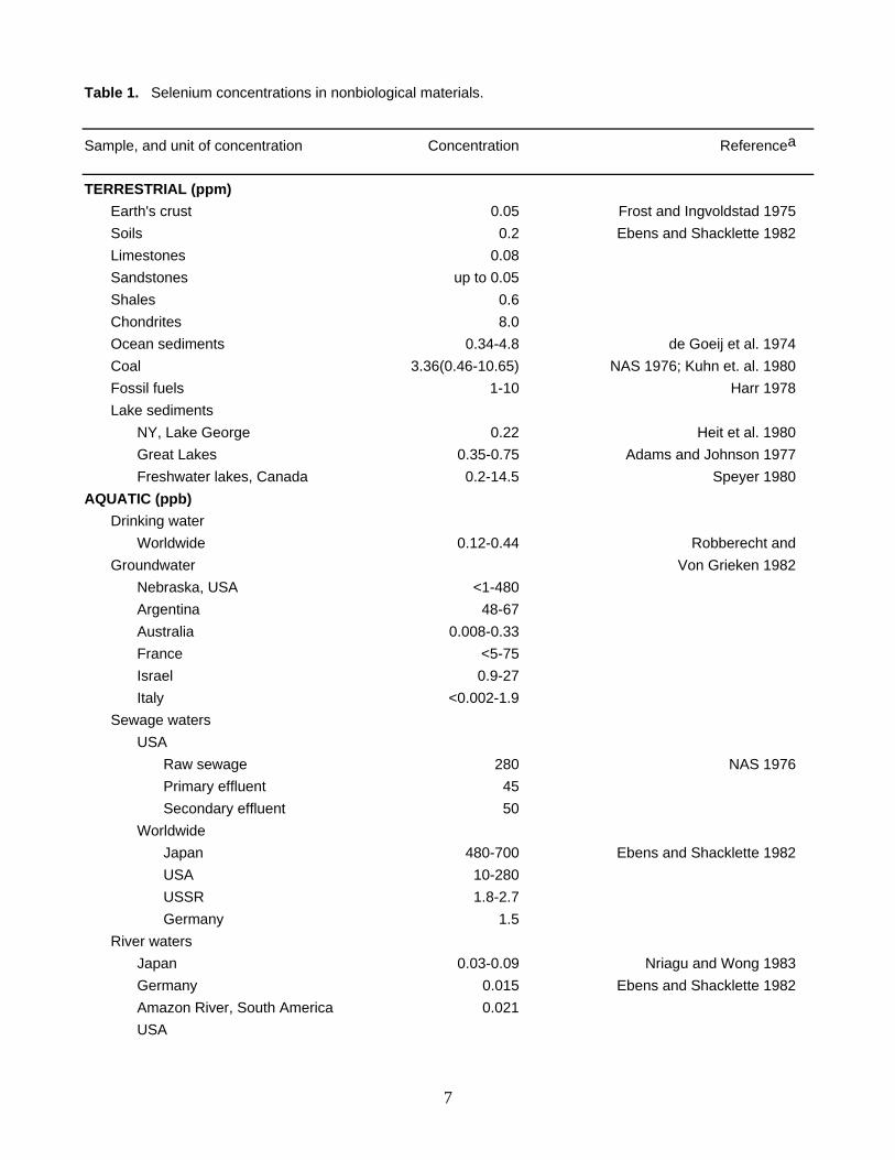
Table 1. Selenium concentrations in nonbiological materials.
Sample, and unit of concentration Concentration Referencea
TERRESTRIAL (ppm) Earth's crust 0.05 Frost and Ingvoldstad 1975 Soils 0.2 Ebens and Shacklette 1982 Limestones 0.08 Sandstones up to 0.05 Shales 0.6 Chondrites 8.0 Ocean sediments 0.34-4.8 de Goeij et al. 1974 Coal 3.36(0.46-10.65) NAS 1976; Kuhn et. al. 1980 Fossil fuels 1-10 Harr 1978 Lake sediments NY, Lake George 0.22 Heit et al. 1980 Great Lakes 0.35-0.75 Adams and Johnson 1977 Freshwater lakes, Canada 0.2-14.5 Speyer 1980 AQUATIC (ppb) Drinking water Worldwide 0.12-0.44 Robberecht and Groundwater Von Grieken 1982 Nebraska, USA <1-480 Argentina 48-67 Australia 0.008-0.33 France <5-75 Israel 0.9-27 Italy <0.002-1.9 Sewage waters USA Raw sewage 280 NAS 1976 Primary effluent 45 Secondary effluent 50 Worldwide Japan 480-700 Ebens and Shacklette 1982 USA 10-280 USSR 1.8-2.7 Germany 1.5 River waters Japan 0.03-0.09 Nriagu and Wong 1983 Germany 0.015 Ebens and Shacklette 1982 Amazon River, South America 0.021 USA
7
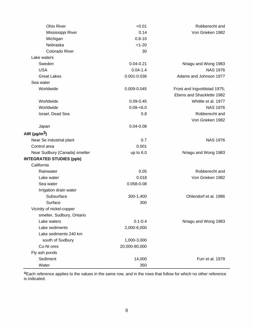
Ohio River <0.01 Robberecht and Mississippi River 0.14 Von Grieken 1982 Michigan 0.8-10 Nebraska <1-20 Colorado River 30 Lake waters Sweden 0.04-0.21 Nriagu and Wong 1983 USA 0.04-1.4 NAS 1976 Great Lakes 0.001-0.036 Adams and Johnson 1977 Sea water Worldwide 0.009-0.045 Frost and Ingvoldstad 1975; Ebens and Shacklette 1982 Worldwide 0.09-0.45 Whittle et al. 1977 Worldwide 0.09-<6.0 NAS 1976 Israel, Dead Sea 0.8 Robberecht and Von Grieken 1982 Japan 0.04-0.08
AIR (µg/m3) Near Se industrial plant 0.7 NAS 1976 Control area 0.001 Near Sudbury (Canada) smelter up to 6.0 Nriagu and Wong 1983 INTEGRATED STUDIES (ppb) California Rainwater 0.05 Robberecht and Lake water 0.018 Von Grieken 1982 Sea water 0.058-0.08 Irrigation drain water Subsurface 300-1,400 Ohlendorf et al. 1986 Surface 300 Vicinity of nickel-copper smelter, Sudbury, Ontario Lake waters 0.1-0.4 Nriagu and Wong 1983 Lake sediments 2,000-6,000 Lake sediments 240 km south of Sudbury 1,000-3,000 Cu-Ni ores 20,000-80,000 Fly ash ponds Sediment 14,000 Furr et al. 1979 Water 350
aEach reference applies to the values in the same row, and in the rows that follow for which no other reference is indicated.
8
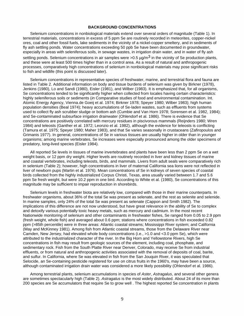
BACKGROUND CONCENTRATIONS Selenium concentrations in nonbiological materials extend over several orders of magnitude (Table 1). In
terrestrial materials, concentrations in excess of 5 ppm Se are routinely recorded in meteorites, copper-nickel ores, coal and other fossil fuels, lake sediments in the vicinity of a nickel-copper smeltery, and in sediments of fly ash settling ponds. Water concentrations exceeding 50 ppb Se have been documented in groundwater, especially in areas with seleniferous soils, in sewage wastes, in irrigation drain water, and in water of fly ash settling ponds. Selenium concentrations in air samples were >0.5 µg/m3 in the vicinity of Se production plants, and these were at least 500 times higher than in a control area. As a result of natural and anthropogenic processes, comparatively high concentrations of selenium in nonbiological materials may pose significant risks to fish and wildlife (this point is discussed later).
Selenium concentrations in representative species of freshwater, marine, and terrestrial flora and fauna are listed in Table 2. Additional information on body and tissue burdens of selenium was given by Birkner (1978), Jenkins (1980), Lo and Sandi (1980), Eisler (1981), and Wilber (1983). It is emphasized that, for all organisms, Se concentrations tended to be significantly higher when collected from locales having certain characteristics: highly seleniferous soils or sediments (in Comparative studies of food and environmental contamination. Int. Atomic Energy Agency, Vienna.de Goeij et al. 1974; Birkner 1978; Speyer 1980; Wilber 1983); high human population densities (Beal 1974); heavy accumulations of Se-laden wastes, such as effluents from systems used to collect fly ash scrubber sludge or bottom ash (Cumbie and Van Horn 1978; Sorensen et al. 1982, 1984); and Se-contaminated subsurface irrigation drainwater (Ohlendorf et al. 1986). There is evidence that Se concentrations are positively correlated with mercury residues in piscivorous mammals (Reijnders 1980; Wren 1984) and teleosts (Ganther et al. 1972; Leonzio et al. 1982), although the evidence for teleosts is conflicting (Tamura et al. 1975; Speyer 1980; Maher 1983), and that Se varies seasonally in crustaceans (Zafiropoulos and Grimanis 1977). In general, concentrations of Se in various tissues are usually higher in older than in younger organisms; among marine vertebrates, Se increases were especially pronounced among the older specimens of predatory, long-lived species (Eisler 1984).
All reported Se levels in tissues of marine invertebrates and plants have been less than 2 ppm Se on a wet weight basis, or 12 ppm dry weight. Higher levels are routinely recorded in liver and kidney tissues of marine and coastal vertebrates, including teleosts, birds, and mammals. Livers from adult seals were comparatively rich in selenium (Table 2); however, high concentrations in liver of maternal California sea lions were not reflected in liver of newborn pups (Martin et al. 1976). Mean concentrations of Se in kidneys of seven species of coastal birds collected from the highly industrialized Corpus Christi, Texas, area usually varied between 1.7 and 5.6 ppm Se fresh weight, but were 10.2 ppm in one bird. According to White et al. (1980), Se concentrations of this magnitude may be sufficient to impair reproduction in shorebirds.
Selenium levels in freshwater biota are relatively low, compared with those in their marine counterparts. In freshwater organisms, about 36% of the total Se was present as selenate, and the rest as selenite and selenide. In marine samples, only 24% of the total Se was present as selenate (Cappon and Smith 1982). The implications of this difference are not now understood, but have great relevance in the ability of Se to complex and detoxify various potentially toxic heavy metals, such as mercury and cadmium. In the most recent Nationwide monitoring of selenium and other contaminants in freshwater fishes, Se ranged from 0.05 to 2.9 ppm (fresh weight, whole fish) and averaged about 0.6 ppm; stations where concentrations in fish exceeded 0.82 ppm (>85th percentile) were in three areas: Atlantic coastal streams; Mississippi River system; and California (May and McKinney 1981). Among fish from Atlantic coastal streams, those from the Delaware River near Camden, New Jersey, had elevated whole body concentrations (i.e., >1.0 and <3.0 ppm Se), which were attributed to the industrialized character of the river. In the Big Horn and Yellowstone Rivers, high Se concentrations in fish may result from geologic sources of the element, including coal, phosphate, and sedimentary rock. Fish from the South Platte River near Denver, Colorado, may receive Se from industrial effluents, or from natural and anthropogenic activities associated with the removal of deposits of coal, barite, and sulfur. In California, where Se was elevated in fish from the San Jouquin River, it was speculated that Selocide, an Se-containing pesticide registered for use on citrus fruits in the 1960's, may have been a source, although contaminated irrigation drain water was considered a more likely possibility (Ohlendorf et al. 1986).
Among terrestrial plants, selenium accumulations in species of Aster, Astragalus, and several other genera are sometimes spectacularly high (Table 2). Astragalus is the most widely distributed. About 24 of its more than 200 species are Se accumulators that require Se to grow well . The highest reported Se concentration in plants
9
was 15,000 ppm dry weight, in loco weed, Astragalus racemosus (Wilber 1983). Consumption of these and other Se-accumulating forage plants by livestock has induced illness and death from selenium poisoning. Even at much lower concentrations, Se may harm animals that eat considerable amounts of the forage. Plants that accumulate Se tend to be deeper rooted than the grasses and survive more severe aridity, thus remaining (unfortunately) as the principal forage for grazing in time of drought (Wilber 1983).
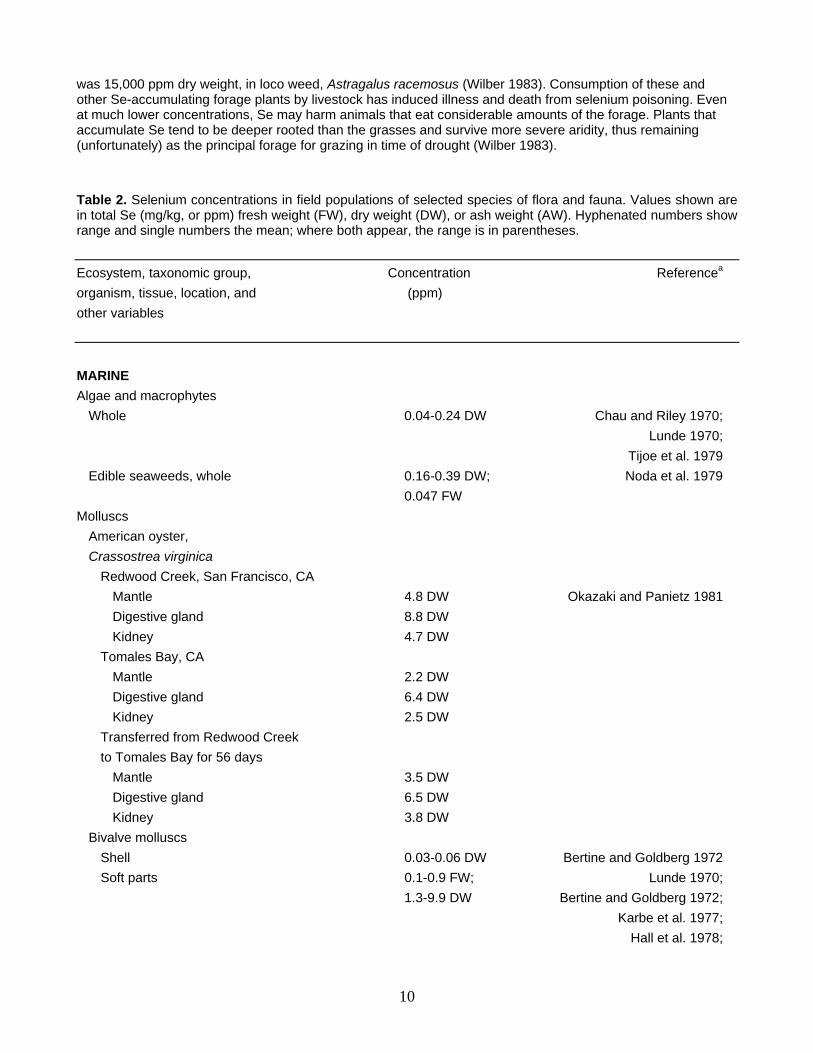
Table 2. Selenium concentrations in field populations of selected species of flora and fauna. Values shown are in total Se (mg/kg, or ppm) fresh weight (FW), dry weight (DW), or ash weight (AW). Hyphenated numbers show range and single numbers the mean; where both appear, the range is in parentheses.
Ecosystem, taxonomic group, Concentration Referencea
organism, tissue, location, and (ppm) other variables
MARINE Algae and macrophytes Whole 0.04-0.24 DW Chau and Riley 1970; Lunde 1970; Tijoe et al. 1979 Edible seaweeds, whole 0.16-0.39 DW; Noda et al. 1979 0.047 FW Molluscs American oyster, Crassostrea virginica Redwood Creek, San Francisco, CA Mantle 4.8 DW Okazaki and Panietz 1981 Digestive gland 8.8 DW Kidney 4.7 DW Tomales Bay, CA Mantle 2.2 DW Digestive gland 6.4 DW Kidney 2.5 DW Transferred from Redwood Creek to Tomales Bay for 56 days Mantle 3.5 DW Digestive gland 6.5 DW Kidney 3.8 DW Bivalve molluscs Shell 0.03-0.06 DW Bertine and Goldberg 1972 Soft parts 0.1-0.9 FW; Lunde 1970; 1.3-9.9 DW Bertine and Goldberg 1972; Karbe et al. 1977; Hall et al. 1978;
10
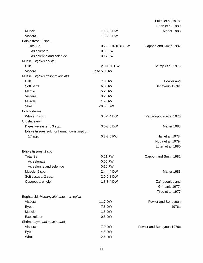
Fukai et al. 1978; Luten et al. 1980 Muscle 1.1-2.3 DW Maher 1983 Viscera 1.6-2.5 DW Edible fresh, 3 spp. Total Se 0.22(0.16-0.31) FW Cappon and Smith 1982 As selenate 0.05 FW As selenite and selenide 0.17 FW Mussel, Mytilus edulis Gills 2.0-16.0 DW Stump et al. 1979 Viscera up to 5.0 DW Mussel, Mytilus galloprovincialis Gills 7.0 DW Fowler and Soft parts 6.0 DW Benayoun 1976c Mantle 5.2 DW Viscera 3.2 DW Muscle 1.9 DW Shell <0.05 DW Echinoderms Whole, 7 spp. 0.8-4.4 DW Papadopoulu et al.1976 Crustaceans Digestive system, 3 spp. 3.0-3.5 DW Maher 1983 Edible tissues sold for human consumption 17 spp. 0.2-2.0 FW Hall et al. 1978; Noda et al. 1979; Luten et al. 1980 Edible tissues, 2 spp. Total Se 0.21 FW Cappon and Smith 1982 As selenate 0.05 FW As selenite and selenide 0.16 FW Muscle, 5 spp. 2.4-4.4 DW Maher 1983 Soft tissues, 2 spp. 2.0-2.8 DW Copepods, whole 1.8-3.4 DW Zafiropoulos and Grimanis 1977; Tijoe et al. 1977 Euphausid, Meganyctiphanes norvegica Viscera 11.7 DW Fowler and Benayoun Eyes 7.8 DW 1976a Muscle 1.8 DW Exoskeleton 0.8 DW Shrimp, Lysmata seticaudata Viscera 7.0 DW Fowler and Benayoun 1976c Eyes 4.8 DW Whole 2.6 DW
11

Muscle 1.9 DW Exoskeleton 1.5 DW Molts 0.3 DW Sharks Muscle 0.2-0.8 FW Glover 1979 Fish Digestive system, 4 spp. 1.0-2.4 DW Maher 1983 Liver 2 spp. 2.6-6.6 DW Grimanis et al. 1978 74 spp. 0.6-5.0 FW de Goeij et al. 1974; Hall et al. 1978; Luten et al. 1980 13 spp. 5.0-30.0 FW Hall et al. 1978 Meals, 3 spp. 1.0-4.0 DW Kifer and Payne 1968 Muscle 4 spp. 0.5-1.5 DW Maher 1983 182 spp. 0.1-2.0 FW Bebbington et al. 1977; Grimanis et al. 1978; Kari and Kauranen 1978; Hall et al. 1978; Noda et al. 1979; Luten et al.1980 5 spp. Total Se 0.4(0.2-0.6) FW Cappon and Smith 1982 As selenate 0.1 FW As selenite and selenide 0.3 FW Whole, 21 spp. 0.3-2.0 FW Kari and Kauranen 1978; Hall et al. 1978; United Nations 1979 Japanese tunas, 4 spp. Liver 10.0-15.0 FW Tamura et al. 1975 White muscle 0.5-1.3 FW Red muscle 3.5-9.1 FW Black marlin, Makaira indica Muscle 0.4-4.3 FW MacKay et al. 1975 Liver 1.4-13.5 FW Blue marlin, Makaira nigricans Kidney 23.0 FW Schultz and Ito 1979 Blood 1.0 FW Gill 1.0 FW Muscle Total Se 3.3(2.5-4.1) FW Cappon and Smith 1982 As selenate 0.2(0.09-0.3) FW As selenite and selenide 3.1(2.4-3.8) FW
12
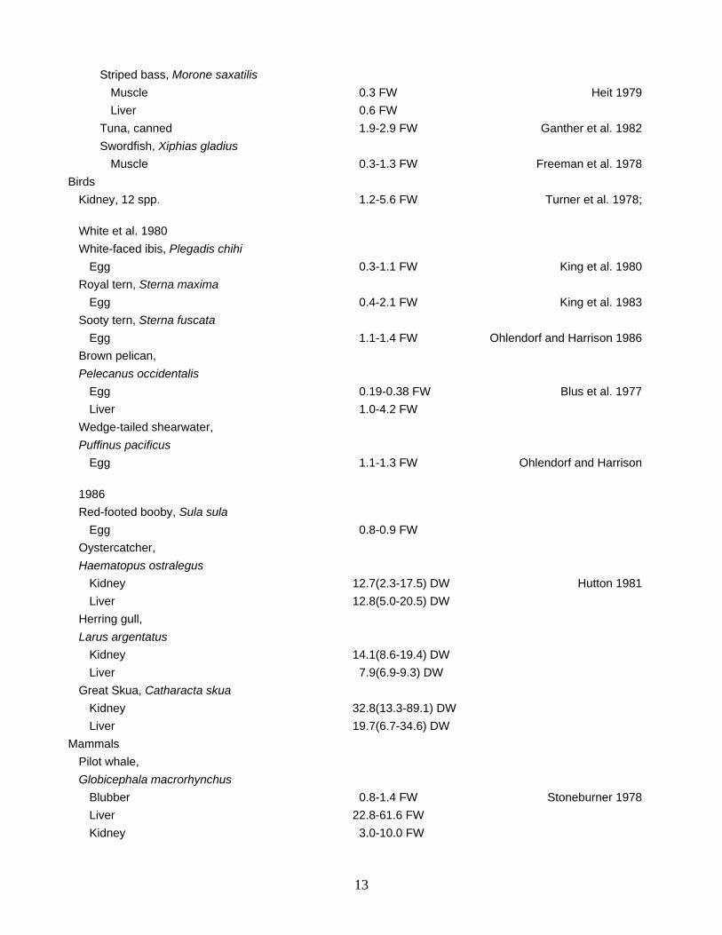
Striped bass, Morone saxatilis Muscle 0.3 FW Heit 1979 Liver 0.6 FW Tuna, canned 1.9-2.9 FW Ganther et al. 1982 Swordfish, Xiphias gladius Muscle 0.3-1.3 FW Freeman et al. 1978 Birds Kidney, 12 spp. 1.2-5.6 FW Turner et al. 1978; White et al. 1980 White-faced ibis, Plegadis chihi Egg 0.3-1.1 FW King et al. 1980 Royal tern, Sterna maxima Egg 0.4-2.1 FW King et al. 1983 Sooty tern, Sterna fuscata Egg 1.1-1.4 FW Ohlendorf and Harrison 1986 Brown pelican, Pelecanus occidentalis Egg 0.19-0.38 FW Blus et al. 1977 Liver 1.0-4.2 FW Wedge-tailed shearwater, Puffinus pacificus Egg 1.1-1.3 FW Ohlendorf and Harrison 1986 Red-footed booby, Sula sula Egg 0.8-0.9 FW Oystercatcher, Haematopus ostralegus Kidney 12.7(2.3-17.5) DW Hutton 1981 Liver 12.8(5.0-20.5) DW Herring gull, Larus argentatus Kidney 14.1(8.6-19.4) DW Liver 7.9(6.9-9.3) DW Great Skua, Catharacta skua Kidney 32.8(13.3-89.1) DW Liver 19.7(6.7-34.6) DW Mammals Pilot whale, Globicephala macrorhynchus Blubber 0.8-1.4 FW Stoneburner 1978 Liver 22.8-61.6 FW Kidney 3.0-10.0 FW
13
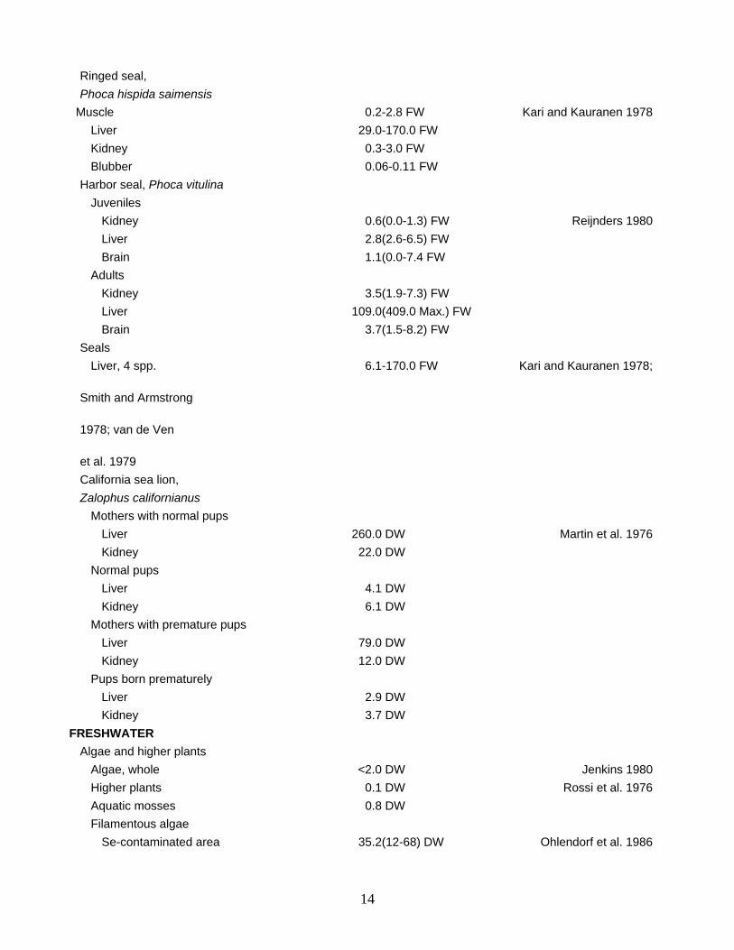
Ringed seal, Phoca hispida saimensis Muscle 0.2-2.8 FW Kari and Kauranen 1978 Liver 29.0-170.0 FW Kidney 0.3-3.0 FW Blubber 0.06-0.11 FW Harbor seal, Phoca vitulina Juveniles Kidney 0.6(0.0-1.3) FW Reijnders 1980 Liver 2.8(2.6-6.5) FW Brain 1.1(0.0-7.4 FW Adults Kidney 3.5(1.9-7.3) FW Liver 109.0(409.0 Max.) FW Brain 3.7(1.5-8.2) FW Seals Liver, 4 spp. 6.1-170.0 FW Kari and Kauranen 1978; Smith and Armstrong 1978; van de Ven et al. 1979 California sea lion, Zalophus californianus Mothers with normal pups Liver 260.0 DW Martin et al. 1976 Kidney 22.0 DW Normal pups Liver 4.1 DW Kidney 6.1 DW Mothers with premature pups Liver 79.0 DW Kidney 12.0 DW Pups born prematurely Liver 2.9 DW Kidney 3.7 DW FRESHWATER Algae and higher plants Algae, whole <2.0 DW Jenkins 1980 Higher plants 0.1 DW Rossi et al. 1976 Aquatic mosses 0.8 DW Filamentous algae Se-contaminated area 35.2(12-68) DW Ohlendorf et al. 1986
14
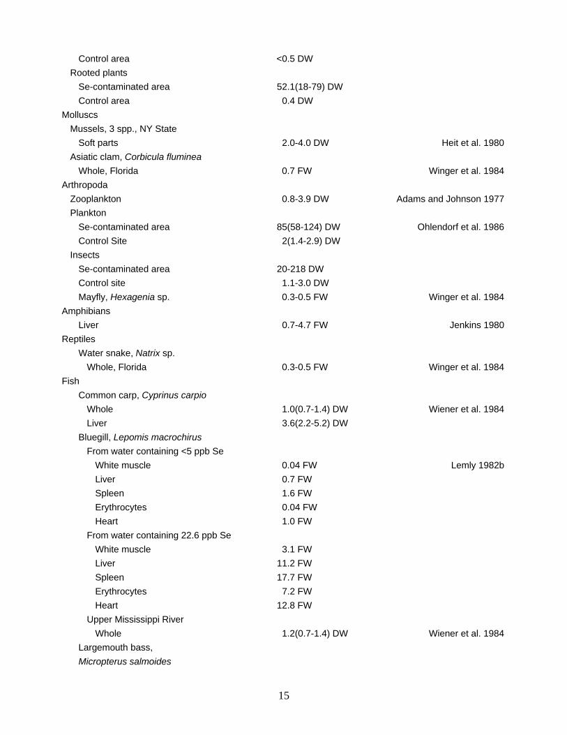
Control area <0.5 DW Rooted plants Se-contaminated area 52.1(18-79) DW Control area 0.4 DW Molluscs Mussels, 3 spp., NY State Soft parts 2.0-4.0 DW Heit et al. 1980 Asiatic clam, Corbicula fluminea Whole, Florida 0.7 FW Winger et al. 1984 Arthropoda Zooplankton 0.8-3.9 DW Adams and Johnson 1977 Plankton Se-contaminated area 85(58-124) DW Ohlendorf et al. 1986 Control Site 2(1.4-2.9) DW Insects Se-contaminated area 20-218 DW Control site 1.1-3.0 DW Mayfly, Hexagenia sp. 0.3-0.5 FW Winger et al. 1984 Amphibians Liver 0.7-4.7 FW Jenkins 1980 Reptiles Water snake, Natrix sp. Whole, Florida 0.3-0.5 FW Winger et al. 1984 Fish Common carp, Cyprinus carpio Whole 1.0(0.7-1.4) DW Wiener et al. 1984 Liver 3.6(2.2-5.2) DW Bluegill, Lepomis macrochirus From water containing <5 ppb Se White muscle 0.04 FW Lemly 1982b Liver 0.7 FW Spleen 1.6 FW Erythrocytes 0.04 FW Heart 1.0 FW From water containing 22.6 ppb Se White muscle 3.1 FW Liver 11.2 FW Spleen 17.7 FW Erythrocytes 7.2 FW Heart 12.8 FW Upper Mississippi River Whole 1.2(0.7-1.4) DW Wiener et al. 1984 Largemouth bass, Micropterus salmoides
15
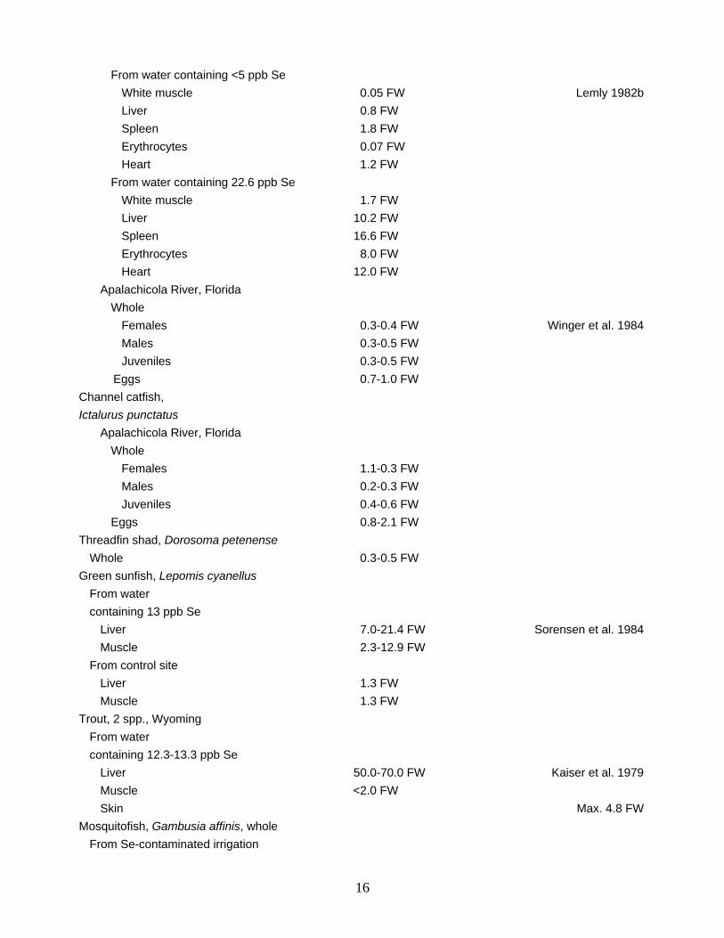
From water containing <5 ppb Se White muscle 0.05 FW Lemly 1982b Liver 0.8 FW Spleen 1.8 FW Erythrocytes 0.07 FW Heart 1.2 FW From water containing 22.6 ppb Se White muscle 1.7 FW Liver 10.2 FW Spleen 16.6 FW Erythrocytes 8.0 FW Heart 12.0 FW Apalachicola River, Florida Whole Females 0.3-0.4 FW Winger et al. 1984 Males 0.3-0.5 FW Juveniles 0.3-0.5 FW Eggs 0.7-1.0 FW Channel catfish, Ictalurus punctatus Apalachicola River, Florida Whole Females 1.1-0.3 FW Males 0.2-0.3 FW Juveniles 0.4-0.6 FW Eggs 0.8-2.1 FW Threadfin shad, Dorosoma petenense Whole 0.3-0.5 FW Green sunfish, Lepomis cyanellus From water containing 13 ppb Se Liver 7.0-21.4 FW Sorensen et al. 1984 Muscle 2.3-12.9 FW From control site Liver 1.3 FW Muscle 1.3 FW Trout, 2 spp., Wyoming From water containing 12.3-13.3 ppb Se Liver 50.0-70.0 FW Kaiser et al. 1979 Muscle <2.0 FW Skin Max. 4.8 FW Mosquitofish, Gambusia affinis, whole From Se-contaminated irrigation
16
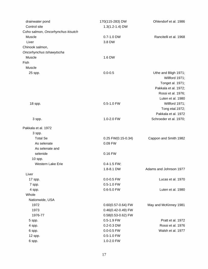
drainwater pond 170(115-283) DW Ohlendorf et al. 1986 Control site 1.3(1.2-1.4) DW Coho salmon, Oncorhynchus kisutch Muscle 0.7-1.0 DW Rancitelli et al. 1968 Liver 3.8 DW Chinook salmon, Oncorhynchus tshawytscha Muscle 1.6 DW Fish Muscle 25 spp. 0.0-0.5 Uthe and Bligh 1971; Willford 1971; Tonget al. 1971; Pakkala et al. 1972; Rossi et al. 1976; Luten et al. 1980 18 spp. 0.5-1.0 FW Willford 1971; Tong etal.1972; Pakkala et al. 1972 3 spp. 1.0-2.0 FW Schroeder et al. 1970; Pakkala et al. 1972 3 spp. Total Se 0.25 FW(0.15-0.34) Cappon and Smith 1982 As selenate 0.09 FW As selenate and selenide 0.16 FW 10 spp. Western Lake Erie 0.4-1.5 FW; 1.8-8.1 DW Adams and Johnson 1977 Liver 17 spp. 0.0-0.5 FW Lucas et al. 1970 7 spp. 0.5-1.0 FW 4 spp. 0.6-5.0 FW Luten et al. 1980 Whole Nationwide, USA 1972 0.60(0.57-0.64) FW May and McKinney 1981 1973 0.46(0.42-0.49) FW 1976-77 0.58(0.53-0.62) FW 5 spp. 0.5-1.9 FW Pratt et al. 1972 4 spp. 0.2-0.3 DW Rossi et al. 1976 6 spp. 0.0-0.5 FW Walsh et al. 1977 12 spp. 0.5-1.0 FW 6 spp. 1.0-2.0 FW
17
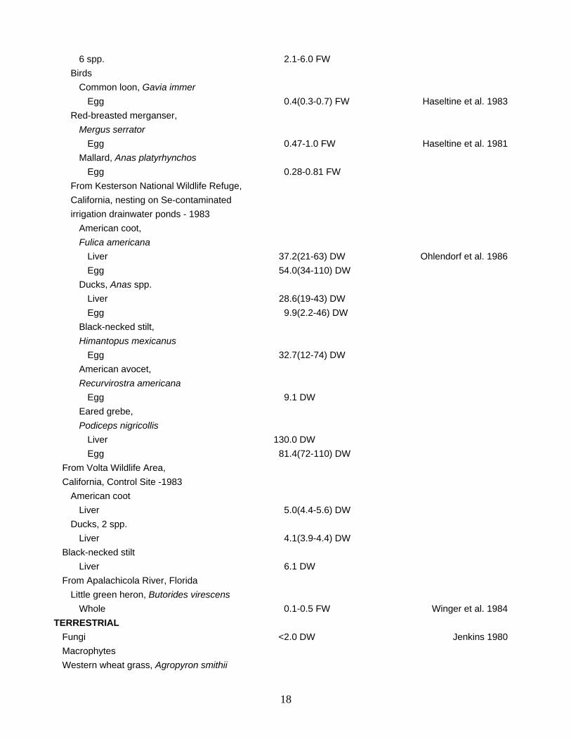
6 spp. 2.1-6.0 FW Birds Common loon, Gavia immer Egg 0.4(0.3-0.7) FW Haseltine et al. 1983 Red-breasted merganser, Mergus serrator Egg 0.47-1.0 FW Haseltine et al. 1981 Mallard, Anas platyrhynchos Egg 0.28-0.81 FW From Kesterson National Wildlife Refuge, California, nesting on Se-contaminated irrigation drainwater ponds - 1983 American coot, Fulica americana Liver 37.2(21-63) DW Ohlendorf et al. 1986 Egg 54.0(34-110) DW Ducks, Anas spp. Liver 28.6(19-43) DW Egg 9.9(2.2-46) DW Black-necked stilt, Himantopus mexicanus Egg 32.7(12-74) DW American avocet, Recurvirostra americana Egg 9.1 DW Eared grebe, Podiceps nigricollis Liver 130.0 DW Egg 81.4(72-110) DW From Volta Wildlife Area, California, Control Site -1983 American coot Liver 5.0(4.4-5.6) DW Ducks, 2 spp. Liver 4.1(3.9-4.4) DW Black-necked stilt Liver 6.1 DW From Apalachicola River, Florida Little green heron, Butorides virescens Whole 0.1-0.5 FW Winger et al. 1984 TERRESTRIAL Fungi <2.0 DW Jenkins 1980 Macrophytes Western wheat grass, Agropyron smithii
18
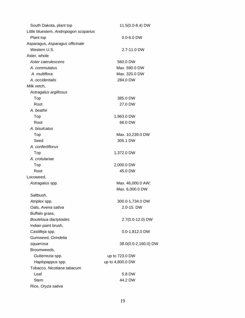
South Dakota, plant top 11.5(0.0-8.4) DW Little bluestem, Andropogon scoparius Plant top 0.0-6.0 DW Asparagus, Asparagus officinale Western U.S. 2.7-11.0 DW Aster, whole Aster caerulescens 560.0 DW A. commutatus Max. 590.0 DW A. multiflora Max. 320.0 DW A. occidentalis 284.0 DW Milk vetch, Astragalus argillosus Top 385.0 DW Root 27.0 DW A. beathii Top 1,963.0 DW Root 66.0 DW A. bisulcatus Top Max. 10,239.0 DW Seed 305.1 DW A. confertiflorus Top 1,372.0 DW A. crotulariae Top 2,000.0 DW Root 45.0 DW Locoweed, Astragalus spp. Max. 46,000.0 AW; Max. 6,000.0 DW Saltbush, Atriplex spp. 300.0-1,734.0 DW Oats, Avena sativa 2.0-15. DW Buffalo grass, Bouteloua dactyloides 2.7(0.0-12.0) DW Indian paint brush, Castilleja spp. 0.0-1,812.0 DW Gumweed, Grindelia squarrosa 38.0(0.0-2,160.0) DW Broomweeds, Gutierrezia spp. up to 723.0 DW Haplopappus spp. up to 4,800.0 DW Tobacco, Nicotiana tabacum Leaf 5.8 DW Stem 44.2 DW Rice, Oryza sativa
19
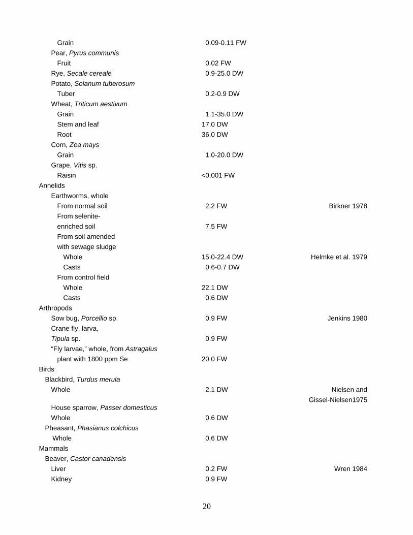
Grain 0.09-0.11 FW Pear, Pyrus communis Fruit 0.02 FW Rye, Secale cereale 0.9-25.0 DW Potato, Solanum tuberosum Tuber 0.2-0.9 DW Wheat, Triticum aestivum Grain 1.1-35.0 DW Stem and leaf 17.0 DW Root 36.0 DW Corn, Zea mays Grain 1.0-20.0 DW Grape, Vitis sp. Raisin <0.001 FW Annelids Earthworms, whole From normal soil 2.2 FW Birkner 1978 From selenite- enriched soil 7.5 FW From soil amended with sewage sludge Whole 15.0-22.4 DW Helmke et al. 1979 Casts 0.6-0.7 DW From control field Whole 22.1 DW Casts 0.6 DW Arthropods Sow bug, Porcellio sp. 0.9 FW Jenkins 1980 Crane fly, larva, Tipula sp. 0.9 FW “Fly larvae,” whole, from Astragalus plant with 1800 ppm Se 20.0 FW Birds Blackbird, Turdus merula Whole 2.1 DW Nielsen and Gissel-Nielsen1975 House sparrow, Passer domesticus Whole 0.6 DW Pheasant, Phasianus colchicus Whole 0.6 DW Mammals Beaver, Castor canadensis Liver 0.2 FW Wren 1984 Kidney 0.9 FW
20
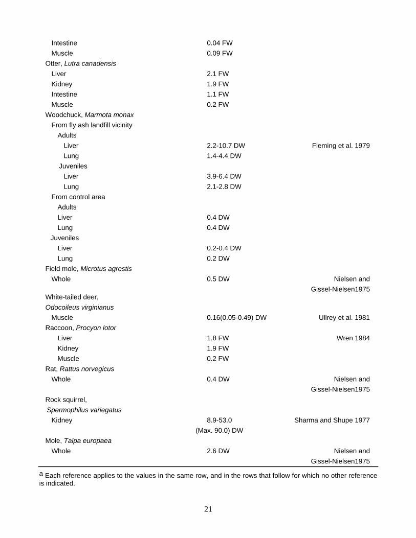
Intestine 0.04 FW Muscle 0.09 FW Otter, Lutra canadensis Liver 2.1 FW Kidney 1.9 FW Intestine 1.1 FW Muscle 0.2 FW Woodchuck, Marmota monax From fly ash landfill vicinity Adults Liver 2.2-10.7 DW Fleming et al. 1979 Lung 1.4-4.4 DW Juveniles Liver 3.9-6.4 DW Lung 2.1-2.8 DW From control area Adults Liver 0.4 DW Lung 0.4 DW Juveniles Liver 0.2-0.4 DW Lung 0.2 DW Field mole, Microtus agrestis Whole 0.5 DW Nielsen and Gissel-Nielsen1975 White-tailed deer, Odocoileus virginianus Muscle 0.16(0.05-0.49) DW Ullrey et al. 1981 Raccoon, Procyon lotor Liver 1.8 FW Wren 1984 Kidney 1.9 FW Muscle 0.2 FW Rat, Rattus norvegicus Whole 0.4 DW Nielsen and Gissel-Nielsen1975 Rock squirrel, Spermophilus variegatus Kidney 8.9-53.0 Sharma and Shupe 1977 (Max. 90.0) DW Mole, Talpa europaea Whole 2.6 DW Nielsen and Gissel-Nielsen1975
a Each reference applies to the values in the same row, and in the rows that follow for which no other reference is indicated.
21
DEFICIENCY AND PROTECTIVE EFFECTS
Selenium is a critical nutrient and a key component of several enzymes. There is a general consensus that Se deficiency in livestock is increasing in many countries, resulting in a need for added Se in the food. Selenium deficiency is considered by some researchers to constitute a greater threat to health than selenium poisoning. Studies with animals and humans have suggested that Se deficiency, in part, underlies susceptibility to cancer, arthritis, hypertension, heart disease, and possibly periodontal disease and cataracts (Frost and Ingvoldstad 1975; Shamberger 1981; Robberecht and Von Grieken 1982). These linkages have not yet been demonstrated conclusively; for example, eye lens cataract was induced in 10-day-old male rats by selenate, selenite, selenomethionine, and selenocystine, presumably through interference with glutathione metabolism (Ostadalova and Babicky 1980). On the other hand, adverse effects of Se inadequacy have been clearly documented for a wide variety of organisms, including bacteria, protozoans, Atlantic salmon, rainbow trout, Japanese quail, ducks, poultry, rats, dogs, horses, sheep, swine, cattle, antelopes, gazelles, deer, monkeys, and humans (Jensen 1968; Frost and Ingvoldstad 1975; Methanococcus vanniellii: culture and effects of selenium and tungsten on growth. J. Bacteriol. 130:1404-1406.
Jones and Stadtman 1975; Fishbein 1977; Harr 1978; Kaiser et al. 1979; Hilton et al. 1980; Shamberger 1981; Bovee and O'Brien 1982; Robberecht and Von Grieken 1982; NRC 1983; Levander 1983, 1984; Morris et al. 1984). Selenium deficiency, whether induced experimentally by use of low Se feeds supplemented with alpha tocopherol or by chronic ingestion of low Se diets, has caused a number of maladies: high embryonic mortality in cattle and sheep; anemia in cattle; poor growth and reproduction in sheep and rats; reduced viability of newly hatched quail; nutritional myopathy (white muscle disease) in sheep, swine, and cattle; hepatic necrosis and lameness in dogs, horses, and breeding bulls; hair loss and sterility in rat offspring; and spermatozoan abnormalities in rats. Deficiencies were usually prevented or reversed by supplements with sodium selenate or selenite at 100 ppb Se in the diet, or 20 µg Se/kg body weight administered parenterally.
The availability of Se to plants may be lessened by modern agricultural practices, eventually contributing to Se deficiency in animal consumers. For example, fertilizers containing nitrogen, sulfur, and phosphorus all influence Se uptake by plants through different modes of action, the net effect being a reduction in Se uptake (Frost and Ingvoldstad 1975). The buildup of sulfur (as sulfates) in the soil, due to acid rain, fertilizers, and other sources, interferes with Se accumulation by crops (Frost and Ingvoldstad 1975). In addition, high dietary levels of various heavy metals (including copper, zinc, silver, and mercury) contribute to Se deficiency in animals (Frost and Ingvoldstad 1975; Harr 1978), presumably as a result of Se binding with the metal into biologically unavailable forms (Harr 1978; Kaiser et al. 1979).
The protective action of selenium against the adverse or lethal effects induced by various metals and metalloids is well documented for a wide variety of plant and animal species. Among marine organisms, for example, selenium protects against toxic levels of mercury in algae (Gotsis 1982), shrimp (Lucu and Skreblin 1982), crabs and oysters (Glickstein 1978), fish (Sheline and Schmidt-Nielsen 1977), and mammals (Koeman et al. 1975). Similar observations have been recorded for copper and marine algae (Gotsis 1982); cadmium and freshwater snails (Wilber 1983), marine crabs (Bjerragaard 1982), earthworms (Helmke et al. 1979; Beyer et al. 1982), and rats (Harr 1978); mercury or methylmercury and rats (Cappon and Smith 1982), eggs of lake trout (Klaverkamp et al. 1983b), freshwater teleosts (Kim et al. 1975, 1977) and (temporarily) Japanese quail (El-Bergearmi et al. 1977, 1982; Beijer and Jernelov 1978); and arsenic and freshwater and marine teleosts (Luten et al. 1980; Orvini et al. 1980). Not all tests were conclusive. Studies with some species of freshwater teleosts demonstrated negligible antagonism of Se against mercury (Klaverkamp et al. 1983a) or cadmium (Duncan and Klaverkamp 1983). Selenium reportedly protects mammals and poikilotherms against poisoning by thallium, the herbicide paraquat, cadmium, mercury, lead, arsenic, and copper (Wilber 1983).
Reasons, to account for the antagonism of Se and heavy metals (here mercury is used as an example) include dietary source and chemical form of Se, influence of sulfur, biological translocation of Se or mercury to less critical body parts, and chemical linkage of Se to mercury on a linear basis. The exact mode of interaction is probably complex and has not yet been resolved. In regard to diet, Se of animal origin and in the form of selenate is less effective than Se from plant and inorganic sources in preventing methylmercury neurotoxicity in experimental animals (Cappon and Smith 1982). Disruption of sulfur metabolism by Se, the sulfur being replaced by seleno-amino acids and other cell constituents containing Se in living organisms, is one probable cause of Se poisoning. It is conceivable that Se-Hg compounds formed within the organism would be sufficiently
22
nonreactive biologically to interfere with sulfur kinetics, presumably -SH groups (Koeman et al. 1975; Beijer and Jernelov 1978; Cappon and Smith 1982; Gotsis 1982). Differential redistribution of Se or Hg to less critical body parts may partly account for observed antagonisms. Pretreatment of marine minnows with Se protects against Hg poisoning and causes a marked redistribution of Hg among organs, presumably to non-critical body parts, and this transfer may partly account for the observed Se-Hg antagonisms in that species (Sheline and Schmidt-Nielsen 1977). Some investigators have reported that Se results in increased Hg accumulations. Increased retention of Hg and other metals may lead to a higher level of biomagnification in the food chain and higher body burden in the individual, which might counteract the positive effect of decreased intoxication (Beijer and Jernelov 1978). Extensive research is under way on the chemical linkage of Se and Hg. In marine mammals and humans, Se and Hg concentrations are closely related, almost linearly in a 1:1 molar ratio, but this relation blurs in teleosts in which Se is in abundance, and fails in birds (Koeman et al. 1975; Beijer and Jernelov 1978; Orvini et al. 1980; Cappon and Smith 1982).
TOXICITY MAMMALS AND BIRDS
“The element selenium can be traced in an orderly sequence from its origin in the earth's crust to specific geological formation, to distribution of specific genera and groups of plants which require the element for their growth, to the accumulation in vegetation, and to its subsequent toxicity to birds or mammals that consume the seleniferous foods” (Rosenfeld and Beath 1964). Selenium poisoning in livestock, discussed here, was largely extracted from reviews by Rosenfeld and Beath (1964), Frost (1972), Fishbein (1977), Harr (1978), Shamberger (1981), NRC (1983), and Wilber (1983).
In livestock, there are three basic types of selenium poisoning: acute, resulting from consumption (usually in a single feeding) of a sufficient quantity of highly seleniferous weeds; “blind staggers,” from consumption of moderately toxic amounts of seleniferous weeds over an extended period of time; and "alkali disease," caused by the consumption of moderately seleniferous grains and forage grasses over a period of several weeks to months.
Acute poisoning is associated with plant materials containing 400 to 800 ppm Se: sheep died when fed amounts of plant material ranging from 8 to 16 g/kg body weight, or about 3.2 to 12.8 mg Se/kg body weight. The minimum lethal dose of Se administered orally as selenite (mg Se per kg body weight) ranged from 3.3 for horses and mules to 11 for cattle and 15 for swine. Other modes of administration were more toxic, e.g., 2 and 1.2 mg Se/ kg body weight given subcutaneously killed swine in 4 hours and 5 days, respectively; and 1.5-6.0 mg Se/kg body weight given intravenously or intraperitoneally to rats and rabbits were fatal. Accidental toxicosis of sheep and cattle from overtreatment with commercial mixtures of Se salts and vitamin E are also documented for Australia and New Zealand. Acute Se poisoning in domestic livestock is characterized by abnormal movements, lowered head, drooped ears, diarrhea, elevated temperature, rapid pulse, labored breathing, bloating with abdominal pain, increased urination, and dilated pupils. Before death, which is due to respiratory failure, there is complete prostration and lethargy. Duration of illness extends from a few hours to several days, depending on the toxicity of plant material ingested. In these cases, Se is distributed by the circulatory system to all body organs, the concentrations being highest in liver, blood, kidney, spleen and brain, and lowest in muscle, skin, hair and bone. Elimination is primarily in the urine; smaller quantities are excreted with the feces, breath, perspiration, and bile. Postmortem examinations indicate many pathological changes in the heart, lungs, rumen, liver, kidney, and other organs. No effective treatment is known for counteracting toxic effects of large amounts of ingested selenium.
Chronic selenosis may be induced by dietary exposure to natural selenite, selenate, or seleniferous feedstuffs at dietary concentrations between 1 ppm (rat) and 44 ppm (horse), or from water containing 0.5 to 2.0 ppm of Se. Cattle fed 0.5 mg Se/kg body weight 3 times weekly lost their appetite; sheep fed up to 75 mg selenite daily developed myocardial degeneration and fibrosis, pulmonary congestion, and edema. The minimum toxic concentration of Se in lifetime exposure of rats (a comparatively sensitive species) fed Se-deficient diets fortified with Se was 0.35 mg Se/kg diet, as judged by changes in liver chemistry; and 0.75 mg Se/kg diet, as judged by longevity, and histological changes in heart, kidney, and spleen. These concentrations are 10 times the nutritional threshold for Se, and about 25% of the minimum lifetime exposure to Se in natural feedstuffs that produces similar effects under the same experimental conditions. Signs of chronic selenosis include skin lesions, lymph channel inflammation, loss of hair and nails, anemia, enlarged organs (spleen, pancreas, liver), fatigue, lassitude, and dizziness. “Blind staggers” is characterized by anorexia, emaciation, and sudden collapse, followed by death. Typically, the upper intestinal tract is ulcerated. In “alkali disease,” in cattle,
23
hogs, and horses that had eaten seleniferous grains, the signs were deformation and sloughing of the hooves, hair loss, lassitude, erosion of the articular cartilages, reduced conception, increased reabsorption of fetuses, and degeneration of heart, kidney, and liver. It is likely that Se displaces sulfur in keratin, resulting in structural changes in hair, nails, and hooves (Fishbein 1977).
Among birds, it appears that domestic chickens are extremely sensitive to Se; reduced hatching of eggs was recorded at 7 to 9 ppm Se in feed (Ort and Latshaw 1978). Similar results were observed by El-Bergearmi et al. (1977) in Japanese quail at 6 and 12 ppm dietary selenite. Studies in progress at the Patuxent Wildlife Research Center with adult mallards indicated that 100 ppm dietary selenium (as sodium selenite) was fatal within one month, but that survival was high at 25 ppm after 3 months. Poor egg hatchability was recorded in the 25 ppm selenite group, but not in the 10 ppm selenite group; however, hatching percent was reduced in eggs of adults fed 10 ppm of Se as selenomethionine (Dr. G. Heinz, pers. comm.). It appears obvious that additional research is needed on the toxicology of organoselenium compounds.
Selenosis in warm blooded organisms is modified by numerous factors, including method of administration, chemical form of Se, dietary composition, and age and needs of the organism. For example, Se administered in natural feedstuffs is only about one-fourth as toxic as are similar exposures in water or purified feeds (Harr 1978). Concurrent ingestion of minerals and rough or high protein feeds reduces Se toxicity, and exposure by diet is less toxic than exposure parenterally or by inhalation.
Many compounds are known to prevent or reduce toxic effects of subacute and chronic selenosis in pigs, beef cattle, and other warmblooded organisms. A partial list includes arsenic, strychnine sulfate, tungsten, germanium, antimony, beet pectin, high-fat diets, ACTH injections, sulfate, increased dietary proteins, lactalbumin, ovalbumin, wheat protein, dried brewer's yeast, desiccated liver, linseed oil meal, glucosamine, hemocysteine, creatine, methionine, and choline. Not all of these compounds afforded equal protection against various Se formulations; the reasons for the difference are not clear, but it appears that the subject of selenoprotective agents warrants additional research effort.
AQUATIC ORGANISMS Among sensitive species of aquatic organisms, death was observed at water concentrations between 60
and 600 ppb Se; early life history stages that were subjected to comparatively lengthy exposures accounted for most of these data (Table 3). Latent mortality after exposure to comparatively high Se concentrations has been documented, but not extensively. For example, all embryos of the zebrafish (Brachydanio rerio) survived exposure to 3,000 ppb of Se during development, but more than 90% of the resultant larvae died soon after hatching; at 1,000 ppb, survival was similar to that in controls (Niimi and LaHam 1975). It has been suggested (EPA 1980) that selenite is more toxic than selenate and is preferentially concentrated over selenate by mussels, Mytilus galloprovincialis (Measures and Burton 1980). Selenite is generally more toxic to early life history stages and effects are most pronounced at elevated temperatures (Klaverkamp et al. 1983a). Also, Se salts may be converted to methylated forms by microorganisms, and these are readily accumulated by aquatic vertebrates (Klaverkamp et al. 1983a). Among freshwater algae species, it has been demonstrated that selenite, selenate, selenomethionine, and selenopurine are all toxic, but that sulphur, as sulphate, has a significant protective role against Se toxicity (Kumar and Prakash 1971). Numerous additional chemical compounds and mixtures probably protect against Se toxicity, much as Se protects against toxic effects of mercury salts and other chemicals, but data are sparse on Se protective agents.
Almost 50 years ago, Ellis et al. (1937) recorded a long list of signs of selenium poisoning in teleosts: loss of equilibrium, lethargy, contraction of dermal chromatophores, loss of coordination, muscle spasms, protruding eyes, swollen abdomen, liver degeneration, reduction in blood hemoglobin and erythrocyte number, and increase in white blood cells. Sorensen et al. (1984), observed most of these signs in Se-poisoned green sunfish together with elevated liver Se concentrations, reduced blood hematocrit, enlarged liver, histopathology of kidney and heart, swollen gill lamellae with extensive cellular vacuolization, and necrotic and degenerating ovarian follicles.
24
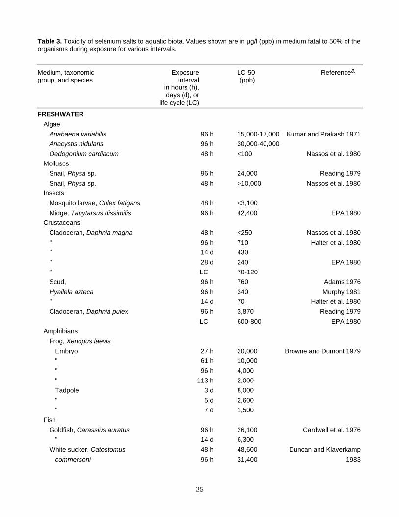
Table 3. Toxicity of selenium salts to aquatic biota. Values shown are in µg/l (ppb) in medium fatal to 50% of the organisms during exposure for various intervals.
Medium, taxonomic Exposure LC-50 Referencea group, and species interval (ppb) in hours (h), days (d), or life cycle (LC)
FRESHWATER Algae Anabaena variabilis 96 h 15,000-17,000 Kumar and Prakash 1971 Anacystis nidulans 96 h 30,000-40,000 Oedogonium cardiacum 48 h <100 Nassos et al. 1980 Molluscs Snail, Physa sp. 96 h 24,000 Reading 1979 Snail, Physa sp. 48 h >10,000 Nassos et al. 1980 Insects Mosquito larvae, Culex fatigans 48 h <3,100 Midge, Tanytarsus dissimilis 96 h 42,400 EPA 1980 Crustaceans Cladoceran, Daphnia magna 48 h <250 Nassos et al. 1980 " 96 h 710 Halter et al. 1980 " 14 d 430 " 28 d 240 EPA 1980 " LC 70-120 Scud, 96 h 760 Adams 1976 Hyallela azteca 96 h 340 Murphy 1981 " 14 d 70 Halter et al. 1980 Cladoceran, Daphnia pulex 96 h 3,870 Reading 1979 LC 600-800 EPA 1980 Amphibians Frog, Xenopus laevis Embryo 27 h 20,000 Browne and Dumont 1979 " 61 h 10,000 " 96 h 4,000 " 113 h 2,000 Tadpole 3 d 8,000 " 5 d 2,600 " 7 d 1,500 Fish Goldfish, Carassius auratus 96 h 26,100 Cardwell et al. 1976 " 14 d 6,300 White sucker, Catostomus 48 h 48,600 Duncan and Klaverkamp commersoni 96 h 31,400 1983
25
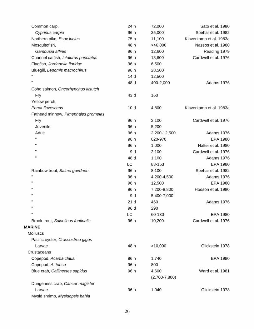
Common carp, 24 h 72,000 Sato et al. 1980 Cyprinus carpio 96 h 35,000 Spehar et al. 1982 Northern pike, Esox lucius 75 h 11,100 Klaverkamp et al. 1983a Mosquitofish, 48 h >>6,000 Nassos et al. 1980 Gambusia affinis 96 h 12,600 Reading 1979 Channel catfish, Ictalurus punctatus 96 h 13,600 Cardwell et al. 1976 Flagfish, Jordanella floridae 96 h 6,500 Bluegill, Lepomis macrochirus 96 h 28,500 " 14 d 12,500 " 48 d 400-2,000 Adams 1976 Coho salmon, Oncorhynchus kisutch Fry 43 d 160 Yellow perch, Perca flavescens 10 d 4,800 Klaverkamp et al. 1983a Fathead minnow, Pimephales promelas Fry 96 h 2,100 Cardwell et al. 1976 Juvenile 96 h 5,200 Adult 96 h 2,200-12,500 Adams 1976 " 96 h 620-970 EPA 1980 " 96 h 1,000 Halter et al. 1980 " 9 d 2,100 Cardwell et al. 1976 " 48 d 1,100 Adams 1976 LC 83-153 EPA 1980 Rainbow trout, Salmo gairdneri 96 h 8,100 Spehar et al. 1982 " 96 h 4,200-4,500 Adams 1976 " 96 h 12,500 EPA 1980 " 96 h 7,200-8,800 Hodson et al. 1980 " 9 d 5,400-7,000 " 21 d 460 Adams 1976 " 96 d 290 " LC 60-130 EPA 1980 Brook trout, Salvelinus fontinalis 96 h 10,200 Cardwell et al. 1976 MARINE Molluscs Pacific oyster, Crassostrea gigas Larvae 48 h >10,000 Glickstein 1978 Crustaceans Copepod, Acartia clausi 96 h 1,740 EPA 1980 Copepod, A. tonsa 96 h 800 Blue crab, Callinectes sapidus 96 h 4,600 Ward et al. 1981 (2,700-7,800) Dungeness crab, Cancer magister Larvae 96 h 1,040 Glickstein 1978 Mysid shrimp, Mysidopsis bahia
26
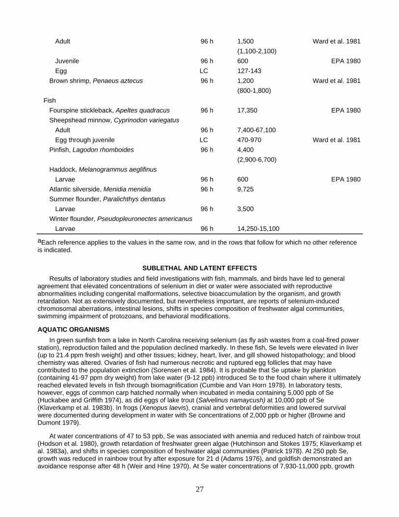
Adult 96 h 1,500 Ward et al. 1981 (1,100-2,100) Juvenile 96 h 600 EPA 1980 Egg LC 127-143 Brown shrimp, Penaeus aztecus 96 h 1,200 Ward et al. 1981 (800-1,800) Fish Fourspine stickleback, Apeltes quadracus 96 h 17,350 EPA 1980 Sheepshead minnow, Cyprinodon variegatus Adult 96 h 7,400-67,100 Egg through juvenile LC 470-970 Ward et al. 1981 Pinfish, Lagodon rhomboides 96 h 4,400 (2,900-6,700) Haddock, Melanogrammus aeglifinus Larvae 96 h 600 EPA 1980 Atlantic silverside, Menidia menidia 96 h 9,725 Summer flounder, Paralichthys dentatus Larvae 96 h 3,500 Winter flounder, Pseudopleuronectes americanus Larvae 96 h 14,250-15,100
aEach reference applies to the values in the same row, and in the rows that follow for which no other reference is indicated.
SUBLETHAL AND LATENT EFFECTS Results of laboratory studies and field investigations with fish, mammals, and birds have led to general
agreement that elevated concentrations of selenium in diet or water were associated with reproductive abnormalities including congenital malformations, selective bioaccumulation by the organism, and growth retardation. Not as extensively documented, but nevertheless important, are reports of selenium-induced chromosomal aberrations, intestinal lesions, shifts in species composition of freshwater algal communities, swimming impairment of protozoans, and behavioral modifications.
AQUATIC ORGANISMS In green sunfish from a lake in North Carolina receiving selenium (as fly ash wastes from a coal-fired power
station), reproduction failed and the population declined markedly. In these fish, Se levels were elevated in liver (up to 21.4 ppm fresh weight) and other tissues; kidney, heart, liver, and gill showed histopathology; and blood chemistry was altered. Ovaries of fish had numerous necrotic and ruptured egg follicles that may have contributed to the population extinction (Sorensen et al. 1984). It is probable that Se uptake by plankton (containing 41-97 ppm dry weight) from lake water (9-12 ppb) introduced Se to the food chain where it ultimately reached elevated levels in fish through biomagnification (Cumbie and Van Horn 1978). In laboratory tests, however, eggs of common carp hatched normally when incubated in media containing 5,000 ppb of Se (Huckabee and Griffith 1974), as did eggs of lake trout (Salvelinus namaycush) at 10,000 ppb of Se (Klaverkamp et al. 1983b). In frogs (Xenopus laevis), cranial and vertebral deformities and lowered survival were documented during development in water with Se concentrations of 2,000 ppb or higher (Browne and Dumont 1979).
At water concentrations of 47 to 53 ppb, Se was associated with anemia and reduced hatch of rainbow trout (Hodson et al. 1980), growth retardation of freshwater green algae (Hutchinson and Stokes 1975; Klaverkamp et al. 1983a), and shifts in species composition of freshwater algal communities (Patrick 1978). At 250 ppb Se, growth was reduced in rainbow trout fry after exposure for 21 d (Adams 1976), and goldfish demonstrated an avoidance response after 48 h (Weir and Hine 1970). At Se water concentrations of 7,930-11,000 ppb, growth
27
was inhibited in freshwater and marine algae (Patrick 1978; EPA 1980), and swimming rate was reduced in the protozoan Tetrahymena pyriformis (Bovee and O'Brien 1982). Eggs of channel catfish exposed to certain metals (including cadmium, mercury, and copper) produced an increased percentage of albino fry; however, eggs exposed to 250 ppb Se produced fry with normal pigmentation (Westernman and Birge 1978).
A significant number of chromosomal aberrations was induced in the edible goby, Boleophthalmus dussumieri, by Se after intramuscular and water exposures (Krishnaja and Rege 1982). Intramuscular injections of Se as low as 0.1 mg/kg body weight, or 3,200 ppb in the water column, were associated with a marked enhancement of polyploid cells 76-96 h postadministration; some deaths were recorded at higher test concentrations. Selenite was more effective than selenate in inducing chromosomal aberrations. The authors concluded that a relatively narrow range of Se concentrations lead to a mutagenic rather than a lethal effect.
Accumulation of Se by aquatic organisms is highly variable. In short-term (48 h) laboratory tests at Se water concentrations of 0.015 to 3.3 ppb, Nassos et al. (1980) reported biological concentration factors (BCF) of 460 for mosquitofish to 32,000 for a freshwater gastropod; values were intermediate for daphnids (2,100), plankton (2,600), and Fundulus kansae (3,300), the freshwater killifish. High BCF's (>680) were recorded for freshwater diatoms subjected to maximum concentrations of 40 ppb Se (Patrick 1978). Livers from rainbow trout and brown trout may contain from 50 to 70 ppm Se fresh weight during lifetime exposure in seleniferous (12.3-13.3 ppb) water, and have BCF values of 3,759 to 5,691 (Kaiser et al. 1979); BCF values were 361 to 390 for skin, and about 180 for muscle (Kaiser et al. 1979). In short-term exposures, most of the Se was probably adsorbed to the body surface (Fowler and Benayoun 1976c), and was then rapidly lost on transfer to Se-free media (Browne and Dumont 1979). In longer exposures, the BCF values in aquatic organisms were lower after immersion in high ambient Se concentrations over extended periods. Thus, marine crabs exposed to a water concentration of 250 ppb of Se for 29 days accumulated Se over water concentration level by a factor of 25 for carapace, and 3.8 for gill; accumulations in muscle and hepatopancreas were negligible. Cadmium in solution enhanced Se uptake (Bjerragaard 1982). Exposure of common carp to 1,000 ppb Se for 85 days resulted in a whole body BCF of 6; additional studies of 7 weeks exposure plus 7 weeks postexposure at Se concentrations between 500 and 5,000 ppb (Sato et al. 1980) yielded a BCF range of 0.6 (5,000 ppb) to 1.8 (500 ppb). Highest BCF's in carp were 50 for kidney and 80 for liver after exposure of the fish to 100 ppb Se for 7 weeks plus 7 weeks in Se-free media. For carp, Se tended to accumulate in kidney, liver, gill, gall bladder, heart, bone, and muscle, in that general order (Sato et al. 1980). Studies with freshwater organisms collected from a farm pond contaminated by fly ash with high Se levels (Furr et al. 1979), and with marine bivalves and nereid worms held for 4 months in seawater flowing through coal fly ash containing 6,200 ppb Se (Ryther et al. 1979), showed that accumulation was slight. Contrasted to this are the observations of Cherry et al. (1976) and Ohlendorf et al. (1986). Cherry and his coworkers collected mosquitofish from a drainage system that received high coal fly ash concentrations at one end and thermal discharges at the other; mosquitofish contained up to 9.0 ppm Se whole body fresh weight. Of 40 elements examined, only Se, zinc, and calcium were accumulated in excess of the levels measured in the water. Ohlendorf et al. found mean residues of 172 ppm dry weight (range 110-280 ppm) in whole mosquitofish from irrigation drainwater ponds contaminated by about 300 ppb of Se; based on a wet/dry factor of 4, the BCF for whole mosquitofish was >91.
Selenium accumulation is modified by water temperature, age of the organism, organ or tissue specificity, mode of administration, and other factors. In the marine mussel, Mytilus galloprovincialis, an increase in water temperature from 13 to 29°C doubled the BCF in 13 days (Fowler and Benayoun 1976b). Mussels preferentially accumulated selenite over selenate (Fowler and Benayoun 1976b); however, mussels did not reach a steady state in 63 days (Fowler and Benayoun 1976c), indicating that Se kinetics in some species are difficult to elucidate in short-term studies. Accumulation rates were higher in small than in large mussels (Fowler and Benayoun 1976b), as they were in freshwater teleosts (Furr et al. 1979). However, the reverse was documented for marine mammals and teleosts (Eisler 1984). When Se was available from both the diet and the medium, concentrations were highest in liver, kidney, and gills of teleosts (Sorensen et al. 1982; Furr et al. 1979; Kaiser et al. 1979), exoskeleton of crustaceans (Fowler and Benayoun 1976c; Bjerragaard 1982), and visceral mass and gills of molluscs (Fowler and Benayoun 1976c). When Se was administered in food to marine shrimps, concentrations were highest in viscera and exoskeleton, suggesting that ingested Se is readily translocated from internal to external tissues (Fowler and Benayoun 1976c). Concentrations of Se in crustaceans usually were higher in fecal pellets than in the diet; fecal pellets may represent a possible biological mechanism for downward vertical transport of Se in the sea (Fowler and Benayoun 1976a), as well as in freshwater environments.
28
The time for 50% excretion of accumulated Se has ranged from 13 to 181 days in various species of marine and freshwater fauna. Biological half-life of Se accumulated from the medium has been estimated at 28 days for carp (Sato et al. 1980), 37 days for the marine euphausid crustacean Meganyctiphanes norvegica (Fowler and Benayoun 1976a), 63 to 81 days for the marine mussel Mytilus galloprovincialis (Fowler and Benayoun 1976b), 58 to 60 days for the marine shrimp Lysmata caudata (Fowler and Benayoun 1976b), and, as reviewed by Stadtman (1974, 1977), 13 days for guppies, 27 days for eels, and 28 days for leeches. Studies by Lemly (1982a) with bluegills and largemouth bass showed elevated tissue levels after exposure to 10 ppb of Se for 120 days. Time for 50% excretion in 30-day elimination trials was about 15 days from gill and erythrocytes; however, there was essentially no elimination from spleen, liver, kidney, or muscle. It appears that research is needed on preferential tissue retention of Se and its implications for biochemical and metabolic transport mechanisms.
TERRESTRIAL INVERTEBRATES Concentrations of Se decreased in whole earthworms from 22.4 to 15.0 ppm (dry weight) as the rate of
sludge application increased from 15 to 60 metric tons/ha. Concentrations of Se in soil and sludge were 0.3 and 0.5 ppm dry weight, respectively (Helmke et al. 1979). Other studies indicated that some metals, notably cadmium, decreased in worms living in soils amended with sewage sludge but that Se concentrations were not affected (Beyer et al. 1982). The biological half life of Se in earthworms is estimated to be 64 days, a period consistent with values of 10 to 81 days documented for ants, birds, mammals, and aquatic biota (Stadtman 1974, 1977).
BIRDS Embryos of the domestic chicken (Gallus domesticus) are extremely sensitive to Se. The hatchability of
eggs is reduced by concentrations of Se in feeds(6 to 9 ppm) that were too low to produce poisoning in other avian species. Dietary Se excess was associated with decreased egg weight, decreased egg production and hatchability, anemia, elevated kidney Se residues in chicks, and a high incidence of grossly deformed embryos with missing or distorted eyes, beaks, wings and feet (Ort and Latshaw 1978; Harr 1979). Selenomethionine was more effective than sodium selenite in raising the Se content of tissues and eggs (Moksnes 1983). Until recently, there was little credible evidence that selenium adversely affected wild birds. However, Ohlendorf et al. (1986) reported severe reproductive effects in ducks (Anas spp.), American coot (Fulica americana), and other species of aquatic birds nesting at irrigation drainwater ponds in the San Joaquin Valley, California. Water in these ponds contained abnormally high concentrations of about 300 ppb of Se, but low or non-detectable levels of silver, chromium, arsenic, cadmium, mercury, lead, and zinc. Of 347 nests examined from this site, about 40% had at least one dead embryo and about 20% had at least one embryo or chick with obvious external anomalies, including missing or abnormal beaks, eyes, wings, legs, or feet. In addition, brain, heart, liver, and skeletal anomalies were recorded. Concentrations of selenium (ppm, dry weight) were 2-110 in eggs and 19-130 in livers of birds, 12-79 in plants, 23-200 in invertebrates, and 110-280 in fish from the ponds, or 7 to 130 times those found at a nearby control area. It was concluded that Se was the probable cause of poor reproduction and developmental abnormalities in the aquatic nesting birds, due to interference with their reproductive processes. The concentrations of Se in breast muscle of coots were sufficiently high (up to 11.0 ppm fresh weight) to induce State agencies to post the area, advising against the consumption of more than one meal per week of this species, or of any coots by children or pregnant women (Ohlendorf et al. 1986).
In studies at the Patuxent Wildlife Research Center, Dr. G. Heinz (pers. comm.) is evaluating effects of dietary Se on mallard reproduction. Ducks given diets containing 100 ppm of inorganic selenite usually died within a month; for some time before death they exhibited reduced food intakes (bordering on repellence) and significant weight loss. In birds receiving 25 ppm, survival was high after 3 months, but growth and reproduction were dramatically reduced. Teratogenic effects were evident at 3 months in ducklings of mallards receiving 10 ppm, and preliminary evidence suggested that selenomethionine at 10 ppm dietary Se was more effective than inorganic selenite in producing developmental abnormalities. Ducks and their progeny appeared normal after 3 months of exposure to 5 ppm and less of dietary selenite.
MAMMALS Harr (1978) and NRC (1983) summarized nonlethal effects of selenium on mammals, including reproductive
anomalies. Selenosis caused congenital malformations in rats, mice, swine, and cattle. In general, young born to females with selenosis were emaciated and unable to nurse. Mice given Se in drinking water reproduced normally for three generations, but litters were fewer and smaller when compared to controls, pups were runts with high mortality before weaning, and most survivors were infertile.
29
In rats, selenium did not induce cirrhosis or neoplasia; however, intestinal lesions were observed among those fed diets containing 0.8 to 1.0 ppm Se during lifetime exposure. The threshold requirement for optimal rat nutrition under similar conditions is about 0.08 ppm, again demonstrating the relatively narrow range separating selenium deficiency from selenium poisoning. Absorption of oral radioselenite by rats was as high as 95 to 100%. A single dose of radioselenite concentrated in the pancreas, intestine, erythrocytes, liver, kidney and testes, in that general order; tissue distributions from chronic exposure were similar. As expected, levels of Se in poisoned rats were highest in liver and kidney. Rats, and probably other mammals, can regulate dietary Se accumulations. Dietary concentrations in excess of 54-84 ppb were usually excreted in urine; however, when Se intake exceeded 1,000 ppb, pulmonary excretion was active. Excretion of Se in feces, bile, saliva, and hair appears to be relatively constant regardless of the amount of exposure. Yonemoto et al. (1983) demonstrated that some selenotoxic effects in mice, including abortion and maternal death, were prevented by prior treatment with vitamin E, but exacerbated by reduced glutathione; the mechanisms for these interactions are unknown, and merit additional research.
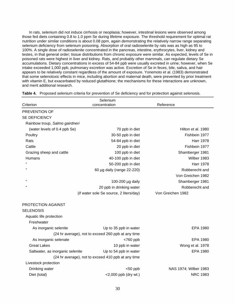
Table 4. Proposed selenium criteria for prevention of Se deficiency and for protection against selenosis.
Selenium Criterion concentration Reference
PREVENTION OF SE DEFICIENCY Rainbow troup, Salmo gairdneri (water levels of 0.4 ppb Se) 70 ppb in diet Hilton et al. 1980 Poultry 30-50 ppb in diet Fishbein 1977 Rats 54-84 ppb in diet Harr 1978 Cattle 20 ppb in diet Fishbein 1977 Grazing sheep and cattle 100 ppb in diet Shamberger 1981 Humans 40-100 ppb in diet Wilber 1983 " 50-200 ppb in diet Harr 1978 " 60 µg daily (range 22-220) Robberecht and Von Greichen 1982 " 100-200 µg daily Shamberger 1981 " 20 ppb in drinking water Robberecht and (if water sole Se source, 2 liters/day) Von Greichen 1982 PROTECTION AGAINST SELENOSIS Aquatic life protection Freshwater As inorganic selenite Up to 35 ppb in water EPA 1980 (24 hr average), not to exceed 260 ppb at any time As inorganic selenate <760 ppb EPA 1980 Great Lakes 10 ppb in water Wong et al. 1978 Saltwater, as inorganic selenite Up to 54 ppb in water EPA 1980 (24 hr average), not to exceed 410 ppb at any time Livestock protection Drinking water <50 ppb NAS 1974; Wilber 1983 Diet (total) <2,000 ppb (dry wt.) NRC 1983
30
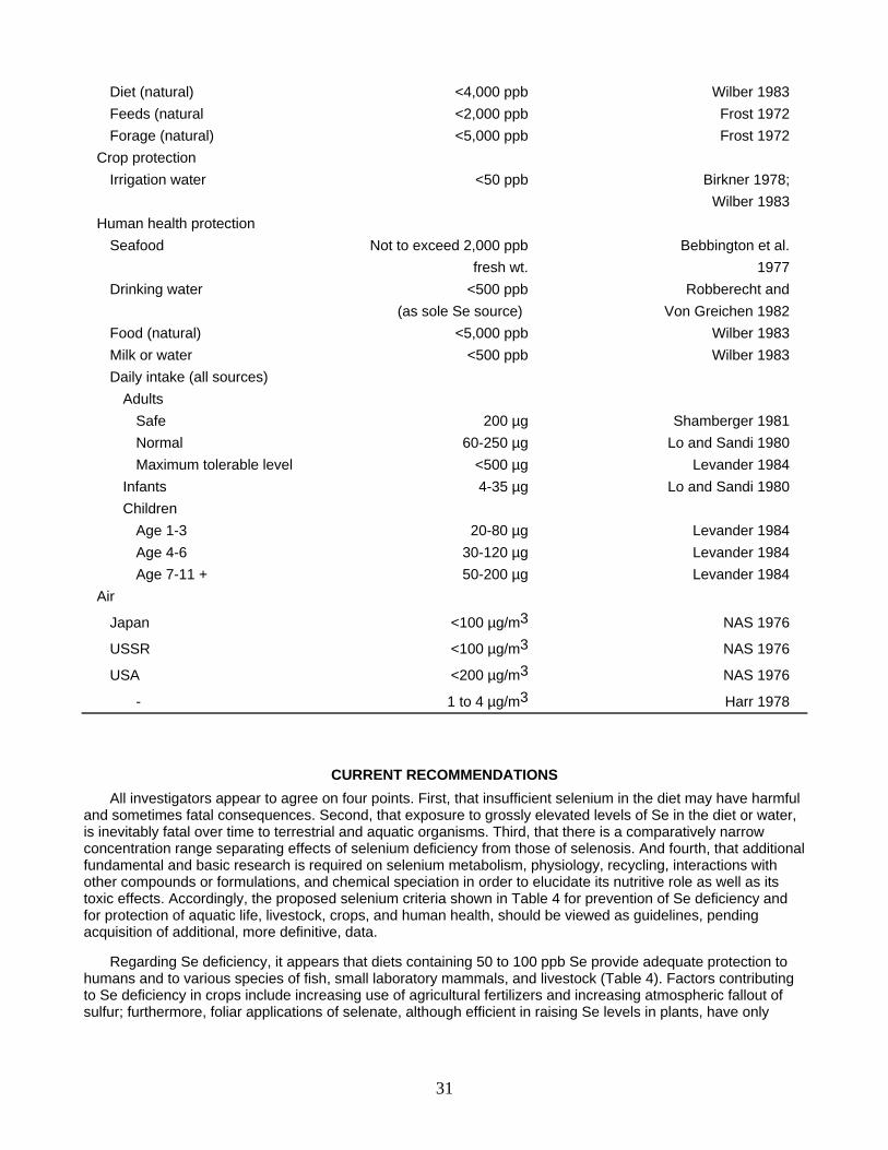
Diet (natural) <4,000 ppb Wilber 1983 Feeds (natural <2,000 ppb Frost 1972 Forage (natural) <5,000 ppb Frost 1972 Crop protection Irrigation water <50 ppb Birkner 1978; Wilber 1983 Human health protection Seafood Not to exceed 2,000 ppb Bebbington et al. fresh wt. 1977 Drinking water <500 ppb Robberecht and (as sole Se source) Von Greichen 1982 Food (natural) <5,000 ppb Wilber 1983 Milk or water <500 ppb Wilber 1983 Daily intake (all sources) Adults Safe 200 µg Shamberger 1981 Normal 60-250 µg Lo and Sandi 1980 Maximum tolerable level <500 µg Levander 1984 Infants 4-35 µg Lo and Sandi 1980 Children Age 1-3 20-80 µg Levander 1984 Age 4-6 30-120 µg Levander 1984 Age 7-11 + 50-200 µg Levander 1984 Air
Japan <100 µg/m3 NAS 1976
USSR <100 µg/m3 NAS 1976
USA <200 µg/m3 NAS 1976
- 1 to 4 µg/m3 Harr 1978
CURRENT RECOMMENDATIONS All investigators appear to agree on four points. First, that insufficient selenium in the diet may have harmful
and sometimes fatal consequences. Second, that exposure to grossly elevated levels of Se in the diet or water, is inevitably fatal over time to terrestrial and aquatic organisms. Third, that there is a comparatively narrow concentration range separating effects of selenium deficiency from those of selenosis. And fourth, that additional fundamental and basic research is required on selenium metabolism, physiology, recycling, interactions with other compounds or formulations, and chemical speciation in order to elucidate its nutritive role as well as its toxic effects. Accordingly, the proposed selenium criteria shown in Table 4 for prevention of Se deficiency and for protection of aquatic life, livestock, crops, and human health, should be viewed as guidelines, pending acquisition of additional, more definitive, data.
Regarding Se deficiency, it appears that diets containing 50 to 100 ppb Se provide adequate protection to humans and to various species of fish, small laboratory mammals, and livestock (Table 4). Factors contributing to Se deficiency in crops include increasing use of agricultural fertilizers and increasing atmospheric fallout of sulfur; furthermore, foliar applications of selenate, although efficient in raising Se levels in plants, have only
31
short-term value (Frost and Ingvoldstad 1975). There is a general consensus that Se deficiency in livestock in many countries is increasing, resulting in a need for added Se in the food chain.
Current recommendations for protection of freshwater aquatic life are that inorganic selenite concentrations in the water should not exceed 35 ppb on a daily average, or 260 ppb at any time (Table 4). These values are higher for saltwater life: 54 ppb daily average, 410 ppb at any time. The concentration range of 35 to 54 ppb recommended for protection of aquatic life is below the range of 60 to 600 ppb that is fatal to various sensitive species of marine and freshwater fauna, and in this respect affords an adequate measure of protection. It is very near the range (47 to 53 ppb) that has been associated with growth inhibition of freshwater algae, anemia and reduced hatch in rainbow trout, and shifts in species composition of freshwater algal communities. But studies by Cumbie and Van Horn (1978) and Sorensen et al. (1984) showed that water concentrations of 9 to 12 ppb of Se were associated with reduced reproduction of freshwater fishes, and their results strongly indicated that some downward modification of the Se freshwater aquatic life protection criterion may be appropriate. Furthermore, high bioconcentration and accumulation of Se from the water column by numerous species of algae, fish, and invertebrates is well documented at levels between 0.015 and 3.3 ppb, which is substantially below the recommended range of 35 to 54 ppb. The significance of Se residues in aquatic biota in terms of bioavailability and Se receptor sites is imperfectly understood, and it appears that much additional research is warranted on Se pharmacokinetics in aquatic environments.
Livestock appear to be protected against selenosis provided that their diets contain less than 4,000 ppb of natural (i.e., nonsupplemented) selenium (Table 4). This concentration is somewhat higher than levels reported for rats (Wilber 1983). Minimum toxic concentrations of Se in lifetime exposure of rats given diets containing natural Se were 1,400 ppb dietary Se, as judged by evidence of liver changes, and 3,000 ppb as estimated from longevity and histological changes in heart, kidney, and spleen. These values were only 350 and 750 ppb, respectively, when rat diets contained purified, rather than natural, Se. This relationship emphasizes that accidental poisoning of livestock, and presumably fish and wildlife, may occur when soils are deliberately supplemented with purified Se, or when soils or aquifers are contaminated as a result of faulty waste disposal practices. Although the concentration of <50 ppb for livestock drinking water and irrigation water for crop protection is inconsistent with that of < 35 ppb for aquatic life protection, neither livestock nor crops appear threatened at the higher level. Since many waterways that abut agricultural lands or areas of high anthropogenic loadings of Se contain valuable and desirable aquatic species, it would appear that some downward modification of the current livestock drinking water concentration of Se is necessary.
Recent field studies demonstrated that migratory waterfowl were heavily and adversely affected while nesting at Se-contaminated irrigation drainwater ponds in California, where food chain organisms contained between 12,000 and 280,000 ppb of Se. The source and fate of Se in irrigation drainwater ponds are largely unknown; they must be determined so that alternate technologies for Se control can be implemented to protect waterfowl in that geographical region.
Aerosol concentrations in excess of 4.0 µg Se/m3 are potentially harmful to human health (Harr 1978); concentrations in excess of this value (6.0 µg Se/m3) were regularly encountered in the vicinity of the smeltery at Sudbury, Canada (Nriagu and Wong 1983). It is not now known whether respiration rates of wildlife, particularly birds, are comparable to those of humans, or whether Se absorption energetics are similar, or whether wildlife species that frequent point sources of air contaminated by high Se levels for protracted periods are at greater risk than humans. Until additional and more conclusive data become available, aerosol concentrations of less than 4.0 µg Se/m3 are recommended for the protection of sensitive species of wildlife.
Acute lethal doses for livestock species ranged from 3,300 µg/kg body weight of Se for horses and mules to 15,000 µg/kg for swine; appetite loss in cattle was noted at 500 µg/kg. For humans, the maximum tolerance level is usually set at 500 µg of Se daily (Lo and Sandi 1980), and the “safe” level at 200 µg/day (Shamberger 1981). Selenium dietary levels for man should not exceed 5,000 ppb; however, recommended maximum dietary levels in other mammals ranged from 1,000 ppb for rats to 4,000 ppb for horses. For all species, including man, there is a tendency to list Se dosage levels in terms of “natural” and “supplemented” levels, with the tacit understanding that “natural” levels are about one-fourth as toxic as “supplemented” values. Given the complexities of Se metabolism and speciation, it appears that greater precision and clarity are necessary in formulating Se criteria if these criteria are to become administratively enforced standards through passage of appropriate legislation.
32
LITERATURE CITED Adams, W. J. 1976. The toxicity and residue dynamics of selenium in fish and aquatic invertebrates. Ph.D.
Thesis, Michigan State Univ., East Lansing. 109 pp.
Adams, W. J., and H. E. Johnson. 1977. Survey of the selenium content in the aquatic biota of western Lake Erie. J. Great Lakes Res. 3:10-14.
Anon. 1975. Preliminary investigation of effects on the environment of boron, indium, nickel, selenium, tin, vanadium and their compounds. Vol. IV. Selenium. U.S. Environ. Protection Agency Rep. 560/2-75-005d. Available as PB - 245 987 from Nat. Tech. Inform. Serv., U.S. Dep. Commer., Springfield, VA. 92 pp.
Beal, A. R. 1974. A study of selenium levels in freshwater fishes of Canada's Central Region. Canada Dep. Environ. Fish. Mar. Serv., Tech. Rep. Ser. CEN/T-74-6:14 pp.
Bebbington, G. N., N. J. Mackay, R. Chvojka, R. J. Williams, A. Dunn, and E. H. Auty. 1977. Heavy metals, selenium and arsenic in nine species of Australian commercial fish. Aust. J. Mar. Freshwater Res. 28:277-286.
Beijer, K., and A. Jernelov. 1978. Ecological aspects of mercury-selenium interactions in the marine environment. Environ. Health Perspec. 25:43-45.
Bertine, K. K., and E. D. Goldberg. 1972. Trace elements in clams, mussels, and shrimp. Limnol. Ocean. 17:877-884.
Beyer, W. N., R. L. Chaney, and B. M. Mulhern. 1982. Heavy metal concentrations in earthworms from soil amended with sewage sludge. J. Environ. Qual. 11:381-385.
Birkner, J. H. 1978. Selenium in aquatic organisms from seleniferous habitats. Ph.D. Thesis, Colorado State Univ., Fort Collins. 121 pp.
Bjerragaard, P. 1982. Accumulation of cadmium and selenium and their mutual interaction in the shore crab Carcinus maenus (L.). Aquat. Toxicol. 2:113-125.
Blus, L. J., B. S. Neely, Jr., T. G. Lamont, and B. Mulhern. 1977. Residues of organochlorines and heavy metals in tissues and eggs of brown pelicans, 1969-73. Pestic. Monitor. J. 11:40-53.
Bovee, E. C., and T. L. O'Brien. 1982. Some effects of selenium, vanadium and zirconium, on the swimming rate of Tetrahymena pyriformis: a bioassay study. Univ. Kans. Sci. Bull. 52 (4):39-44.
Bowen, H. J. M. 1966. Trace elements in biochemistry. Academic Press, New York. 241 pp.
Browne, C. L., and J. N. Dumont. 1979. Toxicity of selenium to developing Xenopus laevis embryos. J. Toxicol. Environ. Health 5:699-709.
Cappon, C. J., and J. C. Smith. 1981. Mercury and selenium content and chemical form in fish muscle. Arch. Environ. Contam. Toxicol. 10:305-319.
Cappon, C. J., and J. C. Smith. 1982. Chemical form and distribution of selenium in edible seafood. J. Anal. Toxicol. 6:10-21.
Cardwell, R. D., D. G. Foreman, T. R. Payne, and D. J. Wilbur. 1976. Acute toxicity of selenium dioxide to freshwater fishes. Arch. Environ. Contam. Toxicol. 4:129-144.
Chau, Y. K., and J. P. Riley. 1965. The determination of selenium in sea water, silicates and marine organisms. Anal. Chim. Acta 33:36-49.
Chau, Y. K., P. T. S. Wong, B. A. Silverberg, P. L. Luxon, and G. A. Bengert. 1976. Methylation of selenium in the aquatic environment. Science 192:1130-1131.
Cherry, D. S., R. K. Guthrie, J. H. Rodgers, Jr., J. Cairns, Jr., and K. L. Dickson. 1976. Responses of mosquitofish (Gambusia affinos) to ash effluent and thermal stress. Trans. Am. Fish. Soc. 105:686-694.
33
Cumbie, P. M., and S. L. Van Horn. 1978. Selenium accumulation associated with fish mortality and reproductive failure. Proc. Annu. Conf. Southeast Assoc. Fish Wildl. Agencies 32:612-624.
de Goeij, J. J. M., V. P. Guinn, D. R. Young, and A. J. Mearns. 1974. Neutron activation analysis trace-element, studies of dover sole liver and marine sediments. Pages 189-200 in Comparative studies of food and environmental contamination. Int. Atomic Energy Agency, Vienna.
Duncan, D. A., and J. F. Klaverkamp. 1983. Tolerance and resistance to cadmium in white suckers Catostomus commersoni previously exposed to cadmium, mercury, zinc, or selenium. Can.-J. Fish. Aquat. Sci. 40:128-138.
Ebens, R. J., and H. T. Shacklette. 1982. Geochemistry of some rocks, mine spoils, stream sediments, soils, plants, and waters in the western energy region of the conterminous United States. U.S. Geol. Surv. Prof. Paper 1237. U.S. Govt. Printing Off., Washington DC. 173 pp.
Eisler, R. 1981. Trace metal concentrations in marine organisms. Pergamon Press, New York. 687 pp.
Eisler, R. 1984. Trace metal changes associated with age of marine vertebrates. Biol. Trace Element Res. 6:165-180.
El-Begearmi, M. M., M. L. Sunde, and H. E- Ganther. 1977. A mutual protective effect of mercury and selenium in Japanese quail. Poult. Sci. 56:313-322.
El-Begearmi, M. M., H. E. Ganther, and M. L. Sunde. 1982. Dietary interaction between methylmercury, selenium, arsenic, and sulfur amino acids in Japanese quail. Poult. Sci. 61:272-279.
Ellis, M. M., H. L. Motley, M. D. Ellis, and R. O. Jones. 1937. Selenium poisoning in fishes. Proc. Soc. Exp. Biol. Med. 36:519-522.
EPA. 1980. Ambient water quality criteria for selenium. U.S. Environ. Protection Agency Rep. 440/5-80-070. 123 pp.
Fishbein, L. 1977. Toxicology of selenium and tellurium. Pages 191-240 in R. A. Goyer and M. A. Mehlman (eds.). Advances in modern toxicology, Vol. 2, Toxicology of trace elements. Hemisphere Publ. Corp., Washington DC.
Fishbein, L. 1983. Environmental selenium and its significance. Fundam. Appl. Toxicol. 3:411-419.
Fleming, W. J., W. H. Gutenmann, and D. J. Lisk. 1979. Selenium in tissues of woodchucks inhabiting fly ash landfills. Bull. Environ. Contam. Toxicol. 21:1-3.
Fowler, S. W., and G. Benayoun. 1976a. Selenium kinetics in marine zooplankton. Mar. Sci. Comm. 2:43-67.
Fowler, S. W., and G. Benayoun. 1976b. Influence of environmental factors on selenium flux in two marine invertebrates. Mar. Biol. 37:59-68.
Fowler, S. W., and G. Benayoun. 1976c. Accumulation and distribution of selenium in mussel and shrimp tissues. Bull. Environ. Contam. Toxicol. 16:339-346.
Freeman, H. C., G. Shum, and J. F. lithe. 1978. The selenium content in swordfish (Xiphias gladius) in relation to total mercury content. J. Environ. Sci. Health A13(3):235-240.
Frost, D. V. 1972. The two faces of selenium. Can selenophobia be cured? CRC Crit. Rev. Toxicol. 1:467-514.
Frost, D. V., and D. Ingvoldstad. 1975. Ecological aspects of selenium and tellurium in human and animal health. Chem. Scr. 8A:96-107.
Fukai, R., B. Oregioni, and D. Vas. 1978. Interlaboratory comparability of measurements of trace elements in marine organisms: results of intercalibration exercise on oyster homogenate. Oceanol. Acta 1:391-396.
34
Furr, A. K., T. F. Parkinson, W. D. Youngs, C. O. Berg, W. H. Gutenmann, I. S. Pakkala, and D. J. Lisk. 1979. Elemental content of aquatic organisms inhabiting a pond contaminated with coal fly ash. N.Y. Fish Game J. 26:154-161.
Ganther, H. E., C. Goudie, M. L. Sunde, M. J. Kopecky, P. Wagner, S. H. Oh, and W. G. Hoekstra. 1972. Selenium: relation to decreased toxicity of methylmercury added to diets containing tuna. Science 175:1122-1124.
Glickstein, N. 1978. Acute toxicity of mercury and selenium to Crassostrea gigas embryos and Cancer magister larvae. Mar. Biol. 49:113-117.
Glover, J. W. 1979. Concentrations of arsenic, selenium and ten heavy metals in school shark, Galeorhinus australis (Macleay), and gummy shark, Mustelus antarcticus Gunt south-eastern Australian waters. Aust. J. Mar. Freshwater Res. 30:505-510.
Gotsis, O. 1982. Combined effects of selenium/mercury and selenium/copper on the cell population of the alga Dunaliella minuta. Mar. Biol. 71:217-222.
Grimanis, A. P., D. Zafiropoulos, and M. Vassilaki-Grimani. 1978. Trace elements in the flesh and liver of two fish species from polluted and unpolluted areas of the Aegean Sea. Environ. Sci. Technol. 12:723-726.
Hall, R. A., E. G. Zook, and G. M. Meaburn. 1978. National Marine Fisheries Service survey of trace elements in the fishery resource. U.S. Dep. Commer. NOAA Tech. Rep. NMFS SSRF-721. 313 pp.
Halter, M. T., W. J. Adams, and H. E. Johnson. 1980. Selenium toxicity to Daphnia magna, Hyallela azteca, and the fathead minnow in hard water. Bull. Environ. Contam. Toxicol. 24:102-107.
Harr, J. R. 1978. Biological effects of selenium. Pages 393-426 in F. W. Oehme (ed.). Toxicity of heavy metals in the environment, Part l. Marcel Dekker, New York.
Haseltine, S. D., G. H. Heinz, W. L. Reichel, and J. F. Moore. 1981. Organochlorine and metal residues in eggs of waterfowl nesting on islands in Lake Michigan off Door County, Wisconsin, 1977-78. Pestic. Monitor. J. 15:90-97.
Haseltine, S. D., J. S. Fair, S. A. Sutcliffe, and D. M. Swineford. 1983. Trends in organochlorine and mercury residues in common loon (Gavia immer) eggs from New Hampshire. Pages 131-141 in R. H. Yahner (ed.), Transactions of the Northeast section, The Wildlife Society, 40th Northeast Fish and Wildlife Conference, May 15-18, 1983, West Dover, VT.
Heit, M. 1979. Variability of the concentrations of seventeen trace elements in the muscle and liver of a single striped bass, Morone saxatilis. Bull. Environ. Contam. Toxicol. 23:1-5.
Heit, M., C. S. Klusek, and K. M. Miller. 1980. Trace element, radionuclide, and polynuclear aromatic hydrocarbon concentrations in Unionidae mussels from northern Lake George. Environ. Sci. Technol. 14:465-468.
Helmke, P. A., W. P. Robarge, R. L. Korotev, and P. J. Schomberg. 1979. Effects of soil-applied sewage sludge on concentrations of elements in earthworms. J. Environ. Qual. 8:322-327.
Hill, C. H. 1975. Interrelationships of selenium with other trace elements. Fed. Proc. 34:2096-2100.
Hilton, J. W., P. V. Hodson, and S. J. Slinger. 1980. The requirement and toxicity of selenium in rainbow trout (Salmo gairdneri). J. Nutr. 110:2527-2535.
Hodson, P. V., D. J. Spry, and B. R. Blunt. 1980. Effects on rainbow trout (Salmo gairdneri) of a chronic exposure to waterborne selenium. Can. J. Fish. Aquat. Sci. 37:233-240.
Huckabee, J. W., and N. A. Griffith. 1974. Toxicity of mercury and selenium to the eggs of carp (Cyprinus carpio). Trans. Am. Fish. Soc. 103:822-825.
35
Hutchinson, T. C., and P. M. Stokes. 1975. Heavy metal toxicity and algal bioassays. Pages 320-343 in S. Barabas (ed.). Water quality parameters. ASTM Spec. Tech. Publ. 573. American Society for Testing and Materials, 1916 Race Street, Philadelphia, PA.
Hutton, M. 1981. Accumulation of heavy metals and selenium in three seabird species from the United Kingdom. Environ. Pollut. 26A:129-145.
Jenkins, D. W. 1980. Biological monitoring of toxic trace metals. Vol. 2. Toxic trace metals in plants and animals of the world. Part III. U.S. Environ. Protection Agency Rep. 600/3-80-0921:1090-1129.
Jensen, L. S. 1968. Selenium deficiency and impaired reproduction in Japanese quail. Proc. Soc. Exp. Biol. Med. 128:970-972.
Jones, J. B., and T. C. Stadtman. 1977. Methanococcus vanniellii: culture and effects of selenium and tungsten on growth. J. Bacteriol. 130:1404-1406.
Kaiser, I. I., P. A. Young, and J. D. Johnson. 1979. Chronic exposure of trout to waters with naturally high selenium levels: effects on transfer RNA methylation. J. Fish. Res. Board Canada 36:689-694.
Karbe, L., Ch. Schnier, and H. O. Slewers. 1977. Trace elements in mussels (Mytilus edulis) from coastal areas of the North Sea and the Baltic. Multielement analyses using instrumental neutron activation analysis. J. Radioanal. Chem. 37:927-943.
Kari, T., and P. Kauranen. 1978. Mercury and selenium contents of seals from fresh and brackish water in Finland. Bull. Environ. Contam. Toxicol. 19:273-280.
Kifer, R. R., and W. L. Payne. 1968. Selenium content of fish meal. Feedstuffs 31 (Aug 68):32.
Kim, J. H., E. Birks, and J. F. Heisinger. 1977. Protective action of selenium against mercury in northern creek chubs. Bull. Environ. Contam. Toxicol. 17:132-136.
King, K. A., D. L. Meeker, and D. M. Swineford. 1980. White-faced ibis populations and pollutants in Texas, 1969-1976. Southwest. Nat. 25:225-240.
King, K. A., C. A. Lefever, and B. M. Mulhern. 1983. Organochlorine and metal residues in royal terns nesting on the central Texas coast. J. Field Ornithol. 54:295-303.
Klaverkamp, J. F., D. A. Hodgkins, and A. Lutz. 1983a. Selenite toxicity and mercury-selenium interactions in juvenile fish. Arch. Environ. Contam. Toxicol. 12:405-413.
Klaverkamp, J. F., W. A. Macdonald, W. R. Lillie, and A. Lutz. 1983b. Joint toxicity of mercury and selenium in salmonid eggs. Arch. Environ. Contam. Toxicol. 12:415-419.
Koeman, J. H., W. S. M. van de Ven, J. J. M. de Goeij, and P. S. Tijoe. 1975. Mercury and selenium in marine mammals and birds. Sci. Total Environ. 3:279-287.
Krishnaja, A. P., and M. S. Rege. 1982. Induction of chromosomal aberrations in fish Boleophthalmus dussumieri after exposure in vivo to mitomycin C and heavy metals mercury, selenium and chromium. Mutation Res. 102:71-82.
Kuhn, J. K., F. L. Fiene, R. A. Cahill, H. J. Gluskoter, and N. F. Shimp. 1980. Abundance of trace and minor elements in organic and mineral fractions of coal. Environ. Geol. Notes 88. Illinois State Geol. Surv. Div., Urbana, IL. 67 pp.
Kumar, H. D., and G. Prakash. 1971. Toxicity of selenium to the blue-green algae, Anacystis nidulans and Anabaena variabilis. Ann. Bot. 35:697-705.
Lakin, H. W. 1973. Selenium in our environment. Pages 96-111 in E. L. Kothny (ed.). Trace elements in the environment. Adv. Chemistry Ser., Am. Chem. Soc., Washington, D.C.
36
Lemly, A. D. 1982a. Response of juvenile centrarchids to sublethal concentrations of waterborne selenium. I. Uptake, tissue distribution, and retention. Aquat. Toxicol. 2:235-282.
Lemly, A. D. 1982b. Determination of selenium, in fish tissues with differential pulse polarography. Environ. Technol. Lett. 3:497-502.
Leonzio, C., S. Focardi, and E. Bacci. 1982. Complementary accumulation of selenium and mercury in fish muscle. Sci. Total Environ. 24:249-254.
Levander, O. A. 1983. Recent developments in selenium nutrition. Pages 147-162 in J. Weininger and C. Briggs (eds.). Nutrition update, Vol. 1. John Wiley, New York.
Levander, O. A. 1984. The importance of selenium in total parenteral nutrition. Bull. N.Y. Acad. Med. 60:144-155.
Lo, M. T., and E. Sandi. 1980. Selenium: occurrence in foods and its toxicological significance. A review. J. Environ. Pathol. Toxicol. 4:193-218.
Lucas, H. F., D. N. Edgington, and P. J. Colby. 1970. Concentrations of trace elements in Great Lakes fishes. J. Fish. Res. Board Canada 27:677-684.
Lucu, C., and M. Skreblin. 1981. Evidence of the interaction of mercury and selenium in the shrimp Palaemon elegans. Mar. Environ. Res. 5:265-274.
Lunde, G. 1970. Analysis of arsenic and selenium in marine raw materials. J. Sci. Food Agric. 21:242-247.
Luten, J. B., A. Ruiter, T. M. Ritskes, A. B. Rauchbaar, and G. Riekwel-Booy. 1980. Mercury and selenium in marine and freshwater fish. J. Food Sci. 45:416-419.
Mackay, N. J., M. N. Kazacos, R. J. Williams, and M. I. Leedow. 1975. Selenium and heavy metals in black marlin. Mar. Pollut. Bull. 6:57-60.
Maher, W. A. 1983. Selenium in marine organisms from St. Vincent's Gulf, South Australia. Mar. Pollut. Bull. 14:35-36.
Martin, J. H., P. D. Elliot, V. C. Anderlini, D. Girvin, S. A. Jacobs, R. W. Risebrough, R. L. Delong, and W. G. Gilmartin. 1976. Mercury-seleniumbromine imbalance in premature parturient California sea lions. Mar. Biol. 35:91-104.
May, T. W., and G. L. McKinney. 1981. Cadmium, lead, mercury, arsenic, and selenium concentrations in freshwater fish, 1976-77 - National Pesticide Monitoring Program. Pestic. Monitor. J. 15:14-38.
Measures, C. I., and J. D. Burton. 1980. The vertical distribution and oxidation states of dissolved selenium in the northeast Atlantic ocean and their relationship to biological processes. Earth Plan. Sci. Lett. 46:385-396.
Moksnes, K. 1983. Selenium deposition in tissues and eggs of laying hens given surplus of selenium as selenomethionine- Acta Vet. Scand. 24:34-44.
Morris, J. G., W. S. Cripe, H. L. Chapman, Jr. , D. F. Walker, J. B. Armstrong, J. D. Alexander, Jr., R. Miranda, A. Sanchez, Jr., B. Sanchez, J. R. Blair-West, and D. A. Denton. 1984. Selenium deficiency in cattle associated with Heinz bodies and anemia. Science 223:491-493.
Murphy, C. P. 1981. Bioaccumulation and toxicity of heavy metals and related trace elements. J. Water Pollut. Control Fed. 53:993-999.
NAS. 1973. Water quality criteria 1972. U.S. Environ. Protection Agency Rep. R3-73-033. 594 pp.
NAS. 1976. Selenium. Comm. Med. Biol. Eff. Environ. Poll., Div. Med. Sci., Natl. Res. Counc., Nat. Acad. Sci., Washington, DC. 203 pp.
37
Nassos, P. A., J. R. Coats, R. L. Metcalf, D. D. Brown, and L. G. Hansen. 1980. Model ecosystem, toxicity, and uptake evaluation of 75Se-selenite. Bull. Environ. Contam. Toxicol. 24:752-758.
Nielsen, M. G., and G. Gissel-Nielsen. 1975. Selenium in soil-animal relationships. Pedobiologia 15:65-67.
Niimi, A. J., and Q. N. LaHam. 1975. Selenium toxicity on the early life stages of zebrafish (Brachydanio rerio). J. Fish. Res. Board Canada 32:803-806.
Noda, K., S. Hirai, K. Sunayashiki, and H. Danbara. 1979. Neutron activation analyses of selenium and mercury in marine products from along the coast of Shikoku Island. Agric. Biol. Chem. 47:1381-1386.
NRC. 1983. Selenium in nutrition. Subcomm. on selenium, Comm. animal nutrition, Board on Agricul., Natl. Res. Counc., National Academy Press, Washington, DC. 174 pp.
Nriagu, J. O., and H. K. Wong. 1983. Selenium pollution of lakes near the smelters at Sudbury, Ontario. Nature 301:55-57.
Ohlendorf, H. M., and C. S. Harrison. 1986. Mercury, selenium, cadmium, and organochlorines in eggs of three Hawaiian seabird species. Environ. Pollut. 11B:169-191.
Ohlendorf, H. M., D. J. Hoffman, M. K. Saiki, and T. W. Aldrich. 1986. Embryonic mortality and abnormalities of aquatic birds: apparent impacts of selenium from irrigation drainwater. Sci. Total Environ. 52:49-63.
Okazaki, R. K., and M. H. Panietz. 1981. Depuration of twelve trace metals in tissues of the oysters Crassostrea gigas and C. virginica. Mar. Biol. 63:113-120.
Ort, J. F., and J. D. Latshaw. 1978. The toxic level of sodium selenite in the diet of laying chickens. J. Nutr. 108:1114-1120.
Orvini, E., V. Caramella-Crespi, and N. Genova. 1980. Activation analysis of As, Hg and Se in some marine organism. Pages 441-448 in J. Albaiges (ed.). Analytical techniques in environmental chemistry. Pergamon Press, New York.
Ostadalova, I., and A. Babicky. 1980. Toxic effect of various selenium compounds on the rat in the early postnatal period. Arch. Toxicol. 45:207-211.
Pakkala, I. S., W. H. Gutenmann, D. J. Lisk, G. E. Burdick, and E. J. Harris. 1972. A survey of the selenium content of fish from 49 New York State waters. Pestic. Monitor. J. 6:107-114.
Papadopoulu, C., G. D. Kanias, and E. M. Kassimati. 1976. Stable elements of radioecological importance in certain echinoderm species. Mar. Pollut. Bull. 7:143-144.
Patrick, R. 1978. Effects of trace metals in the aquatic ecosystem. Am. Sci. 66:185-191.
Prasad, T., S. P. Arora, and R. C. Chopra. 1982. Selenium toxicity as induced by feeding rice husk to buffalo calves: a clinical case report. Indian Vet. J. 59:235-237.
Pratt, D. R., J. S. Bradshaw, and B. West. 1972. Arsenic and selenium analyses in fish. Utah Acad. Arts Sci. Proc., Part 1, 49:23-26.
Rancitelli, L. A., W. A. Haller, and J. A. Cooper. 1968. Trace element variations in silver salmon and king salmon muscle tissue. Rapp. Americain BNWL - 715, Part 2: 42-47.
Reading, J. T. 1979. Acute and chronic effects of selenium on Daphnia pulex. Thesis. Virginia Polytechnic Institute, Blacksburg. 91 pp.
Reddy, C. C., and E. J. Massaro. 1983. Biochemistry of selenium: a brief overview. Fundam. Appl. Toxicol. 3:431-436.
Reijnders, P. J. H. 1980. organochlorine and heavy metal residues in harbour seals from the Wadden Sea and their possible effects on reproduction. Neth. J. Sea Res. 14:30-65.
38
Robberecht, H., and R. Von Grieken. 1982. Selenium in environmental waters: determination, speciation and concentration levels. Talanta 29:823-844.
Rosenfeld, I., and O. A. Beath. 1964. Selenium. Geobotany, biochemistry, toxicity, and nutrition. Academic Press, New York. 411 pp.
Rossi, L. C., G. F. Clemente, and G. Santaroni. 1976. Mercury and selenium distribution in a defined area and in its population. Arch. Environ. Health 31:160-165.
Ryther, J., T. M. Losordo, A. K. Furr, T. F. Parkinson, W. H. Gutenmann, I. S. Pakkala, and D. J. Lisk. 1979. Concentration of elements in marine organisms cultured in seawater flowing through coal-fly ash. Bull. Environ. Contam. Toxicol. 23:207-210.
Sandholm, M. 1973. Biological aspects of selenium: uptake of selenium by SH- groups and different organic materials in the ecosystem. Thesis, Dept. Medicine, Coll. Veterinary Medicine, Helsinki, Finland. 35 pp.
Sato, T., Y. Ose, and T. Sakai. 1980. Toxicological effect of selenium on fish. Environ. Pollut. 21A:217-224.
Schroeder, H. A., D. V. Frost, and J. J. Balassa. 1970. Essential trace metals in man: selenium. J. Chron. Dis. 23:227-243.
Schultz, C. D., and B. M. Ito. 1979. Mercury and selenium in blue marlin, Makaira nigricans, from the Hawaiian Islands. U.S. Natl. Mar. Fish. Serv., Fish Bull. 76:872-879.
Shamberger, R. J. 1981. Selenium in the environment. Sci. Total Environ. 17:59-74.
Sharma, R. P., and J. L. Shupe. 1977. Trace metals in ecosystems: relationships of residues of copper, molybdenum, selenium, and zinc in animal tissues to those in vegetation and soil in the surrounding environment. Pages 595-608 in H. Drucker and R. E. Wildung (eds.). Biological implications of metals in the environment. Avail. as CONF 750929 from Nat. Tech. Infor. Serv., U.S. Dep. Commer., Springfield VA.
Sheline, J., and B. Schmidt-Nielsen. 1977. Methylmercury-selenium interaction in the killifish, Fundulus heteroclitus. Pages 119-130 in F. J. Vernberg, A. Calabrese, F. P. Thur berg and W. B. Vernberg (eds.). Physiological responses of marine biota to pollutants. Academic Press, New York.
Smith, T. G., and F. A. J. Armstrong. 1978. Mercury and selenium in ringed and bearded seal tissues from Arctic Canada. Arctic 31:75-84.
Sorensen, E. B., T. L. Bauer, J. S. Bell, and C. W. Harlan. 1982. Selenium accumulation and cytotoxicity in teleosts following chronic, environmental exposure. Bull. Environ. Toxicol. Contam. 29:688-696.
Sorensen, E. M. B., P. M. Cumbie, T. L. Bauer, J. S. Bell, and C. W. Harlan. 1984. Histopathological, hematological, condition-factor, and organ weight changes associated with selenium accumulation in fish from Belews Lake, North Carolina. Arch. Environ. Contam. Toxicol. 13:153-162.
Spehar, R. L., G. M. Christensen, C. Curtis, A. E. Lemke, T. J. Norberg, and Q. H. Pickering. 1982. Effects of pollution on freshwater fish. J. Water Pollut. Control Fed. 54:877-922.
Speyer, M. R. 1980. Mercury and selenium concentrations in fish, sediments, and water of two northwestern Quebec lakes. Bull. Environ. Contam. Toxicol. 24:427-432.
Stadtman, T. C. 1974. Selenium biochemistry. Science 183:915-921.
Stadtman, T. C. 1977. Biological function of selenium. Nutr. Rev. 35:161-166.
Stoneburner, D. L. 1978. Heavy metals in tissues of stranded short-finned pilot whales. Sci. Total Environ. 9:293-297.
Stump, I. G., J. Kearney, J. M. D'Auria, and J. D. Popham. 1979. Monitoring trace elements in the mussel, Mytilus edulis using X-ray energy spectroscopy. Mar. Pollut. Bull. 10:270-274.
39
Tamura, Y., T. Maki, H. Yamada, Y. Shimamura, S. Ochiai, S. Nishigaki, and Y. Kimura. 1975. Studies on the behavior of accumulation of trace elements in fishes. III. Accumulation of selenium and mercury in various tissues of tuna. Tokyo Toritsu Eisei Kenkyusho Nenpo 26:200-204. (In Japanese. translation available from multilingual Serv., Dep. Secr. State Canada, Fish. Mar. Serv. No. 3994, 1977:11 pp.)
Tijoe, P. S., J. J. M. de Goeij, and M. de Bruin. 1977. Determination of trace elements in dried sea-plant homogenate (SP-M-1) and in dried copepod homogenate (MA-A-1) by means of neutron activation analysis. Interuniv. Reactor Inst. Rept. 133-77-05, Delft, Nederlands:14 pp.
Tong, S. S. C., W. H. Gutenmann, D. J. Lisk, G. E. Burdick, and E. J. Harris. 1972. Trace metals in New York State fish. N.Y. Fish Game J. 19:123-131.
Turner, J. S., S. R. B. Solly, J. C. M. Mol-Krijnen, and V. Shanks. 1978. Organochlorine, fluorine, and heavy-metal levels in some birds from N. Z. estuaries. N. Z. J. Sci. 21:99-102.
Ullrey, D. E., W. G. Youatt, and P. A. Whetter. 1981. Muscle selenium concentrations in Michigan deer. J. Wildl. Manage. 45:534-536.
United Nations. 1979. Review of harmful substances. Reports and studies. No. 2. Jnt. Group Experts Sci. Aspect. Mar. Pollut. GESAMP-76-98245. N.Y.:79 pp.
Uthe, J. F., and E. G. Bligh. 1971. Preliminary survey of heavy metal contamination of Canadian freshwater fish. J. Fish. Res. Board Canada 28:786-788.
van de Ven, W. S. M., J. H. Koeman and A. Svenson. 1979. Mercury and selenium in wild and experimental seals. Chemosphere 8:539-555.
Walsh, D. F., B. L. Berger, and J. R. Bean. 1977. Mercury, arsenic, lead, cadmium and selenium residues in fish, 1971-73. National Pesticide Monitoring Program. Pestic. Monitor. J. 11:5-34.
Ward, G. S., T. A. Hollister, P. T. Heitmuller, and P. R. Parrish. 1981. Acute and chronic toxicity of selenium to estuarine organisms. Northeast Gulf Sci. 4:73-78.
Westernman, A. G., and W. J. Birge. 1978. Accelerated rate of albinism in channel catfish exposed to metals. Prog. Fish-Cult. 40:143-146.
White, D. H., K. A. King, and R. M. Prouty. 1980. Significance of organochlorine and heavy metal residues in wintering shorebirds at Corpus Christi, Texas, 1976-77. Pestic. Monitor. J. 14:58-63.
Whittle, K. J., R. Hardy, A. V. Holden, R. Johnston, and R. J. Pentreath. 1977. Occurrence and fate of organic and inorganic contaminants in marine animals. Ann. N.Y. Acad. Sci. 298:47-79.
Wiener, J. G , G. A. Jackson, T. W. May, and B. P. Cole. 1984. Longitudinal distribution of trace elements (As, Cd, Cr, Hg, Pb, and Se) in fishes and sediments in the upper Mississippi River. Pages 139-170 in J. G. Weiner, R. V. Anderson, and D. R. McConville (eds.). Contaminants in the upper Mississippi River. Butterworth Publ., Stoneham, MA.
Wilber, C. G. 1980. Toxicology of selenium: A review. Clin. Toxicol. 17:171-230.
Wilber, C. G. 1983. Selenium. A potential environmental poison and a necessary food constituent. Charles C. Thomas, Springfield IL. 126 pp.
Willford, W. A. 1971. Heavy metals research in the Great Lakes, 1970-1971. Pages 53-65 in Prevalence and effects of toxic metals in the aquatic environment. Proc. Univ. North Carolina, Rep. 57.
Winger, P. V., C. Sieckman, T. W. May, and W. W. Johnson. 1984. Residues of organochlorine insecticides, polychlorinated biphenyls, and heavy metals in biota from Apalachicola River, Florida, 1978. J. Assoc. Off. Anal. Chem. 67:325-333.
Wong, P. T. S., Y. K. Chau, and P. L. Luxon. 1978. Toxicity of a mixture of metals on freshwater algae. J. Fish. Res. Board Canada 35:479-481.
40
Wren, C. D. 1984 Distribution of metals in tissues of beaver, raccoon and otter from Ontario, Canada. Sci. Total Environ. 34:177-184.
Wrench, J. J., and N. C. Campbell. 1981. Protein bound selenium in some marine organisms. Chemosphere 10:1155-1161.
Yonemoto, J., H. Satoh, S. Himeno, and T. Suzuki. 1983. Toxic effects of sodium selenite on pregnant mice and modification of the effects by Vitamin E or reduced glutathione. Teratology 28:333-340.
Zafiropoulos, D., and A. P. Grimanis. 1977. Trace elements in Acartia clausi from Elefsis Bay of the upper Saronikos Gulf, Greece. Ma . Pollut. Bull. 8:79-81.
Zieve, R. and P. J. Peterson. 1981. Factors influencing the volatilization of selenium from soil. Sci. Total Environ. 19:277-284.
Zingaro, R. A., and W. C. Cooper (eds.). 1974. Selenium. Van Nostrand Reinhold Co., New York. 835 pp.
41