Embed Size (px)
Citation preview
Selenocysteine:
The 21st Amino Acid
Amanda L. Garner
October 5, 2007
C
CH2
CO2H
H2N H
SeH
Sec, U
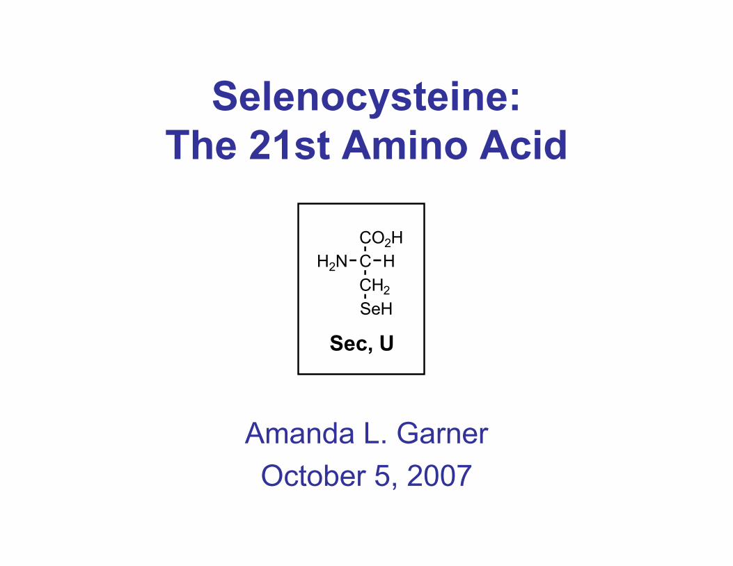
Selenocysteine - Discovery
Chronology:
– Selenium (Se) initially believed to be toxic (more
reactive than sulfur)
– 1954: First report of Se requirement for function of
bacterial formate dehydrogenase
– 1959: First paper addressing selenocysteine (Sec)
– 1972: First biochemical studies on role of Se at
enzyme level
– 1986: Key study implicating UGA as codon for Sec
Se currently regarded as essential nutrient in humans
and mammals (development, immune function, male
reproduction, aging process)
C
CH2
CO2H
H2N H
SeH
Sec, U
Pinsent, J. Biochem. J. 1954, 57, 10. Shum, A. C.; Murphy, J. C. J. Bacteriol. 1972, 110, 447.
Bock, A. PNAS 1986, 83, 4650.
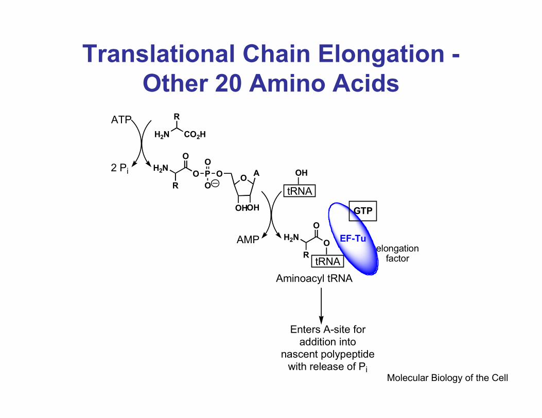
Translational Chain Elongation -
Other 20 Amino Acids
Molecular Biology of the Cell
ATP
2 Pi
H2N CO2H
R
OO
OHOH
AP
O
O
OH2N
O
RtRNA
OH
tRNA
O
O
H2N
R
AMP
Aminoacyl tRNA
EF-Tu
GTP
elongationfactor
Enters A-site for
addition into nascent polypeptide
with release of Pi
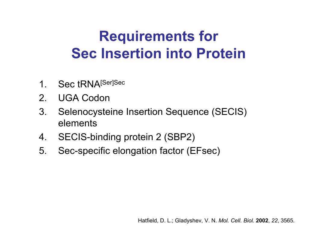
Requirements for
Sec Insertion into Protein
1. Sec tRNA[Ser]Sec
2. UGA Codon
3. Selenocysteine Insertion Sequence (SECIS)
elements
4. SECIS-binding protein 2 (SBP2)
5. Sec-specific elongation factor (EFsec)
Hatfield, D. L.; Gladyshev, V. N. Mol. Cell. Biol. 2002, 22, 3565.
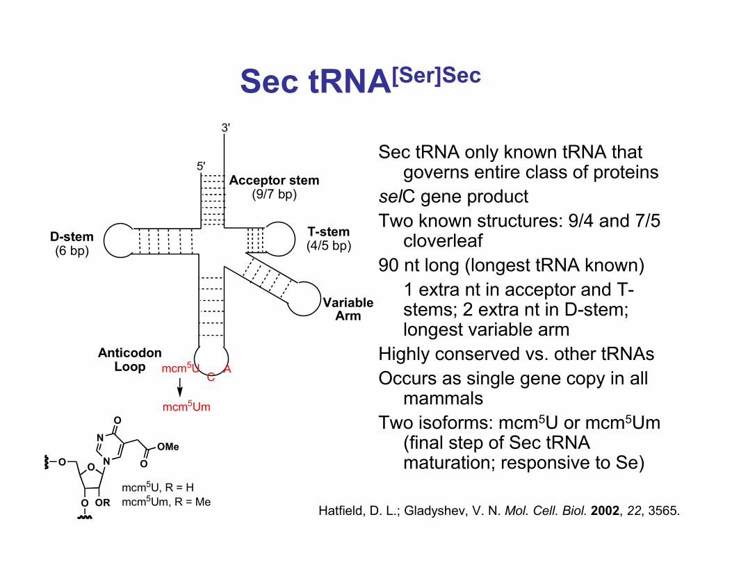
Sec tRNA[Ser]Sec
Sec tRNA only known tRNA that governs entire class of proteins
selC gene product
Two known structures: 9/4 and 7/5 cloverleaf
90 nt long (longest tRNA known)
1 extra nt in acceptor and T-stems; 2 extra nt in D-stem; longest variable arm
Highly conserved vs. other tRNAs
Occurs as single gene copy in all mammals
Two isoforms: mcm5U or mcm5Um (final step of Sec tRNA maturation; responsive to Se)
5'
3'
mcm5U
mcm5Um
CA
Acceptor stem(9/7 bp)
T-stem(4/5 bp)
D-stem(6 bp)
Variable Arm
Anticodon Loop
OO
O OR
N
N
O
OMe
O
mcm5U, R = H
mcm5Um, R = MeHatfield, D. L.; Gladyshev, V. N. Mol. Cell. Biol. 2002, 22, 3565.
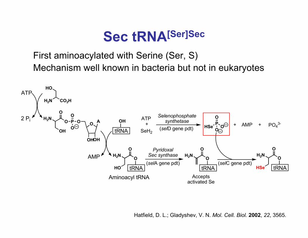
Sec tRNA[Ser]Sec
First aminoacylated with Serine (Ser, S)
Mechanism well known in bacteria but not in eukaryotes
Hatfield, D. L.; Gladyshev, V. N. Mol. Cell. Biol. 2002, 22, 3565.
ATP
2 Pi
H2N CO2H
HO
OO
OHOH
AP
O
O
OH2N
O
OH tRNA
OH
tRNA
O
O
H2N
HO
AMP
Aminoacyl tRNA
Pyridoxal Sec synthase
tRNA
O
O
H2N
tRNA
O
O
H2N
HSe
Accepts activated Se
P
O
OOHSe
ATP+
SeH2
+ +AMP PO43-
(selA gene pdt)
Selenophosphatesynthetase
(selD gene pdt)
(selC gene pdt)
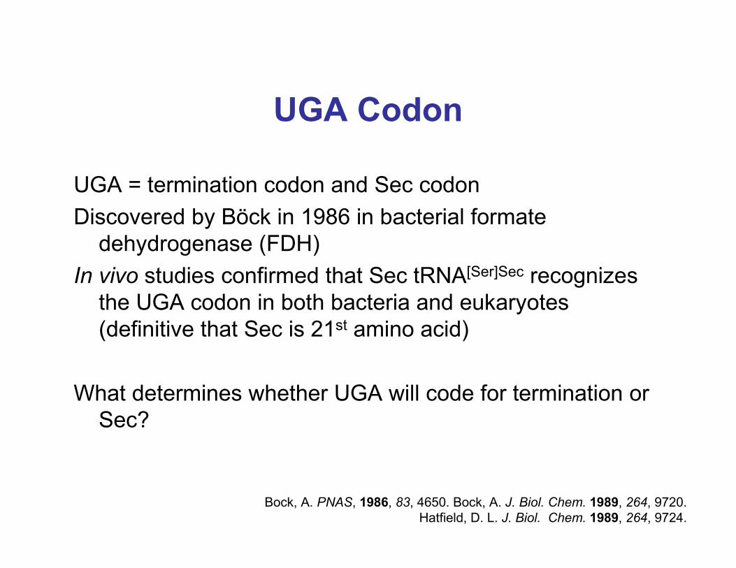
UGA Codon
UGA = termination codon and Sec codon
Discovered by Böck in 1986 in bacterial formate
dehydrogenase (FDH)
In vivo studies confirmed that Sec tRNA[Ser]Sec recognizes
the UGA codon in both bacteria and eukaryotes
(definitive that Sec is 21st amino acid)
What determines whether UGA will code for termination or
Sec?
Bock, A. PNAS, 1986, 83, 4650. Bock, A. J. Biol. Chem. 1989, 264, 9720.
Hatfield, D. L. J. Biol. Chem. 1989, 264, 9724.
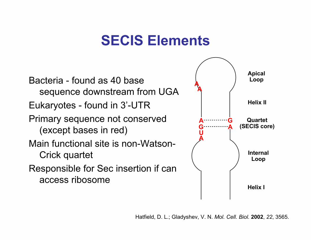
SECIS Elements
Bacteria - found as 40 base
sequence downstream from UGA
Eukaryotes - found in 3’-UTR
Primary sequence not conserved
(except bases in red)
Main functional site is non-Watson-
Crick quartet
Responsible for Sec insertion if can
access ribosome
AGUA
GA
Apical Loop
Helix II
Quartet(SECIS core)
Internal Loop
Helix I
AA
Hatfield, D. L.; Gladyshev, V. N. Mol. Cell. Biol. 2002, 22, 3565.
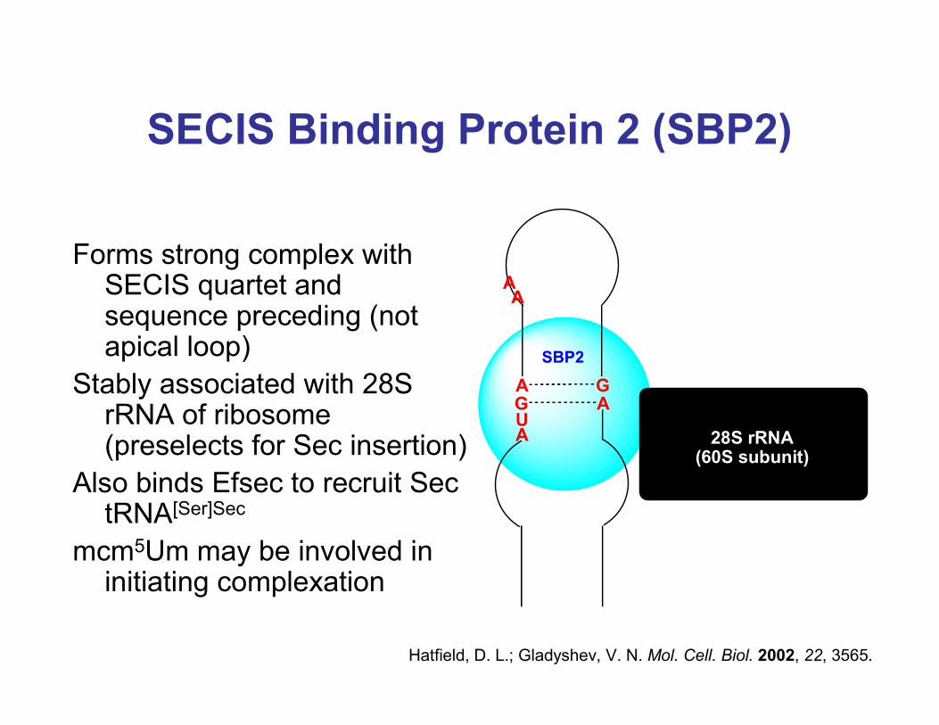
SECIS Binding Protein 2 (SBP2)
Forms strong complex with SECIS quartet and sequence preceding (not apical loop)
Stably associated with 28S rRNA of ribosome (preselects for Sec insertion)
Also binds Efsec to recruit Sec tRNA[Ser]Sec
mcm5Um may be involved in initiating complexation
Hatfield, D. L.; Gladyshev, V. N. Mol. Cell. Biol. 2002, 22, 3565.
AGUA
GA
AA
SBP2
28S rRNA (60S subunit)
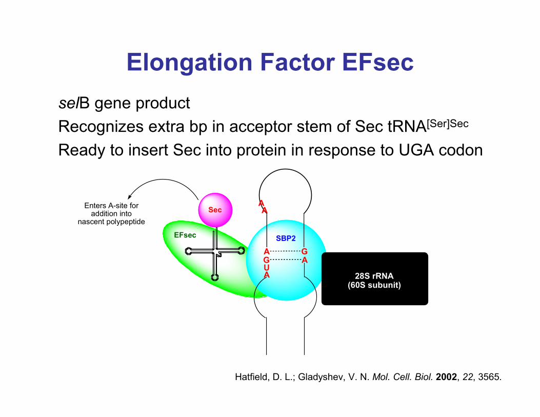
Elongation Factor EFsec
selB gene product
Recognizes extra bp in acceptor stem of Sec tRNA[Ser]Sec
Ready to insert Sec into protein in response to UGA codon
Hatfield, D. L.; Gladyshev, V. N. Mol. Cell. Biol. 2002, 22, 3565.
AGUA
GA
AA
SBP2
28S rRNA (60S subunit)
EFsec
SecEnters A-site for
addition into nascent polypeptide
Selenocysteine - Evolution
Evolution - believed to be a latter addition to the existing
genetic code of 20 amino acids
Sec found in bacteria, archaea, and eukaryotes
Bacteria/archaea - found in proteins responsible for
catabolic processes
Eukaryotes - found in proteins responsible for anabolic
and regulatory pathways
Increased use of Sec in proteins of higher vertebrates
*Sec is an evolutionary advancement
Gladyshev, V. N. BioFactors, 2001, 14, 87.
Examples of Known Selenoproteins
Prokaryotes:
1. Glycine reductase (first known selenoprotein; 1973)
2. Formate dehydrogenase
3. Selenophosphate synthetase
Eukaryotes:
1. Glutathione peroxidase (first known selenoprotein; 1973)
2. Thioredoxin reductase
3. Thyroid hormone deiodinase
4. Selenophosphate synthetase
5. Selenoprotein P
Only 12 eukaryote selenoproteins with known function
25 total selenoproteins contained in the human genome
Gladyshev, V. N. BioFactors, 2001, 14, 87. Burk, R. E., Hill, K. E. Annu. Rev. Nutr. 2005, 25, 215.
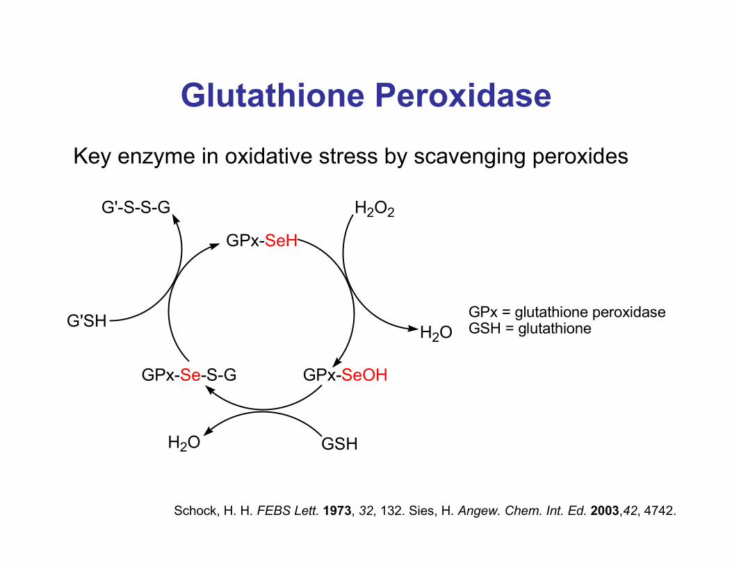
Glutathione Peroxidase
Key enzyme in oxidative stress by scavenging peroxides
Schock, H. H. FEBS Lett. 1973, 32, 132. Sies, H. Angew. Chem. Int. Ed. 2003,42, 4742.
GPx-SeH
H2O2
H2O
GPx-SeOH
GSHH2O
GPx-Se-S-G
G'SH
G'-S-S-G
GPx = glutathione peroxidaseGSH = glutathione
Glutathione Peroxidase Structure
RCSB Protein Data Bank
QuickTime™ and aTIFF (Uncompressed) decompressor
are needed to see this picture.
From human plasma
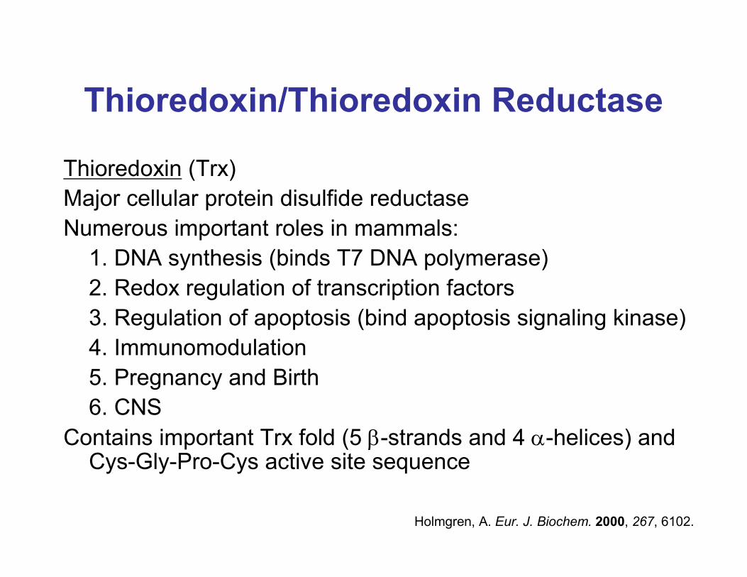
Thioredoxin/Thioredoxin Reductase
Thioredoxin (Trx)
Major cellular protein disulfide reductase
Numerous important roles in mammals:
1. DNA synthesis (binds T7 DNA polymerase)
2. Redox regulation of transcription factors
3. Regulation of apoptosis (bind apoptosis signaling kinase)
4. Immunomodulation
5. Pregnancy and Birth
6. CNS
Contains important Trx fold (5 β-strands and 4 α-helices) and Cys-Gly-Pro-Cys active site sequence
Holmgren, A. Eur. J. Biochem. 2000, 267, 6102.
Thioredoxin Structure
RCSB Protein Data Bank
QuickTime™ and aTIFF (Uncompressed) decompressor
are needed to see this picture.
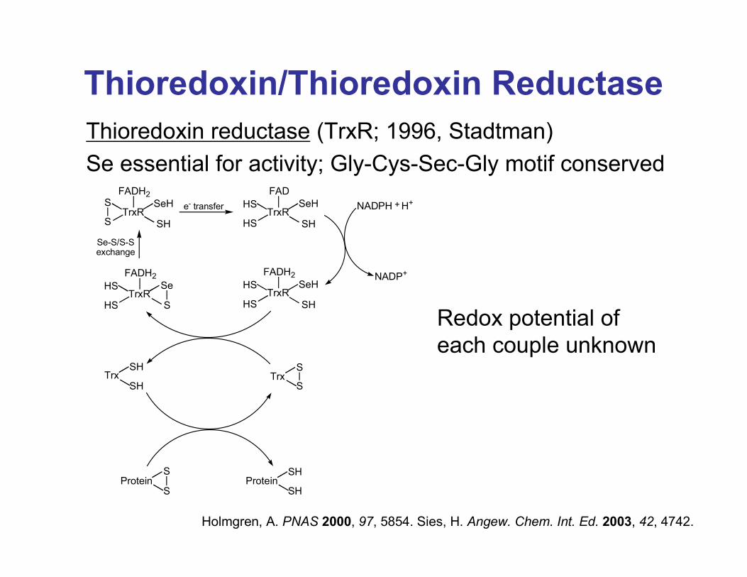
Thioredoxin/Thioredoxin Reductase
Thioredoxin reductase (TrxR; 1996, Stadtman)
Se essential for activity; Gly-Cys-Sec-Gly motif conserved
Holmgren, A. PNAS 2000, 97, 5854. Sies, H. Angew. Chem. Int. Ed. 2003, 42, 4742.
TrxR
FAD
HS
HS
SeH
SHTrxR
FADH2S
S
SeH
SH
TrxR
FADH2
HS
HS
SeH
SHTrxR
FADH2
HS
HS
Se
S
NADPH + H+
NADP+
TrxSH
SHTrx
S
S
ProteinS
SProtein
SH
SH
e- transfer
Se-S/S-Sexchange
Redox potential of
each couple unknown
Thioredoxin Reductase Structure
RCSB Protein Data Bank
QuickTime™ and aTIFF (Uncompressed) decompressor
are needed to see this picture.
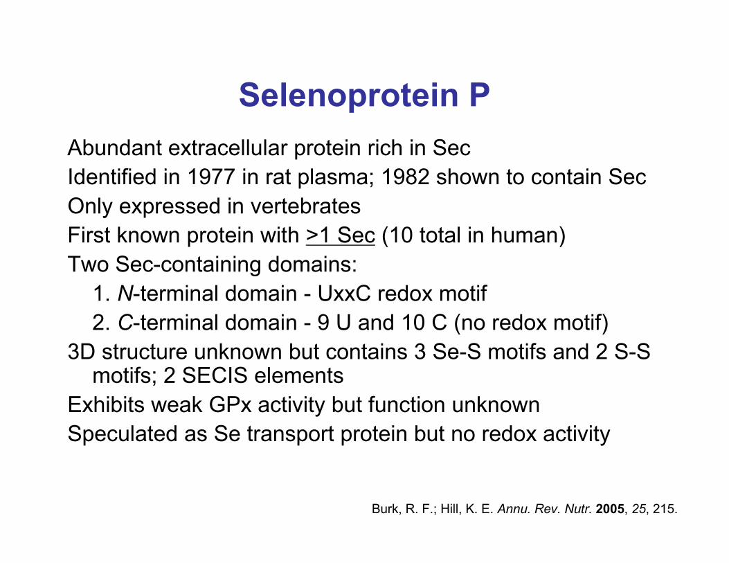
Selenoprotein P
Abundant extracellular protein rich in Sec
Identified in 1977 in rat plasma; 1982 shown to contain Sec
Only expressed in vertebrates
First known protein with >1 Sec (10 total in human)
Two Sec-containing domains:
1. N-terminal domain - UxxC redox motif
2. C-terminal domain - 9 U and 10 C (no redox motif)
3D structure unknown but contains 3 Se-S motifs and 2 S-S motifs; 2 SECIS elements
Exhibits weak GPx activity but function unknown
Speculated as Se transport protein but no redox activity
Burk, R. F.; Hill, K. E. Annu. Rev. Nutr. 2005, 25, 215.
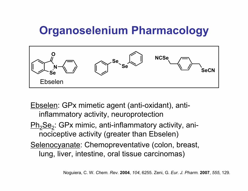
Organoselenium Pharmacology
Ebselen: GPx mimetic agent (anti-oxidant), anti-
inflammatory activity, neuroprotection
Ph2Se2: GPx mimic, anti-inflammatory activity, ani-
nociceptive activity (greater than Ebselen)
Selenocyanate: Chemopreventative (colon, breast,
lung, liver, intestine, oral tissue carcinomas)
SeN
O
Ebselen
SeSe
SeCN
NCSe
Noguiera, C. W. Chem. Rev. 2004, 104, 6255. Zeni, G. Eur. J. Pharm. 2007, 555, 129.
Uniqueness of Selenium
Cys and Sec least abundant aa in proteins
In adult human, 140 g S but only mg quantities of Se
Se more polarizable (better nucleophile, leaving group
and lower redox potential)
RSeH: pKa = 5.7 vs. RSH: pKa = 8.5 (~1 pKa unit
higher in protein)
RSeH largely deprotonated at physiological pH
Sies, H. Angew. Chem. Int. Ed. 2003, 42, 4742. Dawson, P. E. J. Am. Chem. Soc. 2006, 128, 16684.
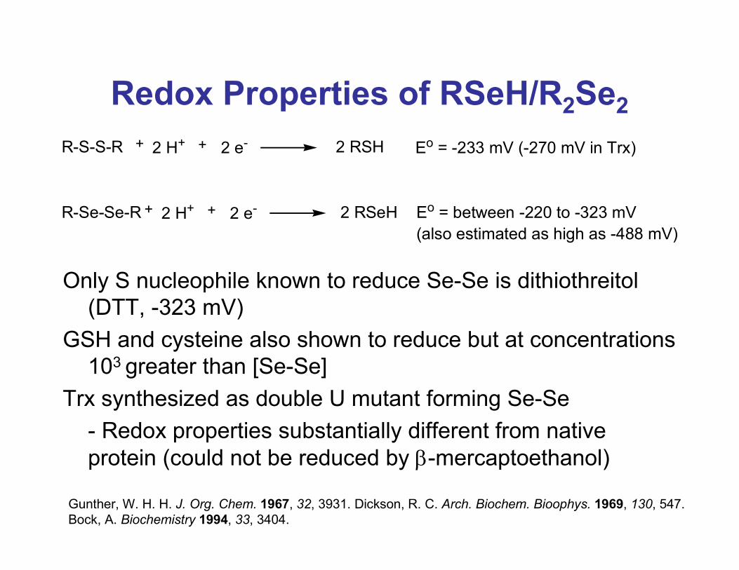
Redox Properties of RSeH/R2Se2
Only S nucleophile known to reduce Se-Se is dithiothreitol
(DTT, -323 mV)
GSH and cysteine also shown to reduce but at concentrations
103 greater than [Se-Se]
Trx synthesized as double U mutant forming Se-Se
- Redox properties substantially different from native
protein (could not be reduced by β-mercaptoethanol)
R-S-S-R + 2 H+ + 2 e- 2 RSH
R-Se-Se-R + 2 H+ + 2 e- 2 RSeH
Eo = -233 mV (-270 mV in Trx)
Eo = between -220 to -323 mV
(also estimated as high as -488 mV)
Gunther, W. H. H. J. Org. Chem. 1967, 32, 3931. Dickson, R. C. Arch. Biochem. Bioophys. 1969, 130, 547.
Bock, A. Biochemistry 1994, 33, 3404.
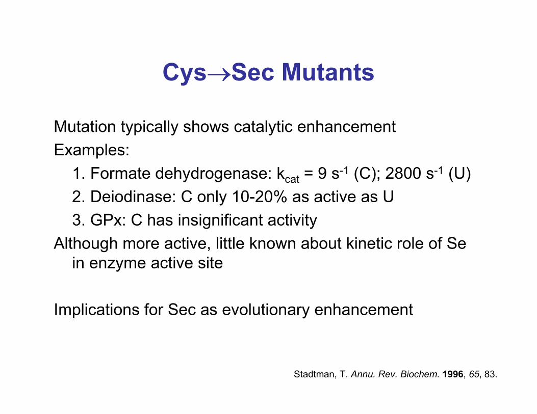
Cys→→→→Sec Mutants
Mutation typically shows catalytic enhancement
Examples:
1. Formate dehydrogenase: kcat = 9 s-1 (C); 2800 s-1 (U)
2. Deiodinase: C only 10-20% as active as U
3. GPx: C has insignificant activity
Although more active, little known about kinetic role of Se
in enzyme active site
Implications for Sec as evolutionary enhancement
Stadtman, T. Annu. Rev. Biochem. 1996, 65, 83.
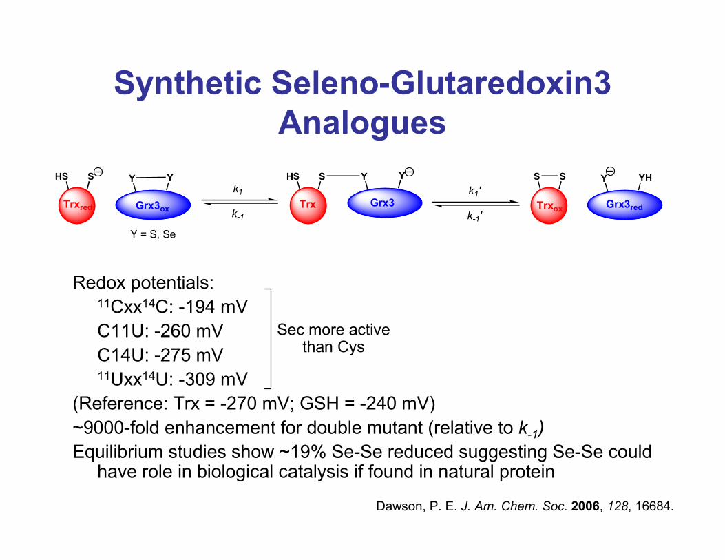
Synthetic Seleno-Glutaredoxin3
Analogues
Redox potentials:11Cxx14C: -194 mV
C11U: -260 mV
C14U: -275 mV11Uxx14U: -309 mV
(Reference: Trx = -270 mV; GSH = -240 mV)
~9000-fold enhancement for double mutant (relative to k-1)
Equilibrium studies show ~19% Se-Se reduced suggesting Se-Se could have role in biological catalysis if found in natural protein
Dawson, P. E. J. Am. Chem. Soc. 2006, 128, 16684.
Trxred Grx3ox
HS S Y Y
Trx Grx3
HS S Y Y
Trxox Grx3red
S S Y YH
Y = S, Se
k1
k-1
k1'
k-1'
Sec more activethan Cys
Identification and Characterization of
a Selenoprotein Family Containing a
Diselenide Bond
Identified SelL, only known selenoprotein containing 2 Sec in UxxU redox motif
Only present in aquatic organisms:
Eukaryotes - 11 fish, 2 ascidian, 2 crustacean, 1 mollusk
Prokaryotes - 2 bacteria
Unknown why only found in aquatic organisms
Close Cys homolog found in mammals
Gladyshev, V. N. PNAS 2007, 104, 13919.
Identification and Characterization
of a Selenoprotein Family
Containing a Diselenide Bond
Structure:
Prx-like domain in C-terminus
Conserved UxxU in N-terminus
2 Sec in loop between β-strand and α-helix (Trx-like)
Single SECIS element for 2 Sec (contains CA vs. AA
as unpaired nt in apical loop)
SelL Structure: Figure 1A, 1B (2 Sec shown in red)
Gladyshev, V. N. PNAS 2007, 104, 13919.
1. Determine if single SECIS element can direct addition of
2 Sec
Express 2 prokaryotic SelL sequences in E. coli via
GST-fused fragment containing both UGA codons,
SECIS element, and C-terminal His tag
(Figure 2A, 2B, 2E)
Identification and Characterization
of a Selenoprotein Family
Containing a Diselenide Bond
Gladyshev, V. N. PNAS 2007, 104, 13919.
2. Confirmation of 2 Sec by mass spectrometry
Method = LC-ESI MS/MS
Mass (from neutral, +2, and +3) = 1039.3 with correct
isotopic distribution
(Figure 3A)
Identification and Characterization
of a Selenoprotein Family
Containing a Diselenide Bond
Gladyshev, V. N. PNAS 2007, 104, 13919.
3. Expression in eukaryote, Danio rerio (zebrafish)
Constructs with C-terminal His-tagged SelL (and
mutants ULPC, CLPU, CLPC)
Transfected into HEK293 and NIH 3T3 cells
(Figure 2C, 3C)
Identification and Characterization
of a Selenoprotein Family
Containing a Diselenide Bond
Gladyshev, V. N. PNAS 2007, 104, 13919.
4. Subcellular localization
Expressed GST-fused D. rerio SelL in NIH 3T3 cells
Cys mutant with GFP at terminus
Observe cytosolic localization
(Supporting Information)
Identification and Characterization
of a Selenoprotein Family
Containing a Diselenide Bond
Gladyshev, V. N. PNAS 2007, 104, 13919.
5. Redox properties of SelL
Expressed in HEK293 cells (Cys→Ser)
a. Method 1 = reduced all S-S with DTT and
alkylated; analyze by SDS-PAGE
Result: all other selenoproteins but mutated SelL
shifted (wt SelL did shift)
b. Method 2 = pretreate with N-ethylmaleimide, then
DTT and alkylation
Result: no SelL shift observed (Se-Se bond!)
(Supporting Information)
Gladyshev, V. N. PNAS 2007, 104, 13919.
Identification and Characterization
of a Selenoprotein Family
Containing a Diselenide Bond
Summary
Isolated first Se-Se containing natural protein, SelL
Demonstrated a single SECIS element can direct the
incorporation of 2 Sec
Many questions remain:
Does a catalytic role exist for Se-Se in proteins?
Why is SelL only found in aquatic organisms?
Do other proteins contain this interesting motif?
Identification and Characterization
of a Selenoprotein Family
Containing a Diselenide Bond
Gladyshev, V. N. PNAS 2007, 104, 13919.