Embed Size (px)
Citation preview

Sense of vision – part 2
Visual pathway
Disorders of the visual field
Pupillary reflex, accommodation reflex
Stereoscopic vision
Colour vision
Successive and simultaneous contrasts
Practical tasks
1. Detection of the central visual acuity
2. Reaction of pupils to light and accommodation stimulus
3. Examination of the colour vision by the use of
pseudoisochromatic charts
4. Successive and simultaneous contrasts
5. Additive mixing of colours by the use of Maxwell´s discs
6. Stereoscopic vision
http://4.bp.blogspot.com/_irl8CO-29xk/SXEXnAPoZ5I/AAAAAAAAAUE/SZ3L7xQ3OSw/S220/180px-CentralScotoma.jpg
http://www.dwp.gov.uk/img/visual-fields.gif
Disorders of the visual field
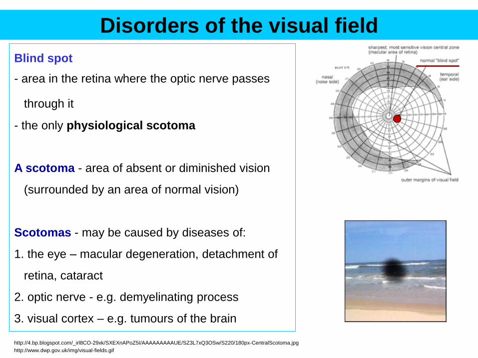
Blind spot
- area in the retina where the optic nerve passes
through it
- the only physiological scotoma
A scotoma - area of absent or diminished vision
(surrounded by an area of normal vision)
Scotomas - may be caused by diseases of:
1. the eye – macular degeneration, detachment of
retina, cataract
2. optic nerve - e.g. demyelinating process
3. visual cortex – e.g. tumours of the brain
http://4.bp.blogspot.com/_irl8CO-29xk/SXEXnAPoZ5I/AAAAAAAAAUE/SZ3L7xQ3OSw/S220/180px-CentralScotoma.jpg
http://www.dwp.gov.uk/img/visual-fields.gif

Blurred central vision -
(in macular degeneration) Tunnel vision (in tumours of hypophysis)
Binasal hemianopsia
Impaired peripheral vision
(in glaucoma)
Disorders of the visual field
http://www.eschenbach-optik.com/en/uploads/RTEmagicC_sehhilfen_makula_lesen_06.jpg.jpg
http://upload.wikimedia.org/wikipedia/commons/thumb/0/0f/Binasalvf.png/230px-Binasalvf.png
http://pituitary.ucla.edu/images/site/Visual3.3.jpg
https://encrypted-tbn0.gstatic.com/images?q=tbn:ANd9GcTMrOczmELpLZpY8vMuueKE-
tmCTvq0y9aGMaWJtHSUE2PCZNZG
https://en.wikipedia.org/wiki/Macular_degeneration

Glaucoma
• progressive optic neuropathies characterized by
degeneration of retinal ganglion cells and resulting
changes in the optic nerve head.
• if untreated may lead to blindness
• aqueous humour - secreted by the ciliary epithelium
into posterior chamber (behind iris)
• circulates through the pupil into the anterior chamber
• drained by the canal of Schlemm into venous system
• constant pressure 22 mm Hg or less
• if aqueous humour is not absorbed - increase of
intraocular pressure impedes blood flow to the retina
https://encrypted-tbn0.gstatic.com/images?q=tbn:ANd9GcTMrOczmELpLZpY8vMuueKE-tmCTvq0y9aGMaWJtHSUE2PCZNZG
http://upload.wikimedia.org/wikipedia/commons/3/3f/Flow_of_aqueous_humour_eye_EDA02.JPG

Cataract
normal lens is transparent
cataract
- slight or complete opacity of the lens (cloudy lens)
- causes obstruction for the passage of light – blurred vision
- often related to aging
http://cd.hpathy.com/wp-content/uploads/2011/09/cataract.jpg
https://encrypted-tbn2.gstatic.com/images?q=tbn:ANd9GcRkAzVaYT1ZGNS7qImsK4dibDosC_bBOwG3pf-Fj1Yq_d1tGR9qTw

- visual images are inverted as they pass
through the lens (nodal point of the
optical system of the eye is in lens)
- the nasal retina receives rays from the
temporal half of the world (hemifield)
- the temporal retina receives rays from
the nasal half of the world (hemifield)
Visual pathway and its disorders
nasal
temporal temporal
http://www.bioon.com/bioline/neurosci/course/bvis1.gif

- visual information leaves the eye by way of the optic nerve (1)
-partial crossing of axons at the optic
chiasm (2)
- nerve fibres from nasal part of retina cross
over
- temporal fibres remain at the same side
- after the chiasm, the axons are called
the optic tract (3)
- the optic tract terminates in the lateral
geniculate nucleus (LGN) - the axons
synapse here
- LGN axons form optic radiations –
terminate in the primary visual cortex, in
occipital lobes
http://www.bioon.com/bioline/neurosci/course/bvis2.gif

left eye right eye
1
2
3
complete
blindness
bitemporal
hemianopsia
(often due to
tumours of hypo-
physis that com-
press the chiasm)
homonymous
hemianopsia
Disorders of the visual pathway
Hemianopsia
• heteronymous – contralateral (bitemporal, binasal)
• homonymous – homolateral (right, left) http://www.bioon.com/bioline/neurosci/course/bvis2.gif

Task: The reaction of pupils to light and accommodation stimulus
Introduction
• the pupil – an opening located in the centre of the iris
• pupillary diameter (2-8 mm) - regulated by tone of small muscles in iris
M. sphincter pupillae (circular muscle)
– its contraction decreases the diameter of pupil (miosis)
– controlled by the parasympathetic NS
M. dilator pupillae (radial muscle)
– its contraction increases the diameter of pupil (mydriasis)
– controlled by the sympathetic NS
The pupillary light reflex
• reflex that controls the diameter of the pupil, in response to the intensity of light that
falls on the retina of the eye (reflex = involuntary, quick, stereotyped response to stimulus)
• greater intensity light - causes myosis (allowing less light in)
• lower intensity light - causes mydriasis (allowing more light in)

Reflex arc for the pupilary light reflex
- light stimulates receptors in retina
- signal is transmitted from retina by n. opticus (afferent fibres), however 10-15 % of
fibres bypass the LGN (leave the primary visual pathway) and terminate in the
Edinger-Westphal nuclei on both sides of the midbrain (therefore both eyes react
to light at one time)
- nerve fibres (efferent, parasympathetic) from the Edinger-Westphal nuclei form the
oculomotor nerve (IIIrd) - terminates in m. constrictor pupilae

The accommodation reflex
- a vision reflex that enables quick transfer of the
focus between near and distant objects
- comprises coordinated changes in
1. pupil size (miosis when focusing closer)
2. lens shape (accommodation when focusing closer)
3. vergence (convergence of the eyeballs – when
focusing to a close dostance)
far object (parallel) close object (convergence)
Pathway for
accommodation reflex
Receptors: rods and cones
Afferent nerve: optic n.
Centre: oculomotor nucleus
Efferent: oculomotor nerve
Effector:
1. constrictor pupillae
2. ciliary muscles
3. m. medial rectus

Task - Procedure
- look at the size of the pupil in normal room illuminaton
1. illuminate the eye by a torch and observe the reaction of the pupil, estimate
approximately the diameter of the pupil
2. switch the torch off and observe the reaction of the pupil
3. shade the other eye with a hand or a note-book, after illumination of the first eye
observe the consensual reaction of pupils
4. stop the illumination of the first eye and observe the consensual reaction of both
eyes
5. ask the examinee to focus on your finger that is moving towards the examinees
nose – observe size of the pupils and vergence of eyes
Result and conclusion
• describe your observations for 1 – 5 (or make a drawing)

Retina
A. Horizontal cells
– horizontal communication of several rods and cones
B. Amacrine cells
– horizontal communication of several ganglion cells
Sclera (outer layer)
Vascular layer
Pigment layer
1
2
3
A
B
- the light sensitive tissue lining the inner surface of the eye
- cells of retina:
1. Receptors
• rods
• cones
2. Bipolar cells – transmission of AP
from the recptor cells to the ganglion cells
3. Ganglion cells
• their axons form n. opticus that transmits the signal to CNS
light

The receptor cells
Cones (7 millions)
• colour vision - photopic vision
• higher threshold – need daylight conditions
• high visual acuity
• maximum density in fovea centralis
• low density outside the fovea
Rods (125 milions)
• operate in reduced light (scotopic vision)
• low threshold for light detection
• no well defined image
• not present in fovea centralis
• maximum in parafoveal region
• in direction to peripheral parts of retina their count rapidly decreases
Fovea
centralis Blind spot - optic nerve disc - no receptors

Yellow spot – Fovea centralis
• the upper layers of retina are „moved to the side“ -
mainly in the central fovea
• the light can more directly reach the receptors
• better image resolution
• because of the pigment layer behind retina – the yellow
spot appears darker in ophtalmoscopy
Fovea centralis
- best image resolution
(ability to recognize details)
1. highest density of receptors
2. highest count of nerve fibres - no
convergence
Periphery
– lower density of receptors, convergence
light
pigment

Examination of the central visual acuity
Introduction
• visual acuity (visus) – ability to see details sharply
• the ability of retina to distinguish two close points as separate and not fused into
one = minimum separabile
• 2 points can be distinguished if 2 sense receptors in retina are stimulated and
between them one receptor remains unstimulated
• visual acuity depends on
– the density of receptors
– the angle of observation (angle of light rays)
• the object you focus on - is imaged in fovea centralis
• fovea centralis – the highest density of receptors, therefore
the best visual acuity (central visual acuity)
• 2 points that are imaged in fovea centralis can be distinguished as 2 if they are
observed under visual angle of 1 minute
1´
receptors
of retina
the observed
points
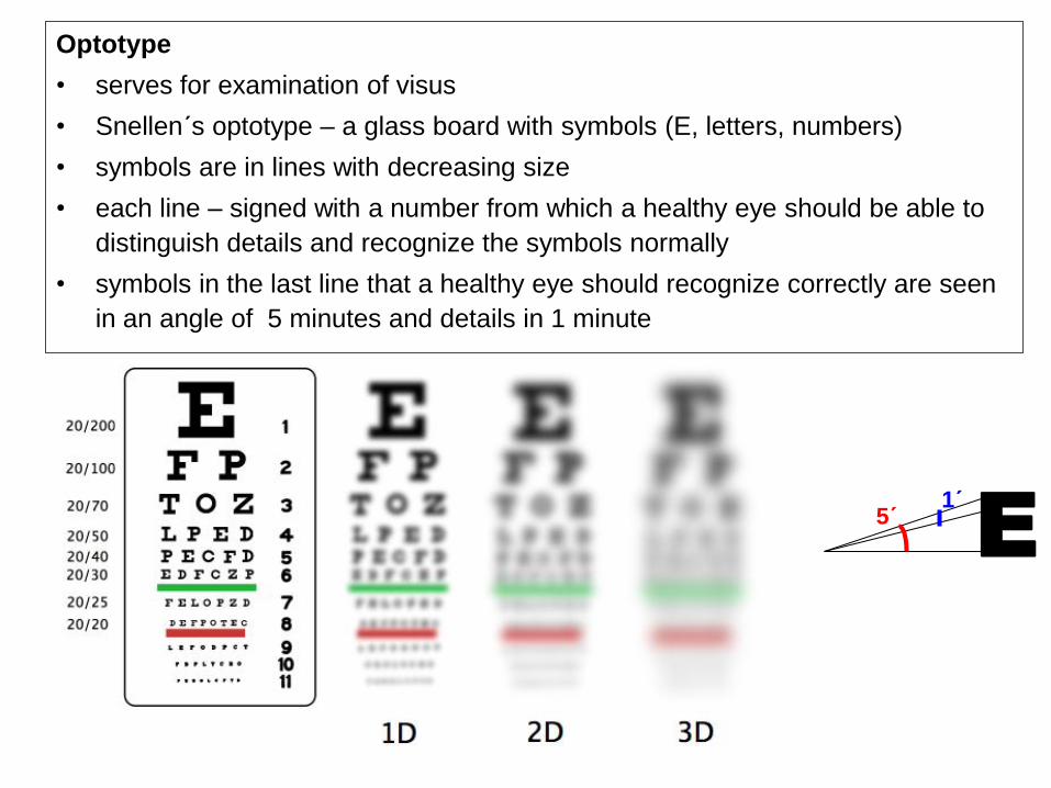
Optotype
• serves for examination of visus
• Snellen´s optotype – a glass board with symbols (E, letters, numbers)
• symbols are in lines with decreasing size
• each line – signed with a number from which a healthy eye should be able to
distinguish details and recognize the symbols normally
• symbols in the last line that a healthy eye should recognize correctly are seen
in an angle of 5 minutes and details in 1 minute
E 1´
5´

Procedure
• examine each eye separately (the non-examined eye is covered)
• switch the optotype on
• according to the type of the optotype the patient stands in distance of 5 or 6 m (d)
• the examinee is asked to read lines of symbols starting from the top continuing to
the bottom (largest → smallest)
• record the number of line that was read without a mistake (D)
Result
• write result in form: visus V=d/D
• d – distance of the patient from the optotype
(5 or 6 meters – depending on the optotype)
• D – number of the last line that was read without a mistake
Conclusion
• evaluate if the visus is normal
• V ≥ 5/5 or 6/6 (≥1) normal visus (normal central visual acuity)
• V < 5/5 or 6/6 (<1) impaired visus Landolt´s optotype
circles with opening

Perception of 3 dimensions
1. Determination by stereopsis – binocular vision
• the eyes are approximately by 6 cm apart
• images at 2 retinas are not identical
- different angle of observation
• object that we focus on is imaged at the same points of both retinas (corresponding points)
(„the same“ spot on both retinas)
• corresponding points in retina have the same cortical projection – two images are fused into one
• two eyes - two images in retina - one perception
• horopter – a circle passing through nodal points of both eyes and the point we focus on
• all points of the horopter circle are imaged to corresponding points
corresponding points

• points outside or inside horopter circle are not
imaged at the corresponding point, but to the
disparate points
• they do not have the same cortical projection
• the image is not sharp
• the bigger the distance from horopter (disparity),
the less sharp the image
• these blurred images contribute to the perception
of depth – 3 dimensional perception
disparate points

2. Determination by sizes of retinal
images of known objects
- distant objects – smaller, close objects –
larger in size
3. Determination by movement parallax
(monocular, motion parallax)
- head produces relatively large apparent
displacement of nearby objects and
relatively small displacement of distant
objects
- quickly moving objects – closer
- slowly moving objects – more distant
http://www.adamdalyonline.com/wp-content/uploads/2011/04/Perspective.jpg
http://www.themonthly.com.au/files/imagecache/home_content_listing_thumbnail/Switchingclubs-Fisher.gif
close
distant

Task: Stereoscopic vision
Principle
- by observation of stereoscopic photographs through a prism of a
stereoscope get the perception of a 3 D image
- stereoscopic photographs – 2 photos of the same object taken in an
angle as seen by 2 eyes

B. fix the eyes at an object in distance approx. 70 cm
- with a finger exert slight lateral pressure on the eyeball
- double image (diplopia) should occur, because an image
was formed on disparate points of retina of the shifted eye
A. put both index fingers in front of your eyes – one closer (20
cm), the other one in more distant position (40 cm)
fix the eyes on the closer finger (now is on the horopter)
- the closer finger is sharp, the further finger is double and blurry
- if you close one eye, one of the further objects disappears
fix the eyes on the further finger (now is on the horopter)
- the closer finger is double and blurry, the further finger is sharp
- if you close one eye, one of the further objects disappears
Procedure
C. place stereoscopic photographs into the holder of stereoscope
- by moving the holder forwards and backwards achieve such a convergence of the
eyes when both photographs fuse into a single image that is perceived as 3-D
Result and conclusions: describe your observation

Strabismus (squint, cross-eye)
• condition in which the eyes are not
properly aligned with each other:
• lack of coordination between
the extraocular muscles
• prevents bringing each point of the visual field
to the same point in retina
• prevents proper binocular vision, which may adversely affect depth perception.
• in a young child’s early efforts to fixate the two eyes on the same object, one
of the eyes fixates satisfactorily while the other fails do so
• abnormal fusion of the images on both retinas (to disparate points)
• the affectes eye becomes supressed and is not used for precise vision

Colour vision
• rods – black/white vision
• cones – colour vision Helmholtz trichromatic theory
• explains the colour vision
• photopigment – chemical substance (opsin) in photoreceptors, that is sensitive to light, chemical changes in photopigments give rise to receptor potential
• cones contain 3 different types of photopigment with different absorption maximum (i.e. maximum sensitivity)
• 420 nm – blue (cyanolab)
• 540 nm – green (cholorolab)
• 560 nm – red (erythrolab)
• all the other colours are a mixture of RGB

• elementary colours: red, green, blue
• additive mixing (in retina or higher centres for vision)
– of 3 elementary colours – stimulation of cones by red, blue and green colour (at the same time) - perception of white colour
• opponent colours = 2 colours which by additive mixing in the same proportion results in perception of white, e.g.
– blue-yellow, red-green
– and many others - any couple of the Maxwell´s triangle
Maxwell´s triangle
w

• human eye can detect almost all gradations of colours when only red, green
and blue monochromatic lights are appropriately mixed
• mixing of colours - red/green/blue cones generate potentials with different amplitude
e.g. orange light 580 nm
- red cones - 99% of their peak stimulation
- green cones - 42 % of their stimulation
- blue cones - no stimulation
e.g. blue light 450 nm
- red cones – 0% of their peak stimulation
- green cones 0 % of their stimulation
- blue cones – 97 % of their stimulation

Disorders of colour vision – Colour blindness
• about 8% of men and 1% of women have colour vision impairment
1. anomalia – (colour weakness) resulting from a deficiency of colour-sensitive pigment
2. anopia – absolute blindness – absence of a colour pigment
• Protanomalia/ protanopia – red colour deficiency/ blindness
• Deuteranomalia/ deuteranopia – green colour deficiency/ blindness
• Tritanomalia/ tritanopia – blue colour deficiency/ blindness
• Trichromatic people – normal vision, anomalia
• Dichromatic people – blindnenss for 1 colour (deuteranomalia is the
most frequent type)
• Monochromatic people– blindness for 2 colours
• Achromatic people – do not recognize any colours at all


Task: Detection of colour blindness by the use
of pseudoisochromatic charts
Principle
• Ishiharas´s peudoisochromatic charts – set of 38 charts composed of
irregular mosaic of circles differing in colour
• an eye with normal colour vision can discriminate figures or lines
• a person with disorder of colour vision
– a/ does not recognize any figure or line
– b/ recognizes different figure than normal eye
• normal eye – 5
• red-green blindness - 2

Procedure
• perform the examination in a room with good illumination
• put the charts in normal reading distance
from the examinee´s eyes in a right angle
• charts 1-25 must be read within 3 seconds
• charts 26-38 are for illiterate persons (children) – must be read within 10 s
• compare the examinee´s results with normal results given (see the text in
Ishihara´s book)
Result
• did the examinee read all charts appropriately?
Conclusion
• is the patient´s colour vision normal?
On line tests for colour vision (Ishihara plates)
http://www.color-blindness.com/ishihara-38-plates-cvd-test/#prettyPhoto/1/

Task: Additive mixing of colours by use
of Maxwell´s discs and monochromatic filters
Principle
• by observing the fast rotation of Maxwell´s discs additive mixing of colours in
retina is achieved
• additive mixing at level of higher centres is achieved by placing of different
monochromatic filters in front of each eye
Procedure
• attach a Maxwell´s disc to an engine
• gradually increase the speed of rotation until additive mixing of colours on the
retina is achieved
• observe individually by each eye and then by both eyes
• repeat procedure with different discs
= simultaneous stimulation of retina by different wavelengths (light of different
colours) which are summed up and result in final perception
possible due to integration abilities of retina or brain

• put a green monochromatic filter in front of one eye and a red monochromatic filter in
front of the other eye
• look at a white surface with both eyes until the perception of final colour is achieved
Result
• describe your observation, draw pictures

Successive and simultaneous contrast
The terms "simultaneous contrast" and "successive contrast" refer to
visual effects in which the appearance of a patch of an object /field is
affected by other fields that are nearby in space and time.
lighter
darker

Tasks
1. Look at a black-white picture, then transfer your look
at a white surface (sheet of paper, wall) – describe
your observation - „the afterimage“ (several pictures
available)
2. Look at a colour picture - then transfer your look at a
white surface – describe your observation – „the
afterimage“ (several pictures available)
Successive contrast
• based on partial adaptation of receptors in retina
• exposure of retina to a colour causes adaptation of the exposed
cells to this colour and leads to higher sensitivity to opponent
colour
• when neutral light is restored (white backgound) a temporary
illusion of a light composed of the "missing" wavelengths (the
complementary colour) is seen
• exposure to white (black) – higher sensitivity to black (white)

Simultaneous contrast
• is based on lateral inhibition of visual neurons
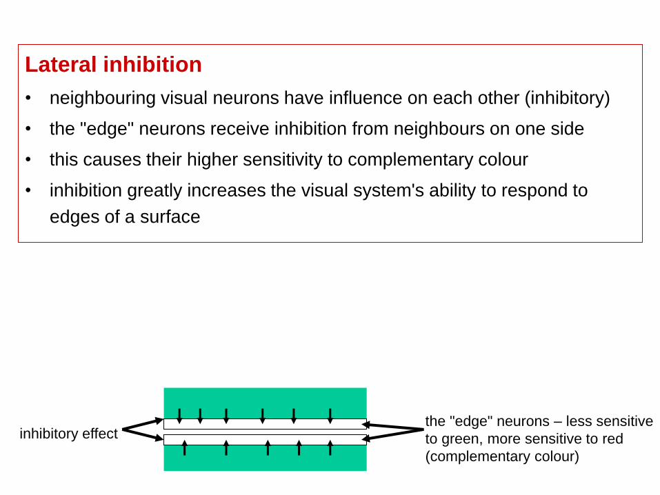
Lateral inhibition
• neighbouring visual neurons have influence on each other (inhibitory)
• the "edge" neurons receive inhibition from neighbours on one side
• this causes their higher sensitivity to complementary field (light, dark)
• inhibition greatly increases the visual system's ability to respond to edges of a
surface
less sensitive to dark, more
sensitive to light
Surrounding field -
inhibitory effect
less sensitive to light, more
sensitive to dark
Tasks
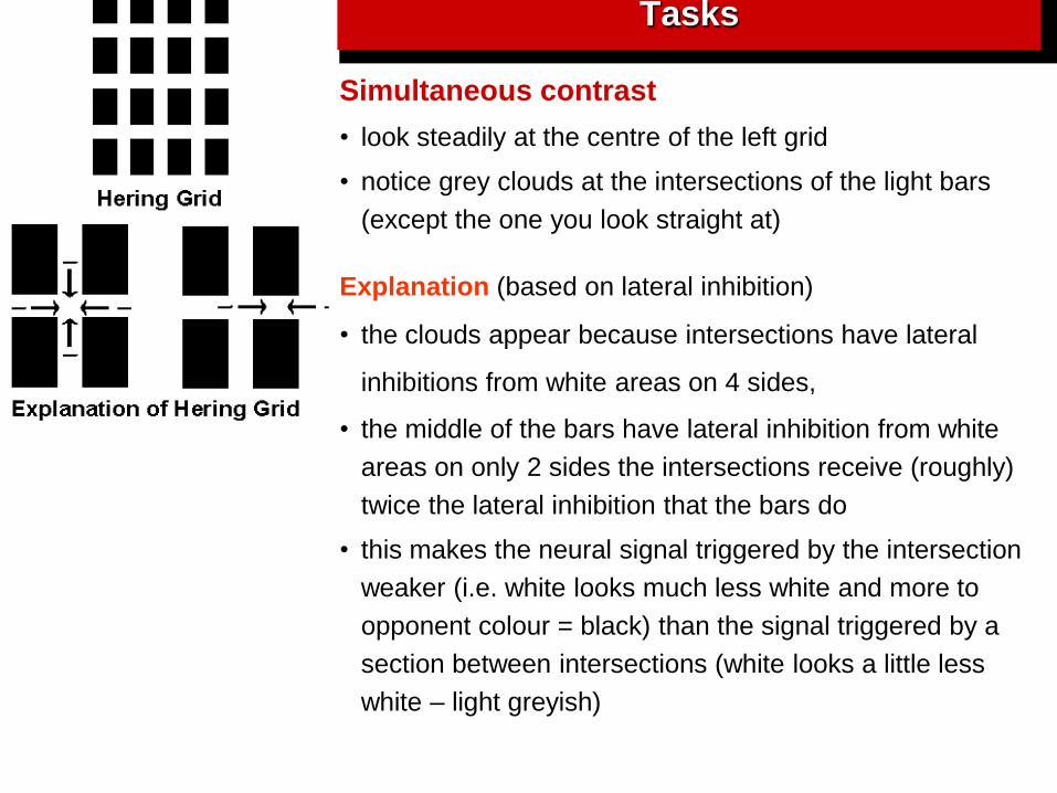
Simultaneous contrast
• look steadily at the centre of the left grid
• notice grey clouds at the intersections of the light bars
(except the one you look straight at)
Explanation (based on lateral inhibition)
• the clouds appear because intersections have lateral
inhibitions from white areas on 4 sides,
• the middle of the bars have lateral inhibition from white
areas on only 2 sides the intersections receive (roughly)
twice the lateral inhibition that the bars do
• this makes the neural signal triggered by the intersection
weaker (i.e. white looks much less white and more to
opponent colour = black) than the signal triggered by a
section between intersections (white looks a little less
white – light greyish)

Simultaneous contrast
• attach the Maxwell´s disc to the engine
• start it, increase frequency of rotation
• the black narrow band of the disc will be perceived in the complementary colour
to the basic colour of the disc
- an intense green light strikes the retina – it makes the green receptors „tired“
- because of links by horizontal cells – it makes also the neghbouting receptors in
the narrow strip tired to green
- when the white strip falls to the „tired“ part of retina, we get perception of the
opponent (complementary colour)
Lateral inhibition
• neighbouring visual neurons have influence on each other (inhibitory)
• the "edge" neurons receive inhibition from neighbours on one side
• this causes their higher sensitivity to complementary colour
• inhibition greatly increases the visual system's ability to respond to
edges of a surface
the "edge" neurons – less sensitive
to green, more sensitive to red
(complementary colour)
inhibitory effect