Embed Size (px)
Citation preview
NANO EXPRESS Open Access
Sensitive Nonenzymatic ElectrochemicalGlucose Detection Based on Hollow PorousNiOGege He1,2,3, Liangliang Tian1,2*, Yanhua Cai1,2, Shenping Wu1, Yongyao Su1,2, Hengqing Yan1,2, Wanrong Pu1,2,Jinkun Zhang1,2 and Lu Li1,2*
Abstract
Transition metal oxides (TMOs) have attracted extensive research attentions as promising electrocatalytic materials.Despite low cost and high stability, the electrocatalytic activity of TMOs still cannot satisfy the requirements ofapplications. Inspired by kinetics, the design of hollow porous structure is considered as a promising strategy toachieve superior electrocatalytic performance. In this work, cubic NiO hollow porous architecture (NiO HPA) wasconstructed through coordinating etching and precipitating (CEP) principle followed by post calcination. Beingemployed to detect glucose, NiO HPA electrode exhibits outstanding electrocatalytic activity in terms of highsensitivity (1323 μA mM−1 cm−2) and low detection limit (0.32 μM). The excellent electrocatalytic activity can beascribed to large specific surface area (SSA), ordered diffusion channels, and accelerated electron transfer ratederived from the unique hollow porous features. The results demonstrate that the NiO HPA could have practicalapplications in the design of nonenzymatic glucose sensors. The construction of hollow porous architectureprovides an effective nanoengineering strategy for high-performance electrocatalysts.
Keywords: NiO, Hollow porous architecture, Coordinating etching and precipitating, Electrochemical sensor,Glucose detection
BackgroundDetection of glucose with fast, accurate, and low-cost process is importance for clinical biochemistry,pharmaceutical analysis, food industry, and environ-mental monitoring [1–3]. Among the multitudinoustechniques, electrochemical detection has been con-sidered as one of the most convenient approachowing to its high sensitivity, low cost, and attractivelower detection limit [4–6]. However, the commonglucose oxidase-based electrochemical sensors arerestricted by the drawback of insufficient stabilityoriginating from the nature of enzymes [7–9]. Toaddress these issues, earth-abundant electrocatalystsbased on TMOs were recommended due to theirlower cost, physicochemical stability, and redoxelectroactivity [10–12]. However, the overall
electrocatalytic activity of conventional TMOs is stillfar away from the requirements of applications. It isstill a challenge to rationally design high-active TMOelectrocatalysts for glucose.Generally, the process of kinetics plays a decisive role
in electrocatalytic activity for established electrocatalyticmaterials. Inspired by the intimate connection betweenkinetics and microstructures, improved electrocatalyticactivity can be achieved by the engineering of micro-structures, including surface area, pore structure, andarchitecture features [13, 14]. The porous structure of-fers large specific surface area (SSA) and providesamounts of active sites. Furthermore, the porous struc-ture also affords enough diffusion channels for analyteand intermediate products, which are beneficial for masstransport process [15, 16]. On the other hand, hollowstructures combining functional shells and inner voidscan offer larger electrolyte-electrode contact area and re-duce the length for both mass and electron transport[17]. Furthermore, the available inner cavities effectively
* Correspondence: [email protected]; [email protected] Institute for New Materials Technology, Chongqing University ofArts and Sciences, Chongqing, People’s Republic of ChinaFull list of author information is available at the end of the article
© The Author(s). 2018 Open Access This article is distributed under the terms of the Creative Commons Attribution 4.0International License (http://creativecommons.org/licenses/by/4.0/), which permits unrestricted use, distribution, andreproduction in any medium, provided you give appropriate credit to the original author(s) and the source, provide a link tothe Creative Commons license, and indicate if changes were made.
He et al. Nanoscale Research Letters (2018) 13:3 DOI 10.1186/s11671-017-2406-0
prevent electroactive nanoparticles from aggregation andaccommodate the volume change and structural strainaccompanied with repeated measurements [18]. In con-clusion, high-active TMO electrocatalysts can be ac-quired through the design of hollow porous architecture.As a typical transition metal oxide, NiO was reported
as an efficient catalyst for electrooxidation of glucosedue to the redox couple of Ni3+/Ni2+ in alkalinemedium, implying potential applications in electrochem-ical glucose sensor. In this work, cubic NiO HPA wasconstructed through a Cu2O-templated coordinatingetching and precipitating (CEP) method and post calcin-ation. The hollow porous structure provides large SSA,well-defined interior voids, abundant ordered transferchannels, and high electron transfer efficiency. Beingemployed to detect glucose, NiO HPA electrode presentshigher sensitivity and lower detection limit compared tobroken NiO HPA (NiO BHPA), demonstrating advan-tages of the hollow porous architecture. This facile strat-egy to construct hollow porous architecture provides avalid method in the development of highly efficientnanomaterials for electrochemical sensors.
ExperimentalMaterialsCuCl2·2H2O, NiCl2·6H2O, Na2S2O3·5H2O, polyvinylpyr-rolidone (PVP, Mw = 40,000), and NaOH were purchasedfrom Chengdu Kelong. Glucose (Glu.), lactose (Lact.),sucrose (Sucr.), fructose (Fruc.), L-ascorbic acid (AA),uric acid (UA), and Nafion solution (5 wt% in mixture oflower aliphatic alcohols and water) were purchased fromSigma-Aldrich without further purification.
Synthesis of Cu2O TemplateThe cubic Cu2O templates were synthesized accordingto our previous work [19]. In this typical procedure,20 ml NaOH (2 M) was added dropwise into 200 mLCuCl2·2H2O (10 mM) under stirring at 55 °C. After0.5 h, 4 mL AA (0.6 M) was introduced dropwise intothe above solution. The suspension was further aged for3 h and washed with water several times by centrifuga-tion. The XRD pattern and SEM and TEM images areshown in Additional file 1: Figure S1.
Synthesis of NiO HPANiO HPA was synthesized by a CEP method. First,Cu2O (10 mg) and NiCl2·6H2O (3 mg) were dispersedinto 10 mL ethanol-water mixed solvent (volume ratio =1:1) for 7 min by ultrasonication. Then, PVP (0.33 g)was added into the solution with vigorous agitation for30 min. Four milliliters Na2S2O3 (1 M) was dropped intothe system; the reaction was proceeded at roomtemperature for 3 h until the color of the suspensionchanged from red to light green. The Ni(OH)2 precursor
was washed several times by warm ethanol-water anddried at room temperature. Finally, NiO HPA was suc-cessively obtained under an air atmosphere at 400 °C for2 h with a slow ramp rate of 1 °C/min. NiO BHPA wasobtained through strong ultrasonic treatment of NiOHPA for 2 h.
Material CharacterizationsThe composition and structure of the products werecharacterized by X-ray diffraction (XRD, Rigaku D/Max-2400). The composition was further analyzed by the X-ray photoelectron spectroscopy (XPS, ESCALAB250Xi)with the C 1s peaks at 284.8 eV as an internal standard.The morphologies and microstructures of the productswere observed using field emission scanning electronmicroscope (FESEM, FEI Quanta 250, Zeiss Gemini 500)and high-resolution transmission electron microscope(HRTEM, FEI F20). Brunauer-Emmett-Teller (BET,Belsort-max) was applied to analyze the specific surfacearea and pore structure.
Electrochemical MeasurementsAll electrochemical measurements were operated in0.1 M NaOH on μIII Autolab electrochemical worksta-tion. A three-electrode configuration with NiO HPA (orNiO BHPA) modified glassy carbon electrode (GCE, Ф= 3 mm) as the working electrodes and Ag/AgCl (in sat-urated KCl) and platinum disk electrode (Ф = 2 mm) asthe reference electrode and counter electrode, respect-ively. Typically, GCE was polished with alumina slurry(3, 0.5, and 0.05 μm). Then, the NiO HPA (10 mg) wasdissolved into a mixture of 0.1 mL Nafion and 0.9 mLdistilled water. Finally, 5 μL of the mixture was droppedonto the pretreated GCE (70.77 μg/cm2) and dried atroom temperature. NiO BHPA-modified GCE was alsoprepared under the same condition to verify the advan-tages of NiO HPA. The modified electrodes were mea-sured by cyclic voltammetry (CV), chronoamperometry(CA), and electrochemical impedance spectroscopy (EIS)to evaluate its electrocatalytic activity. EIS measurementswere carried out over the frequency range between0.01–100 kHz with a perturbation amplitude of 5 mVversus the open-circle potential.
Results and DiscussionCharacterizationsAs shown in Fig. 1a, the diffraction peaks located at37.21°, 43.27°, 62.87°, and 75.42° correspond to (111),(200), (220), and (311) facets of face-centered cubic NiO(JCPDS.no.47-1049) [20]. There are no other diffractionpeaks, indicating the purity of the products. XPS wasfurther employed to analyze the element compositionand oxidation state of NiO HPA. The survey spectrum(Fig. 1b) demonstrates O 1s and Ni 2p peaks at 531.5
He et al. Nanoscale Research Letters (2018) 13:3 Page 2 of 10
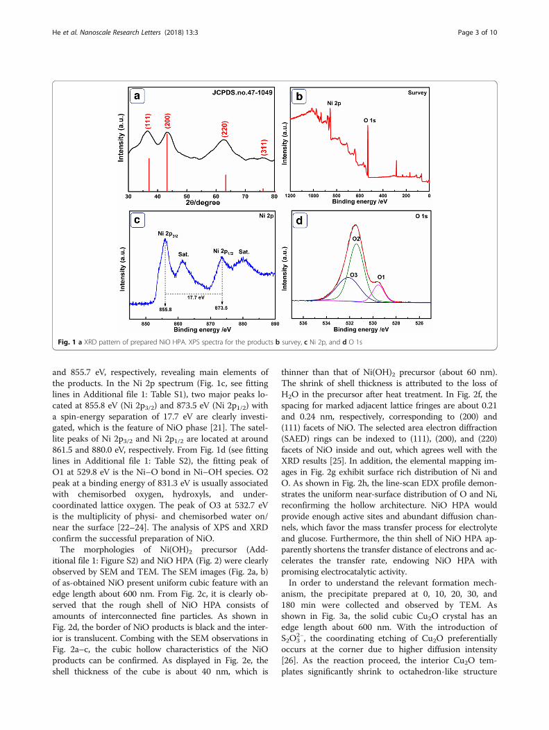
and 855.7 eV, respectively, revealing main elements ofthe products. In the Ni 2p spectrum (Fig. 1c, see fittinglines in Additional file 1: Table S1), two major peaks lo-cated at 855.8 eV (Ni 2p3/2) and 873.5 eV (Ni 2p1/2) witha spin-energy separation of 17.7 eV are clearly investi-gated, which is the feature of NiO phase [21]. The satel-lite peaks of Ni 2p3/2 and Ni 2p1/2 are located at around861.5 and 880.0 eV, respectively. From Fig. 1d (see fittinglines in Additional file 1: Table S2), the fitting peak ofO1 at 529.8 eV is the Ni–O bond in Ni–OH species. O2peak at a binding energy of 831.3 eV is usually associatedwith chemisorbed oxygen, hydroxyls, and under-coordinated lattice oxygen. The peak of O3 at 532.7 eVis the multiplicity of physi- and chemisorbed water on/near the surface [22–24]. The analysis of XPS and XRDconfirm the successful preparation of NiO.The morphologies of Ni(OH)2 precursor (Add-
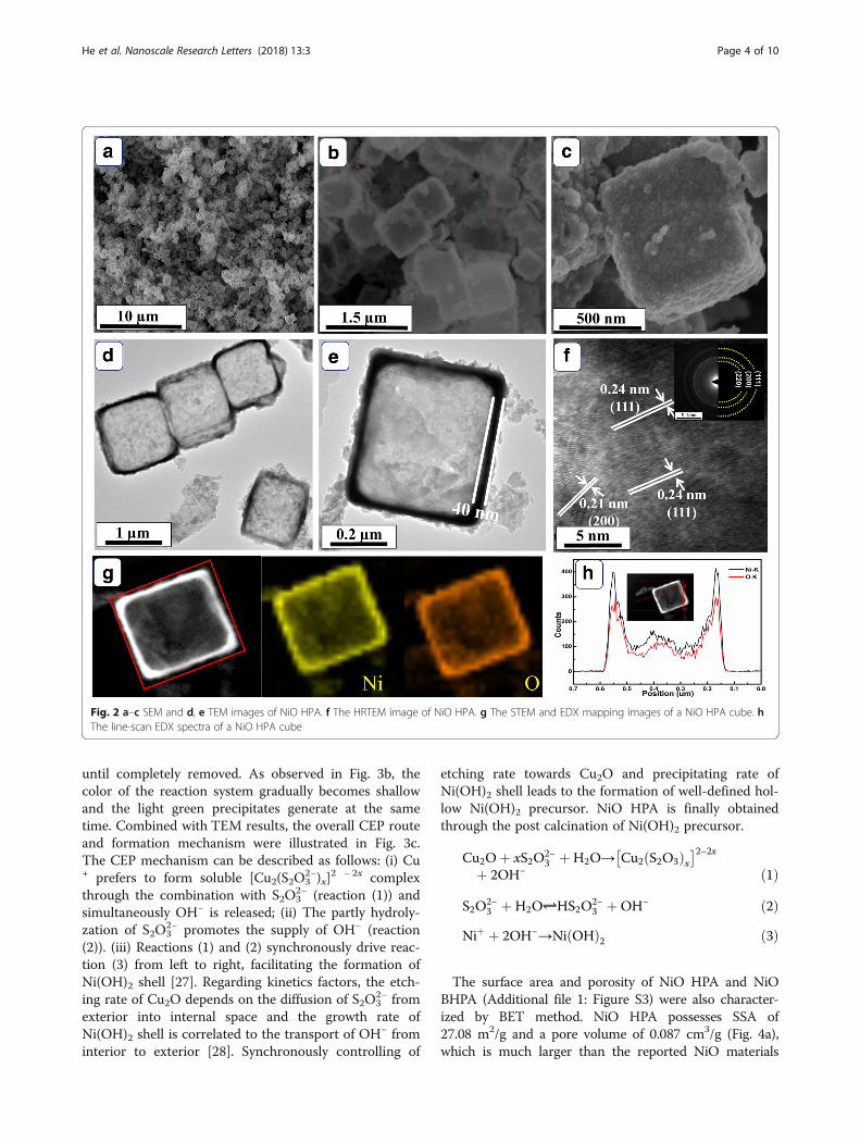
itional file 1: Figure S2) and NiO HPA (Fig. 2) were clearlyobserved by SEM and TEM. The SEM images (Fig. 2a, b)of as-obtained NiO present uniform cubic feature with anedge length about 600 nm. From Fig. 2c, it is clearly ob-served that the rough shell of NiO HPA consists ofamounts of interconnected fine particles. As shown inFig. 2d, the border of NiO products is black and the inter-ior is translucent. Combing with the SEM observations inFig. 2a–c, the cubic hollow characteristics of the NiOproducts can be confirmed. As displayed in Fig. 2e, theshell thickness of the cube is about 40 nm, which is
thinner than that of Ni(OH)2 precursor (about 60 nm).The shrink of shell thickness is attributed to the loss ofH2O in the precursor after heat treatment. In Fig. 2f, thespacing for marked adjacent lattice fringes are about 0.21and 0.24 nm, respectively, corresponding to (200) and(111) facets of NiO. The selected area electron diffraction(SAED) rings can be indexed to (111), (200), and (220)facets of NiO inside and out, which agrees well with theXRD results [25]. In addition, the elemental mapping im-ages in Fig. 2g exhibit surface rich distribution of Ni andO. As shown in Fig. 2h, the line-scan EDX profile demon-strates the uniform near-surface distribution of O and Ni,reconfirming the hollow architecture. NiO HPA wouldprovide enough active sites and abundant diffusion chan-nels, which favor the mass transfer process for electrolyteand glucose. Furthermore, the thin shell of NiO HPA ap-parently shortens the transfer distance of electrons and ac-celerates the transfer rate, endowing NiO HPA withpromising electrocatalytic activity.In order to understand the relevant formation mech-
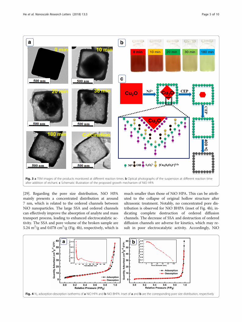
anism, the precipitate prepared at 0, 10, 20, 30, and180 min were collected and observed by TEM. Asshown in Fig. 3a, the solid cubic Cu2O crystal has anedge length about 600 nm. With the introduction ofS2O3
2−, the coordinating etching of Cu2O preferentiallyoccurs at the corner due to higher diffusion intensity[26]. As the reaction proceed, the interior Cu2O tem-plates significantly shrink to octahedron-like structure
Fig. 1 a XRD pattern of prepared NiO HPA. XPS spectra for the products b survey, c Ni 2p, and d O 1s
He et al. Nanoscale Research Letters (2018) 13:3 Page 3 of 10
until completely removed. As observed in Fig. 3b, thecolor of the reaction system gradually becomes shallowand the light green precipitates generate at the sametime. Combined with TEM results, the overall CEP routeand formation mechanism were illustrated in Fig. 3c.The CEP mechanism can be described as follows: (i) Cu+ prefers to form soluble [Cu2(S2O3
2−)x]2 − 2x complex
through the combination with S2O32− (reaction (1)) and
simultaneously OH− is released; (ii) The partly hydroly-zation of S2O3
2− promotes the supply of OH− (reaction(2)). (iii) Reactions (1) and (2) synchronously drive reac-tion (3) from left to right, facilitating the formation ofNi(OH)2 shell [27]. Regarding kinetics factors, the etch-ing rate of Cu2O depends on the diffusion of S2O3
2− fromexterior into internal space and the growth rate ofNi(OH)2 shell is correlated to the transport of OH− frominterior to exterior [28]. Synchronously controlling of
etching rate towards Cu2O and precipitating rate ofNi(OH)2 shell leads to the formation of well-defined hol-low Ni(OH)2 precursor. NiO HPA is finally obtainedthrough the post calcination of Ni(OH)2 precursor.
Cu2Oþ xS2O2−3 þH2O→ Cu2 S2O3ð Þx
� �2−2x
þ 2OH− ð1Þ
S2O2−3 þH2O⇌HS2O
2−3 þOH− ð2Þ
Niþ þ 2OH−→Ni OHð Þ2 ð3Þ
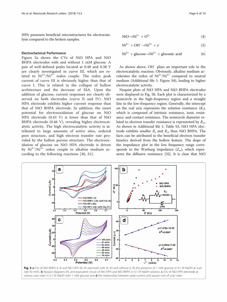
The surface area and porosity of NiO HPA and NiOBHPA (Additional file 1: Figure S3) were also character-ized by BET method. NiO HPA possesses SSA of27.08 m2/g and a pore volume of 0.087 cm3/g (Fig. 4a),which is much larger than the reported NiO materials
Fig. 2 a–c SEM and d, e TEM images of NiO HPA. f The HRTEM image of NiO HPA. g The STEM and EDX mapping images of a NiO HPA cube. hThe line-scan EDX spectra of a NiO HPA cube
He et al. Nanoscale Research Letters (2018) 13:3 Page 4 of 10
[29]. Regarding the pore size distribution, NiO HPAmainly presents a concentrated distribution at around7 nm, which is related to the ordered channels betweenNiO nanoparticles. The large SSA and ordered channelscan effectively improve the absorption of analyte and masstransport process, leading to enhanced electrocatalytic ac-tivity. The SSA and pore volume of the broken sample are5.24 m2/g and 0.078 cm3/g (Fig. 4b), respectively, which is
much smaller than those of NiO HPA. This can be attrib-uted to the collapse of original hollow structure afterultrasonic treatment. Notably, no concentrated pore dis-tribution is observed for NiO BHPA (inset of Fig. 4b), in-dicating complete destruction of ordered diffusionchannels. The decrease of SSA and destruction of ordereddiffusion channels are adverse for kinetics, which may re-sult in poor electrocatalytic activity. Accordingly, NiO
Fig. 3 a TEM images of the products monitored at different reaction times. b Optical photographs of the suspension at different reaction timeafter addition of etchant. c Schematic illustration of the proposed growth mechanism of NiO HPA
Fig. 4 N2 adsorption-desorption isotherms of a NiO HPA and b NiO BHPA. Inset of a and b are the corresponding pore size distribution, respectively
He et al. Nanoscale Research Letters (2018) 13:3 Page 5 of 10
HPA possesses beneficial microstructures for electrocata-lysis compared to the broken samples.
Electrochemical PerformanceFigure 5a shows the CVs of NiO HPA and NiOBHPA electrodes with and without 1 mM glucose. Apair of well-defined peaks located at 0.48 and 0.38 Vare clearly investigated in curve III, which are re-lated to Ni2+/Ni3+ redox couple. The redox peakcurrent of curve III is obviously higher than that ofcurve I. This is related to the collapse of hollowarchitecture and the decrease of SSA. Upon theaddition of glucose, current responses are clearly ob-served on both electrodes (curve II and IV). NiOHPA electrode exhibits higher current response thanthat of NiO BHPA electrode. In addition, the onsetpotential for electrooxidation of glucose on NiOHPA electrode (0.43 V) is lower than that of NiOBHPA electrode (0.46 V), revealing higher electrocat-alytic activity. The high electrocatalytic activity is at-tributed to large amounts of active sites, orderedpore structure, and high electron transfer rate pro-vided by the hollow porous structure. The electroox-idation of glucose on NiO HPA electrode is drivenby Ni2+/Ni3+ redox couple in alkaline medium ac-cording to the following reactions [30, 31]:
NiO→Ni2þ þO2− ð4Þ
Ni2þ þOH−→Ni3þ þ e− ð5Þ
Ni3þ þ glucose→Ni2þ þ gloconic acid ð6Þ
As shown above, OH− plays an important role in theelectrocatalytic reaction. Obviously, alkaline medium ac-celerates the redox of Ni2+/Ni3+ compared to neutralmedium (Additional file 1: Figure S4), leading to higherelectrocatalytic activity.Nyquist plots of NiO HPA and NiO BHPA electrodes
were displayed in Fig. 5b. Each plot is characterized by asemicircle in the high-frequency region and a straightline in the low-frequency region. Generally, the intercepton the real axis represents the solution resistance (Rs),which is composed of intrinsic resistance, ionic resist-ance, and contact resistance. The semicircle diameter re-lated to electron transfer resistance is represented by Rct.As shown in Additional file 1: Table S3, NiO HPA elec-trode exhibits smaller Rs and Rct than NiO BHPA. Thefacts can be attributed to the beneficial electron transferkinetics derived from the hollow feature. The slope ofthe impedance plot in the low frequency range corre-sponds to the Warburg impedance (Zw), which repre-sents the diffusive resistance [32]. It is clear that NiO
Fig. 5 a CVs of NiO BHPA (I, II) and NiO HPA (III, IV) electrode with (II, IV) and without (I, III) the presence of 1 mM glucose in 0.1 M NaOH at scanrate 50 mV/s. b Nyquist diagrams EIS and equivalent circuit of NiO HPA and NiO BHPA in 0.1 M NaOH solution. c CVs of NiO HPA electrode atvarious scan rates in 0.1 M NaOH with 1 mM glucose and d the relationship between peak current and square root of scan rates
He et al. Nanoscale Research Letters (2018) 13:3 Page 6 of 10
HPA favors the diffusion kinetics; however, the NiOBHPA hinders the diffusion of electrolyte. This can beascribed to the destruction of the ordered diffusionchannels after ultrasonic. On the basis of above EIS dis-cussions, NiO HPA electrode is more beneficial for bothelectron and mass transfer kinetics compared to thebroken sample, implying the advantages of NiO HPA asan electrocatalyst for glucose.The kinetics of NiO HPA electrode was determined
from the CVs with different scan rates in 1 mM glucosesolution (Fig. 5c). As depicted in Fig. 5d, the anodic andcathodic peak currents are proportional to the squareroot of scan rates, demonstrating a typical diffusion-controlled dynamic process. Furthermore, no significantpositive/negative shift is observed for anodic/cathodicpeak, implying unimpeded diffusion kinetics originatedfrom the hollow porous structure.
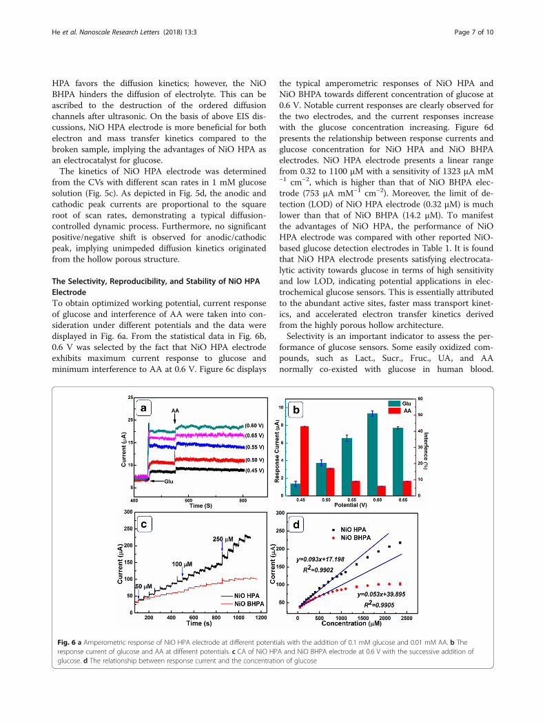
The Selectivity, Reproducibility, and Stability of NiO HPAElectrodeTo obtain optimized working potential, current responseof glucose and interference of AA were taken into con-sideration under different potentials and the data weredisplayed in Fig. 6a. From the statistical data in Fig. 6b,0.6 V was selected by the fact that NiO HPA electrodeexhibits maximum current response to glucose andminimum interference to AA at 0.6 V. Figure 6c displays
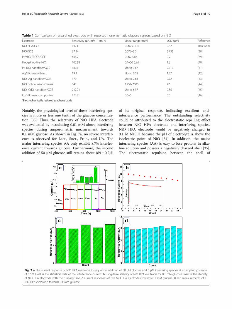
the typical amperometric responses of NiO HPA andNiO BHPA towards different concentration of glucose at0.6 V. Notable current responses are clearly observed forthe two electrodes, and the current responses increasewith the glucose concentration increasing. Figure 6dpresents the relationship between response currents andglucose concentration for NiO HPA and NiO BHPAelectrodes. NiO HPA electrode presents a linear rangefrom 0.32 to 1100 μM with a sensitivity of 1323 μA mM−1 cm−2, which is higher than that of NiO BHPA elec-trode (753 μA mM−1 cm−2). Moreover, the limit of de-tection (LOD) of NiO HPA electrode (0.32 μM) is muchlower than that of NiO BHPA (14.2 μM). To manifestthe advantages of NiO HPA, the performance of NiOHPA electrode was compared with other reported NiO-based glucose detection electrodes in Table 1. It is foundthat NiO HPA electrode presents satisfying electrocata-lytic activity towards glucose in terms of high sensitivityand low LOD, indicating potential applications in elec-trochemical glucose sensors. This is essentially attributedto the abundant active sites, faster mass transport kinet-ics, and accelerated electron transfer kinetics derivedfrom the highly porous hollow architecture.Selectivity is an important indicator to assess the per-
formance of glucose sensors. Some easily oxidized com-pounds, such as Lact., Sucr., Fruc., UA, and AAnormally co-existed with glucose in human blood.
Fig. 6 a Amperometric response of NiO HPA electrode at different potentials with the addition of 0.1 mM glucose and 0.01 mM AA. b Theresponse current of glucose and AA at different potentials. c CA of NiO HPA and NiO BHPA electrode at 0.6 V with the successive addition ofglucose. d The relationship between response current and the concentration of glucose
He et al. Nanoscale Research Letters (2018) 13:3 Page 7 of 10
Notably, the physiological level of these interfering spe-cies is more or less one tenth of the glucose concentra-tion [33]. Thus, the selectivity of NiO HPA electrodewas evaluated by introducing 0.01 mM above interferingspecies during amperometric measurement towards0.1 mM glucose. As shown in Fig. 7a, no severe interfer-ence is observed for Lact., Sucr., Fruc., and UA. Themajor interfering species AA only exhibit 8.7% interfer-ence current towards glucose. Furthermore, the secondaddition of 50 μM glucose still retains about (89 ± 0.2)%
of its original response, indicating excellent anti-interference performance. The outstanding selectivitycould be attributed to the electrostatic repelling effectbetween NiO HPA electrode and interfering species.NiO HPA electrode would be negatively charged in0.1 M NaOH because the pH of electrolyte is above theisoelectric point of NiO [34]. In addition, the majorinterfering species (AA) is easy to lose protons in alka-line solution and possess a negatively charged shell [35].The electrostatic repulsion between the shell of
Table 1 Comparison of researched electrode with reported nonenzymatic glucose sensors based on NiO
Electrode Sensitivity (μA mM−1 cm−2) Linear range (mM) LOD (μM) Reference
NiO HPA/GCE 1323 0.0025–1.10 0.32 This work
NiO/GCE 67.34 0.076–3.0 25.35 [38]
Pt/NiO/ERGOa/GCE 668.2 0.002-5.66 0.2 [39]
Hedgehog-like NiO 1052.8 0.1–50 (μM) 1.2 [40]
Pt–NiO nanofiber/GCE 180.8 Up to 3.67 0.313 [41]
Ag/NiO nanofibers 19.3 Up to 0.59 1.37 [42]
NiO–Ag nanofiber/GCE 170 Up to 2.63 0.72 [43]
NiO hollow nanospheres 343 1500–7000 47 [44]
NiO–CdO nanofiber/GCE 212.71 Up to 6.37 0.35 [45]
Cu/NiO nanocomposites 171.8 0.5–5 0.5 [46]aElectrochemically reduced graphene oxide
Fig. 7 a The current response of NiO HPA electrode to sequential addition of 50 μM glucose and 5 μM interfering species at an applied potentialof 0.6 V. Inset is the statistical data of the interference current. b Long-term stability of NiO HPA electrode for 0.1 mM glucose. Inset is the stabilityof NiO HPA electrode with the running time. c Current responses of five NiO HPA electrodes towards 0.1 mM glucose. d Ten measurements of aNiO HPA electrode towards 0.1 mM glucose
He et al. Nanoscale Research Letters (2018) 13:3 Page 8 of 10
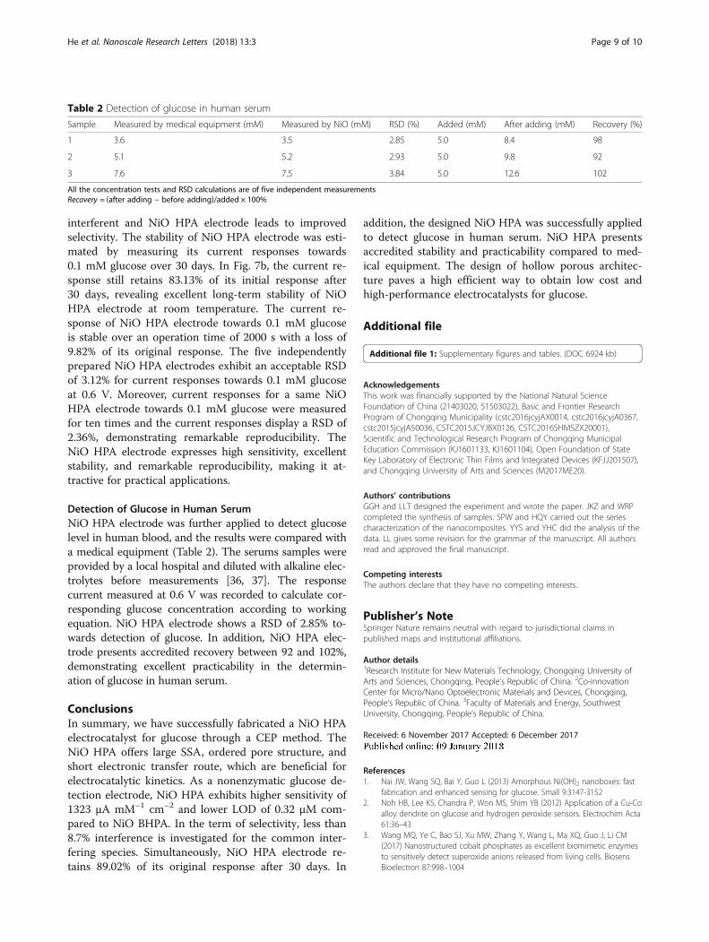
interferent and NiO HPA electrode leads to improvedselectivity. The stability of NiO HPA electrode was esti-mated by measuring its current responses towards0.1 mM glucose over 30 days. In Fig. 7b, the current re-sponse still retains 83.13% of its initial response after30 days, revealing excellent long-term stability of NiOHPA electrode at room temperature. The current re-sponse of NiO HPA electrode towards 0.1 mM glucoseis stable over an operation time of 2000 s with a loss of9.82% of its original response. The five independentlyprepared NiO HPA electrodes exhibit an acceptable RSDof 3.12% for current responses towards 0.1 mM glucoseat 0.6 V. Moreover, current responses for a same NiOHPA electrode towards 0.1 mM glucose were measuredfor ten times and the current responses display a RSD of2.36%, demonstrating remarkable reproducibility. TheNiO HPA electrode expresses high sensitivity, excellentstability, and remarkable reproducibility, making it at-tractive for practical applications.
Detection of Glucose in Human SerumNiO HPA electrode was further applied to detect glucoselevel in human blood, and the results were compared witha medical equipment (Table 2). The serums samples wereprovided by a local hospital and diluted with alkaline elec-trolytes before measurements [36, 37]. The responsecurrent measured at 0.6 V was recorded to calculate cor-responding glucose concentration according to workingequation. NiO HPA electrode shows a RSD of 2.85% to-wards detection of glucose. In addition, NiO HPA elec-trode presents accredited recovery between 92 and 102%,demonstrating excellent practicability in the determin-ation of glucose in human serum.
ConclusionsIn summary, we have successfully fabricated a NiO HPAelectrocatalyst for glucose through a CEP method. TheNiO HPA offers large SSA, ordered pore structure, andshort electronic transfer route, which are beneficial forelectrocatalytic kinetics. As a nonenzymatic glucose de-tection electrode, NiO HPA exhibits higher sensitivity of1323 μA mM−1 cm−2 and lower LOD of 0.32 μM com-pared to NiO BHPA. In the term of selectivity, less than8.7% interference is investigated for the common inter-fering species. Simultaneously, NiO HPA electrode re-tains 89.02% of its original response after 30 days. In
addition, the designed NiO HPA was successfully appliedto detect glucose in human serum. NiO HPA presentsaccredited stability and practicability compared to med-ical equipment. The design of hollow porous architec-ture paves a high efficient way to obtain low cost andhigh-performance electrocatalysts for glucose.
Additional file
Additional file 1: Supplementary figures and tables. (DOC 6924 kb)
AcknowledgementsThis work was financially supported by the National Natural ScienceFoundation of China (21403020, 51503022), Basic and Frontier ResearchProgram of Chongqing Municipality (cstc2016jcyjAX0014, cstc2016jcyjA0367,cstc2015jcyjA50036, CSTC2015JCYJBX0126, CSTC2016SHMSZX20001),Scientific and Technological Research Program of Chongqing MunicipalEducation Commission (KJ1601133, KJ1601104), Open Foundation of StateKey Laboratory of Electronic Thin Films and Integrated Devices (KFJJ201507),and Chongqing University of Arts and Sciences (M2017ME20).
Authors’ contributionsGGH and LLT designed the experiment and wrote the paper. JKZ and WRPcompleted the synthesis of samples. SPW and HQY carried out the seriescharacterization of the nanocomposites. YYS and YHC did the analysis of thedata. LL gives some revision for the grammar of the manuscript. All authorsread and approved the final manuscript.
Competing interestsThe authors declare that they have no competing interests.
Publisher’s NoteSpringer Nature remains neutral with regard to jurisdictional claims inpublished maps and institutional affiliations.
Author details1Research Institute for New Materials Technology, Chongqing University ofArts and Sciences, Chongqing, People’s Republic of China. 2Co-innovationCenter for Micro/Nano Optoelectronic Materials and Devices, Chongqing,People’s Republic of China. 3Faculty of Materials and Energy, SouthwestUniversity, Chongqing, People’s Republic of China.
Received: 6 November 2017 Accepted: 6 December 2017
References1. Nai JW, Wang SQ, Bai Y, Guo L (2013) Amorphous Ni(OH)2 nanoboxes: fast
fabrication and enhanced sensing for glucose. Small 9:3147-31522. Noh HB, Lee KS, Chandra P, Won MS, Shim YB (2012) Application of a Cu-Co
alloy dendrite on glucose and hydrogen peroxide sensors. Electrochim Acta61:36–43
3. Wang MQ, Ye C, Bao SJ, Xu MW, Zhang Y, Wang L, Ma XQ, Guo J, Li CM(2017) Nanostructured cobalt phosphates as excellent biomimetic enzymesto sensitively detect superoxide anions released from living cells. BiosensBioelectron 87:998–1004
Table 2 Detection of glucose in human serum
Sample Measured by medical equipment (mM) Measured by NiO (mM) RSD (%) Added (mM) After adding (mM) Recovery (%)
1 3.6 3.5 2.85 5.0 8.4 98
2 5.1 5.2 2.93 5.0 9.8 92
3 7.6 7.5 3.84 5.0 12.6 102
All the concentration tests and RSD calculations are of five independent measurementsRecovery = (after adding − before adding)/added × 100%
He et al. Nanoscale Research Letters (2018) 13:3 Page 9 of 10
4. Bao SJ, Li CM, Zang JF, Cui XQ, Qiao Y, Guo J (2008) New nanostructuredTiO2 for direct electrochemistry and glucose sensor applications. Adv FunctMater 18:591–599
5. Wang J (2008) Electrochemical glucose biosensors. Chem Rev 108:814–8256. Gouveia-Carida C, Pauliukaite R, Brett CMA (2008) Development of
electrochemical oxidase biosensors based on carbon nanotube-modified carbonfilm electrodes for glucose and ethanol. Electrochim Acta 53:6732–6739
7. Kang X, Mai Z, Zou X, Cai P, Mo J (2007) A sensitive nonenzymatic glucosesensor in alkaline media with a copper nanocluster/multiwall carbonnanotube-modified glassy carbon electrode. Anal Biochem 363:143–150
8. Feng D, Wang F, Chen ZL (2009) Electrochemical glucose sensor based onone-step construction of gold nanoparticle–chitosan composite film.Sensors Actuators B 138:539–544
9. Başkaya G, Yıldız Y, Savk A, Okyay TO, Eriş S, Sert H, Şen F (2017) Rapid,sensitive and reusable detection of glucose by highly monodisperse nickelnanoparticles decorated functionalized multi-walled carbon nanotubes.Biosens Bioelectron 91:728–733
10. Chen C, Xie QJ, Yang DW, Xiao HL, Fu YC, Tan YM, Yao SZ (2013) Recentadvances in electrochemical glucose biosensors: a review. RSC Adv 3:4473–4491
11. Heller A, Feldman B (2008) Electrochemical glucose sensors and theirapplications in diabetes management. Chem Rev 108:2482–2505
12. Scognamiglio V (2013) Nanotechnology in glucose monitoring: advancesand challenges in the last 10 years. Biosens Bioelectron 47:12–25
13. Kaneti YV, Tang J, Salunkhe RR, Jiang XC, Yu A, Wu KCW, Yamauchi Y (2017)Nanoarchitectured design of porous materials and nanocomposites frommetal-organic frameworks. Adv Mater 29:1604898–1604938
14. Xu Y, Tu WG, Zhang BW, Yin SM, Huang YZ, Kraft M, Xu R (2017) Nickelnanoparticles encapsulated in few-layer nitrogen-doped graphene derivedfrom metal-organic frameworks as efficient bifunctional electrocatalysts foroverall water splitting. Adv Mater 29:1605957–1605965
15. Bak SM, Kim KH, Lee CW, Kim KB (2011) Mesoporous nickel/carbon nanotubehybrid material prepared by electroless deposition. J Mater Chem 21:1984–1990
16. Liu H, Wang GX, Liu J, Qiao SZ, Ahnc H (2011) Highly ordered mesoporousNiO anode material for lithium ion batteries with an excellentelectrochemical performance. J Mater Chem 21:3046–3052
17. Ci SQ, Huang TZ, Wen ZH, Cui SM, Mao S, Steeber DA, Chen JH (2014)Nickel oxide hollow microsphere for non-enzyme glucose detection.Biosens Bioelectron 54:251–257
18. Cao CY, Guo W, Cui ZM, Song WG, Cai W (2011) Microwave-assisted gas/liquidinterfacial synthesis of flowerlike NiO hollow nanosphere precursors and theirapplication as supercapacitor electrodes. J Mater Chem 21:3204–3209
19. Tian LL, Zhong XH, Hu WP, Liu BT, Li YF (2014) Fabrication of cubic PtCunanocages and their enhanced electrocatalytic activity towards hydrogenperoxide. Nanoscale Res Lett 9:1–5
20. Kim SI, Lee JS, Ahn HJ, Song HK, Jang JH (2013) Facile route to an efficientNiO supercapacitor with a three-dimensional nanonetwork morphology.ACS Appl Mater Interfaces 5:1596–1603
21. Liang K, Tang XZ, Hu WC (2012) High-performance three-dimensionalnanoporous NiO film as a supercapacitor electrode. J Mater Chem 22:11062–11067
22. Chigane M, Ishikawa M (1998) XRD and XPS characterization ofelectrochromic nickel oxide thin films prepared by electrolysis-chemicaldeposition. J Chem Soc Faraday Trans 94:3665–3670
23. Biesinger MC, Payne BP, Lau LWM, Gerson A, Smart RSC (2009) X-rayphotoelectron spectroscopic chemical state quantification of mixed nickelmetal, oxide and hydroxide systems. Surf Interface Anal 41:324–332
24. Varghese B, Reddy MV, Wu ZY, Lit CS, Hoong TC, Rao GVS, Chowdari BVR,Wee ATS, Lim CT, Sow CH (2008) Fabrication of NiO nanowall electrodes forhigh performance lithium ion battery. Chem Mater 20:3360–3367
25. Zhu H, Gu L, Yu D, Sun YJ, Wan M, Zhang M, Wang L, Wu WW, Yao JM, MLD, Guo SJ (2017) The marriage and integration of nanostructures withdifferent dimensions for synergistic electrocatalysis. Energy Environ Sci 10:321–330
26. Bao HZ, Zhang ZH, Hua Q, Huang WX (2014) Compositions, structures andcatalytic activities of CeO2@Cu2O nanocomposites prepared by thetemplate-assisted method. Langmuir 30:6427–6436
27. Sun SD, Yang ZM (2014) Cu2O-templated strategy for synthesis of definablehollow architectures. Chem Commun 50:7403–7415
28. Sohn JH, Cha HG, Kim CW, Kim DK, Kang YS (2013) Fabrication of hollowmetal oxide nanocrystals by etching cuprous oxide with metal(II) ions:approach to the essential driving force. Nano 5:11227–11233
29. Yang P, Tong XL, Wang GZ, Gao Z, Guo XY, Qin Y (2015) NiO/SiC nanocompositeprepared by atomic layer deposition used as a novel electrocatalyst for non-enzymatic glucose sensing. ACS Appl Mater Interfaces 7:4772–4778
30. Li M, Bo XJ, Mu ZC, Zhang YF, Guo LP (2014) Electrodeposition of nickeloxide and platinum nanoparticles on electrochemically reduced grapheneoxide film as a nonenzymatic glucose sensor. Sensors Actuators B 192:261–268
31. Safavi A, Maleki N, Farjami E (2009) Fabrication of a glucose sensor basedon a novel nanocomposite electrode. Biosens Bioelectron 24:1655–1660
32. Wu CH, Deng SX, Wang H, Sun YX, Liu JB, Yan H (2014) Preparation of novelthree-dimensional NiO/Ultrathin derived graphene hybrid for supercapacitorapplications. ACS Appl Mater Interfaces 6:1106–1112
33. Kong CC, Tang LL, Zhang XZ, Sun SD, Yang SC, Song XP, Yang ZM(2014) Templating synthesis of hollow CuO polyhedron and itsapplication for nonenzymatic glucose detection. J Mater Chem A 2:7306–7312
34. Tyagi M, Tomar M, Gupta V (2014) Glad assisted synthesis of NiO nanorodsfor realization of enzymatic reagentless urea biosensor. Biosens Bioelectron52:196–201
35. Xia KD, Yang C, Chen YL, Tian LL, Su YY, Wang JB, Li L (2017) In situfabrication of Ni(OH)2 flakes on Ni foam through electrochemical corrosionas high sensitive and stable binder-free electrode for glucose sensing.Sensors Actuators B 240:979–987
36. Niu XH, Li X, Pan JM, He YF, Qiu FX, Yan YS (2016) Recent advances in non-enzymatic electrochemical glucose sensors based on non-precious transitionmetal materials: opportunities and challenges. RSC Adv 6:84893–84905
37. Liu MM, Liu R, Chen W (2013) Graphene wrapped Cu2O nanocubes: non-enzymatic electrochemical sensors for the detection of glucose andhydrogen peroxide with enhanced stability. Biosens Bioelectron 45:206–212
38. Wang L, Lu XP, Wen CJ, Xie YZ, Miao LF, Chen SH, Li HB, Li P, Song YH(2015) One-step synthesis of Pt-NiO nanoplate array/reduced grapheneoxide nanocomposites for nonenzymatic glucose sensing. J Mater Chem A3:608–616
39. Li M, Bo XJ, Mu ZC, Zhang YF, Guo LP (2014) Electrodeposition of nickeloxide and platinum nanoparticles on electrochemically reduced grapheneoxide film as a nonenzymatic glucose sensor. Sensors Actuators B Chem192:261–268
40. Soomro RA, Ibupoto ZH, Sirajuddin, Willander M (2015) Electrochemicalsensing of glucose based on novel hedgehog-like NiO nanostructures. SensActuators B 209:966–974
41. Ding Y, Liu YX, Zhang LC, Wang Y, Bellagamba M, Parisi J, Li CM, Lei Y(2011) Sensitive and selective nonenzymatic glucose detection usingfunctional NiO-Pt hybrid nanofibers. Electrochim Acta 58:209–214
42. Ding Y, Wang Y, Su LA, Zhang H, Lei Y (2010) Preparation andcharacterization of NiO-Ag nanofibers, NiO nanofibers, and porous Ag:towards the development of a highly sensitive and selective non-enzymaticglucose sensor. J Mater Chem 20:9918–9926
43. Reddy YAK, Ajitha B, Reddy PS, Reddy MSP, Lee JH (2014) Effect of substratetemperature on structural, optical and electrical properties of sputtered NiO-Ag nanocrystalline thin films. Electron Mater Lett 5:907–913
44. Li CC, Liu YL, Li LM, Du ZF, Xu SJ, Zhang M, Yin XM, Wang TH (2008) Anovel amperometric biosensor based on NiO hollow nanospheres forbiosensing glucose. Talanta 77:455–459
45. Ding Y, Wang Y, Zhang LC, Zhang H, Lei Y (2012) Preparation,characterization and application of novel conductive NiO-CdO nanofiberswith dislocation feature. J Mater Chem 22:980–986
46. Zhang XJ, Gu AX, Wang GF, Huang Y, Ji HQ, Fang B (2011) Porous Cu-NiOmodified glass carbon electrode enhanced nonenzymatic glucoseelectrochemical sensors. Analyst 136:5175–5180
He et al. Nanoscale Research Letters (2018) 13:3 Page 10 of 10