Embed Size (px)
Citation preview
Abstract Renal parenchymal defects (RPD) – scars, hy-poplasia/dyspalsia – in children are a major risk factorfor chronic renal failure. Most authors would agree thatRPD should be detected and followed by a 99mTc-dimercaptosuccinic acid renal scan (DMSA), as ultra-sonography (US) does not seem to be sensitive enoughfor this purpose. However, it might well be that DMSAis too sensitive and detects RPD that are too small to beclinically significant. The purpose of this study was toevaluate the sensitivity of US in identifying patients withclinically significant RPD and in detecting RPD of vari-ous grades as seen by DMSA. In 89 children with abnor-mal DMSA, a second DMSA, US, and other tests forevaluating renal function were performed at least 1 yearafter the first DMSA. The extent of RPD detected byDMSA and US was correlated with renal function pa-rameters. In all 5 patients with diminished renal func-tion, RPD were detected by both DMSA scan and US. In addition, US detected clinically insignificant RPD in48 of 67 cases (71.6%). The present study has shownthat, compared with DMSA, US is sensitive enough todetect clinically significant RPD in children. The substi-tution of DMSA with US would be beneficial, as thiswould eliminate radiation exposure, reduce costs, and in-crease availability.
Keywords Renal parenchymal defects · Ultrasonography · 99mTc-Dimercaptosuccinic acid renalscan · Children · Sensitivity
Introduction
The association between urinary tract infection (UTI),vesicoureteral reflux, and renal scars is well established[1, 2, 3, 4, 5]. Renal parenchymal defects (RPD) – scars,hypoplasia/dysplasia – in children are a major risk factorfor chronic renal failure, hypertension, proteinuria, andcomplications during pregnancy. In the most severecases they can lead to end-stage renal failure [4, 5, 6, 7].Most RPD occur or are discovered at the time of the firstUTI in early childhood, and generally do not tend to in-crease in size [8]. However, according to long-term fol-low-up studies, this is not always the case, as some RPDcan also progress in later childhood [9, 10]. It has beengenerally accepted that after their first UTI, childrenshould be diagnostically evaluated and those with RPDfollowed for several years. However, the precise timingand type of investigation remain controversial. There area number of techniques available for the assessment ofRPD: intravenous urography, 99mTc-dimercaptosuccinicacid renal scan (DMSA), ultrasonography (US) and, to acertain extent, computerized tomography and magneticresonance imaging. Despite the controversy about themost appropriate type of investigation, there is a tenden-cy to choose the least invasive method capable of pro-viding sufficient information. Intravenous urography,long considered the gold standard, has recently been sub-stituted by DMSA and US, because of the radiation haz-ard and side effects caused by the contrast medium.DMSA is the most sensitive method for the detection ofRPD [11, 12, 13, 14], but unfortunately involves a rela-tively high radiation load for the patient and can only beperformed in nuclear medicine departments. AlthoughUS is less sensitive than DMSA in detecting RPD [11,12, 13, 14], it is believed to be harmless and is widelyavailable. Considering the advantages and disadvantages
T. Kersnik Levart (✉) · G. Novljan · R.B. KendaDepartment of Pediatric Nephrology, University Medical Center,Ljubljana, Sloveniae-mail: [email protected].: +386-1-5229600, Fax: +386-1-4311123
J.J. FettichNuclear Medicine Department, University Medical Center, Ljubljana, Slovenia
A. Kenig · D. KljucevsekPediatric Radiology Unit, University Medical Center, Ljubljana, Slovenia
T. Kersnik LevartDepartment of Pediatric Nephrology, University Medical Center,Stare pravde 4, 1000 Ljubljana, Slovenia
Pediatr Nephrol (2002) 17:1059–1062DOI 10.1007/s00467-002-1007-y
O R I G I N A L A RT I C L E
Tanja Kersnik Levart · Anton Kenig · Jure J. FettichDamjana Kljucevsek · Gregor NovljanRajko B. Kenda
Sensitivity of ultrasonography in detecting renal parenchymal defectsin children
Received: 16 May 2002 / Revised: 4 September 2002 / Accepted: 5 September 2002 / Published online: 14 November 2002© IPNA 2002
of these methods, most authors would place DMSA be-fore US because of its higher sensitivity [11, 12, 13, 14].However, DMSA might well be too sensitive, detectingRPD that are too small to be clinically significant. To assess the real value of US in the detection of RPD andits advantages and disadvantages in comparison withDMSA, studies of this kind should also include the eval-uation of renal function parameters. The purpose of thisstudy was to evaluate the sensitivity of US in identifyingpatients with clinically significant RPD and in detectingRPD of various grades as seen by DMSA.
Materials and methods
Eighty-nine children, 63 girls and 26 boys aged 2–19 years (mean8.71, median 9 years), were included in this prospective study.The sample was selected from patients treated for UTI at our de-partment from January 1994 to December 1999. As a part of thatevaluation, a DMSA was also performed at least 6 months afterthe diagnosis of acute UTI. All patients with abnormal DMSAwere included in the present study. The initial DMSA was consid-ered abnormal when at least one of the following criteria was ful-filled: (1) less than 45%/more than 55% relative renal function, (2) abnormal size, shape, contour, or tracer distribution of one orboth kidneys.
DMSA, US, and other tests (blood pressure measurements,analysis of urine and blood for proteinuria, elevated creatinineblood levels, anemia, or electrolyte disturbances) that indirectlyindicate the possible clinical consequences of RPD were per-formed in all patients. The second DMSA was performed at least1 year after the initial test. None of the patients had an active UTIat the time of the investigations.
99mTc-Dimercaptosuccinic acid renal scan
All DMSAs were performed in a standardized fashion. A bodysurface area–adjusted adult-equivalent dose of 80 MBq Tc-99DMSA was administered intravenously to each patient. Imagingwas performed at least 3 h after isotope administration using agamma camera (General Electric MAXI 300) with a high-resolu-tion low-energy collimator. Three planar images were taken: pos-terior and left and right posterior obliques. Relative renal functionwas evaluated on the basis of the posterior image after backgroundcorrection. The images were stored in a computer. Each DMSAwas independently interpreted by two nuclear medicine special-ists, who were unaware of the other's findings. In case of diverg-ing interpretations, the specialists reconciled their findings. In or-der to compare DMSA and US as consistently as possible, a stan-dardized protocol for DMSA image interpretation was developedby modifying the guidelines published by Patel et al. [15]. A scor-ing system for assessing the severity of RPD seen by DMSA wasdesigned and, on the basis thereof, RPD were graded as follows:absent 0 points, mild 1–6 points, moderate 7–11 points, and severe12–18 points. The following parameters were included in the scor-ing system: kidney size (smaller 3 points), relative renal function(40%–44% 3 points, 30%–39% 6 points, 0%–29% 9 points), con-tour [abnormal 1 point for each of the three kidney parts (upperpole, middle part, lower pole)], and defective radionuclide uptake[present 1 point for each of the three kidney parts (upper pole,middle part, lower pole)].
Ultrasonography
All US evaluations were performed on a Toshiba SSA240A or Toshiba SSA370A with convex 3.75- to 6-MHz probes operatingin B mode. The investigations were performed by a pediatric radi-
ologist unacquainted with other findings. In order to compareDMSA and US as consistently as possible, a standardized protocolfor US image interpretation was developed. According to this pro-tocol, US was assessed as normal if the length (between the 3rdand 97th percentile) and shape of the kidney were normal, its con-tours were normal, smooth, and continuous, the echogenicity ofthe renal cortex and medulla was normal, and if the difference insize of the two kidneys was less than 1 cm. US was also consid-ered normal if the kidney was enlarged due to compensatory hy-pertrophy, but of normal shape and smooth, continuous contours,provided the echogenicity of the kidney's renal cortex and medullawas normal. Any different findings rendered US abnormal.
The sensitivity of US in detecting RPD of various grades asseen by DMSA was evaluated using the SPSS for Windows ver-sion 10.1. A chi-squared test was used to evaluate the statisticalsignificance of abnormal US findings on RPD of different gradesas seen by DMSA.
Evaluation of renal function parameters
The patients were considered to have clinically significant RPDwhen at least one of the following criteria was met: arterial hyper-tension, proteinuria, elevated creatinine blood levels, anemia (normocytic, normochromic), or electrolyte disturbance.
Arterial blood pressure was measured three times in 5-min in-tervals and the mean blood pressure was calculated. If the readingsexceeded the 95th percentile for patient age and gender, 48-h ambulatory blood pressure monitoring was performed. Arterial hypertension was defined as more than 50% of measurements exceeding the 95th percentile for patient height.
Spot urine and blood samples were taken to evaluate proteinur-ia, anemia, electrolyte disturbances, or a reduced glomerular filtra-tion rate.
Proteinuria was suspected when proteins were found in thespot urine sample. The proteinuria was further quantitatively eval-uated on the basis of the 24-h urine sample. Significant proteinuriawas defined as a protein excretion of over 4 mg/h per m2.
The glomerular filtration rate was evaluated indirectly on thebasis of blood creatinine levels. When these levels were elevatedfor patient age and gender, the test was repeated and, if elevatedlevels were repeatedly detected, the endogenous clearance of cre-atinine (ECC) or EDTA clearance (EDTA) was measured. Theglomerular filtration rate was considered reduced when ECC orEDTA was below the normal values for patient age and gender.
Anemia (normocytic, normochromic) was defined as a hemo-globin level below the lower limit for age and gender of a patientwith a normal mean corpuscular volume and normal mean corpus-cular hemoglobin. Electrolyte disturbances were defined as a dis-sociation rate of serum electrolyte concentrations above or belownormal values for patient age.
The study was approved by the Medical Ethics Committee andwritten informed consent was obtained from all parents after theyhad been informed of the nature of the study.
Results
The sensitivity of US in identifying patients with clinically significant RPD
In 17 patients with normal kidney function (17 of 89),neither DMSA nor US detected the RPD seen in the firstDMSA performed at least 1 year before their inclusion inthe study. The remaining 72 of 89 patients were classi-fied according to renal function parameters into twogroups. Group A included clinically significant RPD (5 patients) and group B clinically insignificant RPD (67 patients).
1060
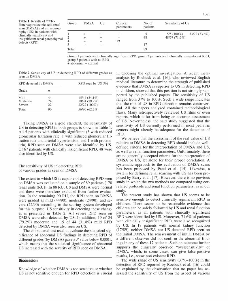
Taking DMSA as a gold standard, the sensitivity ofUS in detecting RPD in both groups is shown in Table 1.All 5 patients with clinically significant (3 with reducedglomerular filtration rate, 1 with reduced glomerular fil-tration rate and arterial hypertension, and 1 with protein-uria) RPD seen on DMSA were also identified by US.Of 67 patients with clinically insignificant RPD, 48 werealso identified by US.
The sensitivity of US in detecting RPD of various grades as seen on DMSA
The extent to which US is capable of detecting RPD seenon DMSA was evaluated on a sample of 89 patients [178renal units (RU)]. In 88 RU, US and DMSA were normaland these were therefore excluded from further evalua-tion. In the remaining 90 RU, the RPD seen on DMSAwere graded as mild (44/90), moderate (24/90), and se-vere (22/90) according to the scoring system developedfor this purpose. US sensitivity in detecting these chang-es is presented in Table 2. All severe RPD seen onDMSA were also detected by US. In addition, 19 of 24(79.2%) moderate and 15 of 44 (31.8%) mild RPD detected by DMSA were also seen on US.
The chi-squared test used to evaluate the statistical sig-nificance of abnormal US findings in detecting RPD ofdifferent grades (by DMSA) gave a P value below 0.0001,which means that the statistical significance of abnormalUS increased with the severity of RPD seen on DMSA.
Discussion
Knowledge of whether DMSA is too sensitive or whetherUS is not sensitive enough for RPD detection is crucial
in choosing the optimal investigation. A recent meta-analysis by Roebuck et al. [16], who reviewed Englishmedical literature to determine the strength of publishedevidence that DMSA is superior to US in detecting RPDin children, showed that this position is not strongly sup-ported by the published papers. The sensitivity of USranged from 37% to 100%. Such a wide range indicatesthat the role of US in RPD detection remains controver-sial. All the papers analyzed contained methodologicalflaws. Many retrospectively reviewed US films or evenreports, which is far from being an accurate assessmentof US. Nevertheless, the said study suggested that thesensitivity of US currently performed in most pediatriccenters might already be adequate for the detection ofRPD.
We believe that the assessment of the real value of USrelative to DMSA in detecting RPD should include well-defined criteria for the interpretation of DMSA and US,as well as renal function parameters. Unfortunately, thereare no generally accepted criteria for the interpretation ofDMSA or US, let alone for their proper correlation. Asystematic approach to the evaluation of DMSA scanshas been proposed by Patel et al. [15]. Likewise, asystem for defining renal scarring with US has been pro-posed by Barry et al. [17]. However, there is no previousstudy in which the two methods are compared using cor-related protocols and renal function parameters, as in ourstudy.
The present study has shown that US seems to be sensitive enough to detect clinically significant RPD inchildren. There seems to be reasonable evidence thatchildren can be safely followed by US and renal functionparameters, as all patients with clinically significantRPD were identified by US. Moreover, 71.6% of patientswith clinically insignificant RPD were also recognizedby US. In 17 patients with normal kidney function(17/89), neither DMSA nor US detected RPD seen onthe initial DMSA. The reassessment of initial DMSA bya different observer did not confirm the abnormal find-ings in any of these 17 patients. Such an outcome furthersupports the clinically observed “oversensitivity” ofDMSA, which, in some cases, can give false-positive results, i.e., show non-existent RPD.
The wide range of US sensitivity (37%–100%) in thedetection of RPD reported by Roebuck et al. [16] couldbe explained by the observation that no paper has as-sessed the sensitivity of US from the aspect of various
1061
Table 1 Results of 99mTc-dimercaptosuccinic acid renalscan (DMSA) and ultrasonog-raphy (US) in patients withclinically significant and insignificant renal parenchymaldefects (RPD)
Group DMSA US Clinical No. of Sensitivity of USparameters patients
1 + + + 5 5/5 (100%) 53/72 (73.6%)2 + + – 48 48/67 (71.6%)+ – – 193 – – – 17Total 89
Group 1 patients with clinically significant RPD, group 2 patients with clinically insignificant RPD,group 3 patients with no RPD+ abnormal, – normal
Table 2 Sensitivity of US in detecting RPD of different grades asseen on DMSA
RPD detected by DMSA RPD seen by US (%)
Grade n
Mild 44 15/44 (34.1%)Moderate 24 19/24 (79.2%)Severe 22 22/22 (100%)Total 90 56/90 (62.2%)
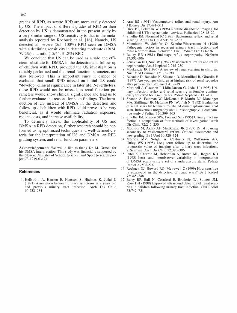
grades of RPD, as severe RPD are more easily detectedby US. The impact of different grades of RPD on theirdetection by US is demonstrated in the present study bya very similar range of US sensitivity to that in the meta-analysis reported by Roebuck et al. [16]. Namely, US detected all severe (5/5, 100%) RPD seen on DMSAwith a declining sensitivity in detecting moderate (19/24,79.2%) and mild (15/44, 31.8%) RPD.
We conclude that US can be used as a safe and effi-cient substitute for DMSA in the detection and follow-upof children with RPD, provided the US investigation isreliably performed and that renal function parameters arealso followed. This is important since it cannot be excluded that small RPD missed on initial US could “develop” clinical significance in later life. Nevertheless,these RPD would not be missed, as renal function pa-rameters would show clinical significance and lead us tofurther evaluate the reasons for such findings. The intro-duction of US instead of DMSA in the detection and follow-up of children with RPD could prove to be verybeneficial, as it would eliminate radiation exposure, reduce costs, and increase availability.
To definitely assess the applicability of US andDMSA in RPD detection, further research should be per-formed using optimized techniques and well-defined cri-teria for the interpretation of US and DMSA, an RPDgrading system, and renal function parameters.
Acknowledgements We would like to thank Dr. M. Grmek forhis DMSA interpretation. This study was financially supported bythe Slovene Ministry of School, Science, and Sport (research pro-ject J3-1219-0312).
References
1. Hellström A, Hanson E, Hansson S, Hjalmas K, Jodal U(1991) Association between urinary symptoms at 7 years oldand previous urinary tract infection. Arch Dis Child66:232–234
2. Arat BS (1991) Vesicoureteric reflux and renal injury. AmJ Kidney Dis 17:491–511
3. Dick PT, Feldman W (1996) Routine diagnostic imaging forchildhood UTI: a systematic overview. Pediatrics 128:15–22
4. Smellie JM, Normand IC (1975) Bacteriuria, reflux, and renalscarring. Arch Dis Child 508:581–585
5. Mannhardt W, Schofer O, Schulte-Wissermann H (1986)Pathogenic factors in recurrent urinary tract infections and renal scar formation in children. Eur J Pediatr 145:330–336
6. Bailey RR (1981) End-stage reflux nephropathy. Nephron27:302–306
7. Senekjian HO, Suki W (1982) Vesicoureteral reflux and refluxnephropathy. Am J Nephrol 2:245–250
8. Mackenzie JR (1996) A review of renal scarring in children.Nucl Med Commun 17:176–190
9. Benador D, Benador N, Slosman D, Mermillod B, Girardin E(1997) Are younger children at highest risk of renal sequelaeafter pyelonephritis? Lancet 4:17–19
10. Martinell J, Claesson I, Lidin-Janson G, Jodal U (1995) Uri-nary infection, reflux and renal scarring in females continu-ously followed for 13–38 years. Pediatr Nephrol 9:131–136
11. Shanon A, Feldman W, McDonald P, Martin DJ, MatzingerMA, Shillinger JF, McLaine PN, Wolfish N (1992) Evaluationof renal scars by technetium-labeled dimercaptosuccinic acidscan, intravenous urography and ultrasonography: a compara-tive study. J Pediatr 120:399–403
12. Smellie JM, Rigden SPA, Precsod NP (1995) Urinary tract in-fection: a comparison of four methods of investigation. ArchDis Child 72:247–250
13. Monsour M, Azmy AF, MacKenzie JR (1987) Renal scarringsecondary to vesicoureteral reflux. Critical assessment andnew grading. Br J Urol 60:320–324
14. Merrick MV, Notghi A, Chalmers N, Wilkinson AG, Uttley WS (1995) Long term follow up to determine the prognostic value of imaging after urinary tract infections. 2. Scarring. Arch Dis Child 72:393–396
15. Patel K, Charron M, Hoberman A, Brown ML, Rogers KD(1993) Intra- and interobserver variability in interpretation of DMSA scans using a set of standardized criteria. PediatrRadiol 23:506–509
16. Roebuck DJ, Howard RG, Metreweli C (1999) How sensitiveis ultrasound in the detection of renal scars? Br J Radiol72:345–348
17. Barry BP, Hall N, Cornford E, Broderic NJ, Somers JM,Rose DH (1998) Improved ultrasound detection of renal scar-ring in children following urinary tract infection. Clin Radiol53:747–751
1062