Embed Size (px)
Citation preview

lable at ScienceDirect
Hearing Research xxx (2016) 1e13
Contents lists avai
Hearing Research
journal homepage: www.elsevier .com/locate/heares
Research Paper
Sensory coding and cognitive processing of sound in Veterans withblast exposure
Scott Bressler a, Hannah Goldberg a, Barbara Shinn-Cunningham a, b, *
a Center for Computational Neuroscience and Neural Technologies (CompNet), Boston University, Boston, MA 02215, USAb Department of Biomedical Engineering, Boston University, Boston, MA 02215, USA
a r t i c l e i n f o
Article history:Received 9 July 2016Received in revised form7 October 2016Accepted 26 October 2016Available online xxx
Keywords:Blast-induced traumatic brain injurySelective attentionEnvelope following responseCochlear neuropathyHidden hearing loss
* Corresponding author. Center for ComputationTechnology, 677 Beacon Street, Boston, MA 02215, US
E-mail address: [email protected] (B. Shinn-Cunningh
http://dx.doi.org/10.1016/j.heares.2016.10.0180378-5955/© 2016 Elsevier B.V. All rights reserved.
Please cite this article in press as: Bressler, SResearch (2016), http://dx.doi.org/10.1016/j.
a b s t r a c t
Recent anecdotal reports from VA audiology clinics as well as a few published studies have identified asub-population of Service Members seeking treatment for problems communicating in everyday, noisylistening environments despite having normal to near-normal hearing thresholds. Because of theirincreased risk of exposure to dangerous levels of prolonged noise and transient explosive blast events,communication problems in these soldiers could be due to either hearing loss (traditional or “hidden”) inthe auditory sensory periphery or from blast-induced injury to cortical networks associated withattention. We found that out of the 14 blast-exposed Service Members recruited for this study, 12 hadhearing thresholds in the normal to near-normal range. A majority of these participants reported havingproblems specifically related to failures with selective attention. Envelope following responses (EFRs)measuring neural coding fidelity of the auditory brainstem to suprathreshold sounds were similar be-tween blast-exposed and non-blast controls. Blast-exposed subjects performed substantially worse thannon-blast controls in an auditory selective attention task in which listeners classified the melodic contour(rising, falling, or “zig-zagging”) of one of three simultaneous, competing tone sequences. Salient pitchand spatial differences made for easy segregation of the three concurrent melodies. Poor performance inthe blast-exposed subjects was associated with weaker evoked response potentials (ERPs) in frontal EEGchannels, as well as a failure of attention to enhance the neural responses evoked by a sequence when itwas the target compared to when it was a distractor. These results suggest that communication problemsin these listeners cannot be explained by compromised sensory representations in the auditory pe-riphery, but rather point to lingering blast-induced damage to cortical networks implicated in the controlof attention. Because all study participants also suffered from post-traumatic disorder (PTSD), follow-upstudies are required to tease apart the contributions of PTSD and blast-induced injury on cognitiveperformance.
© 2016 Elsevier B.V. All rights reserved.
1. Introduction
Anecdotal reports as well as formal studies show that Veteransoften have difficulty with certain auditory tasks, especially thosethat require understanding speech in the presence of competingsounds (Gallun et al., 2012; Lew, 2007; Oleksiak et al., 2012;Saunders et al., 2015). Since nearly every social setting, includinga business meeting, a restaurant, a sporting event, or a party, in-volves simultaneous sounds, such communication difficulties canhave enormous impact on everyday function. Veterans suffering
al Neuroscience and NeuralA.am).
., et al., Sensory coding and coheares.2016.10.018
from this kind of communication challenge may avoid situationsthat make them feel overwhelmed, leading to social isolation athome and reduced productivity at work. Two potential risk factors,both of which are common amongst Veterans, may contribute toproblems communicating in crowded settings: hearing damageassociated with noise exposure and cognitive problems associatedwith blast exposure.
Over $1 billion is spent annually on auditory dysfunction inVeterans, often associated with noise exposure as well as blast;hearing loss has become the most prevalent service-connecteddisability (Fausti et al., 2009). However, in addition to this, since2000, there have been over 347,000 clinically confirmed cases oftraumatic brain injury (TBI) across all branches of the military, 82%falling under the category of mild TBI (Defense and Veterans Brain
gnitive processing of sound in Veterans with blast exposure, Hearing
S. Bressler et al. / Hearing Research xxx (2016) 1e132
Injury Center DVBIC, 2016). Both hearing damage and TBI are formsof the “invisible wounds of war” that are difficult to identify, butnonetheless devastating (Tanielian and Jaycox, 2008). Importantly,being able to understand speech in social settings places demandsboth on basic hearing function and central cognitive function,which may be damaged with TBI. Given how ubiquitous bothhearing damage and TBI are amongst Veterans, it is critical to teaseapart how both contribute to auditory dysfunction in Veterans. Thisis the goal of the current study.
1.1. “Traditional” and “hidden” hearing loss
Hearing loss is traditionally diagnosed when listeners haveelevated hearing thresholds. Such loss is typically the result ofirreversible damage to the hair cells of the cochlea. Damage to theouter hair cells in the cochlea, which are responsible for activelyamplifying cochlear motion, compromises cochlear mechanicalfunction. Sounds that are typically audible but quiet are no longeramplified, and may be inaudible, leading to elevated hearingthresholds. This form of hearing damage is what is diagnosed bycurrent audiological screenings.
Listeners are said to have “normal hearing” as long as they havenormal hearing thresholds. However, recent animal work has beenexploring a subtler (but important) form of hearing loss knownformally as “cochlear synaptopathy.” In mice and guinea pigs,cochlear synapses can be damaged after only moderate levels ofnoise exposure. The damage is thought be the result of excitotox-icity, which leads to destruction of synapses. Over time, this syn-aptic loss results in the degeneration and death of the spiral ganglia(ascending auditory nerve fibers or ANFs) normally enervated bythe missing synapses (Kujawa and Liberman, 2009). Nerve fiberswith low spontaneous firing rates appear to be most susceptible tosuch damage (Furman et al., 2013).
At hearing threshold, the low-spontaneous rate (LSR) fibers thatare most vulnerable to noise damage do not contribute to neuralresponses. However, importantly, these LSR ANFs are critical forencoding amplitude modulation of supra-threshold sounds (short-term fluctuations in the level of clearly audible sounds). Suchsupra-threshold modulation is critical for conveying meaning inongoing sound, especially in non-stationary signals like speech.Interestingly, at exposure levels that can lead to such synaptopathy,cochlear function can remain intact, and the ANFs encodingthreshold-level sounds may respond normally. As a result, standardpure-tone audiograms do not detect this type of hearing loss(Lobarinas et al., 2013), which is colloquially referred to as “hiddenhearing loss” (Schaette and McAlpine, 2011).
Service Members routinely suffer noise exposure, both duringtraining and deployment. Noise exposure can lead not only toclinically recognized hearing deficits in the form of elevatedthresholds, but also to cochlear synaptopathy, which is currentlyundiagnosed (and “hidden”). Thus, a good number of ServiceMembers who do not have “impaired hearing” as defined by cur-rent clinical practice may nonetheless have damaged hearing in theform of “hidden hearing loss.”
Currently, the only direct evidence of cochlear synaptopathy andsubsequent neuropathy in humans is from post-mortem immu-nohistochemical and electron microscopy analysis from temporalbones of five subjects without significant hair cell loss or any his-tory of otologic disease (Viana et al., 2015). Indirect, non-invasiveestimates of hidden hearing loss are being developed; these ef-forts focus on measurements that are sensitive to supra-thresholdneural responses, such as the middle ear muscle reflex, auditorybrainstem response, and envelope following response (EFR)(Bharadwaj et al., 2015; Mehraei et al., 2016; Shaheen et al., 2015;Valero et al., 2016).
Please cite this article in press as: Bressler, S., et al., Sensory coding and coResearch (2016), http://dx.doi.org/10.1016/j.heares.2016.10.018
The EFR, a measure of the fidelity with which the brainstem canfollow periodic oscillations in sound inputs, has recently beenshown to index hidden hearing loss, both in animals (Shaheen et al.,2015), and in humans (Bharadwaj et al., 2015; Ruggles et al., 2011).This measure thus provides a way of assessing supra-thresholdhearing fidelity in listeners with normal hearing thresholds.
1.2. Blast exposure and TBI
For combat troops, the predominant vehicle for injury is expo-sure to explosive blast ordinance (IEDs, mortar rounds). Suchexposure frequently results in blast-induced TBI (Terrio et al.,2009). While the most common symptom associated with TBI isnon-headache and headache pain, Service Members with a historyof TBI often seek treatment for other symptoms that can includesleep disorders, memory loss, cognitive dysfunction, and hearingproblems (Bergemalm and Lyxell, 2009; Farmer et al., 2016; Hooveret al., 2015; Krause et al., 2014; Lew, 2007; Munjal et al., 2010;Oleksiak et al., 2012). These symptoms may resolve within weeksto months after injury (Kwok et al., 2009; Levin et al., 1987);however, in a subset of patients these problems persist and worseninto debilitating post-concussion syndrome (McKee and Robinson,2014). In the most severe cases, patients go on to develop chronictraumatic encephalopathy (CTE), a condition whose symptoms in-cludes problems with impulse control, paranoia, and severedepression; the disease can progress to early-onset dementia andultimately death (McKee and Robinson, 2014; Mez et al., 2013).
Diagnosing mild TBI (mTBI) is complicated and far from an exactscience. Because evidence of injury is often undetectable usingneural imaging techniques (Hoge et al., 2008; Mac Donald et al.,2011), clinicians and combat medical personnel base their di-agnoses, in part, on interviews that focus on whether the patientexperienced a loss of or some form of altered consciousness(Management of Concussion/mTBI Working Group, 2009).Depending on the circumstances surrounding the injury as well aswhether or not trained medical staff were present at or near thetime of injury, this information may be incomplete or inaccurate. Inthe military, it is thought that many such events go unreported(Chapman and Diaz-Arrastia, 2014; Schwab et al., 2007; Tanielianand Jaycox, 2008). In particular, once initial symptoms subside,soldiers are eager to return to duty, and fear the possibility ofdisqualification for medical reasons if they admit to TBI symptoms.Recent work suggests that CTE is a cumulative effect of multiplesub-concussive and concussive events (Baugh et al., 2012), whichhighlights both the need to understand how TBI affects the brainand the need to educate Service Members about the consequencesof TBI.
1.3. Blast exposure and selective auditory attention
The ability to follow a conversation in crowded and noisylistening environments depends critically on the listener's ability tofocus and sustain attention on a speaker of interest while simul-taneously ignoring the interfering sounds that may be present inthe room. This scenario is commonly referred to as the “cocktailparty problem” (Cherry, 1953). Successful communication ineveryday social settings typically strikes a balance between selec-tive attention, where listeners maintain their attention on a singlesound source, and divided attention, where listeners switch theirattention from source to source (talker to talker or talker to TV, forexample). In both cases, in order to ignore unwanted sounds andattend information from an important sound in a scene, a listenermust be able to “segregate” or perceptually separate the soundsmaking up the mixture. Segregation can fail if the listener does nothave an accurate and detailed sensory representation of the
gnitive processing of sound in Veterans with blast exposure, Hearing
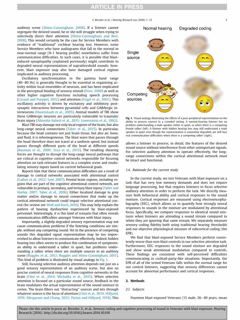
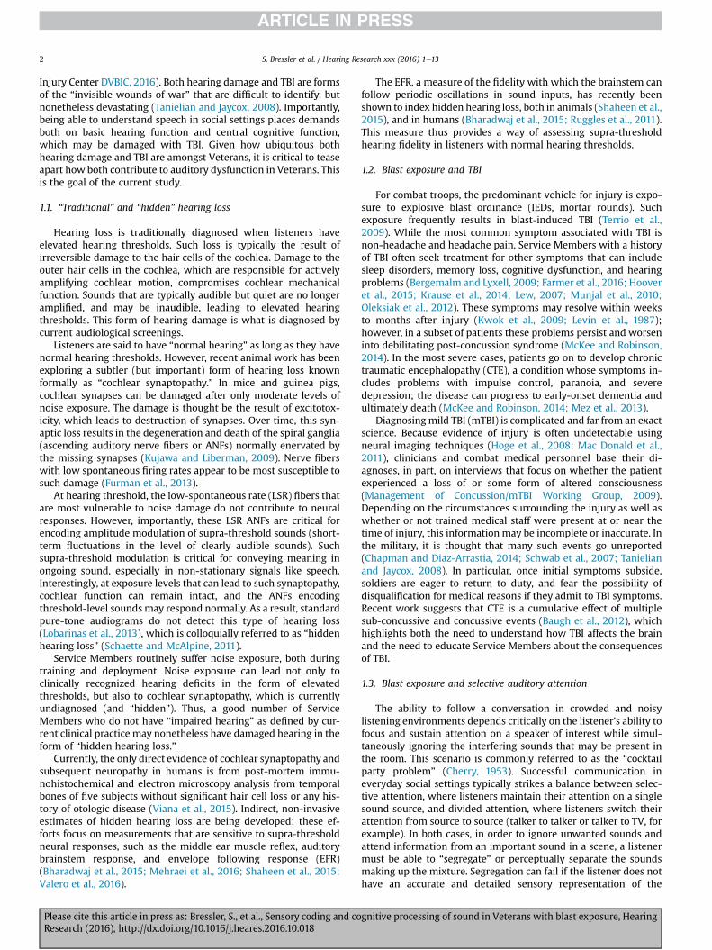
Fig. 1. Visual analogy illustrating the effects of a poor peripheral representation on theability to process sources in a crowded setting. A normal-hearing listener has noproblem understanding a male speaker either in quiet, or when there is a competingfemale talker (left). A listener with hidden hearing loss may still understand a malespeaker in quiet even though the representation is somewhat degraded, yet will facereal communication difficulties when there is a competing talker (right).
S. Bressler et al. / Hearing Research xxx (2016) 1e13 3
auditory scene (Shinn-Cunningham, 2008). If a listener cannotsegregate the desired sound, he or she will struggle when trying toselectively direct their attention (Shinn-Cunningham and Best,2008). This would certainly be the case for Service Members withevidence of “traditional” cochlear hearing loss. However, someService Members who have audiograms that fall in the normal tonear-normal range (H-1 hearing profile) nonetheless suffer fromcommunication difficulties. In such cases, it is possible that blast-induced synaptopathy (explained previously) might contribute todegraded neural representations of suprathreshold sounds; how-ever, blast exposure may also have damaged cortical regionsimplicated in auditory processing.
Oscillatory synchronization in the gamma band range(40e80 Hz) is generally thought to be essential in organizing ac-tivity within local ensembles of neurons, and has been implicatedin the perceptual binding of sensory stimuli (Fries, 2009) as well asother higher cognitive functions including speech processing(Giraud and Poeppel, 2012) and attention (Engel et al., 2001). Thisoscillatory activity is driven by excitatory and inhibitory post-synaptic interactions between pyramidal cells and GABAergic in-terneurons (Hasenstaub et al., 2005). Animal models of TBI showthese GABAergic neurons are particularly vulnerable to traumaticbrain injury (Almeida-Suhett et al., 2015; Lowenstein et al., 1992).
Blast TBI may damage not only local regions of the brain, but alsolong-range neural connections (Taber et al., 2015). In particular,because the head contains not just brain tissue, but also air, bone,and fluid, it is inhomogeneous. The blast wave that passes throughthe head therefore does not travel at a uniform speed, but insteadpasses through different parts of the head at different speeds(Bauman et al., 2009; Sosa et al., 2013). The resulting shearingforces are thought to disrupt the long-range neural pathways thatare critical in cognitive control networks responsible for focusingattention on task-relevant features in a complex scene and modu-lating sensory inputs based on current behavioral goals.
Reports hint that these communication difficulties are a result ofdamage to cortical networks associated with attentional control(Gallun et al., 2012; Lew, 2007). Frontal brain regions, including re-gions that are part of the cognitive attentional control network, arevulnerable to primary, secondary, and tertiary blast injury (Taber andHurley, 2007; Taber et al., 2006). Either localized damage to greymatter and/or damage to neural connections that make up thecortical attentional network could impair selective attentional con-trol (for review see Wolf and Koch, 2016). This may help explain thepattern of hearing dysfunction experienced by blast-exposedpersonnel. Interestingly, it is this kind of scenario that often revealscommunication difficulties amongst Veterans with blast injury.
Importantly, a slightly degraded sensory representation may notcause communication problems if the listening conditions are sim-ple, without any competing sound. Yet in the presence of competingsounds this degraded signal representation may be too impov-erished to allow listeners to communicate effectively. Indeed, hiddenhearing loss often seems to produce this combination of symptoms:an ability to understand a talker in quiet, but problems under-standing a talker when there are multiple sources in an auditoryscene (Ruggles et al., 2012; Ruggles and Shinn-Cunningham, 2011).This kind of problem is illustrated by visual analogy in Fig. 1.
Still, focusing selective auditory attention depends not just on agood sensory representation of an auditory scene, but also onprecise control of neural responses from cognitive networks in thebrain (Choi et al., 2014; Michalka et al., 2015). When selectiveattention is focused on a particular sound source, feedback in thebrain modulates the actual representation of the sound mixture incortex. The brain filters out “distracting” sources and lets throughwhatever source is the focus of attention (Choi et al., 2014; Hillyard,1976; Mesgarani and Chang, 2012; Picton and Hillyard, 1974). This
Please cite this article in press as: Bressler, S., et al., Sensory coding and coResearch (2016), http://dx.doi.org/10.1016/j.heares.2016.10.018
allows a listener to process, in detail, the features of the desiredsound source without interference from other unimportant signals.For selective auditory attention to operate effectively, the long-range connections within the cortical attentional network mustbe intact and functional.
1.4. Rationale for the current study
In the current study, we test Veterans with blast exposure on atask that has very low memory demands and does not requirelanguage processing, but that requires listeners to focus selectiveauditory attention in order to perform the task. We directly mea-sure both behavioral ability and cortical responses to the soundmixture. Cortical responses are measured using electroencepha-lography (EEG), which allows us to quantify how strongly neuralresponses to sounds in the mixture are modulated by attentionalfocus. Specifically, we compare responses to identical sound mix-tures when listeners are attending a sound stream compared towhen they are ignoring that same stream. We separately measuresensory coding fidelity both using traditional hearing thresholdsand our objective physiological measure of subcortical coding (theEFR).
We find that blast-exposed Service Members perform consis-tently worse than non-blast controls in our selective attention task.Furthermore, EEG responses to the sound mixture are degradedand show weak attentional modulation compared to controls.These findings are consistent with self-perceived difficultiescommunicating in cocktail-party-like situations. Importantly, theEFR of all of the tested Veterans falls within the normal range forour control listeners, suggesting that sensory differences cannotaccount for abnormal performance and cortical responses.
2. Methods
2.1. Subjects
Fourteen blast-exposed Veterans (13 male, 26e49 years, mean
gnitive processing of sound in Veterans with blast exposure, Hearing
S. Bressler et al. / Hearing Research xxx (2016) 1e134
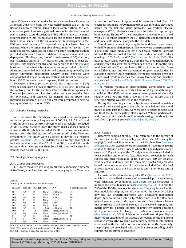
age ¼ 33.5) were referred to the Auditory Neuroscience Laboratoryat Boston University from the Neurorehabilitation Lab at the VABoston Healthcare System, Jamaica Plain Campus (there, the Vet-erans were part of an investigational protocol for the treatment ofpost-traumatic stress disorder, or PTSD). All 14 study participantsreported being within 100 m of at least one explosive blast duringtheir time in service. Of the 14 volunteers, 12 participated in theEEG portion of the study. Six reported having 5 or fewer blast ex-posures, while the remaining six subjects reported having 10 ormore exposures. When possible, the VA Boston Healthcare Systemprovided additional TBI-related data including in-service and pre-deployment TBI diagnoses, loss of consciousness (LOC) duration,post-traumatic amnesia (PTA) duration, and number of blast ex-posures. Data reported for LOC and PTA pertain to the most severeblast event. Individual subject summaries are provided in Table 1.All subjects provided written informed consent as approved by theBoston University Institutional Review Board. Subjects werecompensated at a base hourly rate with an additional performancebonus ($0.02 for each correct response, $7.20 maximum).
Seventeen subjects (6 male, 20e35 years, mean age ¼ 32.5)were selected from a previous study (Choi et al., 2014) to serve asthe control group for the auditory selective attention experiment.These subjects were recruited from advertisements posted at Bos-ton University, and screened for normal hearing (pure tonethresholds less than 25 dB HL). Subjects were presumed to have nohistory of blast exposure or PTSD.
2.2. Objective hearing thresholds
Air conduction thresholds were measured in all participantsfor pulsed pure tones at frequencies of 500, 1 k, 2 k, 3 k, 4 k, and8 kHz in both ears. Control subjects whose thresholds exceeded25 dB HL were excluded from the study. Blast-exposed subjectswhose 4-kHz thresholds exceeded 25 dB HL in any one ear wereexempt from the EEG portion of the study. All of the Veteransremaining in the study were classified as having H-1 hearingprofiles (Smetana, 1999). This is defined as an average hearing lossfor each ear of no more than 25 dB HL at 500, 1 k, and 2 kHz withno individual level greater than 30 dB HL, and no hearing lossgreater than 45 dB HL at 4 kHz.
2.3. Envelope following response
2.3.1. Stimuli and proceduresEFRs were measured in a roughly 40-min session using Biosemi
ActiveTwo system hardware and its accompanying ActiveView data
Table 1Summary of TBI and blast exposure by subject.
Subject# Total TBI Past TBI LOC PTA # Blast Exposures
001 N/A 0 None 5e60 min 31003 2 6 None <2 min 18004 N/A 0 None None 20005 2 0 None 1e4 h 31006 N/A 0 None None 3007 N/A 0 None None 1008 N/A 0 <2 min 5e60 min 18009 2 12 None Dazed/Confused 2010 N/A N/A None Dazed/Confused 3011 N/A 0 <2 min 1e4 h 10114 0 0 None Dazed/Confused 5115 N/A N/A None None 3
N/A: data not available, LOC ¼ loss of consciousness, PTA ¼ post-traumatic amnesia.Total TBI ¼ number of confirmed in-service TBI diagnoses.Past TBI ¼ number of reported pre-deployment TBI.
Please cite this article in press as: Bressler, S., et al., Sensory coding and coResearch (2016), http://dx.doi.org/10.1016/j.heares.2016.10.018
acquisition software. Scalp potentials were recorded from 32electrodes (standard 10/20 montage) plus two reference electrodesplaced on the left and right mastoids. Two vertical electro-oculogram (EOG) electrodes were also included to capture eyeblink events. Timing of critical experimental events was markedwith 5-V TTL pulses sent from the TDT hardware and recorded to anadditional data channel alongside the EEG data.
Stimuli were sinusoidally amplitude-modulated pure tones,with differentmodulation depths. The tones were constructed from4-kHz pure tones modulated by a half-wave rectified, lowpassfiltered 100-Hz sinusoid at four different modulation index valuesequaling 1, 0.63, 0.40, and 0.25 (Bernstein and Trahiotis, 2002). Thepeak-to-peak values were equal across the four modulation depths,and presented at a level that corresponded to 75 dB SPL for the fullymodulated stimuli. The stimuli were 400 ms in durationwith 5-msonset/offset ramps, and were presented in opposing polarities; byaveraging together these responses, the neural response envelopeis measured, while responses that follow temporal fine structureare canceled (Goblick and Pfeiffer, 1969; Skoe and Kraus, 2010; Zhuet al., 2013).
The various modulation depth/polarity combinations werepresented in random order, with a total of 500 presentations percondition. The 4000 stimulus tokens were presented dioticallyroughly every 700 ms with a 100 ms jitter (to ensure that norepetition artifact was present in the responses).
During the recording session, subjects were allowed to watch amovie of their choosing with the subtitles enabled and the soundmuted to help pass the time. We were able to obtain reliable datafor 10 of the 12 participating blast-exposed Veteran participants,and compared it to data from 18 normal-hearing (non-blast) con-trols from a previous study (Bharadwaj et al., 2015).
2.3.2. AnalysisData were sampled at 4096 Hz, re-referenced to the average of
the twomastoid electrodes, and highpass filtered at 70 Hz using theeegfiltfft( ) brick-wall filter function in the EEGLab toolbox (DelormeandMakeig, 2004). Epochs were extracted from�100ms to 450msrelative to stimulus onset. Epochs where the signal dynamic rangeexceeded 150 mV in any of the 32 scalp channels were excluded toreject eyeblink and other artifacts. After epoch rejection, for eachsubject and each modulation depth, 820 trials (410 per polarity)were selected randomly from the remaining epochs. Subjects wholacked the requisite number of trials were excluded from the finalanalysis, leaving a total of 8 blast-exposed and 12 non-blast controlsubjects.
Estimates of the phase-locking value (PLV) (Lachaux et al., 1999),which is a normalized measure of across-trial phase synchrony,were calculated by combining data across the 32 electrodes toimprove the signal to noise ratio (Bharadwaj et al., 2014). From thePLVs of the 100-Hz envelope fundamental frequency for each of thefour modulation depths, we also computed the slope describinghow the PLV strength decreased with decreasing modulationdepth. By cancelling out inter-subject differences due to differencesin head geometry, electrode impedance, and other nuisance factorsthat contribute to the overall strength of the evoked response, thismetric provides a better estimate of brainstem neural encodingfidelity in response to changes in modulation envelope depth(Bharadwaj et al., 2015). Subjects with shallower slopes displaymore robust encoding of the acoustic periodicity in the brainstemresponse, even as the available low frequency envelope informationis reduced with the reduction in modulation depth. Conversely,steep slopes are associated with poor brainstem encoding of thesuprathreshold stimulus envelope.
gnitive processing of sound in Veterans with blast exposure, Hearing

S. Bressler et al. / Hearing Research xxx (2016) 1e13 5
2.4. Subjective self-assessment of hearing
There are anecdotal reports of Service Members with H-1hearing profiles seeking assistance at local VA hospitals for prob-lems communicating in crowded, noisy environments (Oleksiaket al., 2012; Saunders et al., 2015; Saunders and Echt, 2012). Afew published studies have confirmed these reports (Gallun et al.,2012; Lew, 2007). To verify whether we would observe similarfindings, we administered the short form Speech, Spatial, andQualities of Hearing questionnaire (SSQ12) to all study participants(Noble et al., 2013) and compared results for our listeners withthose from a cohort of 103 normal-hearing subjects between theages of 18e25 years of age (Demeester et al., 2012). This surveyinstrument consists of 12 questions designed to return a subjectivemeasure of how well a person hears under several different real-world situations. It evaluates how well a person can 1) followspeech in the presence of noise and competing talkers, and 2)localize and identify sounds, and provides a way of summarizing alistener's subjective assessment of their own hearing abilities.Questions are scored on a Likert scale from 0 to 10.
2.5. Selective auditory attention task
The selective auditory attention task used in this study (Fig. 2)was identical to the one described in Choi et al. (2014), and wasperformed in a separate experimental session from the EFR mea-surement. Briefly, each trial consisted of three simultaneous mel-odies, each simulated from a different lateral angle. Subjects wereinstructed as to what melody to attend using an informativeauditory cue before the start of each trial. They were tasked withidentifying the contour of the attended melody, which was rising,falling, or zig-zagging. Responses were registered using thenumeric keypad on a computer keyboard during a prescribedresponse period.
2.5.1. EquipmentExperimental stimuli were created in MATLAB (The Mathworks,
Natick, MA). The experiment was controlled using the Psychtoolbox3 extension (Brainard, 1997) and TDT Active X Controls fromTucker-Davis Technologies (TDT, Alachua, FL). Auditory and stim-ulus event signals were presented via the System 3 RP 2.1 RealtimeProcessor with an HB7 Headphone Amplifier through ER-1 insertearphones (Etymotic, Elk Grove Village, IL). The stimulus soundlevel was fixed at 70 dB SPL (root-mean-squared). All experimentalsessions were conducted in a 1.7 � 2.0 � 2.0 m sound-attentuated
sdnoces 0.3sm 005sm 005
lavretnI euC Stimulus Interval
600 Hz
320 Hz
180 Hzlagging stream
leading stream
or
Passive
Attend
auditory cue+100µs -100µs
or
Fig. 2. Auditory Selective Attention Melody Detection Task. Subjects were provided with anwere given 1.2 s to enter their response. This example demonstrates an “attend leading” tvisually with a diamond centered over the central fixation point.
Please cite this article in press as: Bressler, S., et al., Sensory coding and coResearch (2016), http://dx.doi.org/10.1016/j.heares.2016.10.018
booth (Model C-14, Eckel Noise Control Technologies, Morrisburg,Ontario, CANADA).
2.5.2. Auditory stimuli and taskEach of the melodies was isochronous, with rhythmically reg-
ular onsets between successive notes, but with rates that differedacrossmelodies. The staggered onsets of the notes in the competingmelodies allowed us to temporally isolate the neural evokedresponse potentials (ERPs) in the EEG responses, which wereevoked by the onsets of the notes in each melody. The envelope ofeach note had a slowly decaying exponential window (100 ms timeconstant) bookended by cosine-squared onset (10ms duration) andoffset (100 ms) ramps.
The melodies were lateralized to come from three different di-rections using interaural time differences (ITDs) of �100, 0,and þ100 ms. The center melody (0 ITD) was a “Distractor,” andconsisted of three notes,1 s in duration; it was always to be ignored.The remaining two lateralized melodies were classified as either“Leading” or “Lagging.” The Leading melody consisted of four notes,600 ms in duration, and started 600 ms after the start of the centermelody. The Lagging melody consisted of three notes, 750 ms induration, and started 150 ms after the leading melody. The laterallocations of the Leading and Lagging melodies were assignedrandomly and separately on each trial (one at �100 ms, the otheratþ100 ms). We previously showed that the onsets of the Distractorand Leading melody draw attention exogenously, and that the ERPstrength does not vary with attentional focus; in contrast, theLaggingmelody, which begins very shortly after the first note of theLeading melody, shows top-down attention effects (Choi et al.,2014). All subsequent notes from both the Leading and Laggingstreams also evoke ERPs whose magnitudes are modulated byattentional focus (Choi et al., 2014).
Each melody was constructed from only two pitches, a high anda low note, with pitches that differed between melodies. Each notecontained six harmonics added in cosine phasedthe fundamentalfrequency and the subsequent 5 harmonics. The magnitudes of theharmonics were inversely proportional to the harmonic number.Subjects were presented with an easier “different pitch” conditionin which the fundamental frequencies of the notes of Leading,Distractor, and Lagging melodies occupied three non-overlappingfrequency ranges (600e726 Hz, 320e387 Hz, and 180e218 Hz,respectively), and a difficult “same pitch” condition in which thefundamental frequencies of all threemelodies were drawn from thesame 320e387 Hz range. Because the blast-exposed Veterans wereunable to successfully negotiate the difficult “same pitch” task,
sdnoces 5.1sm 005
lavretnI esnopseR
Falling
Zig Zagging
Rising
1 2 3Subject Response
“Rising”
ERP to attended Leading melody
informative 500-ms auditory cue 1 s before the 3-s three-melody stimulus. Subjectsrial where the leading melody had a rising melodic contour. Passive trials were cued
gnitive processing of sound in Veterans with blast exposure, Hearing

dB H
L
0
5
10
15
20
25
30500 1k 2k 4k 8k
Freq(Hz)
Left
Right
Fig. 3. Average audiogram of H-1 blast-exposed Service Members (n ¼ 12,mean ± SEM).
S. Bressler et al. / Hearing Research xxx (2016) 1e136
essentially performing at chance level, data from this conditionwere not included in any of our subsequent analyses.
On each trial, each of the streams was randomly chosen to havea melody contour that was rising, falling, or zig-zagging, with equallikelihood (1/3 each). The contours of the three melodies werechosen independently within each trial. If the contour of a givenstream was rising, it started with a low (L) note; if it was falling, itstarted with a high (H) note, and if it was zig-zagging, it could startwith either an L or an H note (with equal likelihood). For all se-quences, the melody changed from its starting value to the othervalue (H or L, respectively) at some random point later in thesequence. For rising and falling sequences, this value was repeatedin all subsequent notes (e.g., valid four-note ascending sequenceswere LHHH, LLHH, and LLLH). In order to ensure that listeners hadto maintain attention on the target stream throughout thesequence, zig-zagging melodies always changed back to the orig-inal note value only for the final note of the melody (e.g., valid four-note zig-zagging sequences were LHHL, LLHL, HLLH, and HHLH).
At the start of each trial, subjects were instructed to fix theirgaze on a dot located in the center of the computer monitor; theywere instructed to maintain their gaze to the fixation dotthroughout each trial. Depending on the trial, subjects weretasked with identifying the melodic contour of one of the later-alized melodies, or to withhold responses entirely. Prior to thestart of the melodies, a cue directed subjects as to what the taskwas in the upcoming trial. For “Attend” trials, the cue was anauditory tone, 500 ms in duration, whose F0, timbre, and locationmatched that of the upcoming melody to be attended. In “Passive”trials, the cue was visual: a diamond (‘⋄’) appeared around thefixation dot for 500 ms. One second after the cue, the 3-s long,three-melody stimulus was presented. 500 ms after the end of thestimulus, a green circle was presented around the central fixationdot to signify the 1.2-s response period. Subjects had to eitherrespond (on Attend trials) or withhold any button presses (onPassive trials) during the response period. Visual feedback wasprovided at the end of each trial. Listeners were rewarded with a$0.02 bonus for each correct response entered in the responseperiod (correct melody contour for Attend trials, no response forPassive trials).
2.5.3. ProcedureExperimental sessions were divided into 12 blocks of 30 trials.
Within each experimental block, subjects were asked to identifythe contours of 12 Leading and 12 Lagging melodies, divided evenlybetween different- and same-pitch conditions. The remaining 6trials were Passive (no response) trials. The presentation order ofthe five different experimental conditions (Attend Leading/different pitch, Attend Lagging/different pitch, Attend Leading/same pitch, Attend Lagging/same pitch, Passive) was randomizedwithin each block separately for each subject.
Subjects were screened with a short 12-trial training sessionthat presented a single melody, without any competing melodies.The training session familiarized subjects with the pacing of thetrials and the keyboard response method, but also was used toensure that subjects could perform the melody classification whena target was presented in isolation: subjects had to score 10 out of12 correct classifications (83.3%) on the single-trial training sessionwithin 3 training runs to be included in the main study. Two Vet-eran Service Members could not successfully complete the single-melody training task, and were excluded on this basis.
2.5.4. Behavioral scoringProportion correct scores were calculated for the Attention
trials. Performance on the Passive trials was used to verify that thesubjects were performing the task as instructed. Inhibition error
Please cite this article in press as: Bressler, S., et al., Sensory coding and coResearch (2016), http://dx.doi.org/10.1016/j.heares.2016.10.018
rate (IE) was quantified as the proportion of Passive trials inwhichsubjects incorrectly entered a response. High rates of inhibitionerrors were interpreted as evidence of problems with impulsecontrol or hypervigilance, a symptom commonly associated withPTSD and TBI (Lagarde et al., 2014; Rosenfeld and Ford, 2010).Finally, the proportion of no-response trials (NR) was calculated asthe percentage of Attend Leading and Attend Lagging trials inwhich subjects failed to register a valid response within the pro-vided response period. Lack of responsiveness during a particulartrial or block of trials was interpreted as a sign of task disen-gagement, e.g., due to momentary lapses (missing the pre-stimulus period cue, for example) or to longer-term changes inlistener state (becoming drowsy or falling asleep during theexperiment).
2.5.5. EEG acquisition and data analysisCortical EEG data were recorded using the same Biosemi
ActiveTwo system hardware setup used for EFR measurements.Data were sampled at 2048 Hz, re-referenced to the average of thetwo mastoid electrodes, and bandpass filtered from 2 to 20 Hzusing a 2048-point zero-phase FIR filter. Eye blink artifacts wereremoved using signal-space projection techniques (Uusitalo andIlmoniemi, 1997). Trial epochs were extracted from �500 ms to3000 ms relative the start of the three-melody stimulus, and sortedbased on the experimental trial type. Any epoch with voltagesexceeding ±100 mV from any of the 32 scalp electrodes was dis-carded to remove other recording artifacts. Attention trials wereclassified as either “Attend Leading” or “Attend Lagging,” collapsingacross target direction. Because behavioral scores from the blast-exposed subject cohort was low, all valid EEG epochs wereincluded in analysis, not just those from trials in which subjectsresponded correctly. After preprocessing and epoch rejection, aminimum of 49 out of a possible 72 trials remained for analysis foreach subject and condition. To ensure a statistically fair comparisonacross subjects and conditions, all final analyses were done byrandomly selecting 49 epochs from amongst all valid epochs foreach subject and condition.
Previous work shows that attention-driven modulation ofneural responses in this selective auditory attention task ismaximal in a montage of five frontal electrodes: AF3, AF4, F3, F4,and Fz (see Fig. 3A from Choi et al., 2014). Therefore, we averagedthese responses across trials for each subject and condition to findthe final ERPs.
gnitive processing of sound in Veterans with blast exposure, Hearing

S. Bressler et al. / Hearing Research xxx (2016) 1e13 7
3. Results
3.1. Objective hearing thresholds were near normal for includedblast-exposed subjects
Twelve (12) of the 14 blast-exposed Military Service Memberswere classified as having H-1 profiles (Smetana, 1999). The twoService Members that had hearing loss greater than the H-1 criteriawere excluded from the EEG portion of the study. Thus, hearingthresholds were near normal in all of the Veterans tested (seeFig. 3).
3.2. EFRs fall within the normal range in the blast-exposed listeners
Fig. 4 analyzes the phase locking value (PLV) (Lachaux et al.,1999) to the 100-Hz envelope (a way to quantify the strength ofthe EFR from the brainstem) as a function of stimulus modulationdepth (decreasing from left to right). The top panel of the figureshows the PLV as a function of envelope modulation depth, whilethe bottom panel shows the slope describing how the PLV changeswith modulation depth (derived from the data in the upper panel).The plots compare results from the blast-exposed Veterans (shownin black) to those from normal-hearing controls (shown in green).
Because these metrics vary significantly across subjects(Bharadwaj et al., 2015; Ruggles et al., 2011), when plotting theresults, we divided each of the groups in half, based on the strengthof the PLVs, defining a “high” group (filled symbols) and a “low”
group (open symbols). For each group, we calculated the across-subject means and standard deviations. This allowed us to betterdemonstrate how great the inter-subject variation is in the PLVstrength in both of the groups, consistent with published reportsdemonstrating that supra-threshold coding fidelity varies signifi-cantly in listeners with normal hearing thresholds. Importantly,while there is a lot of variation within both the blast-exposed andcontrol groups, the across-group differences are small, especiallywhen compared to the within-group differences.
Overall, the PLV decreases monotonically as the modulation
Com
plex
PLV
(n
orm
aliz
ed u
nits
)P
LV S
lope
(nor
mal
ized
uni
ts)
-0.1
-0.05
0
0.05
0.1
∂(Modulation Index)1.0-0.63 0.63-0.40 0.40-0.25
Modulation Index1.00 0.63 0.40 0.25
Control,high (n=6)Control,low (n=6)Blast-Exposed, high (n=3)Blast-Exposed, low (n=5)
Fig. 4. Phase Locking Values (PLVs) to 100-Hz modulated 4-kHz sinusoids. Solid linesin upper portion of the figure are average PLVs as a function of modulation index(upper axis label). Dashed lines in lower portion are the PLV slope estimates as afunction of change in modulation index (lower axis label). Both groups were dividedinto high and low based on the median split (red line) of the PLVs from the non-blastcontrols of the fully modulated stimulus (modulation index ¼ 1.00). Data expressed asmean ± 95% c.i.
Please cite this article in press as: Bressler, S., et al., Sensory coding and coResearch (2016), http://dx.doi.org/10.1016/j.heares.2016.10.018
depth of the stimulus envelope decreases (top panel), with thelargest decline between modulation index values of 1.00 and 0.63.At a modulation index value of 1, PLVs for both the blast and non-blast controls were similar [Wilcoxon rank sum: U ¼ 118,p ¼ 0.1019, z ¼ �1.2706]. Similarly, the PLV slopes were similar inthe blast-exposed and control groups, including the slope calcu-lated from modulation indices of 1.00 to 0.63, where individualdifferences are greatest [Wilcoxon rank sum: U ¼ 99, p ¼ 0.1316,z ¼ 1.1187].
These results suggest that the blast-exposed listeners havesupra-threshold hearing fidelity that overlaps substantially withthat of normal-hearing controls. Thus, if there are differences inperceptual ability between the two groups, it is unlikely to arise dueto differences in sensory coding fidelity.
3.3. Blast-exposed subjects report having trouble in everydaylistening tasks
The SSQ12 questionnaire summarizes subjective self-assessments of hearing ability in understanding speech (ques-tions 1e5), spatial perception (questions 6e8), and sound quality(questions 9e12). Fig. 5 shows box plots (white) of the numericresponses for our subjects and for 103 normal-hearing controlsubjects (green) from a previous study (Demeester et al., 2012).From the control data, we derived 95% confidence intervals, andthen evaluated the number of our blast-exposed Veterans who felloutside this normal range (see numbers below box plots in Fig. 5).
As Fig. 5 shows, the blast-exposed subjects tended to have lower(worse) scores onmany questions, with a large percentage of the 12listeners falling outside the 95% confidence intervals for normal-hearing listeners. These deficits were especially pronouncedwhen listeners were assessing their ability to follow speech in thepresence of interfering sound sources, such as a TV show,competing speech, or in a group setting: for questions 1, 3, 4, and 5,at least 75% of the blast-exposed Veterans fell outside the normalrange. On the speech question related to dividing attention
Q1:
Tal
k 1,
TV
On,
Fol
low
1
Q2:
Tal
k 1,
TV
On-
Follo
w B
oth
Q5:
Gro
up C
onv.
, Not
Mis
s St
art
Q7:
Dis
tanc
eQ
8: M
ovem
ent
Q9:
Seg
rega
tion
Q10
: Ide
ntifi
caito
n of
Sou
nd
Q11
: Qua
lity
& N
atur
alne
ss
Q12
: Lis
teni
ng E
ffort
Q6:
Loc
aliz
atio
n
Q3:
Tal
k 1
& O
ther
s, F
ollo
w 1
Q4:
Tal
k 5
in R
esta
uran
t, Se
e Al
l
9
9
11
12
0
01 1
5
5
3
10
ATTENTION SPATIAL QUALITY
0
2
4
6
8
10
SS
Q S
core
(0−
10)
dediviDevitceleS
Fig. 5. Short form SSQ results (white box plots) compared against published resultsfrom Demeester et al. mean ± standard deviation (green bars with blue mean lines)and extrapolated 95% confidence intervals (thin green bars). Numbers in belowrepresent the number of subjects out of 14 that fell outside the 95% confidence in-tervals derived from published normal-hearing control data.
gnitive processing of sound in Veterans with blast exposure, Hearing

S. Bressler et al. / Hearing Research xxx (2016) 1e138
between two sources (question 2: single talker and TV ondcan youfollow both?), blast-exposed subjects were no better or worse thanyoung normal hearing subjects, a result that may reflect the factthat even the control subjects varied in this self-assessment, withmany control subjects reporting quite low scores.
Results are further summarized in Table 2, which gives themeans and standard deviations of the scores on each question forthe control and the blast-exposed groups, but with the questionsorganized according to the task that is being assessed. A majority ofthe blast-exposed of subjects (75% or more) fell outside the normalrange for the four questions assessing understanding speech in anoisy setting (questions 1, 3, 4, and 5). For the question evaluatingthe ability to segregate simultaneous sources (question 9), 10 out ofthe 14 blast-exposed listeners fell outside the normal range.However, for the other categories, related to spatial hearing andoverall quality of listening experience, the number of blast-exposedsubjects reporting scores outside the 95% confidence interval fornormal-hearing subjects was much smaller (36% or less).
These results confirm that the conditions in which the blast-exposed listeners feel that they have real difficulty are those inwhich there are simultaneous sources. They report having troubleboth when trying to understand the content of speech when thereare competing sounds and when trying to perceptually separatecompeting sounds.
3.4. Blast-exposed service members perform poorly in the selectiveauditory attention task
Blast-exposed Service Members performed substantially worsethan the healthy, non-blast controls in the selective auditoryattention melody classification task. On average, the blast-exposedgroup was equally bad at classifying Leading and Lagging melodies,with correct responses on only 65% of the Attend trials. In contrast,control subjects performed over 95% correct on Attend trials, onaverage. In the blast-exposed group, individual subject perfor-mance varied greatly, from scores that were not significantlydifferent from chance (1/3) up to a maximum of about 90%. Thearcsin transformed percent correct scores were compared using a2-way ANOVAwith factors of Group (Blast Exposed vs. Control) andMelody Type (Leading vs. Lagging). The main effect of Group wassignificant [F(1,57) ¼ 75.04, p ≪ 0.001]; however, neither the maineffect of Melody Type nor the interaction was significant.
Compared to control subjects, blast-exposed subjects alsoexhibited both a larger Inhibition Error rate (failure to withhold aresponse) on Passive trials [Wilcoxon rank sum: U ¼ 200.5,p ¼ 0.0162, z ¼ �2.4036] and a larger No Response rate on Atten-tion trials [ANOVA: F(1,28)¼ 5.19, p¼ 0.0308] (seemiddle and right
Table 2Comparison results for normal-hearing control subjects (n ¼ 103) and blast-exposed subscores fall outside the calculated 95% confidence intervals for the scores from normal-heasubjects reported scores below the derived 95% confidence interval of the normal-hearin
SSQ 12 Item (SSQ 49 index)
Selective Attention Q1: Speech in noise (1.1)Q3: Speech in speech (1.11)Q4: Speech in noise (1.4)Q5: Multiple speech streams (1.12)Q9: Segregation (3.2)
Divided Attention Q2: Multiple speech streams (1.10) “single talker, TV on–follow bothSpatial Q7: Distance and movement (2.9)
Q8: Distance and movement (2.13)Q6: Localization (2.6)
Quality Q11: Quality and naturalness (3.9)Q12: Listening effort (3.14)Q10: Identification of sound (3.7)
a From Demeester et al. (2012).
Please cite this article in press as: Bressler, S., et al., Sensory coding and coResearch (2016), http://dx.doi.org/10.1016/j.heares.2016.10.018
portions of panels in Fig. 6).Individual differences in performance were large and consistent
across Attend Leading and Attend Lagging trials. This is illustratedby Fig. 7, which gives a scatter plot of the scores on the two types oftrials for each subject. This plot emphasizes that the range of scoreswas very large for the blast-exposed subjects (30% to about 90%)compared to the control subjects (ranging from about 90% to 100%).Furthermore, only the two best performers in the blast-exposedgroup had scores that overlapped with the range of scores fromthe control subjects, and these scores fell just at the bottom edge ofthe range from all control subjects (around 90%). However, theinter-subject differences are consistent in both groups (even thecontrol subjects, where the range of scores is small): correlationsbetween Attend Leading and Attend Lagging trials reaches a valueof t16 ¼ 0.78 (p < 10�5) for controls and t11 ¼0.88 (p < 10�4) for theblast-exposed subjects.
3.5. Attentional modulation of ERPs is weak in blast-exposedlisteners
Fig. 8 shows EEG responses to identical sound mixtures for bothcontrol subjects (top panel) and blast-exposed subjects (bottompanel). The top of each panel shows the average scalp distributiontaken at key times that correspond to the expected times of N1peaks in response to different notes in the sound mixture (120 msafter the onset of the notes in Leading and Lagging melodies),separately for Attend Leading (top row), Attend Lagging (bottomrow) trials. The bottom of each panel shows the across-subjectaverage ERP (averaged across frontal-central electrodes AF3, AF4,F3, F4, and Fz), separately for Attend Leading (solid red), AttendLagging (solid blue), and Passive (dashed black) trials. The verticallines in the ERP plots denote the expected N1 times, colored ac-cording to whether the corresponding note onset was in the lead-ing melody (red) or the lagging melody (blue) in the mixture.
As reported in the original control-subject study (Choi et al.,2014), for the control subjects, the N1 peaks evoked by particularnotes in the mixture are relatively large when the correspondingstream is being attended, and relatively small when the samestream is being ignored. In other words, for the top panel, we seelarger peaks in the red traces at the times marked by the verticalred lines and larger peaks in the blue traces at the times marked bythe vertical blue lines. The scalp distributions for the control sub-jects demonstrate that the N1 peaks are strongest over the fronto-central electrodes, as mentioned previously. Here, we reanalyzedthe original ERP data to contrast Attend Leading and Passive trialsand Attend Lagging and Passive trials using a previously developednon-parametric cluster-level analysis method (Maris and
jects (n ¼ 14). The final column reports the number of blast-exposed subjects whosering 18e25 year olds. Numbers in bold highlight that a majority of the blast-exposedg controls.
Normal Hearinga mean ± SD Blast Exposed mean ± SD Blast Exposed n<95% c.i.
9.5 ± 0.7 5.6 ± 1.7 12/149.2 ± 1.1 5.8 ± 2.5 11/148.8 ± 1.2 5.4 ± 2.1 9/149.4 ± 1.2 6.0 ± 2.2 9/149.1 ± 1.3 6.1 ± 2.2 10/14
” 6.2 ± 2.7 4.8 ± 2.0 0/148.1 ± 1.4 6.0 ± 1.9 5/149.2 ± 1.2 7.1 ± 1.9 5/148.7 ± 1.9 7.1 ± 1.6 1/149.6 ± 1.4 7.8 ± 1.5 3/148.5 ± 2.3 5.7 ± 2.1 1/147.5 ± 2.4 7.3 ± 2.2 0/14
gnitive processing of sound in Veterans with blast exposure, Hearing

0
0.1
0.2
0.3
0.4
0.5
0.6
0.7
0.8
0.9
1
0
0.1
0.2
0.3
0.4
0.5
0.6
0.7
0.8
0.9
1
Prop
ortio
n of
“A
tten
d” T
rials
Prop
ortio
n of
“Pa
ssiv
e” T
rials
Leading Lagging Inhibition ErrorsNo Response
Controls (n=17)Blast Exposed (n=12)
F(1,57) = 75.04, p<<0.001***
**
F(1,28) = 8.84p = 0.0061
*
U = 200.5, z = -2.4036p = 0.0162
Fig. 6. Behavioral scores for the selective auditory attention task for both blast-exposed and control subjects. Left panel: percent correct responses for Leading and Lagging melodyidentification on Attend trials. Middle panel: No Response rates for Attend trials, where subjects failed to respond within the allotted response period. Right panel: Inhibition Errorrates for Passive trials, where listeners are supposed to withhold responses.
20 30 40 50 60 70 80 90 100Percent Correct: Attend Lagging
20
30
40
50
60
70
80
90
100
Per
cent
Cor
rect
: Atte
nd L
eadi
ng
τ = 0.78p < 10-5
τ = 0.88p < 10-4
Control (n=17)Blast Exposed (n=12)
Fig. 7. Attend Leading versus Attend Lagging performance for Controls (n ¼ 17, solidgreen) and Blast Exposed (n ¼ 12, open black).
S. Bressler et al. / Hearing Research xxx (2016) 1e13 9
Oostenveld, 2007). Specifically, we analyzed samples within a 240-ms time window starting at the onset of each note, shown belowthe average ERP data as a horizontal bar. The solid areas within eachof these analysis bars denote time periods when the N1 response issignificantly larger when subjects are asked to attend to the cor-responding stream than in the Passive condition.
As previously reported, the N1 responses to the first note of eachof the Leading melodic streams were strong in all three listeningconditions (Attend Leading, Attend Lagging, and Passive), and didnot differ in strength across listening condition (analysis bars areopen for the initial onsets). This result is thought to be the result of
Please cite this article in press as: Bressler, S., et al., Sensory coding and coResearch (2016), http://dx.doi.org/10.1016/j.heares.2016.10.018
robust stimulus-driven exogenous attention (Choi et al., 2014). Inthe selected control subjects, the N1 response to the first note in theLagging stream did not differ significantly across conditions;however, the N1 peaks to the onsets of all subsequent notes in bothof the attended melodies tended to be stronger than the same N1peaks in the Passive trials (p < 0.05). In particular, for all threesubsequent notes in the leading melody and the second note onsetin the lagging melody, significant modulation of the N1 was foundwith attentional focus. For the third note in the lagging melody, thedifference went in the expected direction (the N1 was larger inAttend Lagging than Passive trials); however, this difference failedto reach statistical significance.
For the blast-exposed subjects, the scalp potentials were sub-stantially weaker than for the control group. Furthermore, weobserved no enhancement of the neural representation of anattendedmelody. The same cluster analysis used to compare Attendand Passive conditions in the control group found no significantdifferences when comparing the three N1s evoked by notes in theleading melody for Attend Leading versus Passive trials, and foundno difference in the N1 evoked by the second note in the laggingmelody for Attend Lagging versus Passive trials. The cluster analysisdid find one significant difference within the N1-evoked analysiswindow at the final note in the lagging melody for Attend Laggingversus Passive trials; however, given the latency of this difference, itis unlikely this difference is representative of a true N1 onset-evoked response.
4. Discussion
4.1. Peripheral hearing loss cannot account for poor selectiveattention ability
A majority of our blast-exposed participants complained ofhaving trouble following conversations in situations with multipletalkers or interfering sounds, based on self-report from the short-form SSQ survey. The same survey also suggests that the blast-exposed subjects have problems with perceptually segregatingmultiple sound sources from one another. These findings are
gnitive processing of sound in Veterans with blast exposure, Hearing
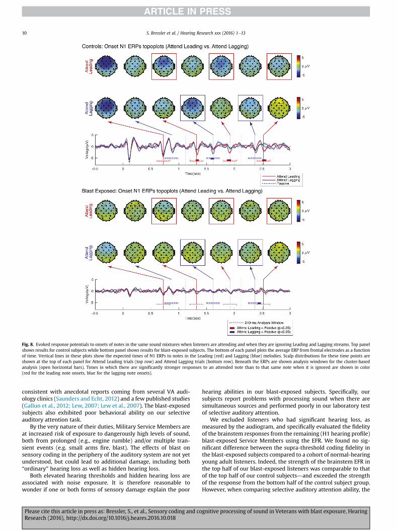
Fig. 8. Evoked response potentials to onsets of notes in the same sound mixtures when listeners are attending and when they are ignoring Leading and Lagging streams. Top panelshows results for control subjects while bottom panel shows results for blast-exposed subjects. The bottom of each panel plots the average ERP from frontal electrodes as a functionof time. Vertical lines in these plots show the expected times of N1 ERPs to notes in the Leading (red) and Lagging (blue) melodies. Scalp distributions for these time points areshown at the top of each panel for Attend Leading trials (top row) and Attend Lagging trials (bottom row). Beneath the ERPs are shown analysis windows for the cluster-basedanalysis (open horizontal bars). Times in which there are significantly stronger responses to an attended note than to that same note when it is ignored are shown in color(red for the leading note onsets, blue for the lagging note onsets).
S. Bressler et al. / Hearing Research xxx (2016) 1e1310
consistent with anecdotal reports coming from several VA audi-ology clinics (Saunders and Echt, 2012) and a few published studies(Gallun et al., 2012; Lew, 2007; Lew et al., 2007). The blast-exposedsubjects also exhibited poor behavioral ability on our selectiveauditory attention task.
By the very nature of their duties, Military Service Members areat increased risk of exposure to dangerously high levels of sound,both from prolonged (e.g., engine rumble) and/or multiple tran-sient events (e.g. small arms fire, blast). The effects of blast onsensory coding in the periphery of the auditory system are not yetunderstood, but could lead to additional damage, including both“ordinary” hearing loss as well as hidden hearing loss.
Both elevated hearing thresholds and hidden hearing loss areassociated with noise exposure. It is therefore reasonable towonder if one or both forms of sensory damage explain the poor
Please cite this article in press as: Bressler, S., et al., Sensory coding and coResearch (2016), http://dx.doi.org/10.1016/j.heares.2016.10.018
hearing abilities in our blast-exposed subjects. Specifically, oursubjects report problems with processing sound when there aresimultaneous sources and performed poorly in our laboratory testof selective auditory attention.
We excluded listeners who had significant hearing loss, asmeasured by the audiogram, and specifically evaluated the fidelityof the brainstem responses from the remaining (H1 hearing profile)blast-exposed Service Members using the EFR. We found no sig-nificant difference between the supra-threshold coding fidelity inthe blast-exposed subjects compared to a cohort of normal-hearingyoung adult listeners. Indeed, the strength of the brainstem EFR inthe top half of our blast-exposed listeners was comparable to thatof the top half of our control subjectsdand exceeded the strengthof the response from the bottom half of the control subject group.However, when comparing selective auditory attention ability, the
gnitive processing of sound in Veterans with blast exposure, Hearing
S. Bressler et al. / Hearing Research xxx (2016) 1e13 11
best of our blast-exposed listeners barely reached the performancelevels of the worst of our control subjects.
In other words, there are significant differences in supra-threshold hearing fidelity amongst the blast-exposed subjects wetested; however, the best of our blast-exposed listeners appear tohave better supra-threshold hearing than the worst of our controllisteners. Despite this, our best blast-exposed listeners performequal to or worse than control listeners on our selective auditoryattention task. Given this, we do not believe that differences insensory coding can explain the poor hearing ability of our blast-exposed listeners.
4.2. Damage to cortical networks may explain poor selectiveauditory attention performance
Problems controlling selective attention are a common symptomassociated with mild traumatic brain injury (Nuwer et al., 2005).Given the importance of communication within diverse brain re-gions thatmake up the network responsible for attentional control, itis possible that the difficulties our blast-exposed subjects experiencein multi-source settings arises due to damage to these cortical net-works, either from focal damage to computational areas importantfor attention, or from damage to white-matter tracks critical forconveying information from one region to another. Fronto-parietalregions are particularly vulnerable to subdural hemorrhage due toblast (Taber et al., 2006), and are also critical for executive control ofattention (Corbetta and Shulman, 2002; Michalka et al., 2015).Another study looking at the effects of blast exposure in subjectswith and without a diagnosis of TBI documented significantly lowerfractional anisotrophy scores in the inferior fronto-occipital fascic-ulus, a fiber tract bundle connecting the ventromedial occipital lobeand the orbitofrontal cortex (Martino et al., 2010). This result sug-gests that blast disrupts long-range connections from fronto-occipital areas involved in attentional processing to sensory andparietal regions that help make up the spatial-attention network(Corbetta and Shulman, 2002; Michalka et al., 2015).
The auditory selective attention task in this study utilizedstimuli that contained salient pitch differences as well as modestspatial differences that allowed normal-hearing control subjects toeasily segregate, select, and analyze whatever stream was to beattended in the mixture of sounds. Normal-hearing non-blastcontrol subjects performed at or near ceiling and exhibitedenhanced neural representations of the onsets to the individualnotes of the attended melody (see Fig. 8, top). In the original study,normal-hearing controls also performed reasonably well when thepitch cue was removed (same pitch condition), which made itharder to focus attention on the correct melody (Choi et al., 2014).In this harder version of the task, performance for the controlsubjects varied from perfect down to chancedcomparable to howour blast-exposed subjects performed when the “redundant” pitchcue was available (and when control subjects performed at orbetter than 90% correct). Importantly, in the original study, theamount of attentional amplification of neural ERPs that an indi-vidual control subject exhibited in the easy, different-pitch condi-tion correlatedwith performance in the same-pitch task. This resultsuggests that the efficacy of attentional control (as measured by themodulation of ERPs based on attentional focus) varies significantlyacross control listeners; when a task is sufficiently easy, all listenersmay do well, regardless of how well they can control attention, butwhen a task is hard, these individual differences in attentionalcontrol determine performance.
Here, blast-exposed subjects performed substantially worsethan normal hearing controls, as if they are particularly bad atcontrolling selective auditory attention. This poor performancewasalso reflected in weaker ERPs; moreover, there was no evidence of
Please cite this article in press as: Bressler, S., et al., Sensory coding and coResearch (2016), http://dx.doi.org/10.1016/j.heares.2016.10.018
neural modulation of ERPs due to attentional focus in the blast-exposed listeners. This result is consistent with the idea thatblast-exposure damages control networks that are critical in se-lective auditory attention tasks. Previous electrophysiological evi-dence suggests that TBI patients are impaired in their ability tofilter out unwanted or irrelevant sensory information (Arciniegaset al., 2014), supporting this kind of explanation. Such impair-ments of executive function certainly could explain why more andmore normal-hearing blast-exposed Service Members are seekingaid in VA-affiliated audiology clinics across the country.
4.3. Caveats
Problems with memory are also associated with TBI; thus, wecannot rule out the possibility that on some trials subjects eitherforgot the cue that described what stream to attend or whether towithhold a response. Similarly, even though the memory load onour task was low, it is possible that memory impairments pre-vented the blast-exposed listeners to hold the note-by-notesequence in memory over the course of the 3-s stimulus, andretain the representation long enough to determine how to answerin the response interval. These types of cognitive failures couldexplain the poor performance and weak neural responses in ourblast-exposed listeners rather than damage to specific attentionalnetworks. Regardless, such deficits are cognitive, rather than sen-sory, in nature.
Indeed, the behavioral deficits of our blast-exposed subjects areunlikely to be associated exclusively with damage to attentionalnetworks. The blast-exposed group was more likely to fail torespond on Attend trials than were the controls. The blast-exposedgroup was also less likely to withhold a response on Passive trialsthan were the controls. These deficits, combined with the lowpercentage of correct responses, suggest that the blast-exposedlisteners had general cognitive deficits that go beyond damagethat is specific to control of selective attention. Cognitive function,in general, depends on communication between pre-frontal exec-utive control regions with other brain structures. It is likely the casethat cortical damage is present in a range of tasks, not just on se-lective auditory attention tasks.
It is well established that post-traumatic stress disorder (PTSD)is comorbid with traumatic brain injury (Hoge et al., 2008). All ofthe blast-exposed subjects recruited for this study were referred toBoston University through the VA Boston Healthcare System as partof an interventional study examining the efficacy of cognitivetherapy on PTSD outcomes. Every study participant in the blastgroup had a PTSD diagnosis; however, not every participant had aconfirmed mTBI diagnosis. Because TBI and PTSD have overlappingsymptomology, it is possible that additional PTSD-specific symp-toms contributed to the behavioral and electrophysiological out-comes of this study. This is a commonly encountered problemwithstudies involving blast injury in military populations. We arecurrently gathering data with active duty Service Members bothwith and without a PTSD diagnosis to tease apart how PTSD andblast may contribute to the deficits we observe here.
While it is possible that damage to cortical grey matter and/orwhite matter connections in attentional control networks explainthe deficits exhibited by our blast-exposed subjects, more work isneeded to rule other reasons for problems with cortical control. Forinstance, PTSD often leads to sleep disorders, drug and alcoholabuse, and other behaviors, which are known to impair cognitiveabilities. Rather than physical damage to brain structures, the dif-ficulties that the blast-exposed subjects have may be caused byshort-term impairments that can be treated through effectivebehavioral modification. This is a possibility that needs furtherinvestigation.
gnitive processing of sound in Veterans with blast exposure, Hearing

S. Bressler et al. / Hearing Research xxx (2016) 1e1312
Finally, it is worth reiterating that results from the normal-hearing controls were from historical data from two previouslypublished studies from our group. Because subjects from these twostudies were recruited through advertisements posted on theBoston University campus, we assumed control participants had noprevious exposure to blast. Additionally, we cannot rule outpossible effects due to differences in education level achieved or tomusical experience, as we did not collect this information in ourinitial surveys of the blast-exposed Service Members and did nothave such information about our control subjects. Our access toblast-exposed Military Service Members was only made possiblethrough generous participant referrals by the NeurorehabilitationLab at VA Boston Healthcare under the direction of Dr. YelenaBogdanova. At the time this study was conducted, this arrangementdid not permit us to directly recruit military personnel from the VABoston Healthcare campus. Ideally, our control group would havebeen a better demographic match to the blast-exposed partici-pants; however, we did not have access to such a pool of partici-pants at that time. The above-mentioned study now underway, aswell as other future studies, need to directly address this issue.
5. Conclusions
Despite the prevalence of noise exposure in the Veteran popu-lation, sensory damage alone cannot account for the behavioral andelectrophysiological deficits we found. Blast-exposed subjects hadnear normal hearing thresholds; they also demonstrated normalsupra-threshold sound coding fidelity. Despite this, their self-reports indicate great difficulties understanding speech in noiseand segregating sounds appearing in a mixture of competingsounds. The blast-exposed Veterans also fail when asked toperform a low-memory load task that requires them to focus se-lective auditory attentiondas well as on other cognitivelydemanding aspects of our experiment, such as withholding re-sponses on certain trials, or making responses within a limited timeperiod. While it is beyond the scope of this study to determine thecause of these deficits, we conclude that cognitive, rather thansensory, factors are likely to blame.
Most importantly, this work demonstrates that blast-exposedVeterans have difficulty understanding sound when there arecompeting, distracting sounds. Given this, blast-exposed militarypersonnel are likely to have difficulty communicating in everydaysocial settings, which can lead to social isolation and depression.Further work to understand the root causes of these cognitivedeficits is critical in order to determine how to treat such problems.
Acknowledgments
The authors thank Dr. Yelena Bogdanova, Sarah Kark, and VivianHo at VA Boston Healthcare System, Jamaica Plain Campus for theirassistancewith subject referrals. This project was supported by NIHRO1 DC009477, and by a National Security Science and EngineeringFellowship to BGSC. The authors declare no competing financialinterests.
References
Almeida-Suhett, C.P., Prager, E.M., Pidoplichko, V., Figueiredo, T.H., Marini, A.M.,Li, Z., Eiden, L.E., Braga, M.F.M., 2015. GABAergic interneuronal loss and reducedinhibitory synaptic transmission in the hippocampal CA1 region after mildtraumatic brain injury. Exp. Neurol. 273, 11e23. http://dx.doi.org/10.1016/j.expneurol.2015.07.028.
Arciniegas, D., Olincy, A., Topkoff, J., McRae, K., Cawthra, E., al, E., 2014. Impairedauditory gating and P50 nonsuppression following traumatic brain injury.J. Neuropsychiatry Clin. Neurosci. 12, 77e85.
Baugh, C.M., Stamm, J.M., Riley, D.O., Gavett, B.E., Shenton, M.E., Lin, A.,Nowinski, C.J., Cantu, R.C., McKee, A.C., Stern, R.A., 2012. Chronic traumatic
Please cite this article in press as: Bressler, S., et al., Sensory coding and coResearch (2016), http://dx.doi.org/10.1016/j.heares.2016.10.018
encephalopathy: neurodegeneration following repetitive concussive and sub-concussive brain trauma. Brain Imaging Behav. 6, 244e254. http://dx.doi.org/10.1007/s11682-012-9164-5.
Bauman, R.A., Ling, G., Tong, L., Januszkiewicz, A., Agoston, D., Delanerolle, N.,Kim, Y., Ritzel, D., Bell, R., Ecklund, J., Armonda, R., Bandak, F., Parks, S., 2009. AnIntroductory Characterization of a Combat-casualty-care Relevant Swine Modelof Closed Head Injury Resulting from Exposure to Explosive Blast. http://dx.doi.org.ezproxy.bu.edu/10.1089/neu.2008.0898.
Bergemalm, P.-O., Lyxell, B., 2009. Appearances are deceptive? Long-term cognitiveand central auditory sequelae from closed head injury. Int. J. Audiol. 44, 39e49.http://dx.doi.org/10.1080/14992020400022546.
Bernstein, L.R., Trahiotis, C., 2002. Enhancing sensitivity to interaural delays at highfrequencies by using “transposed stimuli.” J. Acoust. Soc. Am. 112, 1026e1036.http://dx.doi.org/10.1121/1.1497620.
Bharadwaj, H.M., Bharadwaj, H., Shinn-Cunningham, B.G., Shinn-Cunningham, B.,2014. Rapid acquisition of auditory subcortical steady state responses usingmultichannel recordings. Clin. Neurophysiol. 125, 1878e1888. http://dx.doi.org/10.1016/j.clinph.2014.01.011.
Bharadwaj, H.M., Masud, S., Mehraei, G., Verhulst, S., Shinn-Cunningham, B.G., 2015.Individual differences reveal correlates of hidden hearing deficits. J. Neurosci.35, 2161e2172. http://dx.doi.org/10.1523/JNEUROSCI.3915-14.2015.
Brainard, D.H., 1997. The psychophysics toolbox. Spat. Vis. 10 (4), 433e436.Chapman, J.C., Diaz-Arrastia, R., 2014. Military traumatic brain injury: a review.
Alzheimer's Dementia 10, S97eS104. http://dx.doi.org/10.1016/j.jalz.2014.04.012.
Cherry, E.C., 1953. Some experiments on the recognition of speech, with one andwith two ears. J. Acoust. Soc. Am. 25, 975e979. http://dx.doi.org/10.1121/1.1907229.
Choi, I., Wang, Le, Bharadwaj, H., Shinn-Cunningham, B., 2014. Hearing research.Hear. Res. 314, 10e19. http://dx.doi.org/10.1016/j.heares.2014.04.008.
Corbetta, M., Shulman, G.L., 2002. Control of goal-directed and stimulus-drivenattention in the brain. Nat. Rev. Neurosci. 3, 201e215. http://dx.doi.org/10.1038/nrn755.
Defense and Veterans Brain Injury Center DVBIC, 2016. DoD TBI WorldwideNumbers since 2000.
Delorme, A., Makeig, S., 2004. EEGLAB: an open source toolbox for analysis ofsingle-trial EEG dynamics including independent component analysis.J. Neurosci. Methods 9e21.
Demeester, K., Topsakal, V., Hendrickx, J.-J., Fransen, E., van Laer, L., Van Camp, G.,Van de Heyning, P., Van Wieringen, A., 2012. Hearing disability measured by theSpeech, Spatial, and Qualities of Hearing Scale in clinically normal-hearing andhearing-impaired middle-aged persons, and disability screening by means of areduced SSQ (the SSQ5). Ear Hear. 33, 615e616.
Engel, A., Fries, P., Singer, W., 2001. Dynamic predictions: oscillations and synchronyin topedown processing. Nat. Rev. Neurosci. 2 (10), 704e716. http://dx.doi.org/10.1038/35094565.
Farmer, C.M., Krull, H., Concannon, T.W., Simmons, M., 2016. Characteristics andTreatment Patterns of Service Members with Mild Traumatic Brain Injury.RAND Corporation.
Fausti, S.A., Wilmington, D.J., Gallun, F.J., Myers, P.J., Henry, J.A., 2009. Auditory andvestibular dysfunction associated with blast-related traumatic brain injury.JRRD 46, 797. http://dx.doi.org/10.1682/JRRD.2008.09.0118.
Fries, P., 2009. Neuronal gamma-band synchronization as a fundamental process incortical computation. Annu. Rev. Neurosci. 32, 209e224. http://dx.doi.org/10.1146/annurev.neuro.051508.135603.
Furman, A.C., Kujawa, S.G., Liberman, M.C., 2013. Noise-induced cochlear neurop-athy is selective for fibers with low spontaneous rates. J. Neurophysiol. 110,577e586. http://dx.doi.org/10.1152/jn.00164.2013.
Gallun, F.J., Diedesch, A.C., Kubli, L.R., Walden, T.C., Folmer, R.L., Lewis, M.S.,McDermott, D.J., Fausti, S.A., Leek, M.R., 2012. Performance on tests of centralauditory processing by individuals exposed to high-intensity blasts. J. Rehabil.Res. Dev. 49, 1005e1025.
Giraud, A.-L., Poeppel, D., 2012. Cortical oscillations and speech processing:emerging computational principles and operations. Nat. Neurosci. 15, 511e517.http://dx.doi.org/10.1038/nn.3063.
Goblick Jr., T.J., Pfeiffer, R.R., 1969. Time-domain measurements of cochlear non-linearities using combination click stimuli. J. Acoust. Soc. Am. 46, 924e938.
Hasenstaub, A., Shu, Y., Haider, B., Kraushaar, U., Duque, A., McCormick, D.A., 2005.Inhibitory postsynaptic potentials carry synchronized frequency information inactive cortical networks. Neuron 47, 423e435. http://dx.doi.org/10.1016/j.neuron.2005.06.016.
Hillyard, S., 1976. Auditory evoked potentials during selective listening to dichoticspeech messages. Percept. Psychophys. 20, 236e242.
Hoge, C.W., McGurk, D., Thomas, J.L., Cox, A.L., Engel, C.C., Castro, C.A., 2008. Mildtraumatic brain injury in US soldiers returning from Iraq. N. Engl. J. Med. 358,453e463.
Hoover, E.C., Souza, P.E., Gallun, F.J., 2015. Competing views on abnormal auditoryresults after mild traumatic brain injury. Perspect. Hear Hear Dis. Res. Diagn. 19http://dx.doi.org/10.1044/hhd19.1.12, 12e10.
Krause, M.O., Kennedy, M.R.T., Nelson, P.B., 2014. Masking release, processing speedand listening effort in adults with traumatic brain injury. Brain Inj. 28,1473e1484. http://dx.doi.org/10.3109/02699052.2014.920520.
Kujawa, S.G., Liberman, M.C., 2009. Adding insult to injury: cochlear nervedegeneration after “temporary” noise-induced hearing loss. J. Neurosci. 29,14077e14085. http://dx.doi.org/10.1523/JNEUROSCI.2845-09.2009.
gnitive processing of sound in Veterans with blast exposure, Hearing
S. Bressler et al. / Hearing Research xxx (2016) 1e13 13
Kwok, F.Y., Lee, T.M.C., Leung, C.H.S., Poon, W.S., 2009. Changes of cognitive func-tioning following mild traumatic brain injury over a 3-month period. Brain Inj.22, 740e751. http://dx.doi.org/10.1080/02699050802336989.
Lachaux, J., Rodriguez, E., Martinerie, J., Varela, F., 1999. Measuring phase synchronyin brain signals. Hum. Brain Mapp. 8, 194e208.
Lagarde, E., Salmi, L.-R., Holm, L.W., Contrand, B., Masson, F., Rib�ereau-Gayon, R.,Laborey, M., Cassidy, J.D., 2014. Association of symptoms following mild trau-matic brain injury with posttraumatic stress disorder vs postconcussion syn-drome. JAMA Psychiatry. http://dx.doi.org/10.1001/jamapsychiatry.2014.666.
Levin, H.S., Mattis, S., Ruff, R.M., Eisenberg, H.M., Marshall, L.F., Tabaddor, K.,High, W.M., Frankowski, R.F., 1987. Neurobehavioral outcome following minorhead injury: a three-center study. J. Neurosurg. 66, 234e243. http://dx.doi.org/10.3171/jns.1987.66.2.0234.
Lew, H.L., 2007. Program development and defining characteristics of returningmilitary in a VA Polytrauma Network Site. JRRD 44, 1027e1034. http://dx.doi.org/10.1682/JRRD.2007.05.0073.
Lew, H.L., Jerger, J.F., Guillory, S.B., Henry, J.A., 2007. Auditory dysfunction in trau-matic brain injury. JRRD 44, 921e928. http://dx.doi.org/10.1682/JRRD.2007.09.0140.
Lobarinas, E., Salvi, R., Ding, D., 2013. Insensitivity of the audiogram to carboplatininduced inner hair cell loss in chinchillas. Hear. Res. 302, 113e120. http://dx.doi.org/10.1016/j.heares.2013.03.012.
Lowenstein, D.H., Thomas, M.J., Smith, D.H., McIntosh, T.K., 1992. Selective vulner-ability of dentate hilar neurons following traumatic brain injury: a potentialmechanistic link between head trauma and disorders of the hippocampus.J. Neurosci. 12, 4846e4853.
Mac Donald, C.L., Johnson, A.M., Cooper, D., Nelson, E.C., Werner, N.J., Shimony, J.S.,Snyder, A.Z., Raichle, M.E., Witherow, J.R., Fang, R., Flaherty, S.F., Brody, D.L., 2011.Detection of blast-related traumatic brain injury in U.S. Military personnel.N. Engl. J. Med. 364, 2091e2100. http://dx.doi.org/10.1056/nejmoa1008069.
Management of Concussion/mTBI Working Group, 2009. VA/DoD clinical practiceguideline for management of concussion/mild traumatic brain injury. J. Rehabil.Res. Dev. 46, CP1e68.
Maris, E., Oostenveld, R., 2007. Nonparametric statistical testing of EEG- and MEG-data. J. Neurosci. Methods 164, 177e190. http://dx.doi.org/10.1016/j.jneumeth.2007.03.024.
Martino, J., Brogna, C., Robles, S.G., Vergani, F., Duffau, H., 2010. Anatomic dissectionof the inferior fronto-occipital fasciculus revisited in the lights of brain stimu-lation data. Cortex 46, 691e699. http://dx.doi.org/10.1016/j.cortex.2009.07.015.
McKee, A.C., Robinson, M.E., 2014. Military-related traumatic brain injury andneurodegeneration. Alzheimer's Dementia 10, S242eS253. http://dx.doi.org/10.1016/j.jalz.2014.04.003.
Mehraei, G., Hickox, A.E., Bharadwaj, H.M., Goldberg, H., Verhulst, S., Liberman, M.C.,Shinn-Cunningham, B.G., 2016. Auditory brainstem response latency in noise asa marker of cochlear synaptopathy. J. Neurosci. 36, 3755e3764. http://dx.doi.org/10.1523/JNEUROSCI.4460-15.2016.
Mesgarani, N., Chang, E.F., 2012. Selective cortical representation of attendedspeaker in multi-talker speech perception. Nature 1e5. http://dx.doi.org/10.1038/nature11020.
Mez, J., Stern, R.A., McKee, A.C., 2013. Chronic traumatic encephalopathy: where arewe and where are we going? Curr. Neurol. Neurosci. Rep. 13, 407e412. http://dx.doi.org/10.1007/s11910-013-0407-7.
Michalka, S.W., Kong, L., Rosen, M.L., Shinn-Cunningham, B.G., Somers, D.C., 2015.Short-term memory for space and time flexibly recruit complementarysensory-biased frontal lobe attention networks. Neuron 87, 882e892. http://dx.doi.org/10.1016/j.neuron.2015.07.028.
Munjal, S.K., Panda, N.K., Pathak, A., 2010. Relationship between severity of trau-matic brain injury (TBI) and extent of auditory dysfunction. Brain Inj. 24,525e532. http://dx.doi.org/10.3109/02699050903516872.
Noble, W., Jensen, N.S., Naylor, G., Bhullar, N., 2013. A short form of the Speech,Spatial and Qualities of Hearing scale suitable for clinical use: the SSQ12. Int. J.Audiol. 409e412.
Nuwer, M.R., Hovda, D.A., Schrader, L.M., Vespa, P.M., 2005. Routine and quantita-tive EEG in mild traumatic brain injury. Clin. Neurophysiol. 116, 2001e2025.http://dx.doi.org/10.1016/j.clinph.2005.05.008.
Oleksiak, M., Smith, B.M., Andre, J.R.S., Caughlan, C.M., Steiner, M., 2012. Audio-logical issues and hearing loss among veterans with mild traumatic braininjury. JRRD 49, 995e1010. http://dx.doi.org/10.1682/JRRD.2011.01.0001.
Picton, T.W., Hillyard, S.A., 1974. Human auditory evoked potentials. II: effects ofattention. Electroencephalogr. Clin. Neurophysiol. 36, 191e200.
Rosenfeld, J.V., Ford, N.L., 2010. Bomb blast, mild traumatic brain injury and psy-chiatric morbidity: a review. Injury 41, 437e443. http://dx.doi.org/10.1016/
Please cite this article in press as: Bressler, S., et al., Sensory coding and coResearch (2016), http://dx.doi.org/10.1016/j.heares.2016.10.018
j.injury.2009.11.018.Ruggles, D., Bharadwaj, H., Shinn-Cunningham, B.G., 2012. Why middle-aged lis-
teners have trouble hearing in everyday settings. Curr. Biol. 22, 1417e1422.http://dx.doi.org/10.1016/j.cub.2012.05.025.
Ruggles, D., Bharadwaj, H., Shinn-Cunningham, B.G., 2011. Normal hearing is notenough to guarantee robust encoding of suprathreshold features important ineveryday communication. PNAS 108, 15516e15521.
Ruggles, D., Shinn-Cunningham, B., 2011. Spatial selective auditory attention in thepresence of reverberant energy: individual differences in normal-hearing lis-teners. JARO 12, 395e405. http://dx.doi.org/10.1007/s10162-010-0254-z.
Saunders, G.H., Echt, K.V., 2012. Blast exposure and dual sensory impairment: anevidence review and integrated rehabilitation approach. JRRD 49.
Saunders, G.H., Frederick, M.T., Arnold, M., Silverman, S., Chisolm, T.H., Myers, P.,2015. Auditory difficulties in blast-exposed Veterans with clinically normalhearing. JRRD 52, 343e360. http://dx.doi.org/10.1682/jrrd.2014.11.0275.
Schaette, R., McAlpine, D., 2011. Tinnitus with a normal audiogram: physiologicalevidence for hidden hearing loss and computational model. J. Neurosci. 31,13452e13457. http://dx.doi.org/10.1523/JNEUROSCI.2156-11.2011.
Schwab, K.A., Ivins, B., Cramer, G., Johnson, W., Sluss-Tiller, M., Kiley, K., Lux, W.,Warden, D., 2007. Screening for traumatic brain injury in troops returning fromdeployment in Afghanistan and Iraq: initial investigation of the usefulness of ashort screening tool for traumatic brain injury. J. Head. Trauma Rehabil. 22,377e389. http://dx.doi.org/10.1097/01.HTR.0000300233.98242.87.
Shaheen, L.A., Valero, M.D., Liberman, M.C., 2015. Towards a diagnosis of cochlearneuropathy with envelope following responses. JARO. http://dx.doi.org/10.1007/s10162-015-0539-3.
Shinn-Cunningham, B.G., 2008. Object-based auditory and visual attention. TrendsCogn. Sci. 12, 182e186. http://dx.doi.org/10.1016/j.tics.2008.02.003.
Shinn-Cunningham, B.G., Best, V., 2008. Selective attention in normal and impairedhearing. Trends Amplif. 12, 283e299. http://dx.doi.org/10.1177/1084713808325306.
Skoe, E., Kraus, N., 2010. Auditory brain stem response to complex sounds: atutorial. Ear Hear. 31, 302.
Smetana, W.R., 1999. Predicting Hearing Health Readiness as a Function of the ArmyProfile System Formula and Occupational Safety and Health AdministrationReportable Hearing Loss.
Sosa, M.A.G., De Gasperi, R., Paulino, A.J., Pricop, P.E., Shaughness, M.C., Maudlin-Jeronimo, E., Hall, A.A., Janssen, W.G.M., Yuk, F.J., Dorr, N.P., Dickstein, D.L.,McCarron, R.M., Chavko, M., Hof, P.R., Ahlers, S.T., Elder, G.A., 2013. Blast over-pressure induces shear-related injuries in the brain of rats exposed to a mildtraumatic brain injury. Acta Neuropathol. Commun. 1, 51. http://dx.doi.org/10.1186/2051-5960-1-51.
Taber, K., Hurley, R., 2007. Traumatic axonal injury: atlas of major pathways.J. Neuropsychiatry Clin. Neurosci. 19, ive104.
Taber, K., Warden, D., Hurley, R., 2006. Blast-related traumatic brain injury: what isknown? J. Neuropsychiatry Clin. Neurosci. 18, 141e145.
Taber, K.H., Hurley, R.A., Haswell, C.C., Rowland, J.A., Hurt, S.D., Lamar, C.D.,Morey, R.A., 2015. White matter compromise in veterans exposed to primaryblast forces. J. Head. Trauma Rehabil. 30, E15eE25. http://dx.doi.org/10.1097/HTR.0000000000000030.
Tanielian, T., Jaycox, L.H., 2008. Invisible Wounds of War. RAND Corporation.Terrio, H., Brenner, L.A., Ivins, B.J., Cho, J.M., Helmick, K., Schwab, K., Scally, K.,
Bretthauer, R., Warden, D., 2009. Traumatic brain injury screening: preliminaryfindings in a US Army Brigade Combat Team. J. Head. Trauma Rehabil. 24,14e23.
Uusitalo, M.A., Ilmoniemi, R.J., 1997. Signal-space projection method for separatingMEG or EEG into components. Med. Biol. Eng. Comput. 35, 135e140. http://dx.doi.org/10.1007/BF02534144.
Valero, M.D., Hancock, K.E., Liberman, M.C., 2016. The middle ear muscle reflex inthe diagnosis of cochlear neuropathy. Hear. Res. 332, 29e38. http://dx.doi.org/10.1016/j.heares.2015.11.005.
Viana, L.M., O'Malley, J.T., Burgess, B.J., Jones, D.D., Oliveira, C.A.C.P., Santos, F.,Merchant, S.N., Liberman, L.D., Liberman, M.C., 2015. Cochlear neuropathy inhuman presbycusis: confocal analysis of hidden hearing loss in post-mortemtissue. Hear. Res. 327, 78e88. http://dx.doi.org/10.1016/j.heares.2015.04.014.
Wolf, J.A., Koch, P.F., 2016. Disruption of network synchrony and cognitivedysfunction after traumatic brain injury. Front. Syst. Neurosci. 10, 1629e1714.http://dx.doi.org/10.3389/fnsys.2016.00043.
Zhu, L., Bharadwaj, H., Xia, J., Shinn-Cunningham, B., 2013. A comparison of spectralmagnitude and phase-locking value analyses of the frequency-followingresponse to complex tones. J. Acoust. Soc. Am. 134, 384. http://dx.doi.org/10.1121/1.4807498.
gnitive processing of sound in Veterans with blast exposure, Hearing