Embed Size (px)
Citation preview

CO-1
SentinelregCerebral Protection System During TAVR
February 23 2017Claret Medical IncCirculatory System Devices Panel
CO-2
Introduction
Thomas EngelsVice President of Clinical AffairsClaret Medical Inc
CO-3
Class 2 (proposed) temporary accessory device Placed prior to and removed after Transcatheter Aortic
Valve Replacement (TAVR) TAVR associated with cerebrovascular events1
Embolic Protection Devices (EPD) have been used in carotid stenting for gt15 years
No alternative option available for embolic protection in TAVR
Sentinel investigational in US Sentinel CE Marked 2013
gt3000 TAVR procedure
The Sentinel Cerebral Protection System
1 Smith E et al ldquoCerebral Microinfarcts The Invisible Lesionsrdquo Lancet Neurol 2012 11(3) 272ndash282
CO-4
The Sentinelreg Cerebral Protection System isindicated for use as a cerebral protection deviceto capture and remove embolic material whileperforming transcatheter aortic valve proceduresin order to reduce peri-procedural ischemic braininjuryThe diameters of the arteries at the site of filterplacement should be between 9 ndash 15 mm for thebrachiocephalic and 65 mm ndash 10 mm for the leftcommon carotid arteries
Proposed Sentinel System Indication
CO-5
Animation of the Sentinel System During TAVR
CO-6
Primary Safety 30-Day MACCE vs Performance Goal ndash Achieved
Primary Effectiveness ndash Median New Lesion Volume(DW-MRI) Observed treatment effect ge 30 ndash Achieved Test vs Control ndash Not achieved
Other Relevant Study Outcomes Sentinel system successfully delivered amp retrieved in
94 of patients Major Sentinel access-related complications were rare
(N=1 04) Embolic debris captured in 99 of patients
Safety and Effectiveness Outcomes
CO-7
US Medical Device Classification
Class 1Lowest Risk
eg Surgical Gauze
Class 2Medium Risk
eg BAV
Class 3Highest risk
eg TAVR
Medium risk temporary accessory device De Novo pathway required due to lack of predicate cerebral
protection device De Novo pathway riskbenefit balance on the basis of the totality of
pre-market evidence and post market measures
CO-8
Presentation AgendaBackground Device Description Trial Design Safety and Effectiveness Data
Martin B Leon MDProfessor of MedicineColumbia University Medical Center
HistopathologyRenu Virmani MDPresident CVPath Institute IncClinical Professor George Washington University
History of NeuroprotectionWilliam A Gray MDSystem Chief of the Division of Cardiovascular DiseaseLankenau Medical Center Main Line Health
ConclusionAzin Parhizgar PhDPresident and Chief Executive OfficerClaret Medical Inc
CO-9
Additional ExpertsInterventional Cardiology
Samir Kapadia MDDirector Cardiac Catheterization LaboratoryCleveland Clinic
Susheel Kodali MDDirector Structural Heart amp Valve CenterColumbia University Medical Center
Axel Linke MDCo-director Department of Internal Medicine CardiologyUniversity of Leipzig Heart Center
Roxana Mehran MDProfessor of Medicine CardiologyMount Sinai New York
Neurology and NeurosurgeryMaxim Mokin MD PhDDirector of Neuro Interventional SurgeryUniversity of South Florida Health
Jesse Weinberger MDVascular Neurology SpecialistMount Sinai Hospital
MRI NeuroimagingRobert Zivadinov MD PhDProfessor of Neurology Director Buffalo Neuroimaging Analysis Center
Michael Dwyer PhDDirector Of Technical ImagingBuffalo Neuroimaging Analysis CenterAssistant Professor of NeurologyUniversity of Buffalo
NeurocognitionRonald Lazar PhDProfessor of NeuropsychologyColumbia University Medical Center
StatisticsRoseann White MADirector Pragmatic Clinical Trial StatisticsDuke Clinical Research Institute
CO-10
Background
Martin B Leon MDProfessor of MedicineColumbia University Medical Center
CO-11
Strokes are Considered a Major Complication after TAVR
PARTNER 1A RCT (SAPIEN TAVR vs Surgery) 699 high-risk patients with severe AS N Engl J Med 20113642191-2202
CO-12
Typical Examples of Heavily Calcified Aortic Valves
Radiograph of surgical specimen Autopsy specimen
CO-13
N Engl J Med 20113642191-2202
Technological refinement of transcathetervalves and adjunctive procedures such as the use of embolic protection devices13
will facilitate transcatheter replacement and may improve outcomes but these new devices should be evaluated in controlled trials with randomization against current standard techniques
Strokes are Considered a Major Complication after TAVR
CO-14
In 2015 TAVR accounted for 32 of all Medicare AV replacements in the US
Globally TAVR is expected to grow approximately 4-fold in the next 10 years
TAVR is Projected to Grow in the Next Decade
Courtesy of Dr M Leon TVT 2016 Adapted from Credit Suisse TAVI Comment ndash January 2015
3200071000
289000
2012 2013 2014 2015 2016 2017 2018 2019 2020 2021 2022 2023 2024 2025
CO-15
Strokes After TAVR
Approximately 3 to 7 at 30 days in high surgical risk patients (CEC adjudicated FDA studies)
Up to 85 of strokes occur within 1 week of TAVR Associated with increased 1-year mortality and
reduced quality-of-life Frequency is highly dependent on stroke
definitions (eg VARC-2) and ascertainment methods (eg wwo neurology assessments)
VARC-2 = valve academic research consortium standard definitions (JACC 2012)
CO-16
2621 patients from PARTNER (high and extreme risk) CEC adjudication
Acute-phase (peri-procedural) stroke risk peaked at 2 days with a low constant risk of 08 per year
Strokes After TAVR
Kapadia S et al Circ Cardiovasc Interv 20169e002981
CO-17
Strokes After TAVR (Acute Phase)
Kapadia S et al Circ Cardiovasc Interv 20169e002981
Weeks After TAVR
Neurological Events
(100 patient months)
TF TAVR plusmn 1 Standard ErrorTA TAVR plusmn 1 Standard Error
10 2 3 4
CO-18
Clinical neurologic events Strokes (disabling and non-disabling) Transient ischemic attacks (TIA)
Brain injury on neuro-imaging studies detected by DW-MRI
Neuronal injury without overt symptoms1 which may result in acute or chronic changes in neurocognitive function
Spectrum of Brain Injury Caused by Embolic Material
1 Lansky AJ et al JACC Vol 69 No6 2017
CO-19
Frequent early DW-MRI abnormalities(68-100 of patients) after TAVR from 9 studies
Most patients have multiple infarcts which represent permanent ischemic brain damage
SENTINEL trial based on results from predicate trial (CLEAN-TAVI) Randomized controlled study in 100 patients Single TAVR system Exact MRI methodology was used by the same
core laboratory as is used in the current study
Brain Injury on Neuro-imaging (DW-MRI) after TAVR
CO-20
Sentinel Cerebral Protection System Device Description and Case
CO-21
Protected vs All Territories Intra-cerebral Vasculature
Zhao M et al Regional Cerebral Blood Flow Using Quantitative MR Angiography AJNR 2007281470-1473
Sentinel Placement
RVA~10
RCCA~40 LCCA
~40
Protected blood flow to the brain
LVA~10
Unprotected blood flow to the brain
CO-22
Protected 74 brain volume
Partially Protected 24 brain volume
Unprotected 2 brain volume
Protected and Unprotected Cerebral Vascular Territories
CO-23
Two independent filters capture amp remove embolic material
Polyurethane filter pore size = 140 microm Standard R trans-radial sheath access (6F) One size accommodates most vessel sizes
(brachiocephalic 9-15 mm and left common carotid [LCC] 65-10 mm)
Deflectable compound-curve catheter facilitates cannulation of LCC
Minimal profile in aortic arch (little interaction with other devices)
Sentinel Cerebral Protection System During TAVR
CO-24
Sentinel Cerebral Protection System During TAVR ndash Case
CO-25
SENTINEL Trial Overview
CO-26
SENTINEL Trial Design Overview
SAFETY ARMTAVR with Sentinel
(N=123)
TEST ARMTAVR with Sentinel
(N=121)
CONTROL ARMTAVR Only
(N=119)
Serial MRIs (Baseline Day 2-7 amp Day 30)
Serial Neurocognitive Assessment (Baseline Day 30 amp Day 90)
Histopathology amp Morphometry
Clinical Follow-Up (Neurology Assessments in all patients)
Patients with Severe Symptomatic Aortic Stenosis undergoing TAVR
Patients Randomized (111)(N=363)
CO-27
Patients with symptomatic severe aortic stenosis eligible for treatment with a US commercially approved TAVR system 4 different TAVR systems used (not stratified
during randomization) Acceptable aortic arch anatomy and vessel
diameters without significant stenosis Brachiocephalic diameter 9 -15 mm Left common carotid diameter 65 -10 mm
Key Inclusion Criteria
CO-28
Anatomic Right extremity vasculature not suitable Brachiocephalic left carotid or aortic arch not suitable
Clinical CVA or TIA within 6 months Neurological disease with persistent deficits Carotid disease requiring treatment within 6 weeks Contraindications to MRI Renal insufficiency (CR gt30 mgdL or GFR lt30 ccmin) Severe LV dysfunction (EF lt20) Balloon valvuloplasty (BAV) within 30 days
Key Exclusion Criteria
CO-29
Multicenter Trial 363 Patients at 19 Sites
U Penn(N=23)
Leipzig Heart
Center(N=66)
Morton Plant Hospital(N=8)
University of Washington
(N=15)
Barnes-Jewish
Hospital(N=12)
University of Virginia
(N=5)
Cedars-Sinai Medical Center
(N=73)
University of Texas
Houston(N=6)
Cleveland Clinic(N=38)
Emory UniversityHospital(N=12)
Mass GeneralColumbia (N=57)Weill Cornell (N=2)Mt Sinai (N=10)
AKSt Georg
(N=8)
Sentara NorfolkGeneral (N=1)
St Thomas Hospital
(N=2)
Henry FordHealth System
(N=6)
Washington Hospital Center (N=19)
St LukesHospital
(N=0)
5 Highest Enrolling Sites
CO-30
Study AdministrationCo-Principal Investigators Susheel Kodali MDColumbia University Medical Center
Samir R Kapadia MD Cleveland Clinic
Axel Linke MDCo-director Department of Internal MedicineCardiologyUniversity of Leipzig Heart Center
Clinical Steering Committee ChairmanMartin B Leon MDColumbia University Medical Center
Study Medical MonitorRoxana Mehran MDMount Sinai School of Medicine
Clinical Events CommitteeCardiovascular Research FoundationChair Ozgen Dogan MDNeurologists Jesse Weinberger MDJoshua Willey MD
Data Safety Monitoring BoardCardiovascular Research FoundationChair Blase A Carabello MD
Histopathology Morphometry Core LaboratoryCV Path InstituteChair Renu Virmani MD
MRI Core LaboratoryBuffalo Neuroimaging Analysis Center University of BuffaloChair Robert Zivadinov MD PhD
Neurocognitive Core LaboratoryTananbaum Stroke Center Neurological InstituteColumbia UniversityChair Ronald M Lazar PhD
Sentinel CT Planning CenterCedars-Sinai Medical CenterChair Hasan Jilaihawi MD
Statistical AnalysisDuke Clinical Research Institute Project Director Roseann White MANorth American Science Associates Inc (NAMSA)
CO-31
8
18
29
51 34
9
12
13
25
5360
46
82
0
20
40
60
80
Q42014
Q12015
Q22015
Q32015
Q42015
Q12016
Sapien XT CoreValve Evolut R Sapien 3(N=188)
Valve Type Distribution Over Time
of Valves
(N=93)(N=14)(N=64)
CO-32
Distribution of Valve Types Across Study Arms
293 240 235
187174 169
472554 529
0
20
40
60
80
100
Safety Arm(N=123)
Test Arm(N=121)
Control Arm(N=119)
Sapien 3
Sapien XT
Evolut R
Core Valve33 25 59
No Significant Differences in Valve-type Distribution (p = 071)
CO-33
SENTINEL TrialSafety and Performance
CO-34
SENTINEL Safety PopulationsPatients with Severe Symptomatic Aortic Stenosis Undergoing TAVR
Patients Randomized (111)(N=363)
(N=117) (N=117)
1 No TAVR1 LTFU2 Withdrawal
2 No TAVR2 LTFU 2 Withdrawal
Analyzed ITT
1 No TAVR1 LTFU 6 Withdrawal
(N=111)
(N=123) (N=121)Randomized
Safety Arm Test Arm
(N=119)
TAVR Only
Control Arm
7 No Sentinel2 No Sentinel
(N=115) (N=110)As-Treated
Safety Cohort
CO-35
Non hierarchical MACCE at 30 days All-cause mortality All strokes Acute kidney injury (Stage 3) within 72 hours
Historical MACCE performance goal Weighted average of all FDA pivotal TAVR trials
approved at time of SENTINEL trial initiation = 133 Upper-bound of one-sided 95 CI for MACCE derived
from Safety Cohort (Safety Arm + Test Arm subjects) must be lt183 (133 + 5 non-inferiority margin)
Device cohort (Safety + Test arm) also compared to concurrent randomized Control arm
Primary Safety Endpoint
CO-36
Patient DemographicsSentinel
Control Arm (N=119)
Safety Arm(N=123)
Imaging Arm(N=121)
Age (mean yrs) 82 82 83Female () 55 52 49STS PROM Score (mean ) 62 64 75Previous stroke () 8 4 5Previous TIA () 8 7 7Diabetes () 27 41 38ho atrial fibrillation () 30 35 30Heavily calcified aorta () 3 2 3ho CAD () 54 50 56ho PVD () 16 14 15NYHA IIIIV () 83 85 82Valve area (cm2) 07 plusmn 018 07 plusmn 017 07 plusmn 020Mean aortic valve gradient (mmHg) 42 plusmn 15 44 plusmn 15 41 plusmn 14
CO-37
Sentinel Access and Device Success
Sentinel(Safety + Test)
Sentinel AccessRadial 944Brachial 56
Device SuccessBoth Filters Deployed 944ge One Filter Deployed 996
Reasons for No Sentinel (N=13 56)No TAVR 3Inadequate vascular access 6Late screen failure 3Test patient treated as Control (protocol deviation) 1
Acute delivery and retrieval success Deployment and retrieval of the proximal and distal filters in accessible anatomies (not excessively tortuous or calcified)
CO-38
TAVR Procedural Factors inSENTINEL Study
1 Time elapsed between first arterial access and removal of the last guide from the arterial access sheath2 Time elapsed use of fluoroscopy during TAVR Procedure
Sentinel(Safety + Test) Control P-value
TAVR Procedure Time(Mean Minutes1) 87 74 0013
TAVR Fluoroscopy Time(Mean Minutes2) 19 17 0073
CO-39
74 73 76
0
5
10
15
20
Randomized(N=244)
Analyzed ITT(N=234)
As Treated(N=225)
Primary Safety Endpoint(30-Day MACCE)
183Performance Goal (Including Non-Inferiority Margin)
(p lt 0001)
N=18 N=17 N=17
Error bars represent upper bound of the one-sided 95 Upper CIImputation method based on the logistic regression method Factors used in imputation algorithm age sex BMI history of diabetes history of atrial fibrillation previous stroke with permanent deficit and geography
(p lt 0001) (p lt 0001)
of Patients with an Event
CO-40
74 73 76
0
5
10
15
20
Randomized(N=244)
Analyzed ITT(N=234)
As Treated(N=225)
Safety Endpoint Evaluation(Without Non-Inferiority Margin)
of Patients with an Event
(p = 00025) (p = 00026) (p = 00048)
133Calculated MACCE Rate
Error bars represent upper bound of the one-sided 95 Upper CIImputation method based on the logistic regression method Factors used in imputation algorithm age sex BMI history of diabetes history of atrial fibrillation previous stroke with permanent deficit and geography
N=18 N=17 N=17
CO-41
30-Day MACCE Sentinel vs Concurrent Control (Analyzed ITT)
73
99
0
5
10
15
20
Sentinel (Safety + Test)(N=234)
Control(N=111)
N=17 N=11
of Patients with an Event
Error bars represent upper bound of the one-sided 95 Upper CI
CO-42
30-Day Clinical Safety Results (Analyzed ITT)
Sentinel (Safety + Test)
(N=234)Control(N=111)
P-valueN N Any MACCEdagger patients 17 73 11 99 040Events
Death (all-cause) 3 13 2 18 065Stroke 13 56 10 91 025
Disabling 2 09 1 09 100Non-disabling 11 48 9 82 022
AKI (Stage 3) 1 04 0 0 100TIA 1 04 0 0 100
Sentinel-related complications1 1 04 NA NA NA
1Late brachial artery pseudo-aneurysm treated with thrombin injectiondaggerMACCE defined as Death (any cause) Stroke (any) Acute Kidney Injury (Stage 3)Note MACCE events adjudicated by independent Clinical Events Committee who were blinded to treatment arm
CO-43
Stroke Diagnosis le72 hours(Analyzed ITT)
1304
13
30
45
09
27
82
0
2
4
6
8
10
Day 1 Day 2 Day 3 Total
Sentinel Control
Fisher Exact Test
Days to Stroke
of Patients
p=005263 Reduction
CO-44
Primary Safety Endpoint achieved 30-day Sentinel MACCE vs Performance Goal
(p lt 0001) 30-Day MACCE
Sentinel 73 vs Control 99 30-Day stroke rate
Sentinel 56 vs Control 91 Peri-procedural stroke rate (le72 hours)
Sentinel 30 vs Control 82 One (04) Sentinel-related access site complication
Safety Summary
CO-45
Histopathology
Renu Virmani MDPresident CVPath Institute IncClinical ProfessorGeorge Washington University
CO-46
105 patients with 210 evaluable filters Filters processed and embedded in paraffin and
sectioned Slides classified by thrombus and tissue type
Thrombus (acute and chronic) Valve tissue Calcium nodules Arterial wall (intima or media including necrotic core) Myocardium Foreign material
Histopathologic Analysis of Filters Proximal and Distal
CO-47
Type of Tissue Identified
Organizing
Acute + organizing thrombus Arterial wall + thrombus Valve tissue
Calcium nodules Foreign material + thrombus Myocardium + thrombusMovat pentachrome Movat pentachrome
HampE HampE HampE
HampE
CO-48
Automated analysis for particle size (HALO software)
Five largest tissue samples measured manually in largest and smallest dimensions
Morphology of tissue characterized
Type of Morphometric Analysis Performed
CO-49
SENTINEL HistopathologyTotal Embolic Material by Type
99 98 94
50 50
35
157
1
ANY AcuteThrombusamp TissueForeignMaterial
ArterialWall
ValveTissue
Calcifi-cation
ForeignMaterial
Myo-cardium
OrganizingThrombus
AcuteThrombus
Alone
Patients with Captured Debris ()
Tissue Type
CO-50
Morphometric AnalysisEmbolic Material by Particle Size
14
55
91
99
0 20 40 60 80 100
1
gt=150 um
gt= 500 um
gt= 1000 um
gt=2000 um
Percent of Patients with at Least One Particle of Given Size
ge015 mm
ge05 mm
ge1 mm
ge2 mm
Automated measurement
CO-51
Patient Quartile AnalysisAverage Number of Particles ge05 mm
1 in 4 Patients had 25 Particles ge05 mm in Size
0937
89
251
0
5
10
15
20
25
30
Q1 Q2 Q3 Q4
Average of
ParticlesCapturedge05 mm
PatientQuartiles
Automated measurement
CO-52
Morphometric Analysis Embolic Material by Valve Type ge 05 and ge 1 Millimeter
Manual measurement
76
72
83
100
0 25 50 75 100
CoreValve(N=3)
Evolut R(N=24)
SAPIEN XT(N=20)
SAPIEN 3(N=58)
of Patients with a Particle ge 05 millimeter
of Patients with a Particle ge 1 millimeter
15
34
58
33
0 25 50 75 100
CO-53
Filtered through 40-micron mesh Processed embedded in paraffin Sectioned at 4-6 microns Sections are stained total of 5 sections per filter Assessed by light microscopy
Process MethodologySentinel filters
(with collected debris)CVPath filter
(40 micron pore size)
CO-54
Arterial Wall amp Valve Tissue
Valve tissue
Valve tissueArterial wall
Arterial wallDistal Filter Proximal Filter
CO-55
Calcium Nodules
Distal Filter Proximal Filter
CO-56
Myocardium
Distal Filter Proximal Filter
CO-57
Foreign Material
Distal Filter Proximal Filter
CO-58
Largest Piece ndash Valve and Arterial Wall (54 mm)
Distal Filter
CO-59
TAVR devices are larger stiffer than Sentinel TAVR device features such as exposed metal
frames or flared tubes or tips are prone to interacting with vessel wall
Sentinel vs TAVR CatheterProfile Comparison
Profile in arch
16-20 F
6 F Sentinel
CO-60
Debris From TAVR
TAVR traverses Iliac artery Abdominal aorta Thoracic aorta Aortic arch Ascending aorta
Roberts WC et al Am J Cardiol 2013111(3)448-52
Aortic arch
Thoracic and abdominal aortawith iliac bifurcation
CO-61
Tissue or foreign material combined with acute thrombus was found in 98
Debris captured from all valve types Acute thrombus alone observed in only 1 of
patients Valve tissue and calcium nodules captured in 50
of patients Foreign material captured in 35 of patients 1 in 4 Patients had 25 Particles ge05 mm in size
Histopathology Summary
CO-62
SENTINEL TrialEffectiveness
Martin B Leon MDProfessor of MedicineColumbia University Medical Center
CO-63
Serial 3T scan acquisition at baseline 2-7 days and 30 days on the same scanner
All sites imaging core lab certified according to MRI technologist manual and approved by MRI physicist
Sequences acquired Diffusion weighted (acute changes) T2FLAIR (chronic changes) B0 Field Map High-resolution 3D T1-weighted anatomical image
Scans transferred queried accepted in real time
MRI Methodology and Acquisition Protocol
CO-64
FLAIR ndash attenuated inversion recovery
MRI Analysis of New DWI Lesion Volume and Number
Blinded core lab analysis of all scans
Serial co-registration and subtraction
Artifactdistortion correction
Per-lesion quantification and longitudinal tracking
Baseline DWI 2-7 days DWI Subtraction DWI
343mm3527mm3
4087mm3
Baseline FLAIR 1 Baseline FLAIR 2 Baseline FLAIR 3
DWI ndash diffusion weighted image
CO-65
Patients with Severe Symptomatic Aortic Stenosis undergoing TAVR
Patients Randomized (111)(N=363)
(N=121)
Test Arm
(N=119)
Imaging Cohort
Control Arm
SENTINEL Imaging Study
Randomized
(N=91) (N=98)
Paired Serial DW and FLAIR MRIs(Baseline 2-7days)
11 scan not done10 pacemaker placed
6 Sentinel did not enter vasc1 Sentinel removed prior to TAVR
1 no TAVR1 withdrawal
9 scan not done8 pacemaker placed
2 scan rejected1 no TAVR
1 died
Analyzed ITT
CO-66
Primary Effectiveness Endpoint Median total new lesion volume in protected
territories at Day 2-7 based on DW-MRI Study Success Criterion - Reduction in Median
Total New Lesion Volume (Test vs Control) in protected territories Criterion 1 statistical superiority Criterion 2 observed treatment effect ge30
Primary Effectiveness Endpoint and Success Criteria
CO-67
1091
1740
0
50
100
150
200
Test(N=121)
Control(N=119)
Primary Effectiveness EndpointNew Lesion Volume in Protected Territories
dagger Wilcoxon Test
MedianNew
Lesion Volume
in ProtectedTerritories
(mm3)
Randomizedp = 024dagger
37 Reduction
Imputation method based on the predictive mean matching method Factors used in imputation algorithm based on blinded aggregate data 850 Hounsfield Unitcalcification score BMI Valve type Procedural stroke Prepost dilatation Mean aortic valve gradient
[37423] [34483][37380] [40469]
Analyzed ITTp = 033dagger
42 Reduction
1028
1780
0
50
100
150
200
Test(N=91)
Control(N=98)
IQR
CO-68
Median New Lesion Volume by Territory (Analyzed ITT)
Territory Median New Lesion Volume mm3 [IQR]
P-valuedaggerTest Control
Protected 1028[37423]
1780[34483]
033
Partially Protected 692[0269]
590[0229]
073
Unprotected 0[053]
0[00]
020
All 2940[69786]
3098[100886]
081
dagger Wilcoxon Test
CO-69
Total Lesion Number and Volume for Patients with Stroke in All Territories
Test(Sentinel)
N=5[1 12]
Control(No Protection)
N=9[3 50]
Test(Sentinel)
N=5[81 487]
Control(No protection)
N=9[134 24300]
Lesion Volume mm3
[min max]
0
499
1
500
9500
18500
27500
0
10
20
30
40
50
1
Lesion Number
CO-70
In stroke patients lesion size number and location are ALL important
Renderings of 2-7 day DW-MRI Scansin Control Patients
Size Number Location
3D renderings of 2-7d DW-MRI scans from 3 control stroke patients
CO-71
Post Hoc Analysis of RCTsMeta-Analysis of Effectiveness
CO-72
Test arm results consistent in both studies
SENTINEL underpowered due to Observed lower new lesion volumes in the
control arm Higher variability in control vs design
assumptions
Comparison of CLEAN-TAVI vs SENTINEL Outcomes
ProtectedTerritories
Mean New Lesion Volume mm3
(Coefficient of Variation) Mean ReductionTest Control
CLEAN-TAVI1 474 (172) 1030 (235) 54SENTINEL 413 (190) 696 (363) 41
1 Raw mean calculated and used in the SENTINEL protocol
CO-73
Trials Available for Meta-Analysis of Effectiveness
CLEAN-TAVI MISTRAL-C SENTINEL
Single Blind Yes Yes Yes
Randomized 11 Yes Yes Yes
Independent core lab analysis of DW-MRI Yes Yes Yes
Study Sites 1 SiteEU
4 SitesEU
19 SitesUS amp Europe
Valve Type(s)
CoreValve CoreValveSAPIEN 3
SAPIEN XT
CoreValveSAPIEN 3
SAPIEN XTEvolut R
Number of Patients with DW-MRI data 94 37 189
CO-74
Change (95 CI)[Absolute Difference]
CLEAN-TAVI(N=94)
-527 (-738 -150)[-191]
MISTRAL-C(N=36)
-669 (-894 34)[-45]
SENTINEL(N=189)
-189 (-530 402)[-25]
OVERALL(N=319)
-375 (-576 -80)[-50]
Meta-Analysis of EffectivenessChange in Mean New Lesion Volumes(Protected Territories)
-100 -50 0 50 100 Change Between Test and Control
(95 CI)
FavorsTest
Favors Control
Patient-level data used in analyses
CO-75
Change (95 CI)[Absolute Difference]
CLEAN-TAVI(N=94)
-439 (-672 -41)[-304]
MISTRAL-C(N=36)
-586 (-883 462)[-92]
SENTINEL(N=189)
-14 (-409 645)[-4]
OVERALL(N=319)
-244 (-477 93)[-66]
Meta-Analysis of EffectivenessChange in Mean New Lesion Volumes(All Territories)
Change Between Test and Control(95 CI)
FavorsTest
Favors Control
Patient-level data used in analyses
-100 -50 0 50 100
CO-76
Neurocognitive Sub-Study
CO-77
Methodology
Domain Neurocognitive Test
Attention Digit SpanTrail Making Part A
Verbal Memory Hopkins Verbal Learning Test
Visual Memory Brief Visual Memory Test
Executive FunctionLetter Number SequencingTrail Making Part BRey Complex Figure Test (Copy)
Processing Speed Digit SymbolControlled Oral Word Association
Corrected for the Covariates of Mental Status and Depression
CO-78
(N=93) (N=92)
Patients with Severe Symptomatic Aortic Stenosis undergoing TAVR
Patients Randomized (111)(N=363)
(N=121)
Test Arm
(N=119)
Imaging Cohort
Control Arm
Serial Neurocognition Evaluations(Baseline 30 days)
SENTINEL Trial Design OverviewNeurocognition Sub-study
Randomized
Analyzed ITT
CO-79
Data presented as Mean plusmn SD model adjusted for education and baseline Geriatric Depression Score and baseline Mini Mental State Score
Primary OutcomeZ-score Change at 30 Days (ITT)
SentinelTest
(N=93)Control(N=92) P-value
Composite Z-Score -009 plusmn 044 -003 plusmn 037 042
Components of Z-Score
Attention 014 plusmn 051 003 plusmn 055 018
Executive Function 025 plusmn 086 014 plusmn 086 047
Processing Speed 012 plusmn 039 014 plusmn 043 055
Verbal Memory -032 plusmn 08 -028 plusmn 085 046
Visual Memory -036 plusmn 079 -046 plusmn 091 043
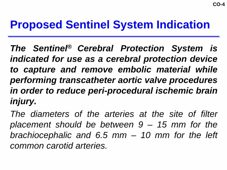
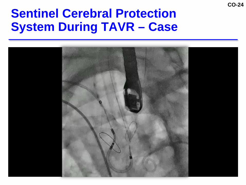
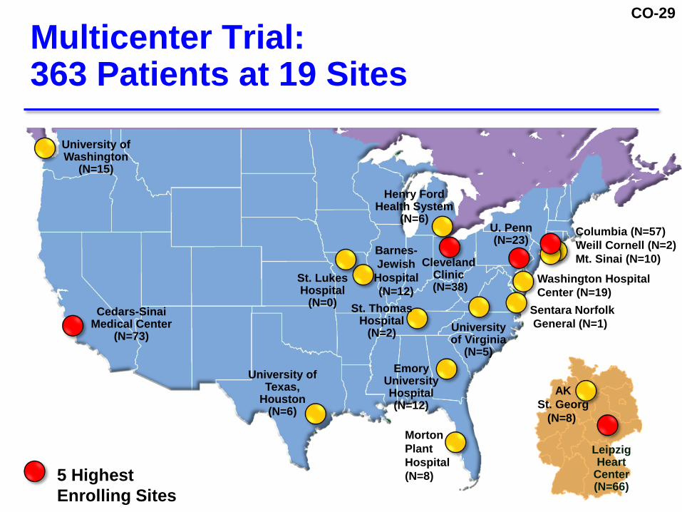
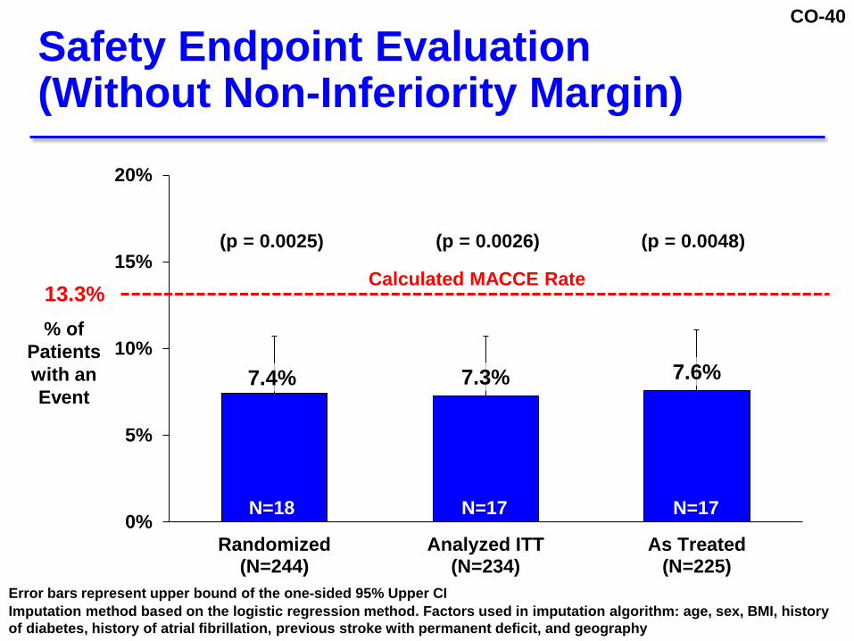
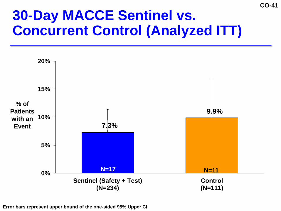
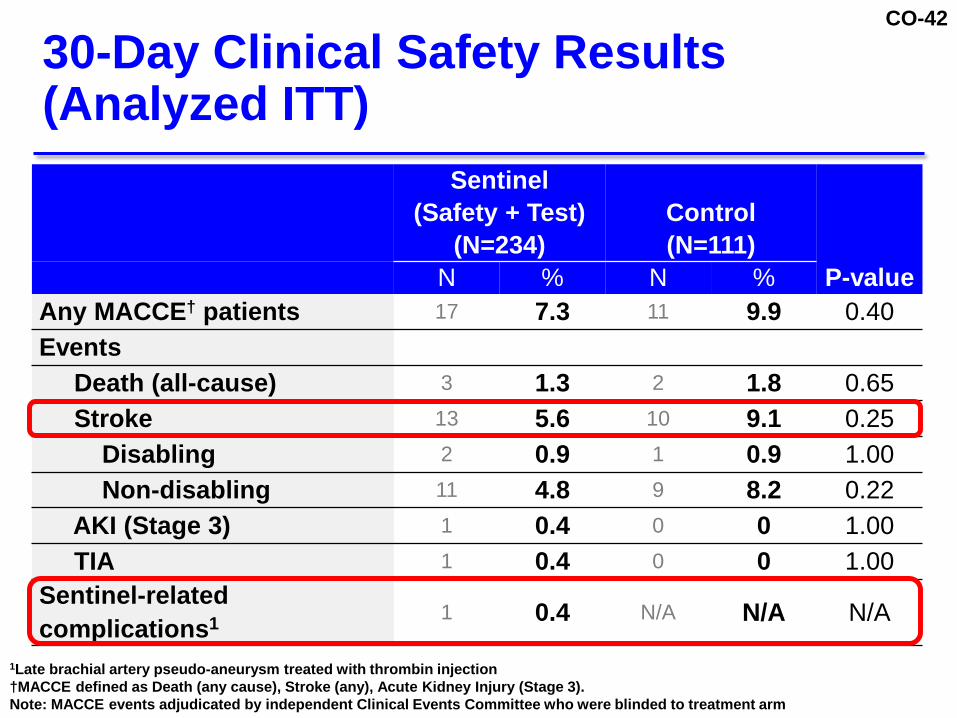
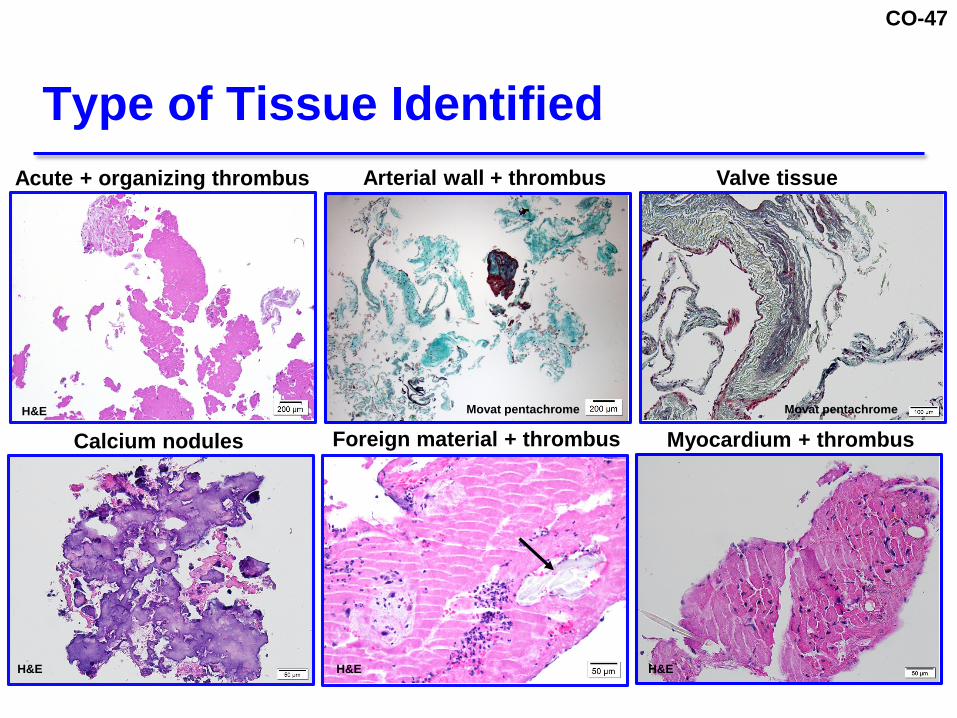
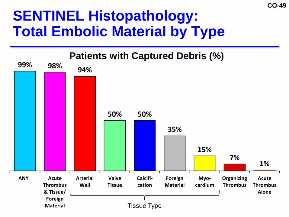
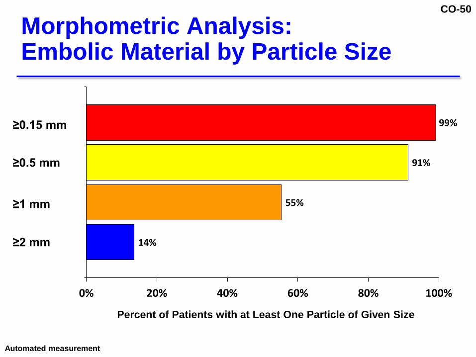
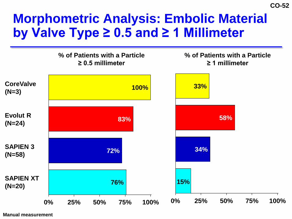
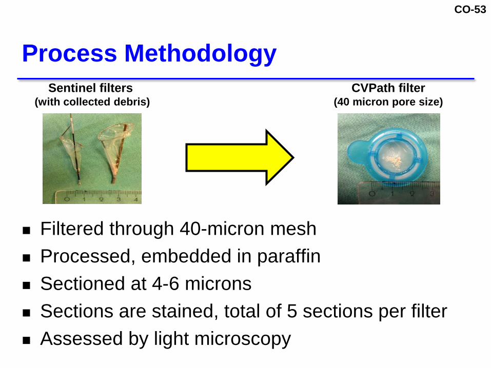
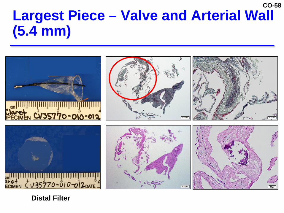
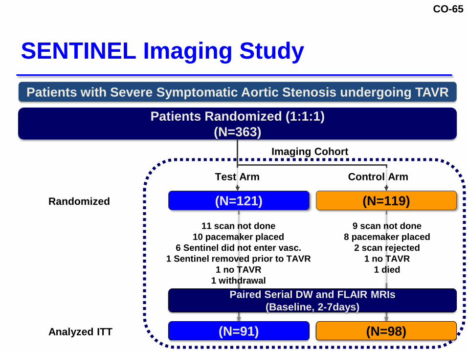
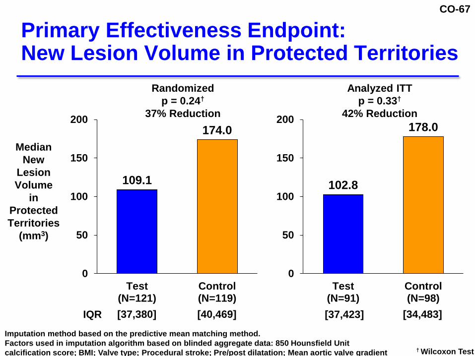

CO-80
Primary Effectiveness ndash Median New Lesion Volume (Protected Territories) Observed treatment effect ge 30 ndash Achieved Test vs Control ndash not achieved
Meta analysis (3 RCTs) provides additional evidence of effectiveness
SENTINEL Trial Effectiveness Summary
CO-81
SENTINEL Results in the Context of Neuroprotection History
William A Gray MDSystem Chief of the Division of Cardiovascular DiseaseMain Line Health
CO-82
Common FeaturesPores ~100-140m
Atraumatic wire frames for centering and sealingDeployed over a 0014rdquo wire from a collapsed state
Cordis Angioguard
Guidant Accunet
eV3Microvena Spider
Abbott VascularMednova
BSCEPI EZ
Accessory Devices Catheter-based Filters Used in Carotid Artery Stenting Are Similar to Sentinel
Claret Sentinel
CO-83
Evaluation metrics are not established Low incidence of clinical endpoints (eg
stroke) limits their utility DW-MRI surrogate is therefore valuable but
still being refined (timing effect of pre-existing abnormalities etc)
DW-MRI lesions ndash relevancy of volume vs number vs location not established
Expected treatment effect of DW-MRI surrogate not established or clinically validated
SENTINEL First RCT in Filter Embolic Protection
CO-84
Vascular trauma from filter embolic protection in CAS is rare
Similarly there was no filter-related vascular trauma reported in SENTINEL Finding is consistent with parallels in filter
construction Dwell times are short
Filters Used in Sentinel and Carotid Artery Stenting Are Safe
CO-85
Both CAS and TAVR EPD Capture Significant Amounts of Liberated Debris
CO-86
Types of embolic material collected by filters
Foam cells Smooth muscle cells Cholesterol Collagenelastin Plateletfibrin
57 Debris Collected in CAS EPDARCHeR Study
57 of samples
contained embolic material
CO-87
Analysis of Particles Collected Per Filter in ARCHeR and in SENTINEL
Filter TypeFrench
Size
Debris Captured
ge20 Particles
Per Patient
RX Accunet 6 Fr 571 241
Sentinel 6 Fr 99 53
1Gray W A et al J Vasc Surg 200644258-69
CO-88
39 Reduction
56
91
0
2
4
6
8
10
12
Sentinel Control
SENTINEL STUDY
EPD with Both CAS and TAVR Demonstrate Similar Stroke Reduction
27
47
0
2
4
6
8
10
12
CAS withEmbolic
Protection
CAS wo EmbolicProtection
Stroke ()
Adapted from Garg et al (2009) Neuroprotection and Stroke Endovascular Thoracic 16 412-427
42 Reduction
CO-89
Carotid artery stent coupled with EPD approval in US in 2004
Approval led to significant increase in use of protected carotid artery stenting 5000 to 75000 50 decrease in overall complication rates
after device approval Improvements likely secondary to Widespread EPD availability Refinements in patient selection and technique
The Impact of Device Approval
CO-90
SENTINEL is the first pivotal multicenter US IDE study to isolate EPD neuroprotective procedural and outcomes
SENTINEL safety profile is consistent with prior carotid artery (CAS) EPD studies
Similar to carotid EPD SENTINEL filter collection resulted in a high percentage of debris capture
Incorporation of Sentinel into TAVR resulted in stroke reduction similar to that seen after adoption of carotid stenting embolic protection
Further outcome improvements possible once TAVR EPD is broadly available
Summary 5 Perspectives
CO-91
Concluding Remarks
Azin Parhizgar PhDPresident and Chief Executive OfficerClaret Medical Inc
CO-92
Claret focused on developing best cerebral protection device to protect from acute embolic ischemic injury or stroke
4-year commercial history outside US SENTINEL first USEU multicenter randomized
controlled EPD trial Provides safety in a rapidly evolving TAVR field
Company Perspective
CO-93
Effectiveness Endpoint Success Criteria ITTNew Lesion Volume in Protected Territories
1028
1780
0
50
100
150
200
Test(N=121)
Control(N=119)
[37423] [34483]
MedianNew
Lesion Volume
in ProtectedTerritories
(mm3)
IQR
42 Reduction
Analyzed ITT
CO-94
Sentinel Debris Type
CO-95
Patient Quartile AnalysisAverage Number of Particles ge05 mm
1 in 4 Patients had 25 Particles ge05 mm in Size
0937
89
251
0
5
10
15
20
25
30
Q1 Q2 Q3 Q4
Average of
ParticlesCapturedge05 mm
Patient Quartiles
CO-96
Primary Safety Endpoint Met(30-Day MACCE)
74 73 76
0
5
10
15
20
Randomized(N=244)
Analyzed ITT(N=234)
As Treated(N=225)
183Performance Goal (Including Non-Inferiority Margin)
N=18 N=17 N=17
133Calculated MACCE Rate
of Patients with an Event
CO-97
Stroke Diagnosis le72 hours (ITT)
1304
13
30
45
09
27
82
0
2
4
6
8
10
Day 1 Day 2 Day 3 Total
Sentinel Control
Fisher Exact Test
Days to Stroke
of Patients
p=005263 Reduction
CO-98
Sentinel is safe with minimal complications injury or
disruption of the TAVR workflow performs as intended reduced the peri-procedural stroke rate
compared to control (3 vs 82) yields an observed treatment of effect of 42 captures a wide spectrum of emboli destined
for the brain in 99 of the patients
Summary
CO-99
Committed to comprehensive training Sentinel safety and technical success
demonstrated that IDE training was effective Elements of training program to mimic IDE study Comprehensive didactic training Hands on learning with anatomical model Proctor up to 5 cases at each site
Post-approval Training Program
CO-100
Close collaboration with FDA in formulating an effective PMS program to ensure a safe commercial roll out
Program to include Post-market registry Collect additional data in a real-world setting A registry or TVT module
Post-Market Surveillance Recommendations
CO-101
SentinelregCerebral Protection System During TAVR
February 23 2017Claret Medical IncCirculatory System Devices Panel
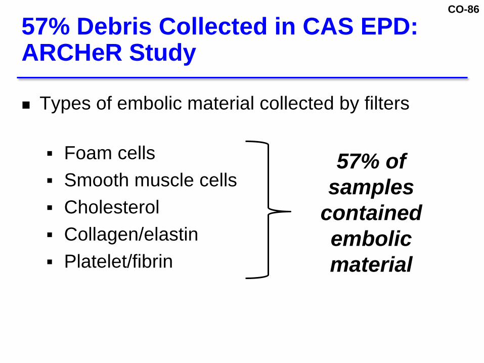

CO-101
SentinelregCerebral Protection System During TAVR
February 23 2017Claret Medical IncCirculatory System Devices Panel