Embed Size (px)
Citation preview
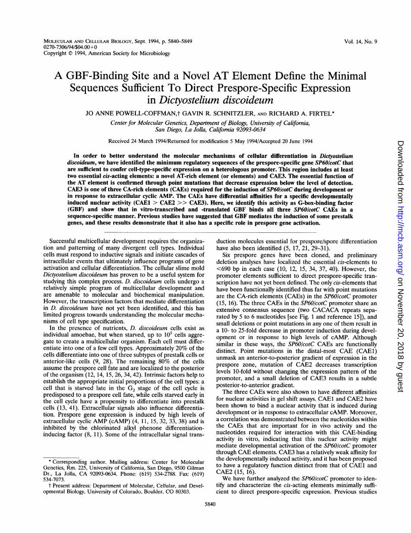
MOLECULAR AND CELLULAR BIOLOGY, Sept. 1994, P. 5840-5849 Vol. 14, No. 90270-7306/94/$04.00+0Copyright © 1994, American Society for Microbiology
A GBF-Binding Site and a Novel AT Element Define the MinimalSequences Sufficient To Direct Prespore-Specific Expression
in Dictyostelium discoideumJO ANNE POWELL-COFFMAN,t GAVIN R. SCHNITZLER, AND RICHARD A. FIRTEL*
Center for Molecular Genetics, Department of Biology, University of Califomia,San Diego, La Jolla, Califomia 92093-0634
Received 24 March 1994/Returned for modification 5 May 1994/Accepted 20 June 1994
In order to better understand the molecular mechanisms of cellular differentiation in Dictyosteliumdiscoideum, we have identified the minimum regulatory sequences of the prespore-specific gene SP60IcotC thatare sufficient to confer cell-type-specific expression on a heterologous promoter. This region includes at leasttwo essential cis-acting elements: a novel AT-rich element (or elements) and CAE3. The essential function ofthe AT element is confirmed through point mutations that decrease expression below the level of detection.CAE3 is one of three CA-rich elements (CAEs) required for the induction of SP60IcotC during development orin response to extracellular cyclic AMP. The CAEs have differential affinities for a specific developmentallyinduced nuclear activity (CAE1 > CAE2 >> CAE3). Here, we identify this activity as G-box-binding factor(GBF) and show that in vitro-transcribed and -translated GBF binds all three SP60IcotC CAEs in asequence-specific manner. Previous studies have suggested that GBF mediates the induction of some prestalkgenes, and these results demonstrate that it also has a specific role in prespore gene activation.
Successful multicellular development requires the organiza-tion and patterning of many divergent cell types. Individualcells must respond to inductive signals and initiate cascades ofintracellular events that ultimately influence programs of geneactivation and cellular differentiation. The cellular slime moldDictyostelium discoideum has proven to be a useful system forstudying this complex process. D. discoideum cells undergo arelatively simple program of multicellular development andare amenable to molecular and biochemical manipulation.However, the transcription factors that mediate differentiationin D. discoideum have not yet been identified, and this haslimited progress towards understanding the molecular mecha-nisms of cell type specification.
In the presence of nutrients, D. discoideum cells exist asindividual amoebae, but when starved, up to 105 cells aggre-gate to create a multicellular organism. Each cell must differ-entiate into one of a few cell types. Approximately 20% of thecells differentiate into one of three subtypes of prestalk cells oranterior-like cells (9, 28). The remaining 80% of the cellsassume the prespore cell fate and are localized to the posteriorof the organism (12, 14, 15, 26, 34, 42). Intrinsic factors help toestablish the appropriate initial proportions of the cell types: acell that is starved late in the G2 stage of the cell cycle ispredisposed to a prespore cell fate, while cells starved early inthe cell cycle have a propensity to differentiate into prestalkcells (13, 41). Extracellular signals also influence differentia-tion. Prespore gene expression is induced by high levels ofextracellular cyclic AMP (cAMP) (4, 11, 15, 32, 33, 38) and isinhibited by the chlorinated alkyl phenone differentiation-inducing factor (8, 11). Some of the intracellular signal trans-
* Corresponding author. Mailing address: Center for MolecularGenetics, Rm. 225, University of California, San Diego, 9500 GilmanDr., La Jolla, CA 92093-0634. Phone: (619) 534-2788. Fax: (619)534-7073.
t Present address: Department of Molecular, Cellular, and Devel-opmental Biology, University of Colorado, Boulder, CO 80303.
duction molecules essential for prespore/spore differentiationhave also been identified (5, 17, 21, 29-31).
Six prespore genes have been cloned, and preliminarydeletion analyses have localized the essential cis-elements to<690 bp in each case (10, 12, 15, 34, 37, 40). However, thepromoter elements sufficient to direct prespore-specific tran-scription have not yet been defined. The only cis-elements thathave been functionally identified thus far with point mutationsare the CA-rich elements (CAEs) in the SP60/cotC promoter(15, 16). The three CAEs in the SP60/cotC promoter share anextensive consensus sequence (two CACACA repeats sepa-rated by 5 to 6 nucleotides [see Fig. 1 and reference 15]), andsmall deletions or point mutations in any one of them result ina 10- to 25-fold decrease in promoter induction during devel-opment or in response to high levels of cAMP. Althoughsimilar in these ways, the SP60/cotC CAEs are functionallydistinct. Point mutations in the distal-most CAE (CAE1)unmask an anterior-to-posterior gradient of expression in theprespore zone, mutation of CAE2 decreases transcriptionlevels 10-fold without changing the expression pattern of thepromoter, and a small deletion of CAE3 results in a subtleposterior-to-anterior gradient.The three CAEs were also shown to have different affinities
for nuclear activities in gel shift assays. CAE1 and CAE2 havebeen shown to bind a nuclear activity that is induced duringdevelopment or in response to extracellular cAMP. Moreover,a correlation was demonstrated between the nucleotides withinthe CAEs that are important for in vivo activity and thenucleotides required for interaction with this CAE-bindingactivity in vitro, indicating that this nuclear activity mightmediate developmental activation of the SP60/cotC promoterthrough CAE elements. CAE3 has a relatively weak affinity forthe developmentally induced activity, and it has been proposedto have a regulatory function distinct from that of CAE1 andCAE2 (15, 16).We have further analyzed the SP60/cotC promoter to iden-
tify and characterize the cis-acting elements minimally suffi-cient to direct prespore-specific expression. Previous studies
5840
on Novem
ber 20, 2018 by guesthttp://m
cb.asm.org/
Dow
nloaded from
PRESPORE GENE REGULATION 5841
have indicated that CAE1 and CAE2 are discrete activatingelements, as large deletions that included CAE1 or CAE2 didnot decrease promoter activity substantially more than pointmutations. We show here that the region around CAE3 ismore complex than CAE1 or CAE2 and that multiple elementsact synergistically to activate transcription. Furthermore, wedelimit the regulatory sequences sufficient to confer cell-type-specific expression to a heterologous promoter, and we dem-onstrate that both CAE3 and a novel 37-bp AT-rich regulatoryregion are required for this function. In addition, we show thatpoint mutations within the AT element prevent gene expres-sion.We identify the developmentally induced CAE-binding ac-
tivity as G-box-binding factor (GBF) and show that it binds thethree SP60/cotC CAEs with sequence specificity. GBF hadbeen described previously as a developmentally regulated,cAMP-induced activity that interacts with cis-elements re-quired for the developmental induction of the prestalk-en-riched genes CP2/pst-cathepsin and ecmB (3, 20). Since oneGBF binding site from DG17 (20) fits the SP60cotC CAEconsensus well, and since GBF has a competition profilesimilar to that of the CAE-binding activity (see Results), wehypothesized that the CAE-binding activity and GBF wereidentical. GBF has recently been purified, and the geneencoding it has been cloned (39). Here, we demonstrate thatGBF copurifies with the CAE-binding activity and that recom-binant GBF binds the SP60/cotC CAEs with properties similarto those of the activity observed in crude nuclear extracts.
MATERIALS AND METHODS
Construction of plasmids. The construction of 5'-deletedSP60/cotC expression vectors was described previously (15).ExoIlI nuclease was used to generate additional 3' deletionsfrom the SpeI site in the 5'A20 promoter, and the deletionswere capped with Hindlll linkers. The NdH endpoint wasmade by filling in the NdeI site with the Klenow fragment ofDNA polymerase I and adding a HindIII linker. To createinternally deleted promoters, 3' deletions were cloned into theBamHI and HindIlI sites upstream of the 5' deletion endpointsin expression vectors. (See Fig. 1 for deletion endpoints.)
3' deletions of 5'A20 were cloned into A15ABam-gal (2) asfollows. The 3'-deleted promoters were subcloned into theBamHI and SpeI sites of Bluescript (Stratagene). The resultantvectors were cut with SpeI, blunt ended with the Klenowfragment, and capped with BglII linkers. The BamHI-BglIIfragments were cloned into the BamHI site of A15ABam-gal,to create the Act15-60A& constructs. To construct Actl5-60-A13A21 and Actl5-60-3(A13A21), the 3'A13 BamHI-HindIIIfragment was cloned into Bluescript. The HindIII site wasfilled in, and BglII linkers were added. This vector was cut withNdeI and ScaI, and the 3'A13-containing fragment was ligatedto the NdeI-ScaI fragment of a 5'A21 pGEM7 subclone. TheBamHI-BglII fragment containing the 5'A21-3'A13 sequenceswas cut out and ligated into A15ABam-gal in one copy or threetandem copies.
Deletion fragments of 5'A21-3'zA13 were generated byPCRs. 5' deletions were amplified from pGEM7 subcloneswith the 3' primer 5'-G'Tl'AGATCTAAAAAATAATTATTAAAT'ITGTTCTATG-3' and the SP6 primer. Internallydeleted fragments were amplified from expression vectors withthe 3' primer and a 5' primer, 5'-GTl''GGATCCATATATATACACTGTGAG-3'. The PCR products were subcloned(BamHI and BglII), sequenced, and cloned as three tan-dem repeats into A15ABam-gal. To create the ANdH-42(b3)mutant, the A42-A11O sequences were removed from the
ANdH-42 subclone (HindlIl and BglII) and replaced with themutant oligonucleotide (mutated nucleotides are underlinedand in boldface) agctTY`I`TGTAAAAAATAGAAAAAATT£QAATAA£QTATl'lI`TTTAgatc.The pSP72N2B vector was designed to allow the conversion
of a HindIlI site to a BamHI site with minimal additionalnucleotides. The HindlIl site of pSP72 (Promega) was filledin with the Klenow fragment of DNA polymerase I, and aHindlll linker was cloned into the blunt-ended BamHI site,recreating BamHI sites on either side of the HindIII site. TheCAE3-containing 3wt oligonucleotide (16) was cloned intopSP72N2B, cut out as a BamHI fragment, and cloned intoA15ABam-gal.
I8-Galactosidase staining. Cells were plated for develop-ment on white Millipore filters placed on phosphate-bufferedagar. At the appropriate times, the organisms were stained for3-galactosidase activity by a modification of previously de-
scribed procedures (7, 15). Filters were removed from the agarand allowed to air dry for 1 min and then placed on Whatmanfilters saturated with 0.5% glutaraldehyde-0.05% Triton X-100in Z buffer. After 5 min, additional fixation solution was addedto the filters to immerse the organisms for 10 min. The cellswere washed in Z buffer and stained with X-Gal (5-bromo-4-chloro-3-indolyl-3-D-galactopyranoside) solution (1 mM potas-sium ferrocyanide, 1 mM potassium ferricyanide, and 0.5 mgX-Gal per ml in Z buffer) for up to 3 h at 37°C.
Luciferase activity measurement. Luciferase activity wasdetermined as previously described (15, 22). All relative ex-pression data are mean values from at least two independentlyderived populations of stable transformants, each assayed in atleast two separate experiments. All values were normalized forthe copy number of the vector, as determined by Southern blothybridization. Copy numbers never varied by more than five-fold, compared with the control construct, and usually variedby less than twofold.
Nuclear extract preparation. Nuclear extracts were pre-pared as described previously (19), except that Tris buffer wasused instead of HEPES (N-2-hydroxyethylpiperazine-N'-2-ethanesulfonic acid), and extracts were not heat treated.Extracts were diluted to 2.5 mg/ml in buffer I50 for precipita-tion at increasing concentrations of ammonium sulfate. Frac-tionation of extracts to enrich for GBF activity is describedelsewhere (39).
In vitro transcribed-translated GBF. The vector R20 con-taining the complete coding region of GBF (39) was linearizedwith ApaI. In vitro transcription and translation were thenperformed with the TNT T3-coupled reticulocyte lysate system(Promega). For binding to CAE3 in vitro, transcription andtranslation reaction mixtures with R20 or a luciferase controlvector were diluted 10-fold in 120 mM KCI affinity columnbuffer (ACB) (5% glycerol, 3.3 mM Tris [pH 8.4], 6.7 mM Tris[pH 7.5], 0.1 mM EDTA, 1 mM dithiothreitol, and 0.05%Triton X-100) and applied to a CP2 G-box affinity columnequilibrated in the same buffer. The column was washed with10 column volumes of 120 mM KCl ACB supplemented with 1mg of bovine serum albumin (BSA) per ml and eluted with 2column volumes of 420 mM KCI ACB with 1 mg of BSA perml. The affinity column was prepared with the complementaryCP2 G-box oligonucleotides 5' GATCCACAGCGGGTGTGTTAAGTTAGaGGTGGGTTTT[ATATAGGT and 5' GATCTCCTATATAAAACCCACCCCTAACTTAACACACCCGCTGTG according to previously described methods (39).
Gel retardation assays. Gel mobility shift assays were per-formed as previously described (16) except as detailed in thefigure legends. The shorter CAE3-containing oligonucleotidedescribed in the text is agctACAAACACACTCCCAACACA
VOL. 14, 1994
on Novem
ber 20, 2018 by guesthttp://m
cb.asm.org/
Dow
nloaded from
5842 POWELL-COFFMAN ET AL.
5 'A2 3
A TCCCATACTA CATTAAAATA TTGTATATC
CAE1 5 'A23-630 AAT'CAGAA;AT TTAAA'CTAAA TTGTTTTTTT TT|CACACAC CCACACAC ATTTACCCA TTITTTTACAT
v 3 Ai' 5'A46_ CAE2
-560 TTCTCATATA AAAATTATCT ATTGTACATT GTTCAATATT CAtC CATTAACACACT TTCAACTCAA
5'A21 3A A2'- 49 TCATT== TCCCTI-C.A.AAAAA ACAATAAAT ATATATACA^;CCCTTGTAATT TCATTA A3GCGATAGAA
__53'2 CAE3-Al' yA5C_3'AL394 CC Nde; 3'A183"AA_ Nc2
51) AAA.'ATTTTA TTT7T(A ACA CACTC AAC ACAC4VC.CATYATGA.AAAC!AAC TCAC ACAAT TYA XTTTGT
v3'A111 3 '5I11 3'A13
-AAAACATAGA ACAAT-,TAA TAATTATTTT '=TCATT T C,ATTATT'AC' IlT1'.'A:TA'I TTATTIATTT
--,,7". A' ''IiTT CA.'iAAAA A AAAAAAGTTA AATTA.TTAC'T -' 'A 'AAAAAAAA
- AS 'A5¢ 5 IXA2"V_A AT T'CA'I'TTAC ATTTTGTTCA TATCCAACTTA A3CiAT-C A.'AT AAA. 1A.kC.TTA'AT
14 CT-T.,V?TT.:TTL AATA'I'TATATA TATATTTATC AA^TATATAA,, TTkATATz,:AA AAAAA.AAAAC; AAATTTAATA
*:CAACATATAAT A'I-'I'TATATAA AATCATAAJ%.T7 TTA"A.%ATA,I'. TAA-T'rTT'FT .kATATT"TAAA :AAl"lCTAATA
IAAAA_:GT'1"I'Tt"tIATIATTTA(,JT AAATTTGTAA -,AT'C'.ATT'I'' I'AAC.Ax;AAA:C ':A.;A'' lA .AAAAASAA1_4
+;1A._FA.>AAA1,AA AAAAAAA}AATG
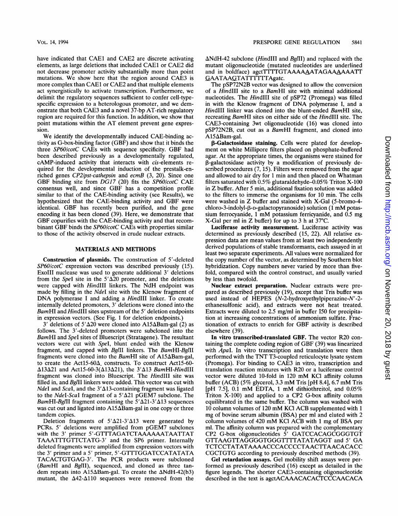
FIG. 1. Deletion endpoints in the SP60/cotC promoter. The threeCAEs are boxed, and the AT element is underlined. The transcrip-tional start site is labelled +1. The Ndel endpoint is also shown (seeMaterials and Methods).
SP60 promoter deletions
600 400
MOL. CELL. BIOL.
CAAGCAtgca (5' overhangs are lowercase). The other SP60!cotC CAE-containing oligonucleotides (16) and the CP2 G-box-containing oligonucleotides (19) have been described pre-viously.
RESULTS
Localization of SP60IcotC promoter elements. Previous anal-yses have shown that 661 bp of SP60/cotC 5' flanking sequenceis sufficient to direct high levels of cell-type-specific expressionand that the three CAEs are required for full developmentalinduction. It was also shown that the CAEs, while necessary fordetectable levels of expression, were not sufficient to directprespore-specific transcription: a large deletion 3' of the CAEscaused a catastrophic (>104-fold) reduction in expression (15,16).
In order to localize those regions of the SP60/cotC promoterrequired for prespore-specific transcription, we assayed aseries of internal deletions with luciferase and lacZ reporterconstructs. Since it was previously shown that the sequencesdownstream of 5'A21 were sufficient to direct prespore expres-sion, although at diminished levels (15), we focused our studieson this region of the promoter. The deletions and the resultantrelative expression levels are listed in Fig. 1 and 2. Removal ofall or part of CAE3 reduced promoter activity 15- to 40-fold(compare full-length with A24-64 and A15-64 or compareA15-55 with A15-NdH). The region immediately upstream of
expression presporelevels expression
200
100 +
CAEI CAE2 CAE3
A17-46 [ [IV] *ILZE
A15-55 * -U L
A24-42 * * I LA
A18-42 * F-E-
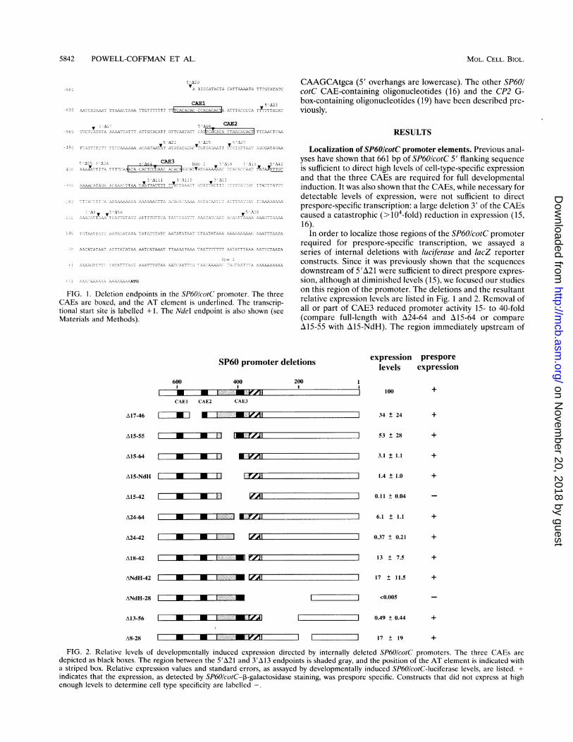
FIG. 2. Relative levels of developmentally induced expression directed by internally deleted SP60/cotC promoters. The three CAEs are
depicted as black boxes. The region between the 5'A21 and 3'A13 endpoints is shaded gray, and the position of the AT element is indicated witha striped box. Relative expression values and standard errors, as assayed by developmentally induced SP60/cotC-luciferase levels, are listed. +indicates that the expression, as detected by SP60/cotC-3-galactosidase staining, was prespore specific. Constructs that did not express at highenough levels to determine cell type specificity are labelled -.
A15-64 m =' V
A15-NdH * *
A15-42 m M l
j 34 ± 24
] 53 ± 28
+
A24-64 * UI' - -.i
3.1 ± 1.1
1.4 ± 1.0
0.11 ± 0.04
ANdH-42
6.1 ± 1.1
0.37 t 0.21
ANdH-28
A13-56
+
13 ± 7.5
A8-28
l lZIZZI
17 ± 11.5 +
<0.005
*F l.-- .a _ ;-YA]
0.49 ± 0.44
17 ± 19
+
r- m
m m 12150.
m
m m
I
I
on Novem
ber 20, 2018 by guesthttp://m
cb.asm.org/
Dow
nloaded from
PRESPORE GENE REGULATION 5843
(-661) CA
A8-A20 E U
SP60 promoter region
,El CAE2 CAE3EZZEI .1 iK
prespore Aexpression
(-204)1111 + A13-A21
SP60 promoter region (3 copies)
(-462) CAE3 (-306)
Al 10-A2A13-A20 A M L7=,#l
A110-A20 +
A1II-A20 * -;;-
A19-A20
A13-A21
1:-4 (-306)
-462) 006)s
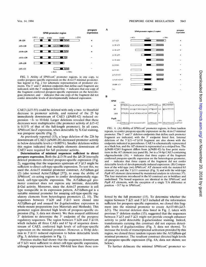
FIG. 3. Ability of SP60/cotC promoter regions, in one copy, toconfer prespore-specific expression on the Actini5 minimal promoter.See legend to Fig. 2 for schematic representation of promoter ele-ments. The 5' and 3' deletion endpoints that define each fragment are
indicated, with the 3' endpoint listed first. + indicates that one copy ofthe fragment conferred prespore-specific expression on the heterolo-gous promoter, and - indicates that one copy of the fragment did notconfer detectable levels of developmentally induced expression.
CAE3 (A15-55) could be deleted with only a two- to threefolddecrease in promoter activity, and removal of the 25 bpimmediately downstream of CAE3 (ANdH-42) reduced ex-
pression -6- to 10-fold. Larger deletions revealed that thesedecreases were multiplicative (the promoter activity of A15-42is 0.11% of that of the full-length promoter). In all cases,SP60/cotC/lacZ expression, when detectable by X-Gal staining,was prespore specific (Fig. 2).As previously reported (15), a large deletion of the 226 bp
downstream of CAE3 (ANdH-28) decreased promoter activityto below detectable levels (<0.005%). Smaller deletions withinthis region indicated that multiple elements downstream ofCAE3 were required for full promoter activity.
Determination of minimal sequences sufficient to directprespore expression. Both the A13-56 and the A8-28 internallydeleted promoters directed prespore-specific expression (Fig.2), suggesting that the sequences upstream of 3'A13 might besufficient to direct cell-type-specific expression. To test this, weused the ActiniS minimal promoter construct, A15ABam-gal(2) (also termed ActinlSABgal [37]), to assay the ability ofSP60/cotC cis-acting regions to confer developmentally regu-lated, cell-type-specific expression. The A15ABam-gal pro-moter construct does not express any intrinsic, detectable1-Gal activity. Moreover, since the Actinl5 promoter is celltype nonspecific in its expression pattern, A15ABam-gal is a
suitable minimal promoter for assaying cell-type-specific reg-ulatory elements from heterologous promoters (2, 10). Thesequences between 5'A20 and 3'A13 were cloned intoA15ABam-gal and assayed for ,B-galactosidase expression inwhole-mount preparations (see Materials and Methods). Thispromoter region directed high levels of prespore-specific ex-
pression (Fig. 3; data not shown). We then assayed additional3' deletions to determine the 3' endpoint of the presporeregulatory sequences. The region between 5'A20 and 3'X1 1 0,which includes all three CAEs and 67 bp of sequence down-stream of CAE3, conferred high levels of cell-type-specificexpression on the minimal promoter. However, a 10-bp dele-tion to 3'A1 11 reduced expression to below detectable levels(Fig. 3; deletion endpoints in Fig. 1).
Previous studies had shown that the sequences downstreamof 5'A21 were sufficient to direct cell-type-specific expression,although expression levels were 500-fold less than those con-
Al 10-A25
Al I0-A55
AI I0-A64
A 24-64)
A 24-42)
A Ndl1-42)
+
_ zzzz
_ s-zzeM +
AiNdI1-42)b3
ANdH-AI I_
3wt EL ZEE:
B
SP60(-355) (-31(9)
MI TTTGTA AAACATAGAACAA AT[AAT AA TTATT[TT
b3 TTTGTA AAAAATAGAAAAA ATITGAAT AA tiTATTTTTT
AIll TTTGTA AAACATAGAACAA ATFUAAT A
PspB-30) -285)AAA TTTGTA GTCAAAAATATT ATTAAT TrATTIATT AATTA I
FIG. 4. (A) Ability of SP60/cotC promoter regions, in three tandemrepeats, to confer prespore-specific expression on the ActiniS minimalpromoter. The 5' and 3' deletion endpoints that define each promoterfragment are indicated, with the 3' endpoint listed first. Internaldeletions of the 5'A21-3'A11O fragment are also shown with theendpoints indicated in parentheses. CAE3 is schematically representedas a black box, and the AT element is represented as a striped box. TheA(NdH-42)b3 fragment differs from A(NdH-42) by four point muta-tions in the AT element (see panel B). 3wt is a 54-bp CAE3-containingoligonucleotide (16). + indicates that three copies of the fragmentconferred prespore-specific expression on the heterologous promoter,and - indicates that three copies of the fragment did not conferdetectable levels of developmentally induced expression. (B) Compar-ison of the wild-type (wt) SP60/cotC AT element with the mutated b3element (A) and the 3M11 1 construct (Fig. 3) and with the wild-typePspB AT element (determined by mutational analysis in reference 37).The four mutations introduced in the b3 construct are in boldface andunderlined. The boxed sequences are identical in the SP60/cotC andPspB AT elements, with the exception of a single T/A difference atposition -317 bp in SP60/cotC.
ferred by the full promoter (15). To determine whether theregion between 5'A21 and 3'A13 included all the informationsufficient for prespore-specific expression, we cloned this frag-ment into the minimal promoter to create Actl5-60-(A13-A21). The internal deletion data listed in Fig. 2, taken withprevious 5' deletion studies (15), suggested that the sequencesbetween 3'A13 and 5'A21 might not provide enough enhanceractivity to yield detectable ,B-galactosidase staining. Indeed,transformants carrying this construct did not express detect-able levels of ,B-galactosidase (Fig. 3; data not shown). Toincrease the levels of transcriptional activation provided by thisregion, we cloned three tandem repeats into the A15ABam-galminimal promoter. The resultant construct directed high levelsof prespore-specific expression (Fig. 4A; data not shown; see
below).To further delineate the minimal SP60/cotC promoter se-
presporeexpression
+
m M
VOL. 14, 1994
on Novem
ber 20, 2018 by guesthttp://m
cb.asm.org/
Dow
nloaded from

5844 POWELL-COFFMAN ET AL.
110-21
a
AN -42
I110X56
AN-421b31
FIG. 5. Spatial pattern of expression conferred by three tandem repeats of SP60cotC promoter regions at the slug and early culminant stagesof development, as detected by 3-galactosidase staining of transformants. In the migrating slug, the prespore cells are localized to the posterior80% of the organism. Once the organism reaches a location suitable for culmination, it forms a second finger or early culminant before the tip cellsfunnel down through the prespore cells and differentiate into stalk. Both developmental stages are shown here for cells transformed with theconstructs Act60-3(110-21), Act 60-3(110-55), Act6O-3[A(NdH-42)], and Act6O-3[A(NdH-42)b3]. For a description of the constructs, see Fig. 4A.
quences sufficient to direct prespore expression, we assayeddeletions of the 157-bp A13-A21 fragment, each as threetandem repeats in the minimal promoter. The results of thesestudies are listed in Fig. 4A. As expected, the A110-A21sequences conferred high levels of prespore expression (Fig.
4A; also pictured in Fig. 5). A 5' deletion of the sequencesupstream of CAE3 did not change the expression pattern, butthe intensity of staining was reduced (see All10-A55 in Fig. 5).The CAE3 sequences were required for detectable expression,as seen in A(24-64), but the 25 bp immediately downstream of
MOL. CELL. BIOL.
on Novem
ber 20, 2018 by guesthttp://m
cb.asm.org/
Dow
nloaded from

VOL14194PRESPORE GENE REGULATION 5845
CAE3, deleted in A(NdH-42), were not essential. By contrast,the AT-rich region between 5'A42 and 3'A11O was absolutelyrequired to direct detectable levels of 3-galactosidase expres-sion [the A(NdH-42) sequences had prespore enhancer activ-ity, but the ANdH-A21 sequences did not] (Fig. 4 and 5).These deletion analyses indicated that two regions of the
SP6O/cotC promoter were required to confer prespore-specificexpression on the minimal promoter: CAE3 and the down-stream AT-rich sequences. As D. discoideum noncoding se-quences are 80 to 95% A/T (25), we were concerned that theAT-rich regulatory region might serve a non-sequence-specificfunction, such as providing proper spacing in the context of theminimal promoter. To address this, we introduced four basechanges (Fig. 4B) into the AT region of the A(NdH-42)fragment and assayed this mutant (termed b3) in the minimalpromoter construct (Fig. 4A). As shown in Fig. 5, the fourpoint mutations in the AT-rich region decreased expression tobelow detectable levels.GBF binds SP6OIcotC CAEs in vitro. Haberstroh et al. (16)
had shown that multiple nuclear activities bound CAE3 invitro. We further examined the specificity of these interactionsin fractionated extracts. The only activity we found to bindspecifically to the CAE3 core sequence formed a complex ofrelative mobility similar to and specificity identical to those ofthe developmentally induced activity previously shown tointeract strongly with CAE1 and CAE2 (16) (Fig. 6A; data notshown). We found that a shorter (30-bp) CAE3-containingoligonucleotide had a slightly higher affinity for this activitythan the 54-bp oligonucleotide used in previous studies (Fig.6A). Similarities in developmental and cAMP regulation,relative mobility, and binding-site sequence prompted us toexamine whether this activity was identical to GBF, which hasbeen shown to mediate the developmental induction of someprestalk genes (3, 6, 18, 20, 35). GBF binding requires twohalf-sites of (T/G/A) G (G/T) G(T/G) G(T/G/A) (19, 20), and theSP60OcotC CAEs each consist of two CACACA half-sitesseparated by 4 to 7 nucleotides (16). In gel mobility shift assays,the CP2Ipst-cathepsin G box (the original GBF binding site)competed strongly for binding to the CAE-binding activity,while a mutant G box (which is nonfunctional for CP2 activa-tion in vivo and GBF binding in vitro) did not (Fig. 6A).Similarly, when the CP2 G box was used as a probe, the CAEscompeted for GBF binding (39) (data not shown). The bindingaffinities were as follows: CAE1 CP2 > CAE 2 > CAE 3 >CP2mut > CAElmut.During the course of these studies, GBF was purified and
the gene encoding it was cloned (39). The developmentallyregulated CAE-binding activity copurified with GBF at alltested stages (Fig. 6B; Fig. 7; data not shown). To determinedirectly whether the CAE-binding activity was GBF, we tran-scribed and translated cloned GBF in reticulocyte lysates andincubated the reaction mixtures with labelled CAE3. Thelysates were enriched for GBF protein by CP2 G-box affinitychromatography (see Materials and Methods). As shown inFig. 7A, recombinant GBF formed a complex with CAE3 thatmigrated at a position similar to that of the complex formed inGBF extract preparations made from developing D. discoi-deum cells. Other proteins in the reticulocyte lysate bound theprobe (see control lanes in Fig. 7A), but formation of the GBFcomplex was dependent upon the addition of the GBF vectorDNA to the reaction. Furthermore, the in vitro-transcribedand -translated GBF had a competition profile similar to thatof the sequence-specific CAE3-binding activity in nuclearextracts: CAE1 and the CP2 G box had a greater affinity forGBF than CAE3, and the mutant CP2 G box competed verypoorly for binding. GBF, transcribed and translated in reticu-
_A _
J - ri r
_-,,, --Xs#,
o. o _
Z.
+a_,_ov--_o_,1 _,1_- _- ___
uIG.6.SP60/cotCCAE-binmimgactivityin nuclearandpartp
B =0 re_CL + +
p
FIG. 6. SP6OIcotC CAE-binding activity in nuclear and partiallypurified GBF extracts. (A) The 54-bp CAE3 oligonucleotide waslabelled and incubated with proteins in a developmental nuclearextract fractionated by precipitation between 20 and 33% ammoniumsulfate. Binding was inhibited with a 25-fold molar excess of theunlabelled oligonucleotides 54-bp CAE3 [CAE3 (long)], 30-bp CAE3(CAE3), CAE1, mutant CAE1(CAE1M), CP2 G box(CP2), andmutant CP2 G box (CP2M). Reactions were assayed by electrophoresison 4% polyacrylamide gels. The position of free probe is indicated byan arrowhead labelled p. Another arrowhead indicates the position ofthe complex formed. (B) Labelled 30-mer CAE3 oligonucleotide wasincubated with GBF that had been partially purified by DNA-celluloseand affinity column chromatography (-2% GBF, by mass of protein).The complex formed was inhibited with unlabelled CAE3 oligonucle-otide.
locyte lysates, also bound specifically to CAE1 and CAE 2
probes (Fig. 7B and C), confirming that the developmentallyregulated activity that binds the three SP60/cotC CAEs is GBF.
DISCUSSIONIn this study, we have analyzed the SP60/cotC promoter and
have delimited the regulatory sequences sufficient to confercell-type-specific expression on a heterologous promoter. Thisregion contains a novel AT-rich element (or elements), theCA-rich repeat CAE3, and neighboring sequences that actsynergistically to induce prespore-specific transcription duringmulticellular development. The SP60OcotC CAEs bind acAMP-induced, developmentally regulated nuclear activity in asequence-specific manner. We have identified this activity asGBF.GBF activates prespore-specific transcription. Full induc-
tion of the SP60OcotC promoter requires all three CAEs. Pointmutations or small deletions in any of the SP60OcotC CAEsdecrease promoter activity 10- to 25-fold, and if all three areremoved in a 5' deletion, promoter expression is not detect-able. Thus, while SP60cotC promoter induction requires atleast one CAE, any one of the three can be deleted, suggestingsome redundancy in their function (16). We have shown herethat a 95-bp SP60/cotC promoter region containing CAE3 is
VOL.14, 1994
on Novem
ber 20, 2018 by guesthttp://m
cb.asm.org/
Dow
nloaded from
5846 POWELL-COFFMAN ET AL.
ACI)iwe.ti,v[ tllfZIn I i§F \)'>.].I'C\ l'itt1%It't<I tt
BI)itv lsituu/it I; F I}T .N
r_c_co7
-_ (10I
*
(.10
I. U
CDicof ostdw/ium (JI express)1t'9. I .ltc.
extract t *et) t .
F_ ~~~~~~~~~~~..// / f f o1~~~~~CA.L
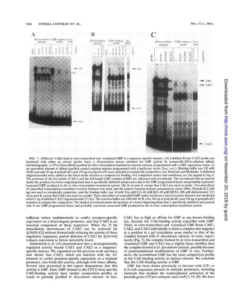
FIG. 7. SP60/cotC CAEs bind in vitro-transcribed and -translated GBF in a sequence-specific manner. (A) Labelled 30-mer CAE3 probe wasincubated with either no extract (probe lane), a Dictyostelium extract enriched for GBF activity by nonspecific-DNA-cellulose affinitychromatography, a CP2-G-box-affinity-purified in vitro transcription-translation reaction mixture programmed with a GBF expression vector, oran equivalent amount of affinity-purified control reaction mixture programmed with a luciferase vector (Luc. vect.). Binding buffer was 120 mMACB, and only 50 ng of poly(dI-dC) and 150 ng of poly(dA-dT) were included as nonspecific competitors (see Materials and Methods). Unlabelledoligonucleotides were added at the listed molar excesses to compete for binding. For competitor names and conditions, sce the legend to Fig. 6.The positions of the free probe (CAE3) and the full-length GBF complex (GBF) are indicated with arrowheads. The arrowhead with an asteriskmarks the position of a lower-migrating band that is specifically inhibited and present only in the GBF-programmed lysate and probably representstruncated GBF produced in the in vitro transcription-translation system. (B) As in part A, except that CAE1 was used as probe. Two microlitersof unpurified transcription-translation reaction mixtures was used, and the control reaction mixture contained no vector DNA. Poly(dI-dC) (200)ng) was used as nonspecific competitor, and the binding buffer was 10 mM Tris (pH 8.5)-80 mM KCl-20 p.M EDTA-200 F.M dithiothreitol. (C)As in part B, except that CAE2 was used as a probe. Three microliters of unpurified GBF and/or luciferase control reaction mixtures was incubatedwith 0.1 ng of labelled CAE2 oligonucleotide (37 bp). The reaction buffer was 100 mM ACB, with 100 ng of poly(dI-dC) and 150 ng of poly(dA-dT)included as nonspecific competitors. The shaded arrowhead marks the position of a lower-migrating band that is specifically inhibited and presentonly in the GBF-programmed lysate and probably represents truncated GBF produced in the in vitro transcription-translation system.
sufficient (when multimerized) to confer prespore-specificexpression on a heterologous promoter, and that CAE3 is anessential component of these sequences. While the 25 bpimmediately downstream of CAE3 can be removed [inA(NdH-42)] without dramatically reducing the activity of theseregulatory sequences, partial deletion of CAE3 [in A(24-64)]reduces expression to below detectable levels.
Haberstroh et al. (16) demonstrated that a developmentallyregulated activity bound CAEI and CAE2 in a sequence-specific manner. We expanded on this previous work and havenow shown that CAE3, which can function with the ATelement to confer prespore-specific expression on a minimalpromoter, also binds this activity, although with lower affinity.Several data presented in this report demonstrate that thisactivity is GBF. First, GBF (bound to the CP2 G box) and theCAE-binding activity have similar competition profiles incrude or partially purified D. discoideum extracts. In fact,
CAEI has as high an affinity for GBF as any known bindingsite. Second, the CAE-binding activity copurifies with GBF.Third, in vitro-transcribed and -translated GBF binds CAEI,CAE2, and CAE3 individually to form a complex that migratesat a position in a gel retardation assay similar to that of thecomplex formed with D. discoideum extracts. In some experi-ments (Fig. 7), the complex formed by in vitro-transcribed and-translated GBF and CAE3 has a slightly faster mobility thanthe complex formed in D. discoideum extracts, possibly becauseof posttranslational modifications of GBF in vivo. Further-more, the recombinant GBF has the same competition profileas the CAE-binding activity in nuclear extracts. We concludethat the CAE-binding activity is GBF.GBF has been shown previously to interact with G/T- or
C/A-rich sequences present in multiple promoters, includingelements that mediate the transcriptional activation of theprestalk genes CP2/pst-cathepsin and ecmB (3, 19, 20). We have
MOL. CELL. BIOL.
on Novem
ber 20, 2018 by guesthttp://m
cb.asm.org/
Dow
nloaded from
PRESPORE GENE REGULATION 5847
shown here, for the first time, that GBF also plays a direct andessential role in the induction of prespore gene expression,through interactions with the CAEs. Because the bindingaffinity of CAE3 for GBF is relatively low, we considered thatCAE3 might function through interaction with DNA-bindingfactors other than GBF. However, we assayed fractionatedextracts and were unable to detect any other activity thatbound CAE3 in a sequence-specific manner. Therefore, webelieve that all three SP60!cotC CAEs function as GBF bindingsites.
Deletions removing both CAEI and CAE2 have been shownto decrease promoter activity substantially more than the sumof the respective decreases due to deletions removing the twoelements individually (16). Haberstroh et al. (16) noted thatthis was suggestive of cooperative interactions between thecomplexes that bound the SP60/cotC CAEs. Consistent withthis model, we find here that a single copy of the A13-A21sequences does not confer detectable levels of expression onthe minimal promoter construct. However, when these se-quences are multimerized, such that there are three copies ofCAE3, the AT element, and flanking sequences, they directhigh levels of prespore-specific transcription.
Previous analyses of CAEs indicated that GBF binding sitesfunction in a context-dependent manner. If CAE1 is deleted ormutated, the remaining SP60/cotC promoter sequences directhigher levels of expression in the anterior region of theprespore zone than in the posterior region. In contrast, dele-tion of CAE3 results in a subtle posterior-to-anterior gradientof expression. Point mutations in CAE2 do not detectablychange the expression pattern (15, 16). These data suggest amodel in which the induction of the SP60/cotC promoter isdownstream of at least two signalling pathways that areactivated in opposing gradients in the prespore zone (16).Thus, the three GBF binding sites, in the context of the fullSP60/cotC promoter, may mediate responses to different sig-nals.The smallest SP60/cotC promoter fragments that specify
prespore-specific expression lack CAE1 and CAE2, yet theseconstructs direct expression uniformly throughout the pre-spore zone (Fig. 4 and 5). This may indicate that sequencesdownstream of the AT element(s) mediate the anterior-to-posterior graded activation reported by Haberstroh et al. (16)in 5' deletion constructs lacking the distal CAEs. We showedthat internal deletion of the 105 bp between 3'A13 and 5'A56decreases expression 200-fold. While these sequences are notabsolutely required for cell-type-specific expression, associatednuclear factors may interact with complexes on CAE3 and theAT element(s) to mediate activation in response to gradedsignals. A second possibility is that multimerization of theSP60/cotC sequences in the ActiS promoter context influencesthe expression pattern within the prespore region. The absenceof a graded response, however, does not detract from theconclusion that CAE3, the AT-element(s), and adjacent flank-ing sequences include all the cis-acting sequences sufficient forprespore-specific transcriptional activation. However, in thecontext of the full promoter, a deletion of CAE3 does notdestroy gene expression or prespore specificity, which mayindicate that any CAE, in conjunction with the AT region, maydirect prespore-specific expression.
Novel AT element is essential for enhancement of presporetranscription. While a GBF binding site is required for SP60/cotC promoter activation, it is not sufficient. The AT-richsequences between 3A I10 (-318 bp) and 5'A42 (-355 bp) arealso necessary to direct expression in prespore cells. The 343bp upstream of 3'A110 confer high levels of prespore-specificexpression on the ActiS minimal promoter. However, a 10-bp
3' deletion of these sequences to 3'A11I decreases expressionto below detectable levels, even though CAEI, CAE2, andCAE3 are left intact. None of the constructs that lack theseAT-rich sequences are capable of directing prespore expres-sion. The sequence-specific function of this element (or ele-ments) is confirmed by the introduction of four point muta-tions into the AT region of the Actl5-60-3z(NdH-42)construct. While the original construct directs prespore-spe-cific expression, the point mutations reduce expression tobelow detectable levels.
In a concurrent analysis of the PspB promoter, we identifieda 46-bp AT-rich region that is essential for high levels ofcell-type-specific expression (37). Comparison of the SP60/cotC and PspB AT elements revealed an extensive consensussequence (Fig. 4B). This suggests that these sequences mayinteract with common regulatory factors that enhance theexpression of these genes in prespore cells. Preliminary dataindicate that there are nuclear activities in fractionated extractsthat bind the SP60/cotC AT element in the presence of 0.5 ,gof poly(dA-dT). Some of these interactions are sequencespecific: the wild-type element competes for binding, but theb3 mutant does not (36). Studies to determine the specificityand regulation of these nuclear factors are currently underway.
Implications for the regulation of prespore differentiation.The data presented here, taken with the analyses of Hjorth etal. (20), indicate that GBF functions in both prestalk andprespore cells. To confirm this, we separated prestalk andprespore cell populations and determined that GBF mRNAand GBF activity are present in both cell types, with anapproximately 2.5-fold enrichment in prespore cells (39; alsodata not shown). Recently, we also demonstrated that gbf nullcells cannot be induced to express any of the late genes and donot execute the developmental program (39). Thus, GBF isrequired for both prestalk- and prespore-specific gene expres-sion, but it is likely that it must interact with other nuclearfactors to activate these late genes.
In this study, we have shown that the GBF binding siteCAE3 cannot confer detectable levels of developmental acti-vation on the Actinl15 minimal promoter, even when multim-erized. Another regulatory sequence, the AT element(s), isalso required for prespore-specific expression. Thus, the acti-vation of promoters in prespore cells may require a coopera-tive interaction between GBF and nuclear factors that recog-nize the AT element(s). Further characterization of thenuclear activities that interact with the AT element(s) willenable us to investigate this hypothesis. We might expect thatthe AT-element binding factors are expressed only in presporecells or that the element may interact with prespore-specificvariants of GBF.
While we have shown that GBF binds G boxes and presporeCAEs (37, 39) (Fig. 7), we are still investigating the extent towhich GBF binding sites are functionally equivalent. In pre-liminary studies, we have found that both CAE1 and CAE3 canreplace the G box in the CP2/pst-cathepsin promoter to directprestalk-enriched transcription (1). This suggests that theconserved region of the CAE sequences can interact with GBFor GBF complexes in prestalk cells. However, we cannotconclude from this that all GBF binding sites could, inassociation with the AT element, direct prespore-specific tran-scription. Before we can address this question definitively, wemust first determine the sequences within or immediatelyadjacent to the CAEs and the G boxes that define theirrespective binding properties. Once this is accomplished, wecan distinguish the functions of the binding sites themselvesfrom those of neighboring elements.
VOL. 14, 1994
on Novem
ber 20, 2018 by guesthttp://m
cb.asm.org/
Dow
nloaded from
5848 POWELL-COFFMAN ET AL.
The sequence of the SP60OcotC CAE flanking sequences
might specify interaction with prespore-specific GBF com-
plexes. Such complexes might be distinguished by posttransla-tional modifications of GBF or by association with prespore-
specific factors. The two-half-site nature of the GBF bindingsites (20) suggests that it binds DNA as a dimer. While GBFcan bind the CP2 G box (39) or any of the three SP60/cotCCAEs (this report) in the absence of heterodimeric partners invitro, it may form functionally distinct heterodimers in vivo.Given the prevalence of heterodimer formation as a mecha-nism of transcriptional regulation in other systems (23, 24, 27),we are investigating this possibility. Now that GBF has beenpurified and cloned, the identification of GBF-associatedproteins is feasible.
ACKNOWLEDGMENTS
We are grateful to Annegrethe Hjorth, who prepared the CP2 G-boxaffinity column resin. We thank Clark Coffman and Sandra Mann forcritical reading of the manuscript and acknowledge the expert secre-
tarial assistance of Jennifer Roth. J.A.P.-C. thanks Linda Haberstrohfor helpful suggestions and for her enthusiasm concerning this subjectand thanks Mineko Maeda for her support during the final stages ofthis work.
This work was supported by USPHS grants to R.A.F.
REFERENCES1. Cao, J., J. A. Powell-Coffman, C. Gaskins, and R. A. Firtel.
Unpublished observations.2. Ceccarelli, A., H. Mahbubani, and J. G. Williams. 1991. Positively
and negatively acting signals regulating stalk cell and anterior-likecell differentiation in Dictyostelium. Cell 65:983-989.
3. Ceccarelli, A., H. J. Mahbubani, R. Insall, G. Schnitzler, R. A.Firtel, and J. G. Williams. 1992. A G-rich sequence elementcommon to Dictyostelium genes which differ radically in theirpatterns of expression. Dev. Biol. 152:188-193.
4. Chisholm, R. L., E. Barklis, and H. F. Lodish. 1984. Mechanism ofsequential induction of cell-type specific mRNAs in Dictyosteliumdifferentiation. Nature (London) 310:67-69.
5. Cubitt, A. B., F. Carrel, S. Dharmawardhane, C. Gaskins, J.Hadwiger, P. Howard, S. K. 0. Mann, K. Okaichi, K. Zhou, andR. A. Firtel. 1992. Molecular genetic analysis of signal transductionpathways controlling multicellular development in Dictyostelium.Cold Spring Harbor Symp. Quant. Biol. LVII:177-192.
6. Datta, S., and R. A. Firtel. 1988. An 80-bp cis-acting regulatoryregion controls cAMP and development regulation of a prestalkgene in Dictyostelium. Genes Dev. 2:294-304.
7. Dingermann, T., N. Reindl, H. Werner, M. Hildebrandt, W.Nellen, A. Harwood, J. Williams, and K. Nerke. 1989. Optimiza-tion and in situ detection of Escherichia coli beta-galactosidasegene expression in Dictyostelium discoideum. Gene 85:353-362.
8. Early, A., and J. Williams. 1988. A Dictyostelium prespore-specificgene is transcriptionally repressed by DIF in vitro. Development103:519-524.
9. Early, A. E., M. J. Gaskell, C. Traynor, and J. G. Williams. 1993.Two distinct populations of prestalk cells within the tip of themigratory Dictyostelium slug with differing fates at culmination.Development 118:353-362.
10. Early, A. E., and J. G. Williams. 1989. Identification of sequences
regulating the transcription of a Dictyostelium gene selectivelyexpressed in prespore cells. Nucleic Acids Res. 17:6473-6484.
11. Fosnaugh, K. L., and W. F. Loomis. 1991. Coordinate regulation ofthe spore coat genes in Dictyostelium discoideum. Dev. Genet.12:123-132.
12. Fosnaugh, K. L., and W. F. Loomis. 1993. Enhancer regionsresponsible for temporal and cell-type-specific expression of a
spore coat gene in Dictyostelium. Dev. Biol. 157:38-48.13. Gomer, R., and R. Firtel. 1987. Cell-autonomous determination of
cell-type choice in Dictyostelium development by cell cycle phase.Science 237:758-762.
14. Gomer, R. H., S. D. Datta, and R. A. Firtel. 1986. Cellular andsubcellular distribution of a cAMP-regulated prestalk protein and
prespore protein in Dictyostelium discoideum. J. Cell Biol. 103:1999-2015.
15. Haberstroh, L., and R. A. Firtel. 1990. A spatial gradient ofexpression of a cAMP-regulated prespore cell-type-specific genein Dictyostelium. Genes Dev. 4:596-612.
16. Haberstroh, L., J. Galindo, and R. A. Firtel. 1991. Developmentaland spatial regulation of a Dictyostelium prespore gene: cis-actingelements and a cAMP-induced, developmentally regulated DNAbinding activity. Development 113:947-958.
17. Hadwiger, J. A., and R. A. Firtel. 1992. Analysis of Ga4, aG-protein subunit required for multicellular development in Dic-tyostelium. Genes Dev. 6:38-49.
18. Hjorth, A., S. Datta, N. Khanna, and R. Firtel. 1988. Analysis of cisand trans elements involved in cAMP-inducible gene expression inDictyostelium discoideum. Dev. Genet. 9:435-454.
19. Hjorth, A. L., N. C. Khanna, and R. A. Firtel. 1989. A trans-actingfactor required for cAMP-induced gene expression in Dictyoste-lium is regulated developmentally and induced by cAMP. GenesDev. 3:747-759.
20. Hjorth, A. L., C. Pears, J. G. Williams, and R. A. Firtel. 1990. Adevelopmentally regulated trans-acting factor recognizes dissimi-lar G/C-rich elements controlling a class of cAMP-inducibleDictyostelium genes. Genes Dev. 4:419-432.
21. Hopper, N. A., C. Anjard, C. D. Reymond, and J. G. Williams.1993. Regulation of terminal differentiation of Dictyostelium byPKA and opposing effects of intracellular and extracellular cAMPon stalk cell differentiation. Development 119:147-154.
22. Howard, P. K., K. G. Ahern, and R. A. Firtel. 1988. Establishmentof a transient expression system for Dictyostelium discoideum.Nucleic Acids Res. 16:2613-2623.
23. Jacob, S. T., and R. R. Reichel. 1993. Control of gene expressionby lipophilic hormones. FASEB J. 7:427-436.
24. Jones, N. 1990. Transcriptional regulation by dimerization: twosides to an incestuous relationship. Cell 61:9-11.
25. Kimmel, A. R., and R. A. Firtel. 1982. The organization andexpression of the Dictyostelium genome, p. 234-324. In W. F.Loomis (ed.), The development of Dictyostelium discoideum. Ac-ademic Press, Inc., New York.
26. Krefft, M., L. Voet, J. Gregg, H. Mairhofer, and K. Williams. 1984.Evidence that positional information is used to establish theprestalk-prespore pattern in Dictyostelium discoideum aggregates.EMBO J. 3:201-206.
27. Lee, K. A. 1992. Dimeric transcription factor families: it takes twoto tango but who decides on partners and venue? J. Cell Sci.103:9-14.
28. Loomis, W. F. (ed.). 1982. The development of Dictyosteliumdiscoideum. Academic Press, Inc., New York.
29. Mann, S. K., and R. A. Firtel. 1991. A developmentally regulated,putative serine/threonine protein kinase is essential for develop-ment in Dictyostelium. Mech. Dev. 35:89-101.
30. Mann, S. K., W. M. Yonemoto, S. S. Taylor, and R. A. Firtel. 1992.DdPK3, which plays essential roles during Dictyostelium develop-ment, encodes the catalytic subunit of cAMP-dependent proteinkinase. Proc. Natl. Acad. Sci. USA 89:10701-10705.
31. Mann, S. K. O., and R. A. Firtel. 1993. cAMP-dependent proteinkinase differentially regulates prestalk and prespore differentiationduring Dictyostelium development. Development 119:135-146.
32. Mehdy, M., and R. Firtel. 1985. A secreted factor and cyclic AMPjointly regulate cell-type-specific gene expression in Dictyosteliumdiscoideum. Mol. Cell. Biol. 5:705-713.
33. Mehdy, M. C., D. Ratner, and R. A. Firtel. 1983. Induction andmodulation of cell type specific gene expression in Dictyostelium.Cell 32:763-771.
34. Ozaki, T., H. Nakao, H. Orii, T. Morio, I. Takeuchi, and M.Tasaka. 1993. Developmental regulation of transcription of anovel prespore-specific gene (Dp87) in Dictyostelium discoideum.Development 117:1299-1308.
35. Pears, C. J., and J. G. Williams. 1988. Multiple copies of a G-richelement upstream of a cAMP-inducible Dictyostelium gene arenecessary but not sufficient for efficient gene expression. NucleicAcids Res. 16:8467-8486.
36. Powell-Coffman, J. A., and R. A. Firtel. Unpublished observations.37. Powell-Coffman, J. A., and R. A. Firtel. Characterization of a novel
MOL. CELL. BIOL.
on Novem
ber 20, 2018 by guesthttp://m
cb.asm.org/
Dow
nloaded from

PRESPORE GENE REGULATION 5849
Dictyostelium discoideum prespore-specific gene, PspB, revealsconserved regulatory sequences. Development, in press.
38. Schaap, P., M. M. Van Lookeren Campagne, R. Van Driel, W.Spek, P. J. Van Haastert, and J. Pinas. 1986. Postaggregativedifferentiation induction by cyclic AMP in Dictyostelium: intracel-lular transduction pathway and requirement for additional stimuli.Dev. Biol. 118:52-63.
39. Schnitzler, G. R., W. H. Fischer, and R. A. Firtel. 1994. Cloningand characterization of the G-box binding factor, an essentialcomponent of the developmental switch between early and latedevelopment in Dictyostelium. Genes Dev. 8:502-514.
40. Tasaka, M., M. Hasegawa, M. Nakata, H. Orii, T. Ozaki, and I.Takeuchi. 1992. Protein binding and DNase-I-hypersensitive sitesin the cis-acting regulatory region of the spore-coat SP96 gene ofDictyostelium. Mech. Dev. 36:105-115.
41. Weier, C., G. Duschl, and C. David. 1984. Dependence ofcell-type proportioning and sorting on cell cycle phase in Dictyo-stelium discoideum. J. Cell Sci. 70:133-146.
42. Williams, J. G., K. T. Duffy, D. P. Lane, S. J. McRobbie, A. J.Harwood, D. Traynor, R. R. Kay, and K. A. Jermyn. 1989. Originsof the prestalk-prespore pattern in Dictyostelium development.Cell 59:1157-1163.
VOL. 14, 1994
on Novem
ber 20, 2018 by guesthttp://m
cb.asm.org/
Dow
nloaded from