Embed Size (px)
Citation preview

JOURNAL OF BACTERIOLOGY, Sept. 1993, p. 5585-5594 Vol. 175, No. 170021-9193/93/175585-10$02.00/0Copyright © 1993, American Society for Microbiology
Sequencing and Characterization of a Gene Cluster Encodingthe Enzymes for L-Rhamnose Metabolism in Escherichia coli
PILAR MORALEJO,1 SUSAN M. EGAN,2 ELENA HIDALGO,' AND JUAN AGUILAR'*Department ofBiochemistry, School ofPharmacy, University of Barcelona, Diagonal 643, 08028 Barcelona,
Spain, 1 and Department of Biology, The Johns Hopkins University, Baltimore, Maryland 212182
Received 9 March 1993/Accepted 22 June 1993
The sequencing of the EcoRI-HindIII fragment complementing mutations in the structural genes of theL-rhamnose regulon of Escherichia coli has permitted identification of the open reading frames correspondingto rhaB, rhaA, and rhaD. The deduced amino acid sequences gave a 425-amino-acid polypeptide correspondingto rhamnulose kinase for rhaB, a 400-amino-acid polypeptide corresponding to rhamnose isomerase for rhaA,and a 274-amino-acid polypeptide corresponding to rhamnulose-1-phosphate aldolase for rhaD. Transcrip-tional fusions of the three putative promoter regions to lacZ showed that only the rhaB leader region acted asa promoter, as indicated by the high 13-galactosidase activity induced by rhamnose, while no significant activityfrom the rhaA and rhaD constructions was detected. The rhaB transcription start site was mapped to -24relative to the start of translation. Mutations in the catabolic genes were used to show that L-rhamnose maydirectly induce rhaBAD transcription.
L-Rhamnose, a methylpentose, is metabolized in Esche-richia coli by a set of enzymes encoded by genes constitutingthe rhamnose regulon, which maps at 88.4 min in thechromosome (2). Four structural genes have been described:rhaA, encoding rhamnose isomerase; rhaB, encoding rham-nulose kinase; rhaD, encoding rhamnulose-1-phosphate al-dolase (32); and rhaT, encoding the rhamnose transportsystem (17). The rhaT gene has been mapped in the rhalocus, separated from rhaA, rhaB, and rhaD by the regula-tory operon rhaC, which has been found to be formed by twopartially overlapping genes, rhaR and rhaS (40). The geneorder of the region, counterclockwise, is glpK... sodA-rhaT-rhaR-rhaS-rhaB-rhaA-rhaD.
In E. coli, rhaT, encoding the transporter (17, 39), andrhaR and rhaS, governing expression (40, 41), have beensequenced and extensively analyzed. In this species, anothermethylpentose, L-fucose, is metabolized by a parallel metab-olic pathway integrated by a set of specific enzymes encodedby the fuc gene cluster, which has been located at 60.2 min(23) and completely sequenced (11, 25). In Salmonellatyphimurium LT2, rhaB, for rhamnulose kinase; rhaC2, oneof the regulatory genes (31); and rhaT, encoding the trans-porter (39) have also been sequenced.Here we present a sequence analysis of three structural
genes of the rhamnose pathway, some experiments involvingtheir expression, and a comparison with the correspondinggene sequences of the L-fucose system.
MATERIALS AND METHODSBacterial strains and growth conditions. The bacterial
strains used in this study are listed in Table 1. Cells weregrown aerobically as described previously (7) on LB orminimal medium. For growth on minimal medium, L-rham-nose, glucose, or L-fucose was added to 0.2%. When indi-cated, X-Gal (5-bromo-4-chloro-3-indolyl-3-D-galactopy-ranoside) was added to 40 ,ug/ml. Ampicillin was used at 100p,g/ml, kanamycin was used at 30 ,ug/ml, and streptomycinwas used at 25 ,ug/ml. For primer extension analysis, the
* Corresponding author.
strains were grown in M10 medium (33) containing 0.4%glycerol, 0.2% Casamino Acids, and 50 ,uM thiamine, in thepresence or absence of 0.2% rhamnose.
Preparation of cell extracts and enzyme assay. For enzymeassay, the cells were harvested at the end of the exponentialphase and the cell extract was prepared as described previ-ously (7) with 10 mM Tris-HCl buffer (pH 7.0). The 0-galac-tosidase activity in strains grown under specified conditionswas assayed as described by Miller, and the values arereported in the units defined by Miller (28).The protein concentration in cell extracts was determined
by the method of Lowry et al. (24) with bovine serumalbumin as the standard.DNA manipulation. Plasmid DNA was routinely prepared
by the boiling method (34). For large-scale preparation, acrude DNA sample was subjected to purification by cesiumchloride-ethidium bromide density gradient centrifugation oron a column (Qiagen GmbH, Dusseldorf, Germany). DNAmanipulations were performed essentially as described bySambrook et al. (34). The DNA sequence was determined byusing the dideoxy-chain termination procedure of Sanger etal. (36). Double-stranded plasmid DNA was used as thetemplate. Plasmid purified with a Qiagen column was usedfor the construction of ordered deletions with the Erase-a-Base system (Promega Biotec, Madison, Wis.). We resolvedthe numerous sequencing gel compressions as describedpreviously (20).
Transcriptional fusions were constructed by inserting theDNA fragments into plasmid pRS550 of Simons et al. (37).Plasmid pRS550 carries a cryptic lac operon and genes thatconfer resistance to both kanamycin and ampicillin. Afterintroduction of the recombinant plasmid into the streptomy-cin-resistant strain MC1061, blue colonies on X-Gal platescontaining ampicillin, kanamycin, and streptomycin wereisolated, and plasmid DNA was sequenced by using the M13primer to ensure that the desired fragment was inserted inthe correct orientation.For the rhaB promoter, a fragment starting with a BglII
site upstream of EcoRI (position 560 in the rhaR-rhaSsequence in reference 40) and ending with a ClaI site(position 392 in Fig. 2) was prepared by digestion of pJB3.1
5585
on March 28, 2019 by guest
http://jb.asm.org/
Dow
nloaded from

5586 MORALEJO ET AL.
TABLE 1. E. coli strains used in this work
Strain Genotype Source orreference
ECL1 HfrCphoA8 reL4U tonA22 T2Y 13(lambda)
DH5a supE44 AlacU169(4080 lacZAM15) BRLhsdRl 7 recAl endAI gyrA96 thi-IrelAI
MC1061 hsdR mcrB araD139 A(araABC-leu) 377679 lacX74 galUgalK rpsL thi
JA121 MC1061(pRS550) This studyJA123 MC1061(pRS550-PrhaB-lacZ) This studyJA124 MC1061(pRS550-PrhaA-lacZ) This studyJA125 MC1061(pRS550-PrhaD-lacZ) This studyJA126 MC1061(pRS550-PrhaBA-lacZ) This studyECL116 F- AlacUl69 endA4 hsdR thi 3ECL339 As ECL116 but A(rha-pfkA)1S 10
zig-l::TnlOECL714 As ECL116 but rhaBlOl 12ECL715 As ECL116 but rhaA502 12ECL716 As ECL116 but rhaD701 12ECL717 As ECL116 but rhaR702 12
(see Fig. 1), blunted, and ligated to vector pRS550 (37).Analogously, the rhaA fusion contained a PvuI-NheI frag-ment (positions 1178 to 1666 in Fig. 2), and the rhaD fusioncontained a SacII-EcoRV fragment (positions 2888 to 3538 inFig. 2) (strains JA123, JA124, and JA125, respectively).Another fusion contained the BglII-NheI fragment encom-passing the whole rhaB gene and the rhaB-rhaA intergenicspace up to the 5' end of rhaA (strain JA126). A control withthe vector pRS550 containing no insertion was strain JA121.RNA preparation and Northern (RNA) blot hybridization.
For the preparation of total RNA, cells of a 25-ml culturegrown up to an A650 of 0.5 or 0.2 as indicated were collectedby centrifugation and resuspended in 125 ,ul of ice-cold 0.3 Msucrose-0.01 M sodium acetate solution (pH 4.5). To thissuspension, 125 p1 of 0.01 M sodium acetate (pH 4.5) with2% (wt/vol) sodium dodecyl sulfate (SDS) was added. Theextract was then heated at 70°C for 3 min. The DNA andproteins were extracted with 1 volume of phenol at 70°C for3 min and then cooled for 15 s in a -80°C bath by means ofa dry ice-acetone freezing mixture. After centrifugation at15,000 x g for 5 min, the supernatant was extracted withphenol at 70°C two times more. The RNA was precipitatedby addition of 1 ml of 100 mM sodium acetate in ethanol andthen washed with 70% ethanol. After centrifugation, thetotal cellular RNA was resuspended in 50 ,ul of 20 mMsodium phosphate buffer (pH 6.5) containing 1 mM EDTA.For the Northern blot hybridization, each RNA sample (10
p,g) was electrophoresed on a 1% agarose-formaldehyde geland transferred to a nylon membrane filter (Schleicher &Schuell) in 1Ox SSPE (34). Prehybridization and hybridiza-tion were carried out in 50% formamide-5 x Denhardt re-
agent-50 mM sodium phosphate (pH 6.5)-10 mM NaCl-0.1% SDS-125 ,ug of sonicated salmon sperm DNA per ml at42°C. The restriction nuclease fragments used as probeswere 3' end labeled with the random-primed DNA labelingkit (Boehringer, Manheim, Germany). Filters were washedtwo times at room temperature and two times at 65°C in 2xSSC (34)-0.5% SDS and then washed once at 50°C and onceat 55°C in 0.1 x SSC. The filters were exposed to X-ray filmsat -70°C with an intensifying screen.Primer extension analysis. RNA was isolated from cells
harvested at an A6. of 0.2 as previously described (33),
except that 10 ml of cells was used and an ethanol precipi-tation was performed following the isopropanol precipita-tion. One microgram of RNA was mixed with 2.5 ng of32P-labeled primer (5'-AAAACGATGGATlT-CGCGCAGCGTCAGGCT-3'), and primer extension reactions were per-formed as described previously (33).
Nucleotide sequence accession number. The nucleotidesequence of the EcoRI-HindIII fragment encompassingrhaB, rhaA, and rhaD has been deposited in the GenBankdata library under accession no. X60472.
RESULTS AND DISCUSSION
Nucleotide sequence analysis and identification of rhaB,rhaA, and rhaD. In a previous study we used plasmid pJB4.1(Fig. 1) to analyze the products of genes rhaB, rhaA, andrhaD (5). The EcoRI-HindIII fragment was inserted inBluescript and designated plasmid pPM.2. This insert wassubcloned in plasmids pPM.2.1 and pPM.2.2, containingfragments EcoRI-BamHI and BamHI-HindIII, respectively.Serial deletions of the three plasmids were obtained andsequenced by the strategy presented in Fig. 1. The DNAwassequenced at least once on each strand. Figure 2 depicts the5,677 bp ofDNA sequenced between the EcoRI and HindIIIrestriction sites as well as the proteins encoded by the fiveopen reading frames (ORFs) found in the sequence.According to previous mapping employing complementa-
tion (5), the first ORF downstream of the EcoRI site corre-sponded to rhaB, encoding rhamnulose kinase. The secondone, which has the BamHI site, corresponded to rhaA,encoding rhamnose isomerase, and the third one, includingthe Sail restriction site, corresponded to rhaD, encodingrhamnulose-1-phosphate aldolase. Two additional unidenti-fied ORFs designated URF1 and URF2 were found betweenthe end of rhaD and the HindIII restriction site.rhaB corresponds to a 1,276-nucleotide ORF which can
encode a 425-amino-acid polypeptide with a calculated mo-lecular mass of 47,708 Da. No -10 or -35 boxes wereapparent upstream from rhaB, but a catabolite repressionprotein recognition inverted repeat could be identified be-tween positions 50 and 65. rhaA corresponds to a 1,200-nucleotide ORF which can encode a polypeptide of 400amino acids with a calculated molecular mass of 44,246 Da,and rhaD corresponds to an 822-nucleotide ORF which canencode a polypeptide of 274 amino acids with a calculatedmolecular mass of 30,149 Da. No evident transcriptioninitiation regulatory signal was apparent in the rhaB-rhaAand rhaA-rhaD intergenic spaces. The N-terminal aminoacid sequences of rhamnose isomerase and rhamnulose-1-phosphate aldolase (4) were in agreement with the assignedORFs. Unfortunately, the N-terminal end of rhamnulosekinase was blocked, and the sequence is not known.Two sequences seem to fulfill the requirements of a
transcription terminator (dashed arrows in Fig. 2) (14, 29).The first one has a 14-bp inverted repeat separated by 4 bpthat could form a stable stem-loop structure with a calcu-lated free energy of stabilization of 24 kcal (ca. 100.4 kJ) andis followed by two thymidine residues. The second one,downstream of rhaD and more likely to be functional, has a10-bp inverted repeat separated by 4 bp, which wouldcorrespond to a stem-loop with a calculated free energy ofstabilization of 14 kcal (ca. 58.5 kJ), followed by fourthymidine residues. This terminator is located 330 bp down-stream from the TAA stop codon, an unusually long distancebetween translation and transcription termination; perhapsthere is posttranscriptional processing, as is the case for the
J. BACTERIOL.
on March 28, 2019 by guest
http://jb.asm.org/
Dow
nloaded from

E. COLI RHAMNOSE REGULON 5587
rhaT rhal rihas ia irha rhaD
,4-,-.Pt I Sma I Eco RI Bam HI
.o --- m-
EcoRI Bamn HI
Sal I
Sal I
Hindm
-M pJB 3.1 (pUC)
I pJB 4.1 (pUC)ME pPM 2 (BS)
pPM 2.1 (BS)
_! pPM 2.2 (BS)
Hind III
>>> > I>
>2X >
>, > -> 2>-X 2X >
"-a>>2X
d4 -a- - 2 X <> <<22XX% <~ 2X E*.< 2X <
~~~~2X<e < ,--.
<2X
x - l~~~~~~~~~~~~~~~~~~~~KbIt It in
FIG. 1. Restriction map of the genomic region containing the rhamnose system. The thin line represents the genomic DNA, while the thicklines correspond to the fragments subcloned into plasmid pUC18 (pUC) or Bluescript (BS). Thick arrows at top show the directions oftranscription and the positions of rha genes. The sequencing strategy for the EcoRI-HindIII fragment encompassing the rhaB, rhaA, and rhaDgenes is also presented. Thin arrows indicate the start point, direction, and extent of sequence determined from each subclone. If a particularsequence was obtained more than once, this is indicated by a number over the arrow.
lactose operon of E. coli (27), or rho-dependent termination(16, 29).
Analysis of DNA sequences of the rhaA-rhaD intergenicfragment showed five 88- to 92-bp repetitions plus an incom-plete sixth one (Fig. 2). Each complete element was acombination of the motifs described by Gilson et al. (18, 19)which appeared sequentially as follows: an REP (PU) palin-dromic unit of 34 bp (Y motif) highly homologous to the REPconsensus previously described (15, 38), a right internalsegment (S motif), a second PU sequence in the oppositeorientation (Z2 motif), and a left internal segment (s motif).The sixth repetition was a Y motif followed by a B-likeexternal fragment. This combination of motifs is in agree-ment with what is generally known as the BIME (bacterialinterspersed mosaic elements) family (18, 19), although thenumber of repetitions found in other intergenic regions isusually lower than that in the one described here. Thefunction of these short, interspersed repetitive DNA se-quences in the regulation of gene expression has been widelydiscussed (26) but remains uncertain. No repetitive se-quences were found in the other intergenic fragments of therhamnose regulon.
Transcription. Total RNA of cells of strain ECL1 grownaerobically on L-rhamnose was prepared as indicated inMaterials and Methods. Northern blot hybridizationsshowed a major RNA of ca. 1.4 kb for an rhaB probe andtranscripts of ca. 1.6 and 2.5 kb for both rhaA and rhaDprobes. A larger probe containing rhaB, rhaA, and part ofrhaD also gave (but only for some mRNA preparations fromearly exponential-phase growth) a minor band which might
correspond to a 3.8-kb transcript (Fig. 3). RNA preparedfrom cells of the same strain grown on L-fucose or glucosegave no band of hybridization with any of the probes used(not shown), indicating the specificity for L-rhamnose of theRNA analysis performed.
Strains carrying transcriptional fusions (see Materials andMethods) were grown under different conditions, and p-ga-lactosidase activity in their extracts was determined. Asshown in Table 2, the promoter of rhaB yielded high activityin growth on L-rhamnose, while the promoters of rhaA andrhaD yielded very low activities. Thus, only the rhaB leaderregion appears to contain an L-rhamnose-inducible pro-moter. The strain containing the rhaBA-lacZ fusion alsodisplayed high activities, indicating that no strict terminationoccurs between these two genes. Growth on glucose gavevery low activities, close to basal levels. Similarly, growthon the isomer sugar L-fucose, which differs only in thestereoconfiguration at carbons 2 and 4, yielded undetectableactivity, indicating no cross induction with this sugar.
In view of these results, we conclude that rhaBAD isprobably transcribed as a single transcription unit and thatthe smaller RNAs observed (Fig. 3) may result from degra-dation or in vivo processing. The role of the differentialmRNA stability in the regulation of gene expression ofrhaBAD, as described for other systems (9, 30, 42), deservesmore study. Moreover, intercistronic transcription termina-tors, such as the one proposed to exist between rhaA andrhaD, could also be acting as gene expression regulators(30). Alternatively, this stem-loop structure could simply
lKb
I
VOL. 175, 1993
on March 28, 2019 by guest
http://jb.asm.org/
Dow
nloaded from

5588 MORALEJO ET AL.
10 20 30 40 50 CRP 60 70 80 90GAATTTTCAGGAAATGCGGTGAGCATCACATCACCACAATTCAGCAAATTGTGAACATCATCACGTTCATCTTTCCCTGGTTGCCAATGG
rhaB100 110 120 Eco RI130 140 150V vv 160 170 180
CCCATTTTCCTGTCAGTAACGAGAAGGTCGCGAATTCAGGCGCTiTTTAGACTGGTCGTAATGAAATTCAGCAGGATCACATTATGACCTM T
190 200 210 220 230 240 250 260 270TTCGCAATTGTGTCGCCGTCGATCTCGGCGCATCCAGTGGGCGCGTGATGCTGGCGCGTTACGAGCGTGAATGCCGCAGCCTGACGCTGCF R N C V A V D L G A S S G R V M L A R Y E R E C R S L T L
280 290 300 310 320 330 340 350 360GCGAAATCCATCGTTTTAACAATGGGCTGCATAGTCAGAACGGCTATGTCACCTGGGATGTGGATAGCCTTGAAAGTGCCATTCGCCTTGR E I H R F N N G L H S Q N G Y V T W D V D S L E S A I R L
370 380 390 ClaI 400 410 420 430 440 450GATTAAACAAGGTGTGCGAGGAAGGGATTCGTATCGATAGCATTGGGATTGATACCTGGGGCGTGGACTTTGTGCTGCTCGACCAACAGGG L N K V C E E G I R I D S I G I D T W G V D F V L L D Q Q
460 470 480 490 500 510 520 530 540GTCAGCGTGTGGGCCTGCCCGTTGCTTATCGCGATAGCCGCACCAATGGCCTAATGGCGCAGGCACAACAACAACTCGGCAAACGCGATAG QR V G L P V A Y R D S R T N G L M A Q A Q Q Q L G K R D
550 560 570 580 590 600 610 620 630TTTATCAACGTAGCGGCATCCAGTTTCTGCCCTTCAATACGCTTTATCAGTTGCGTGCGCTGACGGAGCAACAACCTGAACTTATTCCACI Y Q R S G I QF L P F N T L Y Q L R A L T E Q Q P E L I P
640 650 660 670 680 690 700 710 720ACATTGCTCACGCTCTGCTGATGCCGGATTACTTCAGTTATCGCCTGACCGGCAAGATGAACTGGGAATATACCAACGCCACGACCACGCH I A H A L L M P D Y F S Y R L T G K M N W E Y T N A T T T
730 740 750 760 770 780 790 800 810AACTGGTCAATATCAATAGCGACGACTGGGACGAGTCGCTACTGGCGTGGAGCGGGGCCAACAAAGCCTGGTTTGOTCGCCCGACGCATCQ L V N I N S D D W D E S L L A W S G A N K A W F G R P T H
820 830 840 850 860 870 880 890 900CGAATGTCATAGGTCACTGGATTTGCCCGCAGGGTAATGAGATTCCAGTGGTCGCCGTTGCCAGCCATGATACCGCCAGCGCGGTTATCGP N V I G H W I C PQ G N E I P V V A V A S H D T A S A V I
910 920 930 940 950 960 970 980 990CCTCGCCGTTAAACGGCTCACGTGCTGCTTATCTCTCTTCTGGCACCTGGTCATTGATGGGCTTCGAAAGCCAGACGCCATTTACCAATGA S P L N G S R A A Y L S S G T W S L M G F E S QT P F T N
1000 1010 1020 1030 1040 1050 1060 1070 1080ACACGGCACTGGCAGCCAACATCACCAATGAAGGCGGGGCGGAAGGTCGCTATCGGGTGCTGAAAAATATTATGGGCTTATGGCTGCTTCD T A L A A N I T N E G G A E G R Y R V L K N I M G L W L L
1090 1100 1110 1120 1130 1140 1150 1160 1170AGCGAGTGCTTCAGGAGCAGCAAATCAACGATCTTCCGGCGCTTATCTCCGCGACACAGGCACTTCCGGCTTGCCGCTTCATTATCAATCQR V L Q E Q Q I N D L P A L I S A T Q A L P A C R F I I N
1180pvuI 1190 1200 1210 1220 1230 1240 1250 1260CCAATGACGATCGCTTTATTAATCCTGAGACGATGTGCAGCGAAATTCAGGCTGCGTGTCGGGAAACGGCGCAACCGATCCCGGAAAGTGP N D D R F I N P E T M C S E I Q A A C R E T A P I P E S
1270 1280 1290 1300 1310 1320 1330 1340 1350ATGCTGAACTGGCGCGCTGCATTTTCGACAGTCTGGCGCTGCTGTATGCCGATGTGTTGCATGAGCTGGCGCACGTGCGCGGTGAAGATTD A E L A R C I F D S L A L L Y A D V L H E L A H V R G E D
1360 1370 1380 1390 1400 1410 1420 1430 1440TCTCGCAACTGCTATATTGTCGGCGGAGGCTGCCAGAACACGCTGCTCAACCAGCTATGCGCCGATGCCTGCGGTATTCGGGTGATCGCCF S Q L L Y C R R R L P E H A A Q P A M R R C L R Y S G D R
1450 1460 1470 1480 1490 1500 1510 1520 1530GGGCCTGTTGAAGCCTCGACGCTCGGCAATATCGGCATCCAGTTAATGACGCTGGATGAACTCAACAATGTGGATGATTTCCGTCAGGTCR A C *
FIG. 2. Nucleotide sequence of the EcoRI-HindIII fragment. The coding regions of the ORFs present in the fragment have been translatedand are indicated by the single-letter amino acid code. The restriction sites used in this work are indicated. The inverted repeat constitutinga good catabolite repression protein (CRP) consensus binding site is indicated by heavy underlining. DNA sequences predicted to form hairpinloop structures are shown by dashed arrows. The putative Shine-Dalgarno sequences are underlined. Stop codons are indicated by asterisks.The major transcription start site is indicated by a closed triangle, while open triangles show the two minor or processing transcription startsites found. Dots indicate the position and length of the primer used in primer extension analysis. The locations of motifs in repetitive DNAsequences as described in the text are as follows: Y (>), Z2 (<), S (#), s (*), and B-like (+).
J. BACTERIOL.
on March 28, 2019 by guest
http://jb.asm.org/
Dow
nloaded from

VOL. 1L75,1993 E. COLI RHAMNOSE REGULON
1540 1550 1560 1570 1580 1590 1600 1610 1620GTCAGCACCACCGCGAATCTGACCACCTTTACCCCTAATCCTGACAGTGAAATTGCCCACTATGTGCGCAGATTCACTCTACACGACAGA
rhaA1630 1640 1650 1660 NheI 1670 1680 1690 1700 1710
CAAAGGAGCTTTGCGCATGACCACTCAACTGGAACAGGCCTGGGAGCTAGCGAAACAGCGTTTCGCGGCGGTGGGGATTGATGTCGAGGAM T T Q L E Q A W E L A K Q R F A A V G I D V E E
1720 1730 1740 1750 1760 1770 1780 1790 1800GGCGCTGCGCCAACTTGATCGTTTACCCGTTTCAATGCACTGCTGGCAGGGCGATGATGTTTCCGGTTTTGAAAACCCGGAAGGTTCGCTA L R Q L D R L P V S M H C W Q G D D V S G F E N P E G S L
1810 1820 1830 1840 1850 1860 1870 1880 1890GACCGGGGGGATTCAGGCCACAGGCAATTATCCGGGCAAAGCGCGTAATGCCAGTGAGCTACGTGCCGATCTGGAACAGGCTATGCGGCT
T G G I QA T G N Y P G K A R N A S E L R A D L E Q A M R L
1900 1910 1920 1930 1940 1950 1960 1970 1980GATTCCGGGGCCGAAACGGCTTAATTTACATGCCATCTATCTGGAATCAGATACGCCAGTCTCGCGCGACCAGATCAAACCAGAGCACTT
I P G P K R L N L H A I Y L E S D T P V S R D Q I K P E H F
1990 2000 2010 2020 2030 2040 2050 2060 2070CAAAAACTGGGTTGAATGGGCGAAAGCCAATCAGCTCGGTCTGGATTTTAACCCCTCCTGCTTTTCGCATCCGCTAAGCGCCGATGGCTTK N W V E W A K A N QL G L D F N P S C F S H P L S A D G F
2080 2090 2100 2110 2120 2130 2140 2150 2160TACGCTTTCCCATGCCGACGACAGCATTCGCCAGTTCTGGATTGATCACTGCAAAGCCAGCCGTCGCGTTTCGGCCTATTTTGGCGAGCAT L S H A D D S I R Q F W I D H C K A S R R V S A Y F G E Q
2170 2180 219OBamHI 2200 2210 2220 2230 2240 2250ACTCGGCACACCATCGGTGATGAACATCTGGATCCCGGATGGTATGAAAGATATCACCGTTGACCGTCTCGCCCCGCGTCAGCGTCTGCT
L G T P S V M N I W I P D G M K D I T V D R L A P R Q R L L
2260 2270 2280 2290 2300 2310 2320 2330 2340GGCAGCACTGGATGAGGTGATCAGCGAGAAGCTAAACCCTGCGCACCATATCGACGCCGTTGAGAGCAAATTGTTTGGCATTGGCGCAGAA A L D E V I S E K L N P A H H I D A V E S K L F G I G A E
2350 2360 2370 2380 2390 2400 2410 2420 2430GAGCTACACGGTTGGCTCCAATGAGTTTTACATGGGGTATGCCACCAGCCGCCAGACTGCGCTGTGCCTGGACGCCGGGCACTTCCACCC
S Y T V G S N E F Y M G Y A T S R Q T A L C L D A G H F H P
2440 2450 2460 2470 2480 2490 2500 2510 2520GACTGAAGTGATTTCCGACAAGATTTCCGCCGCCATGCTGTATGTGCCGCAGTTGCTGCTGCACGTCAGCCGTCCGGTTCGCTGGGACAGT E V I S D K I S A A M L Y V P Q L L L H V S R P V R W D S
2530 2540 2550 2560 2570 2580 2590 2600 2610CGATCACGTAGTGCTGCTGGATGATGAAACCCAGGCAATTGCCAGTGAGATTGTGCGTCACGATCTGTTTGACCGGGTGCATATCGGCCTD H V V L L D D E T Q A I A S E I V R H D L F D R V H I G L
2620 2630 2640 2650 2660 2670 2680 2690 2700TGACTTCTTCGATGCCTCTATCAACCGCATTGCCGCGTGGGTCATTGGTACACGCAATATGAAAAAAGCCCTGCTGCGTGCGTTGCTGGAD F F D A S I N R I A A W V I G T R N M K K A L L R A L L E
2710 2720 2730 2740 2750 2760 2770 2780 2790ACCTACCGCTGAGCTGCGCAAGCTGGAAGCGGCGGGCGATTACACTGGCGCGTCTGGCACTGCTGGAAGAGCAGAAATCGTTGCCGTGGC
P T A E L R K L E A A G D Y T G A S G T A G R A E I V A V A
2800 2810 2820 2830 2840 2850 2860 2870 2880AGGCGGTCTGGGAAATGTATTGCCAACGTCACGATACGCCAGCAGGTAGCGAATGGCTGGAGAGCGTGCGGGCTTATGAGAAAGAAATTT
G G L G N V L P T S R Y A S R *
2890sacIi 2900 2910 2920 2930 2940 2950 2960 2970TGAGTCGCCGCGGGTAAACACTGCCGGATGCGGCGCGAGCGCCTTATCCGGCCTACGGGTCGGCAACAGTTGTAGGCCTGATAAGACGCG
»»»»»»»»»»»»»»»»»»»>>>>>>>>>>>>>>>>>## ######### ««««««««««<<<<<<<
2980 2990 3000 3010 3020 3030 3040 3050 3060ACAGCGTCGCATCAGGCATTGATTGCCGGATGCGGCACAAGTGCCTTATCCGGCCTACAGGTCGGCAATAGTTGTAGGCCTGATAAGACG
FIG. 2-Continued.
function in protection from degradation by 3' exoribonu-cleases (6).The rhaB transcription start site was mapped by using
primer extension analysis. We found in strain ECL116 a
major rhamnose-inducible transcription start that mapped to
24 bp upstream of the rhaB translational start site (Fig. 4,lanes 1 and 2). Two minor RNA 5' ends were mapped 18 and19 bp upstream of the rhaB translational start site. It is not
clear whether these represented independent transcriptionstart sites or degradation products of the larger transcript.
5589
on March 28, 2019 by guest
http://jb.asm.org/
Dow
nloaded from

5590 MORALEJO ET AL.
3070 3080 3090 3100 3110 3120 3130 3140 3150CGACACGCATCAGGCATTGATTGCCGGATGCGGCACAAGTGCCTTATCCGGCCTACAGGTCGGCAATAGTTGTAGGCCTGATAAGACGCG«««««««««<<<<<*»»»»»»»»»»»»»»»»»»»>>>>>>>>>#########*###««««««««««<<<<<
3160 3170 3180 3190 3200 3210 3220 3230 3240ACACGCATCAGGCATTGATTGCCGGATGCGGCACAAGTGCCTTATCCGGCCTACAGGTCGGCAACAGTIGTAGGCCTGATAAGACGCGAC««««««««<<<<*»»»»»»»»»»»»»»»»»»»###>>>>>>>>#*#*##### ###«««««««««««<<<<<<
3250 3260 3270 3280 3290 3300 3310 3320 3330AGCATCAGGCATTGATTGCCGGATGCGGCACAAGTGCCTTATCAGGCCTACAGGTCGGCAATAGTTGTAGGCCTGATAAGACGCGACACG««««««<<<<*»»»»»»»»»»»»»»»»»»»#*>>>>>>>>>########### ««««««««««««<<<<<<<
rhaD3340 3350 3360 3370 3380 3390 3400 3410 3420
CATCAGGCATTGATTGCCGGATCGGCCGACGCTTATCCGGCCTACGGGTCGTGCATCGACAACACCGAATTTACAGGAACACAGAACATG<<<<<<<<<<<*>>>>>>>>>>>>>>>>>>>>>>>>>>>>>>>>>>+++++++++++++++ M
3430 3440 3450 3460 3470 3480 3490 3500 3510CAAAACATTACTCAGTCCTGTTGTCCAGGGAATGATCAAAGCCACCACCGACGCCTGGCTGAAAGGCTGGGATGAGCGCAACGGCGGCQN I T QS W F V Q G M I K A T T D A W L K G W D E R N G G
3520 3530 3540EcoRv 3550 3560 3570 3580 3590 3600AACCTGACGCTACGCCTGGATGACGCCGATATCGCACCATATCACGACAATTTCCACCAACAACCGCGCTATATCCCGCTCAGCCAGCCCN L T L R L D D A D I A P Y H D N F H Q Q P R Y I P L S Q P
3610 3620 3630 3640 3650 3660 3670 3680 3690ATGCCTTTACTGGCAAATACACCGTTTATTGTCACCGGCTCGGGCAAATTCTTCCGTAACGTCCAGCTTGATCCTGCGGCTAACTTAGGCM P L L A N T P F I V T G S G K F F R N V L D P A A N L G
3700 SalI 3710 3720 3730 3740 3750 3760 3770 3780ATCGTAAAAGTCGACAGCGACGGCGCGGGCTACCACATTCTTTGGGGGTTAACCAACGAAGCCGTCCCCACTTCCGAACTTCCGGCTCACI V K V D S D G A G Y H I L W G L T N E A V P T S E L P A H
3790 3800 3810 3820 3830 3840 3850 3860 3870TTCCTTTCCCACTGCGAGCGCATTAAAGCCACCAACGGCAAAGATCGGGTGATCATGCACTGCCACGCCACCAACCTGATCGCCCTCACCF L S H C E R I K A T N G K D R V I M H C H A T N L I A L T
3880 3890 3900 3910 3920 3930 3940 3950 3960TATGTACTTGAAAACGACACCGCGGTCTTCACTCGCCAACTGTGGGAAGGCAGCACCGAGTGTCTGGTGGTATTCCCGGATGGCGTTGGCY V L E N D T A V F T R Q L W E G S T E C L V V F P D G V G
3970 3980 3990 4000 4010 4020 4030 4040 4050ATTTTGCCGTGGATGGTGCCCGGCACGGACGAAATCGGCCAGGCGACCGCACAAGAGATGCAAAAACATTCGCTGGTGTTGTGGCCCTTCI L P W M V P G T D E I G Q A T A Q E M Q K H S L V L W P F
4060 407OAvaII 4080 4090 4100 4110 4120 4130 4140CACGGCGTCTTCGGCAGCGGACCGACGCTGGATGAAACCTTCGGTTTAATCGACACCGCAGAAAAATCAGCACAAGTATTAGTGAAGGTTH G V F G S G P T L D E T F G L I D T A E K S A Q V L V K V
4150 4160 4170 4180 4190 4200 4210 4220 4230TATTCGATGGGCGGCATGAAACAGACCATCAGCCGTGAAGAGTTGATAGCGCTCGGCAAGCGTTTCGGCGTTACGCCACTCGCCAGTGCGY S M G G M K Q T I S R E E L I A L G K R F G V T P L A S A
4240 4250 4260 4270 4280 4290 4300 4310 4320CTGGCGCTGTAAGGAGCAAACATGATCCGCAAAGCCTTTGTCATGCAGGTAAACCCCGACGCCCACGAAGAGTATCAGCGTCGGCATAATL A L *
4330 4340 4350 4360 4370 4380 4390 4400 4410CCCATCTGGCCAGAACTGGAAGCAGTGCTGAAATCTCACGGTGCGCATAACTACGCCATCTATCTCGACAAAGCGCGTAATCTGCTGTTT
4420 4430 4440 4450 4460 4470 4480 4490 4500GCCATGGTAGAGATTGAATCTGAAGAACGCTGGAATGCGGTTGCCAGCACTGATG?TIGCCAACGTTOGTGGAAATATATGACCGATGTT
4510 4520 4530 4540 4550ATGCCCGCT>U'AACCCGGHATRAACAGCCCGGTGAGT'AGFGA.C-TGCoAAGAAGTGnue
FIG. 2,-Continuei
Neither transcript was detected in strain ECL339, whichcarries a deletion of the rha region (Fig. 4, lane 3). Prior tothis sequencing of rhaBAD, Tobin and Schleif used Simapping to identify three transcription start points (pl, p2,and p3) in the rhaBAD region (40). The transcription startpoint upstream of the rhaB open reading frame identifiedhere (pB) is distinct from all of the previously identified start
4560 4570 4580 4590'TTACCTGCCGTAATTGCCCAATGTGCCAGAGATGCCT
d. ---- > <-
points. It seems likely that, at least under most conditions,pB defines the major promoter for rhaBAD transcription.The pl and p2 promoters defined by Tobin and Schleif (40)may correspond to the 5' ends of the same breakdownproducts identified in the Northern blot experiments in thiswork. The locations of pl and p2 suggest that rhaBADtranscripts may have been processed between the rhaB and
J. BACTERIOL.
on March 28, 2019 by guest
http://jb.asm.org/
Dow
nloaded from

E. COLI RHAMNOSE REGULON 5591
4600 4610 4620 4630 4640 4650 4660 4670 4680CCGGCACAATATTTTAAATTTCGATTTGAAAAATITAAACATATTACATCTCCCGGTAAGTTATATTTCCCTGATACATTGTGAGTAAATC
4690 4700 4710 4720 4730 4740 4750 4760 4770ACAAAAATAATGAATAACCCATTAATGATTCATGTGGTTTATTTAAATAACCATTATGTGCATTACTCCGCAAATCTGACCTTTCACTCT
4780 4790 4800 4810 4820 4830 4840 4850 4860GTCTCAAATTGTCATTGTTTACCGACATATCGGCCTCTCATCACTTAATCGCTCCACCATTACAATTGATT T
URF14870 4880 4890 4900 4910 4920 4930 4940 4950
CCGATTAATGGCAGCTCTTACTGCAAGCTGTATTGACCTGAATATTCAGGGCAATGGCGCTTATTCCGTTCTGAAGCAGTTGGCGACAATM A A L T A S C I D L N I Q G N G A Y S V L K Q L A T I
4960 4970 4980 4990 5000 5010 5020 5030 5040AGCGTTACAAAACGGTTTTATCACCGACTCACACCAGTTCCTGCAAACCCTGCTGCTGCGCGAAAAAATGCACTCTACTGGATTTGGTTCA L Q N G F I T D S H Q F L Q. T L L L R E K M H S T G F G S
5050 5060 5070 5080 5090 5100 5110 5120 5130CGGTGTCGCCGTGCCGCACGGTAAAAGCGCCTGCGTTAAACAACCGTTCGTATTATTCGCCCGCAAAGCGCAGGCTATTGACTGGAAAGCG V A V P H G K S A C V K Q P F V L F A R K A Q A I D W K A
5140 5150 5160 5170 5180 5190 5200 5210 5220CAGCGATGGCGAAGACGTCAATTGCTGGATCTGCCTCGGCGTGCCGCAAAGCGGCGAAGAGGATCAGGTCAAAATCATCGGCACACTGTG
S D G E D V N C W I C L G V P Q S G E E D Q V K I I G T L C
5230 5240 5250 5260 5270 5280 5290 5300 5310TCGCAAAATTATTCACAAGGAATTTATTCATCAACTGCAACAGGGCGATACCGACCAGGTGCTTGCCTTGTTAAATCAAACCCTCAGCTC
R K I I H K E F I HQ L QQG D T D Q V L A L L N Q T L S SURF2
5320 5330 5340 5350 5360 5370 5380 5390 5400ATAAGGAAGTGGCGATGGAGTCATCCTTACGTATTGTCGCGATCACCAACTGCCCGCCGGGATCGCTCACACCTACATGGTGGCGGAAGC
* M V A E A
5410 5420 5430 5440 5450 5460 5470 5480 5490CCTGGAACAGAAAGCCCGTTCTCTCGGTCATACCATAAAAGTGGAAACTCAAGGGTCCAGTGGCGTTGAAAACCGCTTATCCAGCGAAGA
L E Q K A R S L G H T I K V E T Q G S S G V E N R L S S E E
5500 5510 5520 5530 5540 5550 5560 5570 5580GATTGCCGCTGCCGATTACGTCATTCTCGCTACCGGGCGTGGCCTGAGCGGTGATGATCGCGGGCGGTTTGCCGGGAAGAAAGTTTATGA
I A A A D Y V I L A T G R G L S G D D R G R F A G K K V Y E
5590 5600 5610 5620 5630 5640 5650 5660 5670GATTGCCATCTCCCAGGCGTTGAAAAATATCGACCAGATTTTCAGCGAATTACCGACAAACTCGCAGCTTTTTGCCGCAGATAGCGGCGT
I A I S Q A L K N I D Q I F S E L P T N S Q L F A A D S G V
HindIIIGAAGCTT 5677K L
FIG. 2-Continued.
the rhaA genes. The origin of the p3 transcript (40), whichwould begin after the 3' end of rhaD but with oppositetranscriptional polarity, is unknown at this time.To determine whether L-rhamnose or a metabolite of
L-rhamnose was the direct inducer of the rhaBAD operon,we also determined whether L-rhamnose induced rhaB tran-scription in strains carrying point mutations in rhaA, rhaB,rhaD, or rhaR (Fig. 4, lanes 4 to 7). Mutations in rhaA areexpected to block any catabolism of L-rhamnose, while rhaBmutations prevent formation of L-rhamnulose-1-phosphateand rhaD mutations prevent its further catabolism to dihy-droxy acetone phosphate and L-lactaldehyde. rhaB tran-scription was detected in strains carrying mutations in eachof the structural genes, indicating that L-rhamnose may bethe direct inducer of rhaBAD transcription. The lower levelof transcription in the rhaD mutant strain is likely due to thefact that this strain grew very poorly in the presence ofL-rhamnose. This poor growth is presumably a consequenceof the accumulation of the phosphorylated intermediate
L-rhamnulose-1-phosphate (1). As expected (12), a pointmutation in rhaR abolished rhaBAD transcription (Fig. 4,lane 7).
Sequence similarity with other proteins. The reported se-quences of the L-fucose regulon gene cluster (11, 25) permit-ted a comparison with the functionally analogous enzymesencoded by the L-rhamnose regulon gene cluster whosesequences are presented here. Rhamnulose kinase wasfound to have 25% identity with E. coli fuculose kinase, 18%identity with E. coli xylulose kinase, and 66% identity withthe rhamnulose kinase of S. typhimurium (31). No otherkinase in the EMBL-GenBank data bank showed any ho-mology with the rhaB product of E. coli, according to theTFASTA program.
In the cases of rhamnose isomerase and fucose isomeraseof E. coli, homology was lower, with the alignment display-ing only 15% identity. Likewise, in the cases of rhamnulose-1-phosphate aldolase and fuculose-1-phosphate aldolase, ho-mology was very low, with an identity of 18%. As for
VOL. 175, 1993
on March 28, 2019 by guest
http://jb.asm.org/
Dow
nloaded from

5592 MORALEJO ET AL.
500 bpILIEcoR I EcooRV Avail1E Ca I Pvu I Nhe I Bam Hi Sal I a
rhaB arha B proberha A proberha D proberha BAD probe
1 2 3 4
3.8Kb -
2.5Kb -
1.6Kb -
1.4Kb1
FIG. 3. Northern blots of mRNA from strain ECL1. RNA was isolated from cells grown on rhamnose to anA6m of 0.5 (lanes 1 to 3) or 0.2(lane 4) and hybridized with the probes shown as thick lines in the upper part. A major transcript of 1.4 kb when the rhaB gene probe was usedis apparent (lane 1). Two transcripts of 2.5 and 1.6 kb are present with the rhaA gene probe (lane 2) and with the rhaD gene probe (lane 3). Afull-length transcript of 3.8 kb together with other RNA species appears when the probe used encompasses the three ,*aBAD genes (lane 4).
rhamnulose kinase, no other isomerase or aldolase withsignificant homology to rhamnose isomerase or rhamnulose-1-phosphate aldolase was found.
In spite of the similarity between the reactions catalyzedby the corresponding enzymes in the parallel metabolicpathways for rhamnose and fucose, homologies between thesequences are rather low. Conservation is more stringent inspecific short fragments, which are presumably involved inthe active center, for the kinases but not for the isomerasesor the aldolases. According to Sander and Schneider (35),the 25% identity between rhamnulose kinase and fuculosekinase is at the lower limit at which structural homologycould be inferred, while sequence identities for the corre-
TABLE 2. ,B-Galactosidase activities in strains containingtranscriptional fusions of the rhaB, rhaA, and rhaD
genes and grown under different conditions
,B-Galactosidase activity' of cells grown on:Strain
L-Rhamnose L-Fucose Glucose
JA121 (control) <100 <100 <100JA123 (rhaB-lacZ) 32,600 <100 <100JA124 (rfiaA-lacZ) 1,330 <100 <100JA125 (rhaD-lacZ) 130 <1(X) <100JA126 (rhaBA-lacZ) 24,900 <100 <100
a Enzyme activities are given in Miller units (28).
sponding isomerases or aldolases are below the thresholdpermitting inference of structure homology.The degree of conservation between the genes of the two
systems would be very low if one accepts a divergentevolution for the fucose and rhamnose genetic systems.However, the high homology between rhamnulose kinasesof two different species, E. coli and S. typhimurium, seemsto point to a convergent rather than to a divergent evolution.G+C content and codon usage. The G+C content of rhaB,
rhaA, and rhaD (55.3, 56.3, and 56%, respectively) is signif-icantly higher than the approximately 50% G+C contentwhich is the average of the whole genomes of E. coli K-12and S. typhimurinum. This could be interpreted to indicatethat these genes were transferred to the enteric bacteria froman ancestor with a genome which was G+C rich, as has beenproposed for the A+T-rich rfb region (8, 22). This possiblehorizontal acquisition of the rhamnose system would alsohold true for the rhaB gene of S. typhimurum (31), whichhas a G+C content of 55%. The 50% G+C content of thefucose genes is interesting in view of the lack of homologybetween the corresponding proteins of the rhamnose andfucose systems presented above. If the high G+C content isindicative of an ancestral transfer of genes, the differences inG+C content suggest that each system was of a differentorigin.The high G+C content of the three genes is reflected in the
codon usage. At all codon positions, but particularly at thethird one, there is a strong preference for G or C over A or
J. BACTERIOL.
on March 28, 2019 by guest
http://jb.asm.org/
Dow
nloaded from
E. COLI RHAMNOSE REGULON 5593
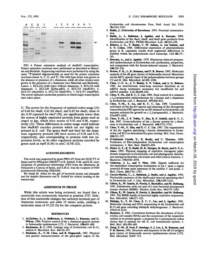
1 2 3 4 5 6 7_ _ _ _ _
-* ~
FIG. 4. Primer extension analysis of rhaBAD transcription.Primer extension reactions were performed as described in Materi-als and Methods. Sequencing reactions were performed by using thesame 32P-labeled oligonucleotide as used for the primer extensionreactions (lanes A, C, G, and T). The wild-type strain was grown inthe absence or presence of L-rhamnose, while all other strains weregrown in the presence of L-rhamnose (see Materials and Methods).Lanes: 1, ECL116 (wild type) without rhamnose; 2, ECL116 withrhamnose; 3, ECL339 [A(rha-pfk)]; 4, ECL714 (rhaB101); 5,ECL715 (rHaA502); 6, ECL716 (rhaD701); 7, ECL717 (rhaR702).The arrow indicates nucleotide position at which major transcriptionstart takes place.
U. The scores for the frequency of optimal codon usage (21)of 0.64 for rhaB, 0.61 for rhaA, and 0.65 for rhaD, close tothe 0.59 reported for rhaT (39), are significantly lower thanthe scores of highly expressed proteins from genes such as
ompA or Ipp, which have scores of 0.92 and 0.98, respec-
tively (21). These differences in codon usage could indicatethat rhaBAD encodes proteins which are not highly ex-
pressed in E. coli. The genes rhaR and rhaS for the rham-nose regulatory proteins (40) have scores of 0.56 and 0.55,respectively, also corresponding to proteins with low ex-
pression levels, as do other regulatory proteins encoded bygenes such as t-pR (0.56) or araC (0.54) (21).
ACKNOWLEDGMENTS
This work was supported by grant PB91-437 from the DGICYT ofSpain and by NIH grant GM18277 to R. Schleif. P.M. and E.H. were
recipients of predoctoral fellowships (FPI) from the Ministerio deEducaci6n y Ciencia of Spain, and S.M.E. was the recipient of NIHpostdoctoral fellowship GM14364.We thank M. Aldea for the gift of bacterial strains and plasmids
and for helpful discussion and R. Schleif for critical reading of themanuscript.
ADDENDUM IN PROOF
While this article was being reviewed, we found that a
nucleotide was erroneously inserted at position 2747. Dele-tion of this nucleotide changes the carboxyl-terminal part ofrhamnose isomerase and adds 19 amino acids, yielding a
molecular mass of 47,231 Da for the complete protein.
REFERENCES
1. Al-Zarban, S., L. Hefferman, J. Nishitani, L. Ransone, and G.Wilcox. 1984. Positive control of the L-rhamnose genetic systemin Salmonella typhimuinum LT2. J. Bacteriol. 158:603-608.
2. Bachmann, B. J. 1990. Linkage map of Escherichia coli K-12,edition 8. Microbiol. Rev. 54:130-197.
3. Backman, K., Y.-M. Chen, and B. Magasanik. 1981. Physicaland genetic characterization of the glnA-glnG region of the
Escherichia coli chromosome. Proc. Natl. Acad. Sci. USA78:3743-3747.
4. Badia, J. (University of Barcelona). 1991. Personal communica-tion.
5. Badia, J., L. Baldoma, J. Aguilar, and A. Boronat. 1989.Identification of the rhaA, rhaB, and rhaD gene products fromEscherichia coli K12. FEMS Microbiol. Lett. 65:253-258.
6. Belasco, J. G., T. Beatty, C. W. Adams, A. von Gabain, andS. N. Cohen. 1985. Differential expression of photosynthesisgenes in R capsulata results from segmental differences instability within the polycistronic rxcA transcript. Cell 40:171-181.
7. Boronat, A., and J. Aguilar. 1979. Rhamnose-induced propane-diol oxidoreductase in Escherichia coli: purification, properties,and comparison with the fucose-induced enzyme. J. Bacteriol.140:320-326.
8. Brown, P. K., L. K. Romana, and P. R. Reeves. 1992. Molecularanalysis of the rib gene cluster of Sabnonella serovar Muenchen(strain M67): genetic basis of the polymorphism between groupsC2 and B. Mol. Microbiol. 6:1385-1394.
9. Chen, C.-Y. A., J. T. Beatty, S. N. Cohen, and J. G. Belasco.1988. An intercistronic stem-loop structure functions as anmRNA decay terminator necessary but insufficient for pufmRNA stability. Cell 52:609-619.
10. Chen, Y.-M., and E. C. C. Lin. 1984. Dual control of a commonL-1,2-propanediol oxidoreductase by L-fucose and L-rhamnosein Escherichia coli. J. Bacteriol. 157:828-832.
11. Chen, Y.-M., Z. Lu, and E. C. C. Lin. 1989. Constitutiveactivation of thefiucAO operon and silencing of the divergentlytranscribedftucPIKoperon by an IS5 element in Escherichia colimutants selected for growth on L-1,2-propanediol. J. Bacteriol.171:6097-6105.
12. Chen, Y.-M., J. F. Tobin, Y. Zhu, R. F. Schleif, and E. C. C.Lin. 1987. Cross-induction of the L-fucose system by L-rham-nose in Escherichia coli. J. Bacteriol. 169:3712-3719.
13. Chen, Y.-M., Y. Zhu, and E. C. C. [An. 1987. The organizationof the fuc regulon specifying L-fucose dissimilation in Esche-richia coli K12 as determined by gene cloning. Mol. Gen. Genet.210:331-337.
14. d'Aubenton Carafa, Y., E. Brody, and C. Thermes. 1990.Prediction of Rho-independent Escherichia coli transcriptionterminators. J. Mol. Biol. 216:835-858.
15. Dimri, G. P., K. E. Rudd, M. K. Morgan, H. Bayat, and G. F.-L.Ames. 1992. Physical mapping of repetitive extragenic palin-dromic sequences in Escherichia coli and phylogenetic distribu-tion among Eschenchia coli strains and other enteric bacteria. J.Bacteriol. 174:4583-4593.
16. Galloway, J. L., and T. Platt. 1988. Signals sufficient forrho-dependent transcription termination at tip t' span a regioncentered 60 base pairs upstream of the earliest 3' end point. J.Biol. Chem. 263:1761-1767.
17. Garcia-Martin, C., L. Baldomfi, J. Badia, and J. Aguilar. 1992.Nucleotide sequence of the rhaR-sod4 interval specifying rhaTin Escherichia coli. J. Gen. Microbiol. 138:1109-1116.
18. Gilson, E., W. Saurin, D. Perrin; S. Bachellier, and M. Hofnung.1991. Palindromic units are part of a new bacterial interspersedmosaic element (BIME). Nucleic Acids Res. 19:1375-1383.
19. Gilson, E., W. Saurin, D. Perrin, S. Bachellier, and M. Hofiung.1991. The BIME family of bacterial highly repetitive sequences.
Res. Microbiol. 142:217-222.20. Hidalgo, E., Y. M. Chen, E. C. C. [An, and J. Aguilar. 1991.
Molecular cloning and DNA sequencing of the Eschenichia coliK-12 ald gene encoding aldehyde dehydrogenase. J. Bacteriol.173:6118-6123.
21. Ikemura, T. 1981. Correlation between the abundance of Esch-erichia coli transfer RNAs and the occurrence of the respectivecodons in its protein genes: a proposal for a synonymous codonchoice that is optimal for the E. coli translational system. J.Mol. Biol. 151:389409.
22. Jiang, X.-M., B. Neal, F. Santiago, S. J. Lee, L. K. Romana, andP. R. Reeves. 1991. Structure and sequence of the rvb (O antigen)gene cluster of Salmwnella serovar typhimurium (strain LT2).Mol. Microbiol. 5:695-713.
A C G T
..: o
VOL. 175, 1993
on March 28, 2019 by guest
http://jb.asm.org/
Dow
nloaded from
5594 MORALEJO ET AL.
23. Lin, E. C. C., and T. T. Wu. 1984. Functional divergence of theL-fucose system in mutants of Escherichia coli, p. 135-163. InR. P. Mortlock (ed.), Microorganisms as model systems forstudying evolution. Plenum, New York.
24. Lowry, 0. H., N. J. Rosebrough, A. L. Farr, and R. J. Randall.1951. Protein measurement with the Folin phenol reagent. J.Biol. Chem. 193:265-275.
25. Lu, Z., and E. C. C. Lin. 1989. The nucleotide sequence ofEscherichia coli genes for L-fucose dissimilation. Nucleic AcidsRes. 17:4883-4884.
26. Lupski, J. R., and G. M. Weinstock. 1992. Short, interspersedrepetitive DNA sequences in prokaryotic genomes. J. Bacteriol.174:4525-4529.
27. McCormick, J. R., J. M. Zengel, and L. Lindahl. 1991. Inter-mediates in the degradation of mRNA from the lactose operonof Escherichia coli. Nucleic Acids Res. 19:2767-2776.
28. Miller, J. H. 1972. Experiments in molecular genetics. ColdSpring Harbor Laboratory Press, Cold Spring Harbor, N.Y.
29. Morgan, W. D., D. G. Bear, B. L. Litchman, and P. H. vonHippel. 1985. A sequence and secondary structure requirementsfor rho-dependent transcription termination. Nucleic Acids Res.13:3739-3754.
30. Newbury, S. F., N. H. Smith, and C. F. Higgins. 1987. Differ-ential mRNA stability controls relative gene expression within apolycistronic operon. Cell 51:1131-1143.
31. Nishitani, J., and G. Wilcox. 1991. Cloning and characterizationof the L-rhamnose regulon in Salmonella typhimurium LT2.Gene 105:37-42.
32. Power, J. 1967. The L-rhamnose genetic system in Escherichiacoli K12. Genetics 55:557-568.
33. Reeder, T., and R. Schleif. 1993. AraC protein can activatetranscription from only one position and when pointed in onlyone direction. J. Mol. Biol. 231:205-218.
34. Sambrook, J., E. F. Fritsch, and T. Maniatis. 1989. Molecularcloning: a laboratory manual. Cold Spring Harbor Laboratory,Cold Spring Harbor, N.Y.
35. Sander, C., and R. Schneider. 1991. Database of homology-derived protein structures and the structural meaning of se-quence alignment. Proteins 9:56-68.
36. Sanger, F., S. Nicklen, and A. R. Coulson. 1977. DNA sequenc-ing with chain-terminating inhibitors. Proc. Natl. Acad. Sci.USA 174:5463-5467.
37. Simons, R. W., F. Houman, and N. Kleckner. 1987. Improvedsingle and multicopy lac-based cloning vectors for protein andoperon fusions. Gene 53:85-96.
38. Stern, M. J., G. F.-L. Ames, N. H. Smith, E. C. Robinson, andC. F. Higgins. 1984. Repetitive extragenic palindromic (REP)sequences: a major component of the bacterial genome. Cell37:1015-1026.
39. Tate, C. G., J. A. R. Muiry, and P. J. F. Henderson. 1992.Mapping, cloning, expression, and sequencing of the rhaT gene,which encodes a novel L-rhamnose-H' transport protein inSalmonella typhimurium and Escherichia coli. J. Biol. Chem.267:6923-6932.
40. Tobin, J. F., and R. F. Schleif. 1987. Positive regulation of theEscherichia coli L-rhamnose operon is mediated by the productsof tandemly repeated regulatory genes. J. Mol. Biol. 196:789-799.
41. Tobin, J. F., and R. F. Schleif. 1990. Purification and propertiesof rhaR, the positive regulator of the L-rhamnose operons ofEscherichia coli. J. Mol. Biol. 211:75-89.
42. Ziemke, P., and J. E. G. McCarthy. 1992. The control ofmRNAstability in Escherichia coli: manipulation of the degradationpathway of the polycistronic atp mRNA. Biochim. Biophys.Acta 1130:297-306.
J. BACTERIOL.
on March 28, 2019 by guest
http://jb.asm.org/
Dow
nloaded from