Embed Size (px)
Citation preview

REVIEW
Serelaxin for the treatment of acute heart failure:a review with a focus on end-organ protectionJavier Dıez1* and Luis M. Ruilope2
1Program of Cardiovascular Diseases, Centre for Applied Medical Research and Department of Cardiology and Cardiac Surgery, University of Navarra Clinic, University of Navarra,Av. Pıo XII 55, Pamplona 31008, Spain; and 2Research Institute, Hypertension Unit, Hospital 12 de Octubre and Department of Public Health and Preventive Medicine, UniversityAutonoma, Madrid, Spain
Received 26 August 2015; revised 15 October 2015; accepted 26 October 2015; online publish-ahead-of-print 26 November 2015
Acute heart failure (AHF) is a complex clinical syndrome characterized by fluid overload and haemodynamic abnormalities (short-term clinicalconsequences) and the development of end-organ damage (long-term consequences). Current therapies for the treatment of AHF, such asloop diuretics and vasodilators, help to relieve haemodynamic imbalance and congestion, but have not been shown to prevent (and may evencontribute to) end-organ damage, or to provide long-term clinical benefit. Serelaxin is the recombinant form of human relaxin-2, a naturallyoccurring hormone involved in mediating haemodynamic changes during pregnancy. Preclinical and clinical studies have investigated the effectsmediated by serelaxin and the suitability of this agent for the treatment of patients with AHF. Data suggest that serelaxin acts via multiple path-ways to improve haemodynamics at the vascular, cardiac, and renal level and provide effective congestion relief. In addition, this novel agent mayprotect the heart, kidneys, and liver from damage by inhibiting inflammation, oxidative stress, cell death, and tissue fibrosis, and stimulatingangiogenesis. Serelaxin may therefore improve both short- and long-term outcomes in patients with AHF. In this review, we examine the uniquemechanisms underlying the potential benefits of serelaxin for the treatment of AHF, in particular, those involved in mediating end-organprotection.- - - - - - - - - - - - - - - - - - - - - - - - - - - - - - - - - - - - - - - - - - - - - - - - - - - - - - - - - - - - - - - - - - - - - - - - - - - - - - - - - - - - - - - - - - - - - - - - - - - - - - - - - - - - - - - - - - - - - - - - - - - - - - - - - - - - - - - - - - - - - - - - - - - - - - - - - - -Keywords Serelaxin † Acute heart failure † Congestion relief † Organ protection † Long-term outcomes
IntroductionHeart failure (HF) is a chronic condition, punctuated by acute epi-sodes, which affects as many as one in five people aged 70–80years.1,2 In acute heart failure (AHF), rapid worsening of the signsand symptoms of HF results in the requirement for urgent therapyand, frequently, hospitalization.3 The frequency of AHF episodes in-creases with disease progression, resulting in high rates of hospital-ization and an increased risk of mortality.3 As such, AHF places asignificant burden on both patients and healthcare systems.4
Pathophysiologically, it is known that AHF involves both haemo-dynamic abnormalities and end-organ damage (Figure 1).5 – 12
Haemodynamic abnormalities result in early clinical features ofcongestion,2,13 – 15 whereas end-organ damage may contribute tolong-term morbidity and mortality.16
Current therapies for AHF include loop diuretics and vasodila-tors, agents which stimulate vasodilation and diuresis to relievehaemodynamic abnormalities.4,10,17 – 19 However, none of theseagents have been shown to prevent end-organ damage, and their
use may be associated with detrimental effects on numerous organs,thereby contributing to long-term morbidity and mortality.20 – 22
As a result, new therapies for the treatment of AHF should relievecongestion to improve short-term clinical consequences and pro-vide organ protection to positively impact the long-term clinicalconsequences of AHF.
Human relaxin-2 is the major form of the hormone relaxin, whichhas vital roles during pregnancy.23,24 Relaxin-2 binds primarily torelaxin family peptide receptor 1 (RXFP1), located in the heart, kid-neys, and vasculature, to activate numerous cellular pathways.16,25–27
Serelaxin has been manufactured as the recombinant form of humanrelaxin-2 and is currently under investigation for the treatment ofAHF.27,28
In this review, we briefly describe the unique mechanisms under-lying the ability of serelaxin to relieve congestion and, therefore,mediate short-term beneficial effects in patients with AHF. Wealso examine, in detail, the novel mechanisms by which serelaxin,unlike current treatments, may limit end-organ damage and thus,provide long-term treatment benefit in patients with AHF.
* Corresponding author. Tel: +34 948 194700, Fax: +34 948 194716, Email: [email protected]
& The Author 2015. Published by Oxford University Press on behalf of the European Society of Cardiology.This is an Open Access article distributed under the terms of the Creative Commons Attribution Non-Commercial License (http://creativecommons.org/licenses/by-nc/4.0/), whichpermits non-commercial re-use, distribution, and reproduction in any medium, provided the original work is properly cited. For commercial re-use, please [email protected].
European Heart Journal – Cardiovascular Pharmacotherapy (2016) 2, 119–130doi:10.1093/ehjcvp/pvv046

Serelaxin for the treatmentof acute heart failure: key clinicaldataThe safety and efficacy of serelaxin for the treatment of patientswith AHF has been determined in the preliminary RELAXin in AcuteHeart Failure (pre-RELAX-AHF) and RELAXin in Acute Heart Fail-ure (RELAX-AHF) clinical trials. In the phase IIb pre-RELAX-AHFtrial, serelaxin (30 mg/kg/day 48-h infusion) resulted in a positiveeffect on dyspnoea compared with placebo.29 In the phase IIIRELAX-AHF trial, serelaxin (30 mg/kg/day 48-h infusion), whencompared with placebo, significantly improved the primary efficacyendpoint of dyspnoea relief by the visual analogue scale area underthe curve to Day 5, with numerical improvement observed in theprimary endpoint of dyspnoea as assessed by the Likert scale at 6,12, and 24 h.30 Serelaxin treatment improved signs and symptomsof congestion and length of hospital stay compared with placeboin the RELAX-AHF study, although, no significant improvement inthe two secondary endpoints of days alive and out of hospital, andcardiovascular (CV) death or rehospitalization for HF or renalfailure through Day 60 was observed.30 In both studies, serelaxindemonstrated favourable effects on longer-term clinical outcomes,such as CV and all-cause mortality through Day 180 compared withplacebo (Figure 2).29–31 In the RELAX-AHF study, elevated levels oftroponin T, cystatin C, aspartate aminotransferase (AST), alanineaminotransferase (ALT), and N-terminal pro-B-type natriureticpeptide (NT-proBNP) were associated with an increased risk ofall-cause mortality through Day 180 (Figure 3).31 Serelaxin treat-ment, when compared with placebo, was associated with lowerlevels of these biomarkers, indicating that serelaxin may protectorgans from further damage following AHF hospitalization.31
Overall, serelaxin had a favourable safety and tolerability profilecompared with placebo.29,30
Although promising, pre-RELAX-AHF and RELAX-AHF studieswere not powered to detect changes in mortality, thus adequatelydesigned follow-up studies are needed. A second phase IIItrial, RELAX-AHF-2, is ongoing and will further investigate the
safety and efficacy of serelaxin for the treatment of patients withAHF, including the mortality benefit observed in previous clinicaltrials.32
Serelaxin and correction ofhaemodynamic imbalanceObservations from preclinical and clinical studies indicate thatserelaxin acts via multiple mechanisms to correct haemodynamic
Figure 1 The ‘continuum’ of pathophysiological changes associated with acute heart failure that may lead to both short- and long-term effectson the heart and other end organs.5 –12
Figure 2 Risk for all-cause mortality through Day 180 inPre-RELAX-AHF and RELAX-AHF.31 AHF, acute heart failure;RELAX-AHF, RELAXin in Acute Heart Failure; Pre-RELAX-AHF,preliminary RELAXin in Acute Heart Failure. Reproduced underthe terms of the Elsevier user license (http://www.elsevier.com/about/open-access/open-access-policies/oa-license-policy/elsevier-user-license) for Metra et al.31
J. Dıez and L.M. Ruilope120

Figure 3 All-cause mortality through Day 180 in RELAX-AHF by markers of organ damage/dysfunction: troponin T (A); cystatin C (B); AST (C);ALT (D), and NT-proBNP (E).31 ALT, alanine aminotransferase; AST, aspartate aminotransferase; CI, confidence interval; HR, hazard ratio;NT-proBNP, N-terminal pro-B-type natriuretic peptide. Reproduced under the terms of the Elsevier user license (http://www.elsevier.com/about/open-access/open-access-policies/oa-license-policy/elsevier-user-license) for Metra et al.31
Serelaxin for the treatment of acute heart failure 121

imbalance and relieve congestion, as described in Table 1.30,33 – 52
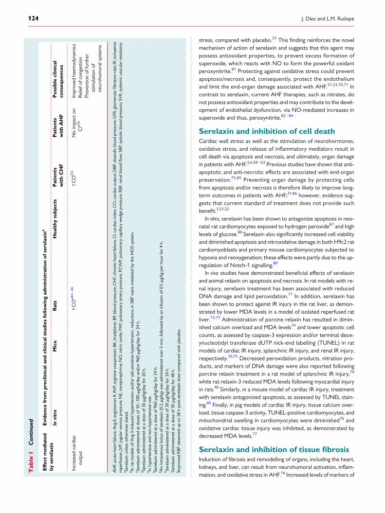
For instance, serelaxin is thought to stimulate vasorelaxatorysystems and counteract vasoconstrictor systems, to mediate bothrapid and sustained vasorelaxation53 (Figure 4),28,43,54 and thus,improve haemodynamics and alleviate congestion. Evidencesuggests that serelaxin also increases arterial compliance40,42 anddecreases systemic vascular resistance,35,36,44 – 46 which couldincrease capacitance to prevent fluid redistribution to the lungsand improve haemodynamic abnormalities, aiding decongestion inAHF.8 Interestingly, in contrast to vasodilators such as nitroglycerin,which primarily act via direct venodilation,55 the vasorelaxatoryaction of serelaxin is thought to predominantly affect arteries.45
In addition to inducing vasorelaxation, serelaxin treatment hasbeen shown to reduce cardiac pressures and to preserve orimprove cardiac and renal function,30,33 – 36,41,44 – 52 which is likelyto help restore haemodynamics, relieve congestion (via mechanismswhich may include the prevention of fluid redistribution), andprevent further stimulation of neurohumoral systems in AHF.2,56
In addition, the renal effects of serelaxin may be associated withlong-term renal protection, which warrants further investigation.
Serelaxin treatment and thelimitation of end-organ damage
Serelaxin interferes with the mechanismsunderlying the development of end-organdamageIn patients with AHF, haemodynamic alterations stimulate a numberof systemic mechanisms, including the adrenergic system, vasoactivehormones, inflammation, and oxidative stress which, in turn, alterthe local mechanisms controlling cell death, tissue repair, and vesselfunction, contributing to the development of cardiac, renal, hepatic,vascular, and other organ damage.2,6,57 – 64 The available evidencesuggests that serelaxin may interfere with these systemic and localmechanisms to limit end-organ damage.
Serelaxin and inhibition of inflammationDamage to organs including the heart, kidneys, and liver occurs earlyin AHF and has long-term consequences.16,65 Inflammatory activationcan contribute to organ injury, in addition to vascular dysfunction andfluid overload.8,16 For instance, in patients with newly diagnosed HF,levels of tumour necrosis factor alpha (TNF-a), interleukin (IL)-6, andCD14 were elevated on the third day of initial hospitalization and as-sociated with impaired function of the left atrium and more advancedleft ventricular (LV) systolic and diastolic dysfunction.66
Changes in inflammatory pathways have been determined in anumber of studies following the administration of serelaxin. In hu-man umbilical vein endothelial cells incubated with serelaxin,TNF-a-induced upregulation of vascular cell adhesion molecule 1(VCAM-1) and platelet endothelial cell adhesion molecule was di-minished, along with C-C chemokine receptor type 2 and monocytechemotactic protein 1 levels, and monocyte adhesion to the cells.67
In addition, serelaxin inhibited basophil function, via nitric oxide syn-thase activation, to reduce histamine release and prevent the rise inintracellular calcium that stimulates granule release.68,69
In rats subjected to cardiac, renal, hepatic, or splanchnic ischae-mia–reperfusion (IR) injury, treatment with serelaxin or porcine re-laxin diminished myeloperoxidase activity, a marker of inflammatoryleukocyte infiltration.70 – 74 Serelaxin treatment decreased expres-sion of inflammatory mediators and adhesion molecules includingintercellular adhesion molecule-1 (ICAM-1), IL-1b, IL-18, andTNF-a in rats subjected to renal IR injury,71 while porcine relaxindownregulated expression of adhesion molecules P-selectin,E-selectin, VCAM, and ICAM-1 in a rat model of splanchnic IR in-jury,70 as well as TNF-a expression in a rat model of renal IR in-jury.75 In addition, porcine relaxin treatment was associated with areduction in the number of neutrophils and inhibition of mast cellgranule release in a rat model of cardiac IR injury.74 Similarly, reduc-tions in myeloperoxidase levels and cardiac mast cell degranulationwere evident following the administration of serelaxin in a pig modelof cardiac IR injury.76,77
Inhibiting the inflammatory response in patients with AHF maydecrease fluid overload to relieve congestion and positively impactvascular, myocardial, renal, and hepatic injury and dysfunc-tion8,71,73,75,77 and consequently, improve long-term outcomes.The anti-inflammatory actions of serelaxin distinguish this agentfrom current AHF therapies, such as nitrates, that have not beenshown to improve long-term outcomes in patients with AHF21
and are therefore unlikely to inhibit inflammation.
Serelaxin and reduction of oxidative stressIncreased oxidative stress results from the dominance of reactiveoxygen species (ROS) such as superoxide over endogenous antioxi-dant defence mechanisms.78 In patients with AHF, oxidative stresscan result in myocardial, renal, and hepatic injury and remodelling.16
Neurohormones contribute to the activation of ROS in AHF, whilemitochondrial calcium overload and dysfunction (via leaky type 2ryanodine receptors) may lead to increased release of ROS inHF78,79 and reperfusion-induced inflammation may contribute tooxidative cardiac tissue injury.24
In vitro studies, animal models and clinical studies have investigatedthe effects of animal relaxin and serelaxin on oxidative stress. In vitro,porcine relaxin was found to reduce the production of superoxideanions from human neutrophils.68 In rats with renal or splanchnic IRinjury, serelaxin treatment was associated with increased levels ofthe antioxidant enzymes manganese and copper–zinc superoxidedismutase71 and diminished consumption of superoxide dismutase,lipid peroxidation, and markers of deoxyribonucleic acid (DNA)damage including 8-hydroxy-2′-deoxyguanosine and poly-ADP-ribosylated DNA.70 In addition, serelaxin decreased hydrogenperoxide and thiobarbituric acid-reactive substance (TBARs) excre-tion and consequently, oxidative stress, in rats with angiotensinII-induced hypertension.33 In the same experimental model, serelax-in treatment reduced nicotinamide adenine dinucleotide phosphate(NADPH) oxidase activity (i.e. superoxide anion generation) andexcretion of TBARs and 8-isoprostane (markers of oxidative stress),and restored nitric oxide (NO) oxidation product excretion.80 Fi-nally, serelaxin was found to decrease levels of malondialdehyde(MDA), a marker of oxygen-free radical-mediated cell damage, ina porcine model of cardiac IR injury.76
In patients with AHF, serelaxin treatment (30 mg/kg/day, 48-h in-fusion) significantly reduced levels of uric acid, a marker of oxidative
J. Dıez and L.M. Ruilope122

. . . . . . . . . . . . . . . . . . . . . . . . . . . . . . . . . . . . . . . . . . . . . . . . . . . . . . . . . . . . . . . . . . . . . . . . . . . . . . . . . . . . . . . . . . . . . . . . . . . . . . . . . . . . . . . . . . . . . . . . . . . . . . . . . . . . . . . . . . . . . . . . . . . . . . . . . . . . . . . . . . . . . . . . . . . . . . . . . . . . . . . . . . . . . . . . . . . . . . . . . . . . . . . . . . . .
. . . . . . . . . . . . . . . . . . . . . . . . . . . . . . . . . . . . . . . . . . . . . . . . . . . . . . . . . . . . . . . . . . . . . . . . . . . . . . . . . . . . . . . . . . . . . . . . . . . . . . . . . . . . . . . . . . . . . . . . . . . . . . . . . . . . . . . . . . . . . . . . . . . . . . . . . . . . . . . . . . . . . . . . . . . . . . . . . . . . . . . . . . . . . . . . . . . . . . . . . . . . . . . . . . . . . . . . . . . . . . . . . . . . . . . . . . . . .
Table 1 Effects mediated by serelaxin that may alleviate haemodynamic imbalance and relieve congestion in patients with AHF30,33 – 52
Effect mediatedby serelaxin
Evidence from preclinical and clinical studies following administration of serelaxina
In vitro Mice Rats Healthy subjects Patientswith CHF
Patientswith AHF
Possible clinicalconsequences
Reduction ofcardiacpressures
�SBPb33,34 (includingporcine relaxin)
�DBPc35
�PCWPc35
�SBPc35
�PAPc35
�DBPd36
�PCWPd36
�SBPd36
�PAPd36
�JVPd30
Improved haemodynamicsRelief of congestionPrevention of further
stimulation ofneurohumoral systems
Stimulation ofvasorelaxation
Blunted responses of ratmesenteric arteries tovasoconstriction induced byAVP and NE37 (rat relaxin)
Vasorelaxation of small humanresistance arteries38
�Coronary flow/�NOgeneration in isolated guineapig hearts subject to IR injury39
(porcine relaxin)
�Arterialcompliance40
Blunted response tovasoconstriction and �BPinduced by Ang II33,41
(including porcine relaxin)�Wall stiffness42
�Arterial compliance42
�Rapid and sustainedBK-mediated vasorelaxationof mesenteric arteries43
Improved haemodynamicsRelief of congestionPossible prevention of
fluid redistribution
Reduction of SVR �SVRe44–46 �SVRf35 �SVRd36 VasorelaxationImproved haemodynamicsRelief of congestionPossible prevention of
fluid redistribution
Preservation ofdiuresis andnatriuresis
�Urinary excretion of sodium47
�Salt sensitivityb34
(porcine relaxin)�Urinary flow rate47
�Renal clearance,fractional excretionand urinaryexcretion ofsodiumg48
No effect on urinaryflow rateg48
No effect on urinaryexcretion ofsodium or urinaryflow rateh49
Neutral effecton diureticresponsei50
Preservation of renalfunction
Improved haemodynamicsPossible prevention of
fluid redistribution
Increased RBF andpreservation ofGFR
�GFR41,51,52
�RBF41,47,51,52�RBFg48
No effect on GFRg48�RBFh,j49
No effect on GFRh49Preservation of renal
functionPossible long-term renal
protection
Continued
Serelaxinfor
thetreatm
entofacute
heartfailure
123
stress, compared with placebo.31 This finding reinforces the novelmechanism of action of serelaxin and suggests that this agent maypossess antioxidant properties, to prevent excess formation ofsuperoxide, which reacts with NO to form the powerful oxidantperoxynitrite.81 Protecting against oxidative stress could preventapoptosis/necrosis and, consequently, protect the endotheliumand limit the end-organ damage associated with AHF.31,33,70,71 Incontrast to serelaxin, current AHF therapies, such as nitrates, donot possess antioxidant properties and may contribute to the devel-opment of endothelial dysfunction, via NO-mediated increases insuperoxide and thus, peroxynitrite.82 –84
Serelaxin and inhibition of cell deathCardiac wall stress as well as the stimulation of neurohormones,oxidative stress, and release of inflammatory mediators result incell death via apoptosis and necrosis, and ultimately, organ damagein patients with AHF.2,6,58–63 Previous studies have shown that anti-apoptotic and anti-necrotic effects are associated with end-organpreservation.75,85 Preventing organ damage by protecting cellsfrom apoptosis and/or necrosis is therefore likely to improve long-term outcomes in patients with AHF;31,86 however, evidence sug-gests that current standard of treatment does not provide suchbenefit.3,21,22
In vitro, serelaxin has been shown to antagonize apoptosis in neo-natal rat cardiomyocytes exposed to hydrogen peroxide87 and highlevels of glucose.88 Serelaxin also significantly increased cell viabilityand diminished apoptosis and nitroxidative damage in both H9c2 ratcardiomyoblasts and primary mouse cardiomyocytes subjected tohypoxia and reoxygenation; these effects were partly due to the up-regulation of Notch-1 signalling.89
In vivo studies have demonstrated beneficial effects of serelaxinand animal relaxin on apoptosis and necrosis. In rat models with re-nal injury, serelaxin treatment has been associated with reducedDNA damage and lipid peroxidation.71 In addition, serelaxin hasbeen shown to protect against IR injury in the rat liver, as demon-strated by lower MDA levels in a model of isolated reperfused ratliver.72,73 Administration of porcine relaxin has resulted in dimin-ished calcium overload and MDA levels74 and lower apoptotic cellcounts, as assessed by caspase-3 expression and/or terminal deox-ynucleotidyl transferase dUTP nick-end labelling (TUNEL) in ratmodels of cardiac IR injury, splanchnic IR injury, and renal IR injury,respectively.70,75 Decreased peroxidation products, nitration pro-ducts, and markers of DNA damage were also reported followingporcine relaxin treatment in a rat model of splanchnic IR injury,70
while rat relaxin-3 reduced MDA levels following myocardial injuryin rats.90 Similarly, in a mouse model of cardiac IR injury, treatmentwith serelaxin antagonized apoptosis, as assessed by TUNEL stain-ing.85 Finally, in pig models of cardiac IR injury, tissue calcium over-load, tissue caspase-3 activity, TUNEL-positive cardiomyocytes, andmitochondrial swelling in cardiomyocytes were diminished76 andoxidative cardiac tissue injury was inhibited, as demonstrated bydecreased MDA levels.77
Serelaxin and inhibition of tissue fibrosisInduction of fibrosis and remodelling of organs, including the heart,kidneys, and liver, can result from neurohumoral activation, inflam-mation, and oxidative stress in AHF.16 Increased levels of markers of
....
....
....
....
....
....
....
....
....
....
....
....
....
....
....
....
....
....
....
....
....
....
....
....
....
....
....
....
....
....
....
....
....
....
....
....
....
....
....
....
....
....
....
....
....
....
....
....
....
....
....
....
....
....
....
....
....
....
....
....
....
....
....
....
....
....
....
....
....
....
....
....
....
....
....
....
....
....
....
....
....
....
....
....
....
....
....
....
....
....
....
....
....
....
....
....
....
....
....
....
....
....
....
....
....
....
....
....
....
....
....
....
.
Tab
le1
Con
tinu
ed
Eff
ect
med
iate
dby
sere
laxi
nE
vide
nce
fro
mpr
eclin
ical
and
clin
ical
stud
ies
follo
win
gad
min
istr
atio
no
fser
elax
ina
Invi
tro
Mic
eR
ats
Hea
lthy
subj
ects
Pat
ient
sw
ith
CH
FP
atie
nts
wit
hA
HF
Po
ssib
lecl
inic
alco
nseq
uenc
es
Incr
ease
dca
rdia
cou
tput
�CO
e44
–46
�CO
f35
No
impa
cton
CId
36
Impr
oved
haem
odyn
amic
sR
elie
fofc
onge
stio
nPr
even
tion
offu
rthe
rst
imul
atio
nof
neur
ohum
oral
syst
ems
AH
F,ac
ute
hear
tfai
lure
;Ang
II,an
giot
ensin
II;A
VP,
argi
nine
vaso
pres
sin;
BK,b
rady
kini
n;BP
,blo
odpr
essu
re;C
HF,
chro
nic
hear
tfai
lure
,CI,
card
iac
inde
x;C
O,c
ardi
acou
tput
;DBP
,dia
stol
icbl
ood
pres
sure
;GFR
,glo
mer
ular
filtr
atio
nra
te;I
R,i
scha
emia
repe
rfus
ion;
JVP,
jugu
lar
veno
uspr
essu
re;N
E,no
repi
neph
rine
;NO
,nitr
icox
ide;
PAP,
pulm
onar
yar
tery
pres
sure
;PC
WP,
pulm
onar
yca
pilla
ryw
edge
pres
sure
;RBF
,ren
albl
ood
flow
;SBP
,sys
tolic
bloo
dpr
essu
re;S
VR
,sys
tem
icva
scul
arre
sist
ance
.a Se
rela
xin
unle
ssot
herw
isest
ated
.b In
rat
mod
els
ofA
ngII-
indu
ced
hype
rten
sion
and/
orsa
lt-se
nsiti
vehy
pert
ensio
n,re
duct
ions
inSB
Pw
ere
med
iate
dby
the
NO
Ssy
stem
.c Se
rela
xin
adm
inist
ered
atdo
ses
of10
–10
0m
g/kg
/day
and/
or96
0m
g/kg
/day
for
24h.
d Sere
laxi
nad
min
ister
edat
ado
seof
30m
g/kg
/day
for
20h.
e Inhy
pert
ensi
vean
dno
n-hy
pert
ensiv
era
ts.
f Sere
laxi
nad
min
ister
edat
ado
seof
960m
g/kg
/day
for
24h.
g An
intr
aven
ous
bolu
sof
sere
laxi
n(0
.2m
g/kg
)w
asad
min
ister
edov
er5
min
,fol
low
edby
anin
fusio
nof
0.5m
g/kg
per
hour
for
4h.
h Sere
laxi
nad
min
ister
edat
ado
seof
30m
g/kg
/day
for
24h.
i Sere
laxi
nad
min
ister
edat
ado
seof
30m
g/kg
/day
for
48h.
j Impr
oved
RBF
obse
rved
upto
28h
post
-ser
elax
indo
seco
mpa
red
with
plac
ebo.
J. Dıez and L.M. Ruilope124
extracellular matrix turnover, including matrix metalloproteinase(MMP)-2, tissue inhibitor of MMP (TIMP)-1, and procollagen typeIII N-terminal peptides, have been observed during the first 24 hof hospital admission for HF decompensation.6 In addition, failinghearts, when compared with non-failing hearts, have demonstrateddysregulation of microRNA expression, which is thought to contrib-ute to myocardial remodelling in HF.91
In vitro, serelaxin inhibited transforming growth factor beta(TGF-b) and/or TIMPs in human hepatic stellate cells and humandermal fibroblasts,92,93 and increased expression of MMPs, includingMMP-1, -2, -9, and -13, via mechanisms including the NO pathway, inhuman dermal fibroblasts92,94 and rat renal myofibroblasts.94 Pro-duction of collagen was found to decrease in rat atrial and ventricu-lar fibroblasts95,96 and human scleroderma fibroblasts97 followingadministration of serelaxin. In addition, serelaxin treatment downre-gulated activation of human renal fibroblasts,98 rat renal fibroblastfunction,99 and differentiation of rat renal fibroblasts to myofibro-blasts,100 to inhibit renal fibrogenesis.
The potential anti-fibrotic and anti-hypertrophic actions of ser-elaxin have also been assessed in vivo. Serelaxin treatment reducedventricular collagen accumulation in mice,95 cardiac fibrosis inmouse models of myocardial infarction-induced IR injury,85 and
isoprenaline-induced cardiac injury when compared with theangiotensin-converting enzyme inhibitor enalapril.101 In the latterstudy, combined administration of enalapril and serelaxin dimin-ished cardiac fibrosis two-fold compared with enalapril alone,and the inhibitory effects of serelaxin were mediated by TGF-bdownregulation.101 In ageing rats and in rat models of hypertensionand diabetic cardiomyopathy, administration of serelaxin de-creased LV and kidney collagen content,52,102,103 fibroblast differ-entiation in the left ventricle,103 and atrial remodelling,104 as well ascardiac hypertrophy via inhibition of extracellular signal-regulatedkinase.105 In addition, porcine relaxin diminished renal fibrosis in arat model of salt-sensitive hypertension34 and rat relaxin-3 amelio-rated cardiac fibrosis in rats with isoproterenol-induced myocar-dial injury.90
Inhibiting fibrosis and hypertrophy is likely to be beneficial inpatients with AHF, and may be associated with reduced fibrosis inorgans, including the heart, vessels, kidneys, and liver, as well asthe limitation of organ damage and improvement of long-term prog-nosis.16,34,103 The anti-fibrotic effects of serelaxin may differentiatethis agent from current treatments for AHF, such as nitrates, that donot protect end organs from further damage,2,21,22 and are there-fore unlikely to inhibit tissue fibrosis.
Figure 4 Time-dependent effects of intravenously administered serelaxin on vasoactive systems that result in vasorelaxation.28,43,54 A, timeafter serelaxin administration, when the hormone is detectable in the blood ranges from minutes to hours; B, time after serelaxin administration,when the hormone is not detected in the blood ranges from 1 to several days; Ang II, angiotensin II; AVP, arginine vasopressin; BK, bradykinin;COX2, cyclo-oxygenase 2; EDHF, endothelium-derived hyperpolarizing factor; eNOS, endothelial nitric oxide synthase; ET, endothelin; ET-BR,endothelin receptor type B; MMP, metalloproteinase; NE, norepinephrine; nNOS, neuronal nitric oxide synthase; RXFP1, relaxin/insulin-like familypeptide receptor 1; TGF-b, transforming growth factor b; VEGF, vascular endothelial growth factor.
Serelaxin for the treatment of acute heart failure 125

Serelaxin and stimulation of angiogenesisUsing imaging techniques, significant reductions in perfused smallmicrovessels have been demonstrated in tissues from patientswith AHF106 compared with control subjects.107 In addition, theperipheral tissue oxygen extraction rate (an inverse index of tissuemicrovascular perfusion) is increased in patients with AHF com-pared with those with chronic stable HF.108 Of interest, this param-eter improved with AHF therapy, in parallel with the amelioration ofcongestion and haemodynamic parameters.108 Therefore, altera-tions in microcirculation may play an important role in organ dam-age in AHF.
Angiogenesis can facilitate tissue repair and serelaxin may medi-ate pro-angiogenic effects, unlike current treatments for AHF, as as-sessed in vitro and in animal models. Serelaxin has been reported tostimulate NO production from, and migration of, human endothelialprogenitor cells in vitro, and to increase the number of circulating hu-man endothelial progenitor cells and stimulate vascularization inmice.109 In addition, studies with H2 relaxin and serelaxin have ob-served increased expression of the angiogenic cytokine vascularendothelial growth factor (VEGF) in a cyclic adenosine monopho-sphate (cAMP)-dependent manner,110 stimulation of angiogenesisat ischaemic cardiac sites, and induction of expression of VEGF inrodents and pigs.85,111,112 This induction of angiogenesis could min-imize further organ damage and repair injury, particularly of themyocardium, in patients with AHF.16
Serelaxin and effective protectionof end-organsAs previously mentioned, serelaxin treatment, in contrast to currenttherapies, interferes with the systemic and local mechanisms under-lying the development of organ damage, and thus, may protect endorgans in patients with AHF.3,21,22,75,85
Cardiac protectionEarly cardiomyocyte injury and stress and LV dysfunction resultfrom AHF.95,113,114 Cardiomyocyte injury and loss can be detectedby measuring troponin T levels, which are elevated in HF,60,61 whileincreased levels of NT-proBNP indicate ventricular wall stress.115 Inpatients with AHF, increased levels of troponin T may be detectedupon hospital admission and in the 6–12 h following admission.86
In vitro studies, animal models and clinical studies have investigatedthe cardioprotective properties of serelaxin and porcine relaxin. Invitro, administration of porcine relaxin has been reported to diminishIR injury in isolated reperfused guinea pig hearts, as determined bydecreased calcium overload and MDA production,39 in addition toinfarct size in a rat model of IR injury.74 Serelaxin treatment also re-duced markers of cardiomyocyte damage, including troponin T, cre-atine kinase-MB, and myoglobin, as well as cardiac injury in pigmodels of IR injury.76,77
In patients with AHF, serelaxin (30 mg/kg/day for 20 or 48 h) de-creased levels of troponin T and NT-proBNP.31,36 Similarly,NT-proBNP levels were diminished following serelaxin treatment(10–100 and 960 mg/kg/day for 24 h) in patients with chronic heartfailure (CHF).35 These data imply that the unique mechanism of ac-tion of serelaxin may be associated with the preservation of cardiac
function in patients with AHF. Although further assessment of thishypothesis is needed, this finding contrasts with the effects of nitratetreatment, which is thought to contribute to cardiac injury by redu-cing blood pressure and organ perfusion.2,22
In addition to protecting cardiomyocytes from injury and death,serelaxin has been reported to modulate ionic currents in cardiaccells.104,116 Although the translation of these findings into the clinicrequires further studies, it is interesting to note that recently, in theRELAX-AHF study, serelaxin treatment reduced mortality fromother CV causes and sudden deaths, without impact on HF deaths.117
Renal protectionRenal dysfunction is common in patients with AHF62 and may be ex-acerbated by nitrate treatment, which can cause hypotension andsubsequently, renal hypoperfusion and injury.2 Renal damage anddysfunction is a major predictor of poor outcomes in AHF27 andcan be detected via increased levels of serum creatinine, cystatinC, uric acid, and blood urea nitrogen (BUN), as well as reducedestimated glomerular filtration rate (GFR).4,16,31,118 Elevated levelsof serum creatinine, cystatin C, uric acid, and BUN have beenreported in patients with AHF in the 48 h following hospitaladmission.31,119
Data from preclinical and clinical studies are available concerningthe impact of serelaxin treatment on kidney function and protec-tion. For example, in rats, serelaxin treatment increased GFR and re-nal blood flow, and protected against renal IR injury and glomerulardysfunction,41,47,51,52,71 whereas porcine relaxin decreased levels ofcreatinine and BUN in rats subjected to renal IR injury.75
In healthy subjects, serelaxin increased renal blood flow, but didnot impact GFR,48 an effect also observed following administrationof serelaxin (30 mg/kg/day for 24 h) in patients with CHF whencompared with placebo, suggesting that serelaxin treatment reducesthe increase in filtration fraction to mediate beneficial renal haemo-dynamic effects.49
In patients with AHF, serelaxin (30 mg/kg/day for 48 h) reducedlevels of cystatin C, uric acid, BUN, and serum creatinine,31 and in-creased creatinine clearance (30 mg/kg/day for 20 h).36 Decreasedserum creatinine was also reported after infusion of serelaxin(10–100 and 960 mg/kg/day for 24 h) in patients with CHF.35
Consequently, serelaxin may prevent worsening renal function, aproperty which differentiates this novel agent from vasodilatortreatment in AHF.
Hepatic protectionHepatic injury and cell death can occur during AHF,58,120 withelevated markers of hepatic dysfunction, including AST and ALT,which are also predictors of mortality and worsening HF, reportedwithin 48 h of hospitalization for AHF.31,121 Studies have demon-strated that serelaxin may mediate hepatic protection, asobserved by diminished IR injury in rat liver72,73 and decreased levelsof AST and ALT in patients with AHF following serelaxin treatment(30 mg/kg/day for 48 h).31
Vascular and other organ protectionDamage to the vasculature and other organs may occur in patientswith AHF57,122 and nitrate therapy may increase endothelial dys-function further in these patients via increased oxidative stress.82
J. Dıez and L.M. Ruilope126
Organ preservation and vasoprotective properties may distin-guish serelaxin from classical vasodilators for the treatment ofAHF and improve outcomes in these patients.16,123 For instance,treatment with serelaxin has been associated with improved endo-thelial function in rat aortic endothelial cells57 and decreases in ves-sel size, wall thickening, cross-sectional area, and collagen content inspontaneously hypertensive rats,124 while porcine relaxin has pro-vided endothelial protection in a rat model of splanchnic IR injury.70
Furthermore, studies in the rat brain have shown that serelaxintreatment reduced ischaemic cell damage in brain slices, as well asinfarct size in vivo, determined 4 h following ischaemia.125 – 127 In add-ition, administration of serelaxin has resulted in diminished IR injuryin rat lungs.128,129
Conclusions and perspectivesAHF poses a significant burden to patients and healthcare systems.The precise mechanisms underlying this condition are poorly under-stood, but it is clear that a variety of pathophysiological processesare involved, which result in both haemodynamic abnormalitiesand end-organ damage. Current therapies available for the treat-ment of AHF moderately address the haemodynamic changesassociated with the short-term effects of this condition, to alleviatecongestion. However, no currently approved agent has demonstratedtrue benefit on the long-term outcomes of AHF. As such, there is anunmet medical need in AHF; a need for therapies that address boththe short- and long-term effects of this condition.
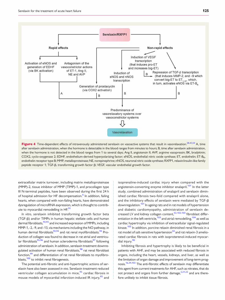
Preclinical and clinical data have highlighted serelaxin as a prom-ising treatment of both the short- and long-term consequences ofAHF. In contrast to classical vasodilators, serelaxin may act at thevascular, cardiac, and renal level to improve haemodynamics and ef-fectively relieve congestion. Moreover, available data suggest thatserelaxin may provide organ protection via inhibition of
inflammation, oxidative stress, cell death, and tissue fibrosis, and in-duction of angiogenesis (Figure 5),24,31,33,71– 74,76,77,80,90 to improvethe long-term prognosis of these patients, as observed in clinicaltrials to date.
Additional clinical data are required to confirm the potential ben-efits of serelaxin for the treatment of AHF. A second phase III study,RELAX-AHF 2, began in September 2013 and will further assess theeffects of serelaxin on CV mortality in patients with AHF.32 Futureexperimental research efforts should aim to establish animal modelsof AHF, in which the mechanisms underlying the efficacy of serelaxinfor the treatment of this condition could be studied. Meanwhile, fur-ther preclinical studies are required to investigate the pharmacoki-netic and pharmacodynamic properties of serelaxin in this patientpopulation.
Authors’ ContributionsJ.D. and L.R. designed, jointly reviewed, and revised the initial draftand subsequent versions of the manuscript, and both agreed on thefinal version submitted for publication.
AcknowledgementsThe authors thank Hannah Birchby and Rebecca Douglas(CircleScience, an Ashfield Company, part of UDG Healthcareplc), for providing writing/editorial assistance, which was fundedby Novartis Pharma AG, Basel, Switzerland.
FundingThe writing/editorial support was funded by Novartis Pharma AG,Basel, Switzerland. The sponsor reviewed the initial draft and subse-quent versions of the manuscript for own data accuracy and for pro-prietary evaluation. Funding to pay the Open Access publication
Figure 5 Serelaxin activates multiple pathways to improve haemodynamics and may protect cells and organs via anti-apoptotic/necrotic, anti-inflammatory, anti-fibrotic, antioxidant, and pro-angiogenic effects.24,31,33,71 –74,76,77,80,90 ET, endothelin; ET-BR, endothelin receptor type B; MDA,malondialdehyde; MMP, metalloproteinase; NADPH, nicotinamide adenine dinucleotide phosphate-oxidase; NO, nitric oxide; NOS, nitric oxidesynthase; TBARs, thiobarbituric acid-reactive substance; TGF-b, transforming growth factor b; TNF-a, tumour necrosis factor a; VEGF, vascularendothelial growth factor. Adapted from Teichman et al.24 Reproduced under the terms of the Creative Commons Attribution Non-commercialLicense for open access.
Serelaxin for the treatment of acute heart failure 127

charges for this article was provided by Novartis Pharma AG, Basel,Switzerland.
Conflict of interest: J.D. has served as an advisor and as a speakerfor Novartis, Merck, Sharp and Dohme, and Abbvie. L.R. has servedas an advisor and as a speaker for Novartis.
References1. Dickstein K, Cohen-Solal A, Filippatos G, McMurray JJ, Ponikowski P,
Poole-Wilson PA, Stromberg A, van Veldhuisen DJ, Atar D, Hoes AW,Keren A, Mebazaa A, Nieminen M, Priori SG, Swedberg K; ESC Committee forPractice Guidelines (CPG). ESC guidelines for the diagnosis and treatmentof acute and chronic heart failure 2008: the Task Force for the Diagnosis andTreatment of Acute and Chronic Heart Failure 2008 of the European Societyof Cardiology. Developed in collaboration with the Heart Failure Associationof the ESC (HFA) and endorsed by the European Society of Intensive CareMedicine (ESICM). Eur Heart J 2008;29:2388–2442.
2. Gheorghiade M, Pang PS. Acute heart failure syndromes. J Am Coll Cardiol 2009;53:557–573.
3. Gheorghiade M, De LL, Fonarow GC, Filippatos G, Metra M, Francis GS. Patho-physiologic targets in the early phase of acute heart failure syndromes. Am J Cardiol2005;96:11G–17G.
4. McMurray JJ, Adamopoulos S, Anker SD, Auricchio A, Bohm M, Dickstein K,Falk V, Filippatos G, Fonseca C, Gomez-Sanchez MA, Jaarsma T, Køber L,Lip GY, Maggioni AP, Parkhomenko A, Pieske BM, Popescu BA, Rønnevik PK,Rutten FH, Schwitter J, Seferovic P, Stepinska J, Trindade PT, Voors AA,Zannad F, Zeiher A; ESC Committee for Practice Guidelines. ESC guidelinesfor the diagnosis and treatment of acute and chronic heart failure 2012:the Task Force for the Diagnosis and Treatment of Acute and Chronic Heart Fail-ure 2012 of the European Society of Cardiology. Developed in collaboration withthe Heart Failure Association (HFA) of the ESC. Eur Heart J 2012;33:1787–1847.
5. Cotter G, Felker GM, Adams KF, Milo-Cotter O, O’Connor CM. The pathophysi-ology of acute heart failure—is it all about fluid accumulation? Am Heart J 2008;155:9–18.
6. Biolo A, Fisch M, Balog J, Chao T, Schulze PC, Ooi H, Siwik D, Colucci WS.Episodes of acute heart failure syndrome are associated with increased levels oftroponin and extracellular matrix markers. Circ Heart Fail 2010;3:44–50.
7. Bott-Flugel L, Weig HJ, Uhlein H, Nabauer M, Laugwitz KL, Seyfarth M. Quantita-tive analysis of apoptotic markers in human end-stage heart failure. Eur J Heart Fail2008;10:129–132.
8. Cotter G, Metra M, Milo-Cotter O, Dittrich HC, Gheorghiade M. Fluid overloadin acute heart failure—re-distribution and other mechanisms beyond fluid accu-mulation. Eur J Heart Fail 2008;10:165–169.
9. Feng Q, Wang S. Cardiomyocytic apoptosis and heart failure. J Geriatr Cardiol2008;5:1–6.
10. Hunt SA, Abraham WT, Chin MH, Feldman AM, Francis GS, Ganiats TG, Jessup M,Konstam MA, Mancini DM, Michl K, Oates JA, Rahko PS, Silver MA,Stevenson LW, Yancy CW; American College of Cardiology Foundation;American Heart Association. 2009 Focused update incorporated into the ACC/AHA 2005 Guidelines for the Diagnosis and Management of Heart Failure inAdults: a report of the American College of Cardiology Foundation/AmericanHeart Association Task Force on Practice Guidelines developed in collaborationwith the International Society for Heart and Lung Transplantation. J Am Coll Cardiol2009;53:e1–e90.
11. Oikonomou E, Tousoulis D, Siasos G, Zaromitidou M, Papavassiliou AG,Stefanadis C. The role of inflammation in heart failure: new therapeutic ap-proaches. Hellenic J Cardiol 2011;52:30–40.
12. Tsutsui H, Kinugawa S, Matsushima S. Oxidative stress and heart failure. Am J Phy-siol Heart Circ Physiol 2011;301:H2181–H2190.
13. Gheorghiade M, Filippatos G, De LL, Burnett J. Congestion in acute heart failuresyndromes: an essential target of evaluation and treatment. Am J Med 2006;119:S3–S10.
14. Goldberg RJ, Spencer FA, Szklo-Coxe M, Tisminetzky M, Yarzebski J, Lessard D,Gore JM, Gaasch W. Symptom presentation in patients hospitalized with acuteheart failure. Clin Cardiol 2010;33:E73–E80.
15. Adams KF Jr, Fonarow GC, Emerman CL, LeJemtel TH, Costanzo MR,Abraham WT, Berkowitz RL, Galvao M, Horton DP; ADHERE Scientific AdvisoryCommittee and Investigators. Characteristics and outcomes of patients hospita-lized for heart failure in the United States: rationale, design, and preliminary ob-servations from the first 100,000 cases in the Acute Decompensated HeartFailure National Registry (ADHERE). Am Heart J 2005;149:209–216.
16. Dıez J. Serelaxin: a novel therapy for acute heart failure with a range of hemo-dynamic and non-hemodynamic actions. Am J Cardiovasc Drugs 2014;14:275–285.
17. Konecke LL. Clinical trial of bumetanide versus furosemide in patients with con-gestive heart failure. J Clin Pharmacol 1981;21:688–690.
18. Bolognese L, Sarasso G, Rognoni G, Makmur J, Fornaro G, Perucca A, Rossi P.Sustained beneficial hemodynamic effects of low transdermal nitroglycerin dosescompared with placebo in patients with congestive heart failure. Clin Cardiol 1988;11:79–85.
19. Stroobandt R, Dodion L, Kesteloot H. Clinical efficacy of torasemide, a new diur-etic agent, in patients with acute heart failure: a double blind comparison with fur-osemide. Arch Int Pharmacodyn Ther 1982;260:151–158.
20. Gheorghiade M, Zannad F, Sopko G, Klein L, Pina IL, Konstam MA, Massie BM,Roland E, Targum S, Collins SP, Filippatos G, Tavazzi L; International WorkingGroup on Acute Heart Failure Syndromes. Acute heart failure syndromes: cur-rent state and framework for future research. Circulation 2005;112:3958–3968.
21. Stough WG, O’Connor CM, Gheorghiade M. Overview of current noninodila-tor therapies for acute heart failure syndromes. Am J Cardiol 2005;96:41G–46G.
22. Metra M, Teerlink JR, Voors AA, Felker GM, Milo-Cotter O, Weatherley B,Dittrich H, Cotter G. Vasodilators in the treatment of acute heart failure: whatwe know, what we don’t. Heart Fail Rev 2009;14:299–307.
23. Sherwood OD. Relaxin’s physiological roles and other diverse actions. Endocr Rev2004;25:205–234.
24. Teichman SL, Unemori E, Teerlink JR, Cotter G, Metra M. Relaxin: review of biol-ogy and potential role in treating heart failure. Curr Heart Fail Rep 2010;7:75–82.
25. Grossman J, Frishman WH. Relaxin: a new approach for the treatment of acutecongestive heart failure. Cardiol Rev 2010;18:305–312.
26. Sarwar M, Samuel CS, Bathgate RA, Stewart DR, Summers RJ. Serelaxin-mediatedsignal transduction in human vascular cells: bell-shaped concentration-responsecurves reflect differential coupling to G proteins. Br J Pharmacol 2015;172:1005–1019.
27. Teichman SL, Unemori E, Dschietzig T, Conrad K, Voors AA, Teerlink JR,Felker GM, Metra M, Cotter G. Relaxin, a pleiotropic vasodilator for the treat-ment of heart failure. Heart Fail Rev 2009;14:321–329.
28. Bathgate RA, Halls ML, van der Westhuizen ET, Callander GE, Kocan M,Summers RJ. Relaxin family peptides and their receptors. Physiol Rev 2013;93:405–480.
29. Teerlink JR, Metra M, Felker GM, Ponikowski P, Voors AA, Weatherley BD,Marmor A, Katz A, Grzybowski J, Unemori E, Teichman SL, Cotter G. Relaxinfor the treatment of patients with acute heart failure (Pre-RELAX-AHF): a multi-centre, randomised, placebo-controlled, parallel-group, dose-finding phase IIbstudy. Lancet 2009;373:1429–1439.
30. Teerlink JR, Cotter G, Davison BA, Felker GM, Filippatos G, Greenberg BH,Ponikowski P, Unemori E, Voors AA, Adams KF Jr, Dorobantu MI, Grinfeld LR,Jondeau G, Marmor A, Masip J, Pang PS, Werdan K, Teichman SL, Trapani A,Bush CA, Saini R, Schumacher C, Severin TM, Metra M; RELAXin in Acute HeartFailure (RELAX-AHF) Investigators. Serelaxin, recombinant human relaxin-2, fortreatment of acute heart failure (RELAX-AHF): a randomised, placebo-controlledtrial. Lancet 2013;381:29–39.
31. Metra M, Cotter G, Davison BA, Felker GM, Filippatos G, Greenberg BH,Ponikowski P, Unemori E, Voors AA, Adams KF Jr, Dorobantu MI, Grinfeld L,Jondeau G, Marmor A, Masip J, Pang PS, Werdan K, Prescott MF, Edwards C,Teichman SL, Trapani A, Bush CA, Saini R, Schumacher C, Severin T,Teerlink JR; RELAX-AHF Investigators. Effect of serelaxin on cardiac, renal, andhepatic biomarkers in the Relaxin in Acute Heart Failure (RELAX-AHF) develop-ment program: correlation with outcomes. J Am Coll Cardiol 2013;61:196–206.
32. Clinicaltrials.gov. NCT01870778. Efficacy, safety and tolerability of serelaxinwhen added to standard therapy in AHF (RELAX-AHF-2). https://clinicaltrialsgov/ct2/show/NCT01870778 (19 May 2015).
33. Sasser JM, Molnar M, Baylis C. Relaxin ameliorates hypertension and increases nitricoxide metabolite excretion in angiotensin II but not N(omega)-nitro-L-arginine me-thyl ester hypertensive rats. Hypertension 2011;58:197–204.
34. Yoshida T, Kumagai H, Suzuki A, Kobayashi N, Ohkawa S, Odamaki M, Kohsaka T,Yamamoto T, Ikegaya N. Relaxin ameliorates salt-sensitive hypertension and renalfibrosis. Nephrol Dial Transplant 2012;27:2190–2197.
35. Dschietzig T, Teichman S, Unemori E, Wood S, Boehmer J, Richter C, Baumann G,Stangl K. Intravenous recombinant human relaxin in compensated heart failure: asafety, tolerability, and pharmacodynamic trial. J Card Fail 2009;15:182–190.
36. Ponikowski P, Mitrovic V, Ruda M, Fernandez A, Voors AA, Vishnevsky A,Cotter G, Milo O, Laessing U, Zhang Y, Dahlke M, Zymlinski R, Metra M.A randomized, double-blind, placebo-controlled, multicentre study to assesshaemodynamic effects of serelaxin in patients with acute heart failure. Eur HeartJ 2014;35:431–441.
37. Massicotte G, Parent A, St-Louis J. Blunted responses to vasoconstrictors in mes-enteric vasculature but not in portal vein of spontaneously hypertensive rats trea-ted with relaxin. Proc Soc Exp Biol Med 1989;190:254–259.
J. Dıez and L.M. Ruilope128

38. Fisher C, MacLean M, Morecroft I, Seed A, Johnston F, Hillier C, McMurray J. Is thepregnancy hormone relaxin also a vasodilator peptide secreted by the heart?Circulation 2002;106:292–295.
39. Masini E, Bani D, Bello MG, Bigazzi M, Mannaioni PF, Sacchi TB. Relaxin counter-acts myocardial damage induced by ischemia-reperfusion in isolated guinea pighearts: evidence for an involvement of nitric oxide. Endocrinology 1997;138:4713–4720.
40. Debrah DO, Debrah JE, Haney JL, McGuane JT, Sacks MS, Conrad KP, Shroff SG.Relaxin regulates vascular wall remodeling and passive mechanical properties inmice. J Appl Physiol 2011;111:260–271.
41. Danielson LA, Sherwood OD, Conrad KP. Relaxin is a potent renal vasodilator inconscious rats. J Clin Invest 1999;103:525–533.
42. Jelinic M, Leo CH, Post Uiterweer ED, Sandow SL, Gooi JH, Wlodek ME,Conrad KP, Parkington H, Tare M, Parry LJ. Localization of relaxin receptors inarteries and veins, and region-specific increases in compliance and bradykinin-mediated relaxation after in vivo serelaxin treatment. FASEB J 2014;28:275–287.
43. Leo CH, Jelinic M, Parkington HC, Tare M, Parry LJ. Acute intravenous injection ofserelaxin (recombinant human relaxin-2) causes rapid and sustained bradykinin-mediated vasorelaxation. J Am Heart Assoc 2014;3:e000493.
44. Conrad KP, Debrah DO, Novak J, Danielson LA, Shroff SG. Relaxin modifies sys-temic arterial resistance and compliance in conscious, nonpregnant rats. Endocrin-ology 2004;145:3289–3296.
45. Debrah DO, Conrad KP, Jeyabalan A, Danielson LA, Shroff SG. Relaxin increasescardiac output and reduces systemic arterial load in hypertensive rats. Hyperten-sion 2005;46:745–750.
46. Debrah DO, Conrad KP, Novak J, Danielson LA, Shroff SG. Recombinant humanrelaxin (rhRLX) modifies systemic arterial properties in conscious rats irrespect-ive of gender, but in a biphasic fashion. Ann N Y Acad Sci 2005;1041:155–162.
47. Bogzil AH, Eardley R, Ashton N. Relaxin-induced changes in renal sodium excre-tion in the anesthetized male rat. Am J Physiol Regul Integr Comp Physiol 2005;288:R322–R328.
48. Smith MC, Danielson LA, Conrad KP, Davison JM. Influence of recombinant hu-man relaxin on renal hemodynamics in healthy volunteers. J Am Soc Nephrol 2006;17:3192–3197.
49. Voors AA, Dahlke M, Meyer S, Stepinska J, Gottlieb SS, Jones A, Zhang Y,Laurent D, Slart RH, Navis GJ. Renal hemodynamic effects of serelaxin in patientswith chronic heart failure: a randomized, placebo-controlled study. Circ Heart Fail2014;7:994–1002.
50. Voors AA, Davison BA, Teerlink JR, Felker GM, Cotter G, Filippatos G,Greenberg BH, Pang PS, Levin B, Hua TA, Severin T, Ponikowski P, Metra M;RELAX-AHF Investigators. Diuretic response in patients with acute decompen-sated heart failure: characteristics and clinical outcome-an analysis fromRELAX-AHF. Eur J Heart Fail 2014;16:1230–1240.
51. Danielson LA, Kercher LJ, Conrad KP. Impact of gender and endothelin on renalvasodilation and hyperfiltration induced by relaxin in conscious rats. Am J PhysiolRegul Integr Comp Physiol 2000;279:R1298–R1304.
52. Danielson LA, Welford A, Harris A. Relaxin improves renal function and histologyin aging Munich Wistar rats. J Am Soc Nephrol 2006;17:1325–1333.
53. Conrad KP. Unveiling the vasodilatory actions and mechanisms of relaxin. Hyper-tension 2010;56:2–9.
54. Du XJ, Hewitson TD, Nguyen MN, Samuel CS. Therapeutic effects of serelaxin inacute heart failure. Circ J 2014;78:542–552.
55. Hollenberg SM. Vasodilators in acute heart failure. Heart Fail Rev 2007;12:143–147.
56. Kemp CD, Conte JV. The pathophysiology of heart failure. Cardiovasc Pathol 2012;21:365–371.
57. Dschietzig T, Brecht A, Bartsch C, Baumann G, Stangl K, Alexiou K. Relaxin im-proves TNF-alpha-induced endothelial dysfunction: the role of glucocorticoid re-ceptor and phosphatidylinositol 3-kinase signalling. Cardiovasc Res 2012;95:97–107.
58. Giallourakis CC, Rosenberg PM, Friedman LS. The liver in heart failure. Clin LiverDis 2002;6:947–967.
59. Missov E, Calzolari C, Pau B. Circulating cardiac troponin I in severe congestiveheart failure. Circulation 1997;96:2953–2958.
60. Januzzi JL Jr, Filippatos G, Nieminen M, Gheorghiade M. Troponin elevation in pa-tients with heart failure: on behalf of the third Universal Definition of MyocardialInfarction Global Task Force: Heart Failure Section. Eur Heart J 2012;33:2265–2271.
61. Kociol RD, Pang PS, Gheorghiade M, Fonarow GC, O’Connor CM, Felker GM.Troponin elevation in heart failure prevalence, mechanisms, and clinical implica-tions. J Am Coll Cardiol 2010;56:1071–1078.
62. Ronco C, Haapio M, House AA, Anavekar N, Bellomo R. Cardiorenal syndrome.J Am Coll Cardiol 2008;52:1527–1539.
63. Ronco C, Cicoira M, McCullough PA. Cardiorenal syndrome type 1: pathophysio-logical crosstalk leading to combined heart and kidney dysfunction in the setting ofacutely decompensated heart failure. J Am Coll Cardiol 2012;60:1031–1042.
64. Santulli G. Adrenal signaling in heart failure: something more than a distant ship’ssmoke on the horizon. Hypertension 2014;63:215–216.
65. Du XJ, Bathgate RA, Samuel CS, Dart AM, Summers RJ. Cardiovascular effects ofrelaxin: from basic science to clinical therapy. Nat Rev Cardiol 2010;7:48–58.
66. Chrysohoou C, Pitsavos C, Barbetseas J, Kotroyiannis I, Brili S, Vasiliadou K,Papadimitriou L, Stefanadis C. Chronic systemic inflammation accompanies im-paired ventricular diastolic function, detected by Doppler imaging, in patientswith newly diagnosed systolic heart failure (Hellenic Heart Failure Study). HeartVessels 2009;24:22–26.
67. Brecht A, Bartsch C, Baumann G, Stangl K, Dschietzig T. Relaxin inhibits earlysteps in vascular inflammation. Regul Pept 2011;166:76–82.
68. Masini E, Nistri S, Vannacci A, Bani ST, Novelli A, Bani D. Relaxin inhibits the ac-tivation of human neutrophils: involvement of the nitric oxide pathway. Endocrin-ology 2004;145:1106–1112.
69. Bani D, Baronti R, Vannacci A, Bigazzi M, Sacchi TB, Mannaioni PF, Masini E.Inhibitory effects of relaxin on human basophils activated by stimulation of theFc epsilon receptor. The role of nitric oxide. Int Immunopharmacol 2002;2:1195–1204.
70. Masini E, Cuzzocrea S, Mazzon E, Muia C, Vannacci A, Fabrizi F, Bani D. Protectiveeffects of relaxin in ischemia/reperfusion-induced intestinal injury due to splanch-nic artery occlusion. Br J Pharmacol 2006;148:1124–1132.
71. Collino M, Rogazzo M, Pini A, Benetti E, Rosa AC, Chiazza F, Fantozzi R, Bani D,Masini E. Acute treatment with relaxin protects the kidney against ischaemia/reperfusion injury. J Cell Mol Med 2013;17:1494–1505.
72. Boehnert MU, Hilbig H, Armbruster FP. Relaxin as an additional protective sub-stance in preserving and reperfusion solution for liver transplantation, shown in amodel of isolated perfused rat liver. Ann N Y Acad Sci 2005;1041:434–440.
73. Boehnert MU, Armbruster FP, Hilbig H. Relaxin as a protective substance in thepreserving solution for liver transplantation: spectrophotometric in vivo imagingof local oxygen supply in an isolated perfused rat liver model. Ann N Y Acad Sci2009;1160:320–321.
74. Bani D, Masini E, Bello MG, Bigazzi M, Sacchi TB. Relaxin protects against myocar-dial injury caused by ischemia and reperfusion in rat heart. Am J Pathol 1998;152:1367–1376.
75. Yoshida T, Kumagai H, Kohsaka T, Ikegaya N. Relaxin protects against renalischemia-reperfusion injury. Am J Physiol Renal Physiol 2013;305:F1169–F1176.
76. Perna AM, Masini E, Nistri S, Briganti V, Chiappini L, Stefano P, Bigazzi M,Pieroni C, Bani Sacchi T, Bani D. Novel drug development opportunity for relaxinin acute myocardial infarction: evidences from a swine model. FASEB J 2005;19:1525–1527.
77. Nistri S, Cinci L, Perna AM, Masini E, Mastroianni R, Bani D. Relaxin induces mastcell inhibition and reduces ventricular arrhythmias in a swine model of acute myo-cardial infarction. Pharmacol Res 2008;57:43–48.
78. van Kimmenade RR, Januzzi JL Jr. Emerging biomarkers in heart failure. Clin Chem2012;58:127–138.
79. Santulli G, Xie W, Reiken SR, Marks AR. Mitochondrial calcium overload is a keydeterminant in heart failure. Proc Natl Acad Sci USA 2015;112:11389–11394.
80. Sasser JM, Cunningham MW Jr, Baylis C. Serelaxin reduces oxidative stress andasymmetric dimethylarginine in angiotensin II induced hypertension. Am J PhysiolRenal Physiol 2014;307:F1355–F1362.
81. Pacher P, Beckman JS, Liaudet L. Nitric oxide and peroxynitrite in health and dis-ease. Physiol Rev 2007;87:315–424.
82. Munzel T, Daiber A, Gori T. Nitrate therapy: new aspects concerning molecularaction and tolerance. Circulation 2011;123:2132–2144.
83. Gori T, Parker JD. Nitrate tolerance: a unifying hypothesis. Circulation 2002;106:2510–2513.
84. Daiber A, Mulsch A, Hink U, Mollnau H, Warnholtz A, Oelze M, Munzel T. Theoxidative stress concept of nitrate tolerance and the antioxidant properties of hy-dralazine. Am J Cardiol 2005;96:25i–36i.
85. Samuel CS, Cendrawan S, Gao XM, Ming Z, Zhao C, Kiriazis H, Xu Q,Tregear GW, Bathgate RA, Du XJ. Relaxin remodels fibrotic healing followingmyocardial infarction. Lab Invest 2011;91:675–690.
86. Metra M, Bettari L, Pagani F, Lazzarini V, Lombardi C, Carubelli V, Bonetti G,Bugatti S, Parrinello G, Caimi L, Felker GM, Dei Cas L. Troponin T levels in pa-tients with acute heart failure: clinical and prognostic significance of their detec-tion and release during hospitalisation. Clin Res Cardiol 2012;101:663–672.
87. Moore XL, Tan SL, Lo CY, Fang L, Su YD, Gao XM, Woodcock EA, Summers RJ,Tregear GW, Bathgate RA, Du XJ. Relaxin antagonizes hypertrophy and apoptosisin neonatal rat cardiomyocytes. Endocrinology 2007;148:1582–1589.
88. Zhang X, Ma X, Zhao M, Zhang B, Chi J, Liu W, Chen W, Fu Y, Liu Y, Yin X. H2 andH3 relaxin inhibit high glucose-induced apoptosis in neonatal rat ventricular myo-cytes. Biochimie 2015;108:59–67.
Serelaxin for the treatment of acute heart failure 129

89. Boccalini G, Sassoli C, Formigli L, Bani D, Nistri S. Relaxin protects cardiac musclecells from hypoxia/reoxygenation injury: involvement of the Notch-1 pathway.FASEB J 2015;29:239–249.
90. Zhang J, Qi YF, Geng B, Pan CS, Zhao J, Chen L, Yang J, Chang JK, Tang CS. Effect ofrelaxin on myocardial ischemia injury induced by isoproterenol. Peptides 2005;26:1632–1639.
91. Wronska A, Kurkowska-Jastrzebska I, Santulli G. Application of microRNAs indiagnosis and treatment of cardiovascular disease. Acta Physiol 2015;213:60–83.
92. Unemori EN, Amento EP. Relaxin modulates synthesis and secretion of procolla-genase and collagen by human dermal fibroblasts. J Biol Chem 1990;265:10681–10685.
93. Williams EJ, Benyon RC, Trim N, Hadwin R, Grove BH, Arthur MJ, Unemori EN,Iredale JP. Relaxin inhibits effective collagen deposition by cultured hepatic stellatecells and decreases rat liver fibrosis in vivo. Gut 2001;49:577–583.
94. Chow BS, Chew EG, Zhao C, Bathgate RA, Hewitson TD, Samuel CS. Relaxin sig-nals through a RXFP1-pERK-nNOS-NO-cGMP-dependent pathway to up-regulate matrix metalloproteinases: the additional involvement of iNOS. PLoSONE 2012;7:e42714.
95. Samuel CS, Unemori EN, Mookerjee I, Bathgate RA, Layfield SL, Mak J,Tregear GW, Du XJ. Relaxin modulates cardiac fibroblast proliferation, differen-tiation, and collagen production and reverses cardiac fibrosis in vivo. Endocrinology2004;145:4125–4133.
96. Mookerjee I, Unemori EN, Du XJ, Tregear GW, Samuel CS. Relaxin modulatesfibroblast function, collagen production, and matrix metalloproteinase-2 expres-sion by cardiac fibroblasts. Ann N Y Acad Sci 2005;1041:190–193.
97. Unemori EN, Bauer EA, Amento EP. Relaxin alone and in conjunction withinterferon-gamma decreases collagen synthesis by cultured human sclerodermafibroblasts. J Invest Dermatol 1992;99:337–342.
98. Heeg MH, Koziolek MJ, Vasko R, Schaefer L, Sharma K, Muller GA, Strutz F. Theantifibrotic effects of relaxin in human renal fibroblasts are mediated in part byinhibition of the Smad2 pathway. Kidney Int 2005;68:96–109.
99. Masterson R, Hewitson TD, Kelynack K, Martic M, Parry L, Bathgate R, Darby I,Becker G. Relaxin down-regulates renal fibroblast function and promotes matrixremodelling in vitro. Nephrol Dial Transplant 2004;19:544–552.
100. Mookerjee I, Hewitson TD, Halls ML, Summers RJ, Mathai ML, Bathgate RA,Tregear GW, Samuel CS. Relaxin inhibits renal myofibroblast differentiation viaRXFP1, the nitric oxide pathway, and Smad2. FASEB J 2009;23:1219–1229.
101. Samuel CS, Bodaragama H, Chew JY, Widdop RE, Royce SG, Hewitson TD. Ser-elaxin is a more efficacious antifibrotic than enalapril in an experimental model ofheart disease. Hypertension 2014;64:315–322.
102. Samuel CS, Hewitson TD, Zhang Y, Kelly DJ. Relaxin ameliorates fibrosis inexperimental diabetic cardiomyopathy. Endocrinology 2008;149:3286–3293.
103. Lekgabe ED, Kiriazis H, Zhao C, Xu Q, Moore XL, Su Y, Bathgate RA, Du XJ,Samuel CS. Relaxin reverses cardiac and renal fibrosis in spontaneously hypertensiverats. Hypertension 2005;46:412–418.
104. Parikh A, Patel D, McTiernan CF, Xiang W, Haney J, Yang L, Lin B, Kaplan AD,Bett GC, Rasmusson RL, Shroff SG, Schwartzman D, Salama G. Relaxin sup-presses atrial fibrillation by reversing fibrosis and myocyte hypertrophy and in-creasing conduction velocity and sodium current in spontaneously hypertensiverat hearts. Circ Res 2013;113:313–321.
105. Dschietzig T, Bartsch C, Kinkel T, Baumann G, Stangl K. Myocardial relaxin coun-teracts hypertrophy in hypertensive rats. Ann N Y Acad Sci 2005;1041:441–443.
106. Lauten A, Ferrari M, Goebel B, Rademacher W, Schumm J, Uth O, Kiehntopf M,Figulla HR, Jung C. Microvascular tissue perfusion is impaired in acutely decom-pensated heart failure and improves following standard treatment. Eur J HeartFail 2011;13:711–717.
107. De Backer D, Creteur J, Dubois MJ, Sakr Y, Vincent JL. Microvascular alterations inpatients with acute severe heart failure and cardiogenic shock. Am Heart J 2004;147:91–99.
108. Hogan CJ, Ward KR, Kontos MC, Thacker LR, Pittman R. Peripheral tissue oxy-genation improves during ED treatment of acute heart failure. Am J Emerg Med2012;30:196–202.
109. Segal MS, Sautina L, Li S, Diao Y, Agoulnik AI, Kielczewski J, McGuane JT,Grant MB, Conrad KP. Relaxin increases human endothelial progenitor cell NOand migration and vasculogenesis in mice. Blood 2012;119:629–636.
110. Unemori EN, Erikson ME, Rocco SE, Sutherland KM, Parsell DA, Mak J, Grove BH.Relaxin stimulates expression of vascular endothelial growth factor in normalhuman endometrial cells in vitro and is associated with menometrorrhagia inwomen. Hum Reprod 1999;14:800–806.
111. Formigli L, Perna AM, Meacci E, Cinci L, Margheri M, Nistri S, Tani A, Silvertown J,Orlandini G, Porciani C, Zecchi-Orlandini S, Medin J, Bani D. Paracrine effects oftransplanted myoblasts and relaxin on post-infarction heart remodelling. J Cell MolMed 2007;11:1087–1100.
112. Unemori EN, Lewis M, Constant J, Arnold G, Grove BH, Normand J,Deshpande U, Salles A, Pickford LB, Erikson ME, Hunt TK, Huang X. Relaxin in-duces vascular endothelial growth factor expression and angiogenesis selectivelyat wound sites. Wound Repair Regen 2000;8:361–370.
113. Gonzalez A, Ravassa S, Beaumont J, Lopez B, Diez J. New targets to treat thestructural remodeling of the myocardium. J Am Coll Cardiol 2011;58:1833–1843.
114. Kandasamy AD, Chow AK, Ali MA, Schulz R. Matrix metalloproteinase-2 and myo-cardial oxidative stress injury: beyond the matrix. Cardiovasc Res 2010;85:413–423.
115. Hall C. NT-ProBNP: the mechanism behind the marker. J Card Fail 2005;11:S81–S83.
116. Han X, Habuchi Y, Giles WR. Relaxin increases heart rate by modulating calciumcurrent in cardiac pacemaker cells. Circ Res 1994;74:537–541.
117. Felker GM, Teerlink JR, Butler J, Hernandez AF, Miller AB, Cotter G, Davison BA,Filippatos G, Greenberg BH, Ponikowski P, Voors AA, Hua TA, Severin TM,Unemori E, Metra M. Effect of serelaxin on mode of death in acute heart failure:results from the RELAX-AHF study. J Am Coll Cardiol 2014;64:1591–1598.
118. Metra M, Cotter G, Gheorghiade M, Dei CL, Voors AA. The role of the kidney inheart failure. Eur Heart J 2012;33:2135–2142.
119. Lassus JP, Nieminen MS, Peuhkurinen K, Pulkki K, Siirila-Waris K, Sund R,Harjola VP; FINN-AKVA study group. Markers of renal function and acute kidneyinjury in acute heart failure: definitions and impact on outcomes of the cardiorenalsyndrome. Eur Heart J 2010;31:2791–2798.
120. Nikolaou M, Parissis J, Yilmaz MB, Seronde MF, Kivikko M, Laribi S, Paugam-Burtz C, Cai D, Pohjanjousi P, Laterre PF, Deye N, Poder P, Cohen-Solal A,Mebazaa A. Liver function abnormalities, clinical profile, and outcome in acute de-compensated heart failure. Eur Heart J 2013;34:742–749.
121. van Deursen V, Edwards C, Cotter G, Davison BA, Damman K, Teerlink JR,Metra M, Felker GM, Ponikowski P, Unemori E, Severin T, Voors AA. Liver func-tion, in-hospital, and post-discharge clinical outcome in patients with acute heartfailure-results from the relaxin for the treatment of patients with acute heart fail-ure study. J Card Fail 2014;20:407–413.
122. de la Torre JC. Cardiovascular risk factors promote brain hypoperfusion leadingto cognitive decline and dementia. Cardiovasc Psychiatry Neurol 2012;2012:367516.
123. Bani D. Relaxin as a natural agent for vascular health. Vasc Health Risk Manag 2008;4:515–524.
124. Xu Q, Chakravorty A, Bathgate RA, Dart AM, Du XJ. Relaxin therapy reverseslarge artery remodeling and improves arterial compliance in senescent spontan-eously hypertensive rats. Hypertension 2010;55:1260–1266.
125. Wilson BC, Milne P, Saleh TM. Relaxin pretreatment decreases infarct size inmale rats after middle cerebral artery occlusion. Ann N Y Acad Sci 2005;1041:223–228.
126. Wilson BC, Connell B, Saleh TM. Relaxin-induced reduction of infarct size in malerats receiving MCAO is dependent on nitric oxide synthesis and not estrogenicmechanisms. Neurosci Lett 2006;393:160–164.
127. Wilson BC, Rappaport R. An in vitro study of the protective effect of relaxin onbrain tissue under ischemic stress. Ann N Y Acad Sci 2009;1160:265–268.
128. Alexiou K, Matschke K, Westphal A, Stangl K, Dschietzig T. Relaxin is a candidatedrug for lung preservation: relaxin-induced protection of rat lungs fromischemia-reperfusion injury. J Heart Lung Transplant 2010;29:454–460.
129. Alexiou K, Wilbring M, Matschke K, Dschietzig T. Relaxin protects rat lungs fromischemia-reperfusion injury via inducible NO synthase: role of ERK-1/2, PI3 K, andforkhead transcription factor FKHRL1. PLoS ONE 2013;8:e75592.
J. Dıez and L.M. Ruilope130


![Prevention of Worsening Heart Failure by Serelaxin in ... Failure/RELAX-AHF (2)].pdf · Prevention of Worsening Heart Failure by Serelaxin in Patients Admitted for Acute Heart Failure:](https://img.pdfslide.net/doc/110x75/5cce5b0088c9934c718cf98b/prevention-of-worsening-heart-failure-by-serelaxin-in-failurerelax-ahf-2pdf.jpg)