Embed Size (px)
Citation preview

Sérgio L. Morelhão & Andrea AntunesInstituto de Física, USP(Institute of Physics, Univ. of Sao Paulo)morelhao @ if.usp.br

Disease:- Alterations in optical/physiological properties of ocular tissues
Ophthalmic Diseases and X-rays
Ocular Tissues:- Those responsible for processing the visible light inside theeye, such as retina, cornea and crystalline (eye lens)
Cataract:- Transparency loss of the eye lens. One of the most commondisease, leading cause of eye surgery (8-10 million/year)
X-ray imaging: what are the benefits?- Short wavelength, low scattering, good resolution/penetrationratio, fast data collection ( 50 samples/day )
Drugs, R&D:- For corrective/preventive treatments new tools are requiredwith sensitivity to extended changes in the tissue & feasible onlarge ensembles

Cataract
Cataract: eye lens opacity ...Opacity: visible light scattering ...
http
://w
ww
.tedm
ontg
omer
y.co
m/th
e_ey
e/ey
epho
tos/
inde
x.ht
ml
Scattering centers of visible light ?
Any tissue damage, even external ones(a.f.i. UV exposure)
Physiological changes
Causes: aging, drugs, diabetes, congenital, ...
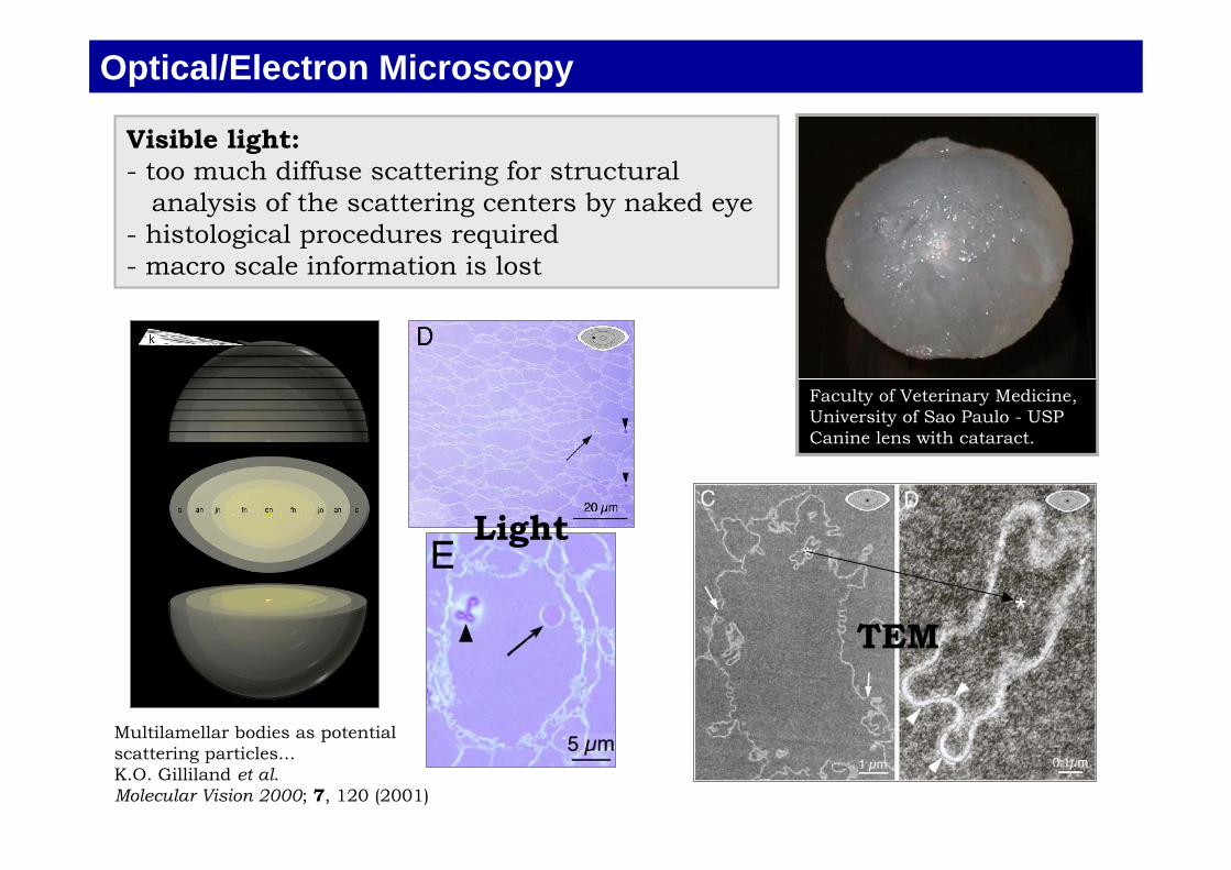
Optical/Electron Microscopy
Visible light:- too much diffuse scattering for structural analysis of the scattering centers by naked eye- histological procedures required- macro scale information is lost
Faculty of Veterinary Medicine,University of Sao Paulo - USPCanine lens with cataract.
Multilamellar bodies as potentialscattering particles...K.O. Gilliland et al.Molecular Vision 2000; 7, 120 (2001)
Light
TEM

Optical / Electron microscopies are limited for investigating: distributions of scattering center and density fluctuations
on extended tissues (cm scale/entire lenses)
Scanning Electron Microscopy
Images of human cataractous lens...W.L. Jongebloed. Scanning Microscopy 12, 653 (1998)
Nuclear fiber cell compaction...Christopher D. Freel BMC Ophthalmology 3, 1 (2003)

rad1
101.0
20keVE , 10 1
tan
6
e
e
e
e
5e
μ≈αΔ
=δΔ⇒=ρρΔ
ρρΔ
δ=δΔ=Δ
≈≅ρΓ=δ
δ−=
αΔ
=αΔ
−
−
nn
nnn
Why X-rays?
X-ray can see through entire lenses: - minimum of scattering (angular spreading ≈ λ/D )- refraction and diffusion scattering are angular resolved processes - selective mapping of: • density fluctuations and • distributions of scattering centers
Refraction
(index of refraction)
(Snell's Law)
Small angle scattering

Refraction
Half width = 0.58λ/D

Diffraction enhanced X-ray imaging (DEI)a) Experimental setup at X15A (NSLS).
b) Incident beam (0), absorption (1), refraction (2), and diffuse scattering (3).
c) Analyzer window(acceptance angle forBragg diffraction in theanalyzer crystal)
P: center of reflection curve (absorption)
S: shoulder (refraction) (ΔI/I ~ 30% per μrad)
T: tail (diffuse scat.) |Δθ| > 5μrad, I/I0 < 2%
Z. Zhong et al., Phys. Res. A 450, 556 (2000)
FWHM = 3.2μrad @ 18keV / 1.5 μrad @ 40 keV

DEI @ LNLS (10.7KeV)
"High contrast radiography of normal and cataractous canine lenses"A. Antunes, M.G. Hönnicke, C. Cusatis and S.L. MorelhãoJ. Phys. D: Appl. Phys. 38 A85-A88 (2005)
"Diffraction Enhanced X-Ray Imaging of Mammals Crystalline Lens"A. Antunes, M. G. Hönnicke, A. M. V. Safatle, C. Cusatis, P. S. Moraes Barros and S. L. MorelhãoNucl. Instr. Meth. B 238, 28-31 (2005)
FWHM = 50μrad

Healthy Cataractous
DEI of entire lenses (@ 20 KeV)
Air
Healthy Cataractous
Water
Med. Phys. 33, 2338 (2006)

Discovery of calcificated tissue in the eye lens (canine)
Calcificated tissues(Ca, Micro X-ray fluorescence)
Refraction: Δθ = -1.2 μrad
Diffusescattering:
Absorption(beforeanalyzer)
Side view1mm
Δθ = -4.5μrad
Mass absorption image

Localized calcification and the partial cataract cases
Refraction: Δθ = -1.5 μrad
Diffuse scattering: Δθ = -4.5μrad
Air bubble
-3.2
-1.2
-0.2
1.8
6.8
Δθ (μrad)
D < 5μm
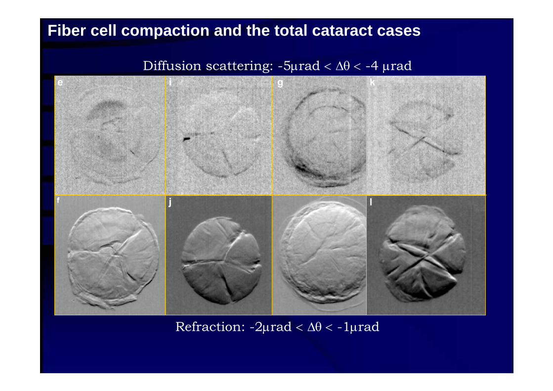
Fiber cell compaction and the total cataract cases
Diffusion scattering: -5μrad < Δθ < -4 μrad
Refraction: -2μrad < Δθ < -1μrad

White/dark contrast due to compaction of fiber cells
Left shoulder of the analyzer window

Cortical fiber-cell organization and low-density fissures
Size reduction of fiber-cells promotescompaction towards sites A and low-density fissures at sites B.
Refractionimage
Low-densityfissure

3D configuration of lesions- localization of calcificated tissues, i.e. nuclear/cortical- extension of fiber compaction
Correlations to be established- ophthalmic exams (partial/total opacity) and type of lesions- calcification:
inductive agents and susceptible mammal species - potential causes of cataract and type of lesions- information from other techniques
Multi-disciplinary approach - to fully understand the disease- histological procedures; microscopic analysis - UV and Raman spectroscopy; protein/molecular content- X-ray fluorescence; elemental analysis
Classification scheme of cataract by DEI

Dr. Andrea Antunes- 4 years of Young Researcher fellowship, FAPESP- to explore the multi-disciplinary aspect of the
ophthalmic diseases- Institute of Physics, Dept. Applied Physics, USP
Researchers and Institutes
Prof. Paulo S.M. Barros and Dr. Angélica Safatle- Faculty of Veterinary Medicine, USP- cataract surgery in mammals- patient clinical records- ophthalmic classification, i.e. extension of opacity
Dr. Maria Ines Borella - Institute of Biomedical Sciences, USP- histological analysis- lab. for handling tissues (microtome)

Dr. Marcia Temperini - Institute of Chemistry, USP- Raman spectroscopy- Laser Phys. Lett. 2, 356 (2005)
Prof. Maria Luisa de Carvalho- University of Lisbon, Portugal- Institute of Atomic Physics- X-ray fluorescence
Researchers and Institutes
Acknowledgements- Z. Zhong, C. Parham- Brazilian agency CNPq
(proc. No. 150329/2003-2 and 301617/95-3)- Research founding offices PRP and CCInt of USP,- Use of X15A beamline at the NSLS,
U.S. Department of EnergyNo. DE-AC02-98CH10886 and byNIH grant R01 AR48292.