Embed Size (px)
Citation preview
Copyrightⓒ2013 Journal of the Korean Society of Magnetic Resonance in Medicine 181
Acute renal failure (ARF) remains a significant causeof morbidity and mortality in adults and children.Although developing a treatment for ARF has been achallenge, there have been some advances in preventa-tive strategies and supportive measures (1). Celltherapy, including the use of stem cells and progenitor
INTRODUCTION�Received; March 25, 2013�Revised; May 22, 2013�Accepted; June 25, 2013This work was supported by the Inje Research and ScholarshipFoundation in 2010.Corresponding author : Sang Yong Lee, M.D.Department of Diagnostic Radiology, Chonbuk National UniversityHospital & Medical School, 634-18, Keumam-dong, Jeonju, Jeonbuk,561-712, Korea.Tel. 82-63-250-1178, Fax. 82-63-272-0481 E-mail : [email protected]
Serial MR Imaging of Magnetically Labeled HumenUmbilical Vein Endothelial Cells in Acute Renal FailureRat Model
Sun Joo Lee1, Sang Yong Lee2, Kyung Pyo Kang3, Won Kim3, Sung Kwang Park3
1Department of Radiology, College of Medicine, Inje University, Busan Paik Hospital, Busan, Korea2Department of Diagnostic Radiology, Chonbuk National University Hospital & Medical School, Jeonbuk, Korea3Department of Internal Medicine Chonbuk National University Hospital & Medical School, Jeonbuk, Korea
Purpose : To evaluate the usefulness of in vivo magnetic resonance (MR) imaging for tracking intravenously injectedsuperparamagnetic iron oxide (SPIO)-labeled human umbilical vein endothelial cells (HUVECs) in an acute renal failure(ARF) rat model.
Materials and Methods: HUVECs were labeled with SPIO and poly-L-lysine (PLL) complex. Relaxation rates at 1.5-T MR,cell viability, and labeling stability were assessed. HUVECs were injected into the tail vein of ARF rats (labeled cells in 10rats, unlabeled cells in 2 rats). Follow-up serial T2*-weighted gradient-echo MR imaging was performed at 1, 3, 5 and 7days after injection, and the MR findings were compared with histologic findings.
Results: There was an average of 98.4±2.4% Prussian blue stain-positive cells after labeling with SPIO-PLL complex. Relaxation rates (R2*) of all cultured HUVECs at day 3 and 5 were not markedly decreased compared withthat at day 1. The stability of SPIO in HUVECs was maintained during the proliferation of HUVECs in culture media. In thepresence of left unilateral renal artery ischemia, T2*-weighted MR imaging performed 1 day after the intravenous injec-tion of labeled HUVECs revealed a significant signal intensity (SI) loss exclusively in the left renal outer medulla regions, butnot in the right kidney. The MR imaging findings at days 3, 5 and 7 after intravenous injection of HUVECs showed a SI lossin the outer medulla regions of the ischemically injured kidney, but the SI progressively recovered with time and the rightkidney did not have a significant change in SI in the same period. Upon histologic analysis, the SI loss on MR images wascorrespondent to the presence of Prussian blue stained cells, primarily in the renal outer medulla.
Conclusion: MR imaging appears to be useful for in vivo monitoring of intravenously injected SPIO-labeled HUVECs in anischemically injured rat kidney.
Index words : Magnetic resonance imaging (MRI)∙Superparamagnetic iron oxide (SPIO)Human umbilical vein endothelial cells (HUVECs)∙Cell tracking
www.ksmrm.org JKSMRM 17(3) : 181-191, 2013
pISSN 1226-9751 / eISSN 2288-3800 http://dx.doi.org/10.13104/jksmrm.2013.17.3.181
Original Article
cells, has recently attracted a great deal of interest as anew therapeutic strategy for ARF (2). Recently,Brodsky et al. (3) demonstrated that implantation ofhuman umbilical vein endothelial cells (HUVECs)resulted in functional protection from ischemic ARF.Important considerations for improving cell-basedtherapy for renal failure include the delivery of cells tothe site of renal injury and successful engraftment.Noninvasive imaging techniques allow in vivo monitor-ing of cell location and can therefore be of great valuefor improving cell-based therapy for renal failure.
Evaluation of transplanted or injected therapeuticcells is currently conducted by histologic analysis.
Although histologic analysis allows direct visualiza-tion of therapeutic cells on tissue, it cannot be used forin vivo cell tracking. Advances in cell labelingtechniques have allowed many researchers to trackcells in vivo with molecular imaging such as biolumi-nescence, nuclear or MR imaging. Nowadays, there isan increasing interest in in vivo cell tracking withhigh-spatial-resolution magnetic resonance (MR)imaging due to its ability to depict detailed anatomy(4, 5). Superparamagnetic iron oxide (SPIO) particles,which have been used as a contrast agent in clinicalmedical imaging, are already being used to label cellsfor in vivo MR cell tracking (6-11). Different classesof transfection agents such as polycationic amines,dendrimers, and lipid-based agents have beenemployed to efficiently load cultured cells withenough SPIO for in vivo MR imaging (12). It has beenreported that PLL, a polycationic amine, has littlecytotoxicity and can be used for effective transfectionefficiency in mammalian cells (12). The utility of MRcell tracking with SPIO-labeled stem cells has beenshown in the experimental monitoring of cell-basedtherapies for myocardial infarction (9, 13), adult andneonatal mice brain,10 and a rat model of nephropa-thy (11). MR cell tracking imaging with inflammatorycells has also been applied to conditions such as graftrejection after transplantation (14) and adhesion ofnatural killer cells to tumors (15). To our knowledge,no study using MR imaging has been performed ontracking intravenously injected SPIO-labeled endothe-lial cells in rats with induced ARF, which may have arole in the recovery of deteriorated renal function.
Thus, the purpose of this study was to evaluate theusefulness of in vivo MR imaging for monitoring the
distribution of HUVECs after intravenous injection ina rat model for unilateral ischemic acute renal failure.
1. In Vitro ExperimentsCell Cultures
Human umbilical vein endothelial cells (HUVECs)were prepared from human umbilical cords by collage-nase digestion as previously described (16). Theendothelial identity of the cultures was confirmed bythe detection of factor VIII by immunofluoresence.HUVECs were maintained in M199 medium supple-mented with 20% (vol/vol) fetal bovine serum at 37℃in a 5% CO2 atmosphere. The primary cultured cellsin this study were used between passages 2 and 4.
Cell Labeling with Ferumoxides-PLL complexes andCellTracker
PLL hydrobromide (Sigma, St Louis, MO, USA) wasused as the transfection agent. The PLL (1.5 mg/mL)was added to the culture media at a dilution of 1:1,000and mixed with ferumoxides (Feridex 50 μg/mL;Wayne, NJ, USA) for 60 minutes at room temperatureon a rotating shaker. These culture media containingthe ferumoxides-PLL complex were added to the cellssuch that the final concentration of ferumoxides was 25μg/mL and the final dilution of PLL was 1:2,000. Thecell cultures were kept overnight at 37℃ in a 95% airper 5% CO2 atmosphere (17). After labeling, cellswere washed two times and resuspended in completemedia. Cell viability was maintained at greater than95% in all instances and was monitored before andafter labeling. There was no difference observed in cellviability (95%) between the ferumoxides-PLL labeledand unlabeled HUVECs.
Before injection, HUVECs were loaded with Green-fluorescent fluorescein diacetate (CellTracker,Molecular Probes, Eugene, OR) according to themanufacture’s protocol. Briefly, confluent HUVECswere lifted with trypsin-EDTA and suspended.CellTracker (10 μM) was added to the HUVECs andHUVECS were incubated for 15 minutes at 37℃.After centrifugation at 1,500 rpm and 4℃, M199media was added, and the HUVECs were incubated anadditional 15 minutes at 37℃. After being washed
MATERIALS AND METHODS
182 JKSMRM 17(3) : 181-191, 2013
http://www.ksmrm.org http://dx.doi.org/10.13104/jksmrm.2013.17.3.181
two times with PBS, HUVECs were resuspended inPBS at a final concentration of 6×10⁶ cells/mL. Thecell suspension was kept on ice until transplantation,but for no longer than 30 minutes. Fluorescent imagesof the labeled cells were obtained to confirm thethoroughness of the cell labeling.
Labeling Stability Study in HUVECsTo study labeling stability, cultured HUVECs were
divided into three groups, each with the same numberof cells (1 × 10⁶ cells each). HUVECs were labeled bymeans of incubation with ferumoxides-PLL complexesovernight and then were replated in a 10-cm culturedish. After 1, 3, and 5 days, all HUVECs were washedthree times, treated with trypsin, and harvested. Aftercentrifugation, HUVECs were then suspended in PBSfor the MR relaxation measurements. All sampleswere placed in a head coil provided by the manufac-turer and imaged with a clinical 1.5-T MR imager(Magnetom Symphony, Siemens Medical Systems,Erlangen, Germany). The T2* relaxation time wasdetermined by using a multiple gradient-echosequence, with a fixed repetition time of 1000 msec,flip angle of 30。, and 5 echoes starting at 7.5 msec.R2* (1/T2*) was deduced by fitting a single exponen-tial function to the signal intensity versus TE data (TEof 7.5 - 60 msec and fixed TR). It has been wellrecognized that R2* depends linearly on the globaliron concentration of the sample (18).
2. In Vivo ExperimentsInbred male Sprague-Dawley rats (Charles River
Korea, Seoul, Korea) were maintained on standardlaboratory chow and water ad libitum. ARF wasinduced by unilateral (left) renal artery ischemiafollowed by the reperfusion of blood flow. Briefly, rats(n=12) were anesthetized with an intramuscularinjection of 100 mg/kg ketamine and 10 mg/kgxylazine. The animals were placed on a heating tableto maintain constant body temperature. A midlineincision was made and the left renal pedicle wasexposed and clamped for 40 minutes. After the clampwas removed, the kidney was inspected for restorationof blood flow. After abdominal wall closure, HUVECs(6 × 10⁶ cells) labeled with SPIO and CellTrackeraccording to the above methods were diluted in 300-400 μL PBS and injected into the tail vein of the ARF
rats. Rats (n=8) injected with labeled HUVECs wereimaged at 1, 3, 5, and 7 days after injection with a1.5-T MR imager. Rats (n=2) injected with unlabeledHUVECs and sham-operated rats (n=2) injected withlabeled HUVECs were imaged 1 day after injection toevaluate signal intensity changes in the kidney. Atdays 1, 3, 5, and 7, the animals were sacrificed andboth kidneys were harvested for histologic examina-tion.
3. MR Imaging All in vivo MR images of the rats were obtained by a
1.5-T clinical MR imager (Magnetom Symphony,Siemens Medical Systems, Erlangen, Germany) using asmall flexible coil with a width of 17 cm provided bythe manufacturer. Rats were positioned prone in theflexible coil. Then, fat-suppressed, coronal, andtransverse MR images were obtained using a gradient-echo sequence with 500/13 (TR/TE, msec), 30。flipangle, 75 mm field of view, 2.5 mm section thickness,256 × 192 matrix, and eight signals were acquired toevaluate signal decay in the kidneys. Both kidneyswere imaged on the same imaging plane. MR imagesof the rats were obtained after 1 × 10⁶ HUVECs wereinjected through tail veins at days 1, 3, 5, and 7. Afterinjection of labeled HUVECs, low signal intensity wasobserved predominantly in the outer medulla of theleft kidney, in which ARF had been induced. Signalintensity values at the outer medulla and cortex of theleft kidney were measured to calculate the ratio ofsignal decrease due to the presence of labeledHUVECs. Signal intensity values for low signalintensity regions were measured on serial MR imagesusing the region of interest in the outer medulla of theleft kidney. Also signal intensity values for high signalintensity were measured in the cortex of the leftkidney. The ratio of signal decrease (Srsd) in the outermedulla of the left kidney was determined by usingthe equation Srsd = [(Sco - Som)/ Sco] × 100, whereSco is the signal intensity value measured in the renalcortex and Som is the signal intensity value measuredin the outer medulla of the kidney. The ratio of signaldecrease in the right kidney was also measured withthe same method as in the left kidney.
http://dx.doi.org/10.13104/jksmrm.2013.17.3.181 http://www.ksmrm.org
Serial MR Imaging of Magnetically Labeled Humen Umbilical Vein Endothelial Cells in Acute Renal Failure Rat Model � Sun Joo Lee, et al. 183
4. AnalysisHistologic Examination
Renal tissue was prepared after in vivo MRIexamination. The tissue blocks of rat kidney werefixed in 4% paraformaldehyde and processed forparaffin embedding. Histopathology was examined on5 μm slides stained by hematoxylin and eosin, and
Prussian blue staining for iron was used by implement-ing standard methods on the kidney sections from theferumoxides-PLL labeled group (n = 8 with ARF, n = 2sham operation) and unlabeled controls (n = 2). Toexamine Cell Tracker-labeled HUVECs, a fluorescentmicroscope (Carl Zeiss, Gottingen, Germany) wasused on fixed frozen sections of kidney tissue.
184 JKSMRM 17(3) : 181-191, 2013
http://www.ksmrm.org http://dx.doi.org/10.13104/jksmrm.2013.17.3.181
a b
c
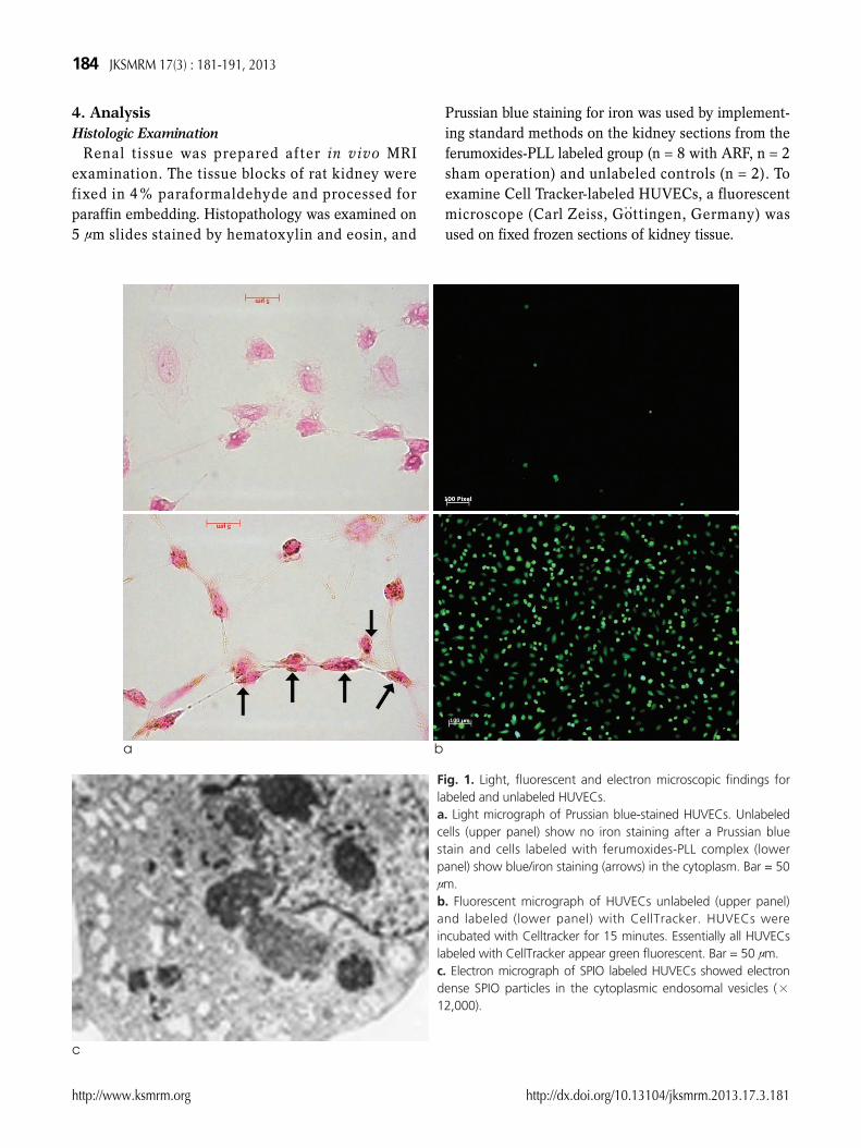
Fig. 1. Light, fluorescent and electron microscopic findings forlabeled and unlabeled HUVECs. a. Light micrograph of Prussian blue-stained HUVECs. Unlabeledcells (upper panel) show no iron staining after a Prussian bluestain and cells labeled with ferumoxides-PLL complex (lowerpanel) show blue/iron staining (arrows) in the cytoplasm. Bar = 50μm. b. Fluorescent micrograph of HUVECs unlabeled (upper panel)and labeled (lower panel) with CellTracker. HUVECs wereincubated with Celltracker for 15 minutes. Essentially all HUVECslabeled with CellTracker appear green fluorescent. Bar = 50 μm. c. Electron micrograph of SPIO labeled HUVECs showed electrondense SPIO particles in the cytoplasmic endosomal vesicles (×12,000).
1. In Vitro ExperimentsHUVECs with SPIO-PLL and CellTracker
Prussian blue stain for iron was performed onunlabeled and ferumoxides-PLL labeled HUVECs inculture (Fig. 1a). On average, there were 98.4±2.4%Prussian blue stain-positive cells after labeling withferumoxides-PLL and 2.4±0.2% Prussian blue stain-positive cells without labeling with ferumoxides-PLL.With fluorescent microscopy, essentially all HUVECslabeled with CellTracker appeared green fluorescent,while unlabeled HUVECs were rarely green fluores-cent (Fig. 1b). Electron microscopic examinationshowed the presence of SPIO nanoparticles in theendosomal vesicles of the HUVECs (Fig. 1c).
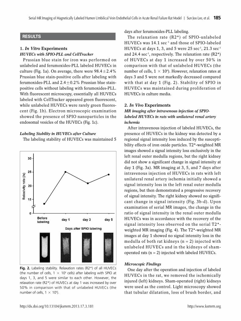
Labeling Stability in HUVECs after CultureThe labeling stability of HUVECs was maintained 5
days after ferumoxides-PLL labeling. The relaxation rate (R2*) of SPIO-unlabeled
HUVECs was 14.1 sec-1 and those of SPIO-labeledHUVECs at days 1, 3, and 5 were 25 sec-1, 21.3 sec-1
and 24.4 sec-1, respectively. The relaxation rate (R2*)of HUVECs at day 1 increased by over 50% incomparison with that of unlabeled HUVECs (thenumber of cells, 1 × 10⁶). However, relaxation rates atdays 3 and 5 were not markedly decreased comparedwith that at day 1 (Fig. 2). Stability of SPIO inHUVECs was maintained during proliferation ofHUVECs in culture media.
2. In Vivo Experiments MR imaging after intravenous injection of SPIO-labeled HUVECs in rats with unilateral renal arteryischemia
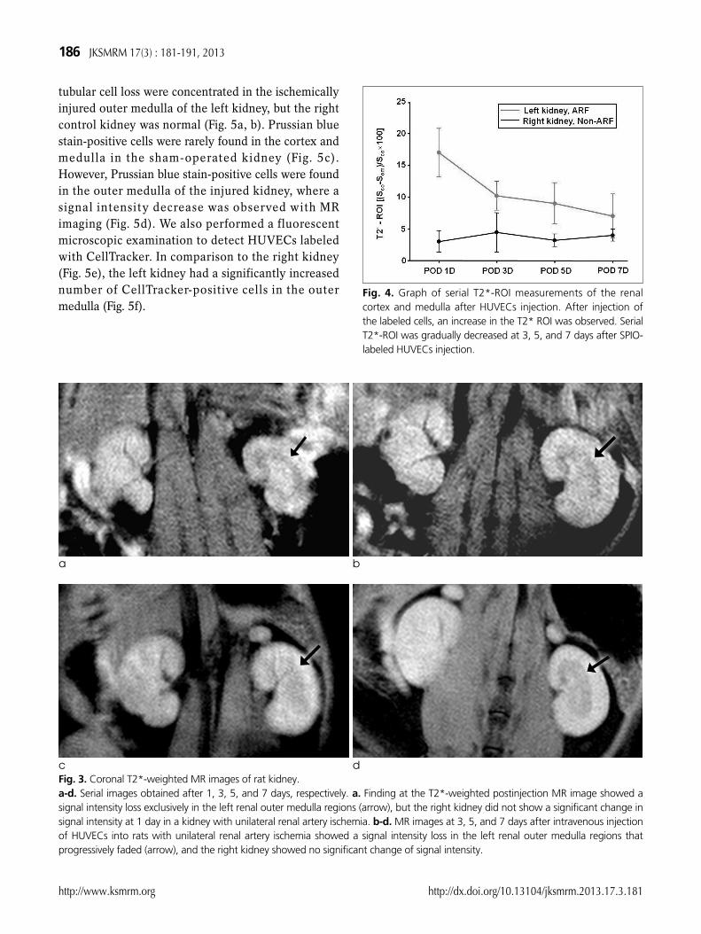
After intravenous injection of labeled HUVECs, thepresence of HUVECs in the kidney was detected by aregional signal intensity loss induced by the suscepti-bility effects of iron oxide particles. T2*-weighted MRimages showed a signal intensity loss exclusively in theleft renal outer medulla regions, but the right kidneydid not show a significant change in signal intensity atday 1 (Fig. 3a). MR imaging at 3, 5, and 7 days afterintravenous injection of HUVECs in rats with leftunilateral renal artery ischemia initially showed asignal intensity loss in the left renal outer medullaregions, but then demonstrated a progressive recoveryof signal intensity. The right kidney showed no signifi-cant change in signal intensity (Fig. 3b-d). Uponexamination of serial MR images, the change in theratio of signal intensity in the renal outer medullaHUVECs was in accordance with the recovery of thesignal intensity loss observed on the serial T2*-weighted MR imaging (Fig. 4). The T2*-weighted MRimages at day 1 showed no signal intensity loss in themedulla of both rat kidneys (n = 2) injected withunlabeled HUVECs and in the kidneys of sham-operated rats (n = 2) injected with labeled HUVECs.
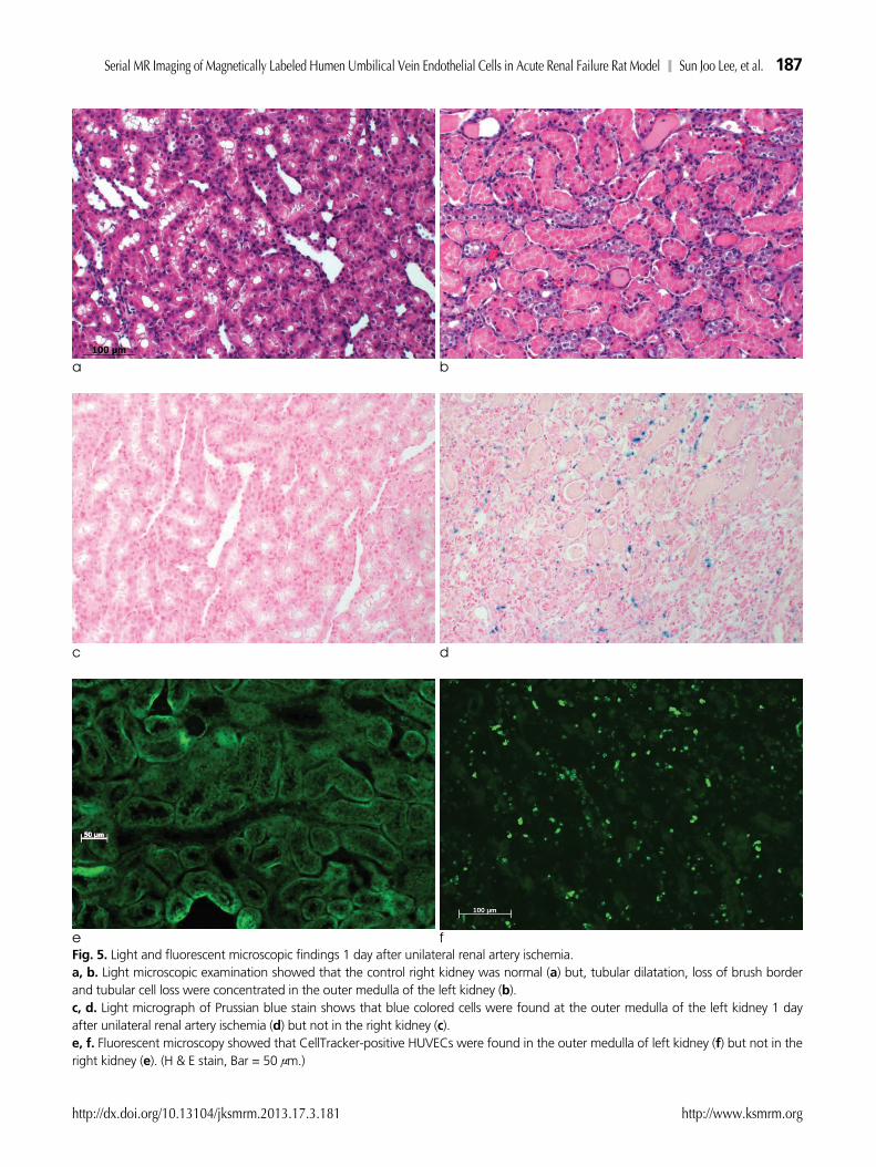
Microscopic FindingsOne day after the operation and injection of labeled
HUVECs in the rat, we removed the ischemicallyinjured (left) kidneys. Sham-operated (right) kidneyswere used as the control. Light microscopy showedthat tubular dilatation, loss of brush border, and
RESULTS
http://dx.doi.org/10.13104/jksmrm.2013.17.3.181 http://www.ksmrm.org
Serial MR Imaging of Magnetically Labeled Humen Umbilical Vein Endothelial Cells in Acute Renal Failure Rat Model � Sun Joo Lee, et al. 185
Fig. 2. Labeling stability. Relaxation rates (R2*) of all HUVECs(the number of cells, 1 × 10⁶ cells) after labeling with SPIO atdays 1, 3, and 5 were similar to each other. However, therelaxation rate (R2*) of HUVECs at day 1 was increased by over50% in comparison with that of unlabeled HUVECs (thenumber of cells, 1 × 10⁶).
tubular cell loss were concentrated in the ischemicallyinjured outer medulla of the left kidney, but the rightcontrol kidney was normal (Fig. 5a, b). Prussian bluestain-positive cells were rarely found in the cortex andmedulla in the sham-operated kidney (Fig. 5c).However, Prussian blue stain-positive cells were foundin the outer medulla of the injured kidney, where asignal intensity decrease was observed with MRimaging (Fig. 5d). We also performed a fluorescentmicroscopic examination to detect HUVECs labeledwith CellTracker. In comparison to the right kidney(Fig. 5e), the left kidney had a significantly increasednumber of CellTracker-positive cells in the outermedulla (Fig. 5f).
186 JKSMRM 17(3) : 181-191, 2013
http://www.ksmrm.org http://dx.doi.org/10.13104/jksmrm.2013.17.3.181
a b
c dFig. 3. Coronal T2*-weighted MR images of rat kidney. a-d. Serial images obtained after 1, 3, 5, and 7 days, respectively. a. Finding at the T2*-weighted postinjection MR image showed asignal intensity loss exclusively in the left renal outer medulla regions (arrow), but the right kidney did not show a significant change insignal intensity at 1 day in a kidney with unilateral renal artery ischemia. b-d. MR images at 3, 5, and 7 days after intravenous injectionof HUVECs into rats with unilateral renal artery ischemia showed a signal intensity loss in the left renal outer medulla regions thatprogressively faded (arrow), and the right kidney showed no significant change of signal intensity.
Fig. 4. Graph of serial T2*-ROI measurements of the renalcortex and medulla after HUVECs injection. After injection ofthe labeled cells, an increase in the T2* ROI was observed. SerialT2*-ROI was gradually decreased at 3, 5, and 7 days after SPIO-labeled HUVECs injection.
http://dx.doi.org/10.13104/jksmrm.2013.17.3.181 http://www.ksmrm.org
Serial MR Imaging of Magnetically Labeled Humen Umbilical Vein Endothelial Cells in Acute Renal Failure Rat Model � Sun Joo Lee, et al. 187
a b
c d
e fFig. 5. Light and fluorescent microscopic findings 1 day after unilateral renal artery ischemia. a, b. Light microscopic examination showed that the control right kidney was normal (a) but, tubular dilatation, loss of brush borderand tubular cell loss were concentrated in the outer medulla of the left kidney (b). c, d. Light micrograph of Prussian blue stain shows that blue colored cells were found at the outer medulla of the left kidney 1 dayafter unilateral renal artery ischemia (d) but not in the right kidney (c). e, f. Fluorescent microscopy showed that CellTracker-positive HUVECs were found in the outer medulla of left kidney (f) but not in theright kidney (e). (H & E stain, Bar = 50 μm.)
188 JKSMRM 17(3) : 181-191, 2013
http://www.ksmrm.org http://dx.doi.org/10.13104/jksmrm.2013.17.3.181
a b
c d
e fFig. 6. MR images and light and fluorescent microscopic findings of an ischemically injured kidney after injection of SPIO-labeledHUVECs at days 1 and 5. MR imaging examination on day 1 showed a signal intensity loss in the left renal outer medulla regions (a).Prussian blue stain-positive cells were found at the outer medulla of the left kidney (c). By fluorescent microscopic examination, theCellTracker-positive HUVECs were found in the outer medulla of the left kidney (e). There was a strong correlation between signalintensity loss area and the location of Prussian blue stain-positive cells and CellTracker-positive HUVECs. MR imaging 5 days afterinjection of HUVECs showed a decreased signal intensity loss in the left renal outer medulla region (b). Prussian blue stain-positive cellsand CellTracker-positive HUVECs were found in the same area, but slightly decreased in numbers compared to day 1 (d and f).
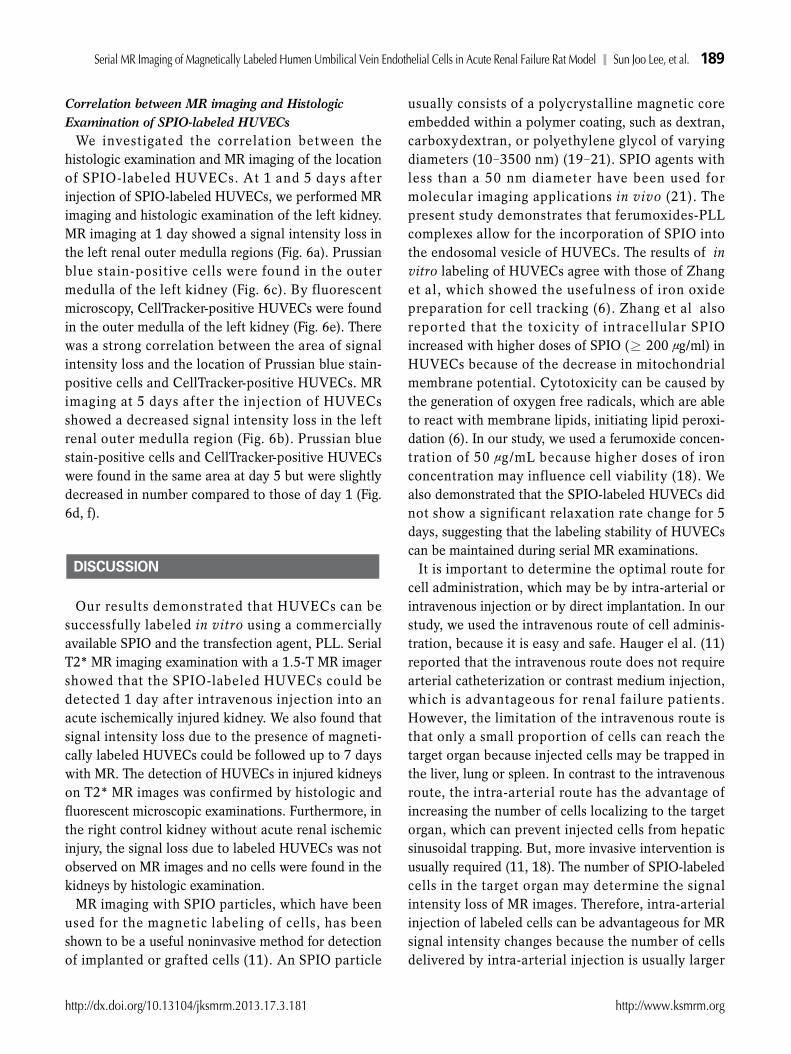
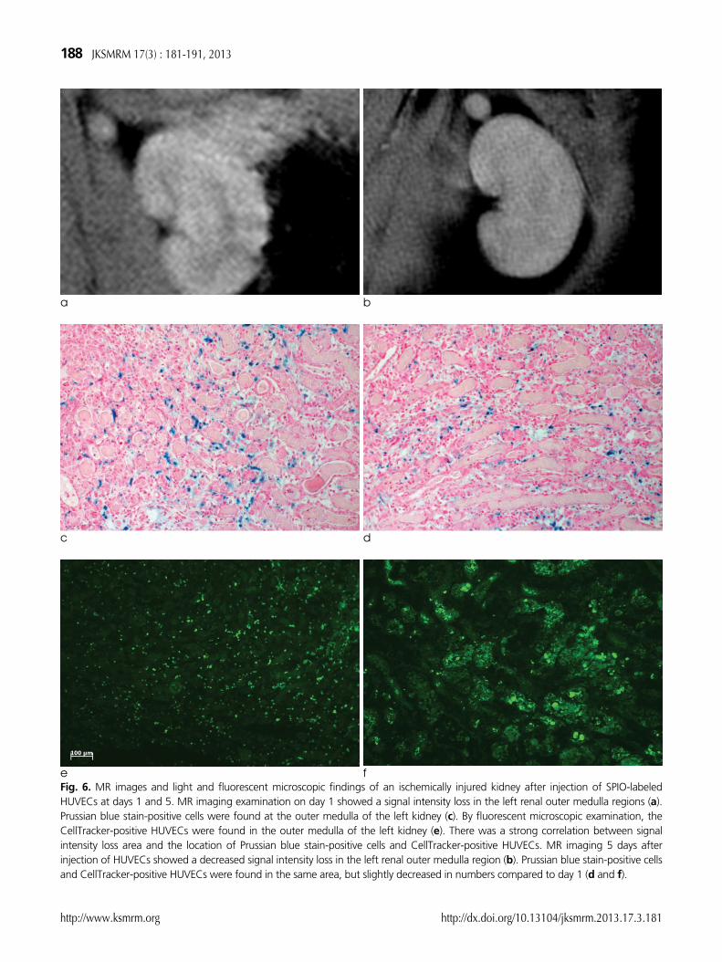
Correlation between MR imaging and HistologicExamination of SPIO-labeled HUVECs
We investigated the correlation between thehistologic examination and MR imaging of the locationof SPIO-labeled HUVECs. At 1 and 5 days afterinjection of SPIO-labeled HUVECs, we performed MRimaging and histologic examination of the left kidney.MR imaging at 1 day showed a signal intensity loss inthe left renal outer medulla regions (Fig. 6a). Prussianblue stain-positive cells were found in the outermedulla of the left kidney (Fig. 6c). By fluorescentmicroscopy, CellTracker-positive HUVECs were foundin the outer medulla of the left kidney (Fig. 6e). Therewas a strong correlation between the area of signalintensity loss and the location of Prussian blue stain-positive cells and CellTracker-positive HUVECs. MRimaging at 5 days after the injection of HUVECsshowed a decreased signal intensity loss in the leftrenal outer medulla region (Fig. 6b). Prussian bluestain-positive cells and CellTracker-positive HUVECswere found in the same area at day 5 but were slightlydecreased in number compared to those of day 1 (Fig.6d, f).
Our results demonstrated that HUVECs can besuccessfully labeled in vitro using a commerciallyavailable SPIO and the transfection agent, PLL. SerialT2* MR imaging examination with a 1.5-T MR imagershowed that the SPIO-labeled HUVECs could bedetected 1 day after intravenous injection into anacute ischemically injured kidney. We also found thatsignal intensity loss due to the presence of magneti-cally labeled HUVECs could be followed up to 7 dayswith MR. The detection of HUVECs in injured kidneyson T2* MR images was confirmed by histologic andfluorescent microscopic examinations. Furthermore, inthe right control kidney without acute renal ischemicinjury, the signal loss due to labeled HUVECs was notobserved on MR images and no cells were found in thekidneys by histologic examination.
MR imaging with SPIO particles, which have beenused for the magnetic labeling of cells, has beenshown to be a useful noninvasive method for detectionof implanted or grafted cells (11). An SPIO particle
usually consists of a polycrystalline magnetic coreembedded within a polymer coating, such as dextran,carboxydextran, or polyethylene glycol of varyingdiameters (10-3500 nm) (19-21). SPIO agents withless than a 50 nm diameter have been used formolecular imaging applications in vivo (21). Thepresent study demonstrates that ferumoxides-PLLcomplexes allow for the incorporation of SPIO intothe endosomal vesicle of HUVECs. The results of invitro labeling of HUVECs agree with those of Zhanget al, which showed the usefulness of iron oxidepreparation for cell tracking (6). Zhang et al alsoreported that the toxicity of intracellular SPIOincreased with higher doses of SPIO (≥ 200 μg/ml) inHUVECs because of the decrease in mitochondrialmembrane potential. Cytotoxicity can be caused bythe generation of oxygen free radicals, which are ableto react with membrane lipids, initiating lipid peroxi-dation (6). In our study, we used a ferumoxide concen-tration of 50 μg/mL because higher doses of ironconcentration may influence cell viability (18). Wealso demonstrated that the SPIO-labeled HUVECs didnot show a significant relaxation rate change for 5days, suggesting that the labeling stability of HUVECscan be maintained during serial MR examinations.
It is important to determine the optimal route forcell administration, which may be by intra-arterial orintravenous injection or by direct implantation. In ourstudy, we used the intravenous route of cell adminis-tration, because it is easy and safe. Hauger el al. (11)reported that the intravenous route does not requirearterial catheterization or contrast medium injection,which is advantageous for renal failure patients.However, the limitation of the intravenous route isthat only a small proportion of cells can reach thetarget organ because injected cells may be trapped inthe liver, lung or spleen. In contrast to the intravenousroute, the intra-arterial route has the advantage ofincreasing the number of cells localizing to the targetorgan, which can prevent injected cells from hepaticsinusoidal trapping. But, more invasive intervention isusually required (11, 18). The number of SPIO-labeledcells in the target organ may determine the signalintensity loss of MR images. Therefore, intra-arterialinjection of labeled cells can be advantageous for MRsignal intensity changes because the number of cellsdelivered by intra-arterial injection is usually larger
DISCUSSION
http://dx.doi.org/10.13104/jksmrm.2013.17.3.181 http://www.ksmrm.org
Serial MR Imaging of Magnetically Labeled Humen Umbilical Vein Endothelial Cells in Acute Renal Failure Rat Model � Sun Joo Lee, et al. 189
than that by intravenous injection. We injected 1 ×10⁶ SPIO-labeled HUVECs intravenously to detectcells localizing to the kidney, and serial MR imagesshowed a signal intensity loss in the acute ischemicallyinjured kidney.
In acute renal ischemia, proximal tubular epithelialcell injury is a major pathophysiologic event (1). But,renal vascular changes including renal vascularendothelial injury and dysfunction may occur simulta-neously after ischemic acute renal failure (22, 23). Ithas been demonstrated that tissue ischemia andcytokines mobilize bone marrow-derived endothelialprogenitor cells (24) and that circulating endothelialcells of hematopoietic origin are recruited to theischemic areas to form adult blood vessels (25).Interestingly, our results show that labeled HUVECswere found only in the ischemically injured leftkidney, but not in the sham-operated right kidney. Ourresults suggest that unilateral ischemic injury signifi-cantly stimulates the recruitment of transplantedendothelial cells to the injured kidney.
Although we did not analyze the functional changesafter ARF, we used in vivo cell tracking with SPIOparticles and a commercially available 1.5-T MRimaging system to show that HUVECs may beengrafted in the outer medulla of an injured kidney forsystemic treatment of renal failure. To our knowledge,this is the first report of systemic intravenous cellgrafting and in vivo MR imaging of SPIO-labeledHUVECs within an ischemically injured kidney.
There were several limitations in this study. Wedemonstrated only the localization of SPIO-labeledHUVECs in an acute ischemically injured kidney withMR imaging. Uncovering the mechanism by whichHUVECs localize to the ischemically injured kidneywas beyond the scope of this study. Also, we followedthe presence of SPIO-labeled HUVECs for up to 7days, but the recovery period for acute ischemic renalfailure may exceed 7 days. Further, we did not analyzethe relationship between HUVEC recruitment in theinjured kidney and renal function improvement.
MR imaging appears to be useful for in vivomonitoring of intravenously injected SPIO-labeledHUVECs in an ischemically injured rat kidney, whichhas been used as a model of cell therapy.
References
1. Thadhani R, Pascual M, Bonventre JV. Acute renal failure. NEngl J Med 1996;334:1448-1460
2. Rookmaaker MB, Verhaar MC, van Zonneveld AJ, Rabelink TJ.Progenitor cells in the kidney: biology and therapeutic perspec-tives. Kidney Int 2004;66:518-522
3. Brodsky SV, Yamamoto T, Tada T, et al. Endothelial dysfunc-tion in ischemic acute renal failure: rescue by transplantedendothelial cells. Am J Physiol Renal Physiol 2002;282:F1140-F1149
4. Sun R, Dittrich J, Le-Huu M, et al. Physical and biologicalcharacterization of superparamagnetic iron oxide- andultrasmall superparamagnetic iron oxide-labeled cells: a compar-ison. Invest Radiol 2005;40:504-513
5. Frank JA, Anderson SA, Kalsih H, et al. Methods for magneti-cally labeling stem and other cells for detection by in vivomagnetic resonance imaging. Cytotherapy 2004;6:621-625
6. Zhang, Z, van den Bos EJ, Wielopolsk PA, et al. In vitroimaging of single living human umbilical vein endothelial cellswith a clinical 3.0-T MRI scanner. Magma 2005;18:175-185
7. Bulte JW, Zhang S, van Gelderen P, et al. Neurotransplantationof magnetically labeled oligodendrocyte progenitors: magneticresonance tracking of cell migration and myelination. Proc NatlAcad Sci USA 1999;96:15256-15261
8. Bulte JW, Arbab AS, Douglas T, Frank JA. Preparation ofmagnetically labeled cells for cell tracking by magneticresonance imaging. Methods Enzymol 2004;386:275-299
9. Kraitchman DL, Heldman AW, Atalar E, et al. In vivo magneticresonance imaging of mesenchymal stem cells in myocardialinfarction. Circulation 2003;107:2290-2293
10. Magnitsky S, Watson DJ, Walton RM, et al. In vivo and ex vivoMRI detection of localized and disseminated neural stem cellgrafts in the mouse brain. Neuroimage 2005;26:744-754
11. Hauger O, Frost EE, van Heeswijk R, et al. MR evaluation of theglomerular homing of magnetically labeled mesenchymal stemcells in a rat model of nephropathy. Radiology 2006;238:200-210
12. Arbab AS, Yocum GT, Wilson LB, et al. Comparison oftransfection agents in forming complexes with ferumoxides, celllabeling efficiency, and cellular viability. Mol Imaging 2004;3:24-32
13. Himes N, Min JY, Lee R, et al. In vivo MRI of embryonic stemcells in a mouse model of myocardial infarction. Magn ResonMed 2004;52:1214-1219
14. Ho C, Hitchens TK. A non-invasive approach to detecting organrejection by MRI: monitoring the accumulation of immune cellsat the transplanted organ. Curr Pharm Biotechnol 2004;5:551-566
15. Kircher MF, Allport JR, Graves EE, et al. In vivo high resolutionthree-dimensional imaging of antigen-specific cytotoxic T-lymphocyte trafficking to tumors. Cancer Res 2003;63:6838-6846
16. Sung MJ, Kim W, Ahn SY, et al. Protective effect of alpha-lipoicacid in lipopolysaccharide-induced endothelial fractalkineexpression. Circ Res 2005;97:880-890
17. Arbab AS, Bashaw LA, Miller BR, et al. Characterization ofbiophysical and metabolic properties of cells labeled withsuperparamagnetic iron oxide nanoparticles and transfection
190 JKSMRM 17(3) : 181-191, 2013
http://www.ksmrm.org http://dx.doi.org/10.13104/jksmrm.2013.17.3.181
agent for cellular MR imaging. Radiology 2003;229:838-84618. Bos C, Delmas Y, Desmouliere A, et al. In vivo MR imaging of
intravascularly injected magnetically labeled mesenchymal stemcells in rat kidney and liver. Radiology 2004;233:781-789
19. Jung CW, Jacobs P. Physical and chemical properties ofsuperparamagnetic iron oxide MR contrast agents: ferumoxides,ferumoxtran, ferumoxsil. Magn Reson Imaging 1995;13:661-674
20. Artemov D. Molecular magnetic resonance imaging withtargeted contrast agents. J Cell Biochem 2003;90:518-524
21. Thorek DL, Chen AK, Czupryna J, Tsourkas A. Superpara-magnetic iron oxide nanoparticle probes for molecular imaging.Ann Biomed Eng 2006;34:23-38
22. Basile DP, Donohoe D, Roethe K, Osborn JL. Renal ischemic
injury results in permanent damage to peritubular capillariesand influences long-term function. Am J Physiol Renal Physiol2001;281:F887-F899
23. Sutton TA, Fisher CJ, Molitoris BA. Microvascular endothelialinjury and dysfunction during ischemic acute renal failure.Kidney Int 2002;62:1539-1549
24. Takahashi T, Kalka C, Masuda H, et al. Ischemia- and cytokine-induced mobilization of bone marrow-derived endothelialprogenitor cells for neovascularization. Nat Med 1999; 5:434-438
25. Crosby JR, Kaminski WE, Schatteman G, et al. Endothelial cellsof hematopoietic origin make a significant contribution to adultblood vessel formation. Circ Res 2000;87:728-730
http://dx.doi.org/10.13104/jksmrm.2013.17.3.181 http://www.ksmrm.org
Serial MR Imaging of Magnetically Labeled Humen Umbilical Vein Endothelial Cells in Acute Renal Failure Rat Model � Sun Joo Lee, et al. 191
통신저자 : 이상용, (561-712) 전북 전주시 덕진구 건지로 20, 전북 학교 의학전문 학원 상의학과Tel. (063) 250-1178 Fax. (063) 272-0481 E-mail: [email protected]
급성신부전쥐모델에서자기표지된인간제 정맥내피세포의연속자기공명 상
1인제 학교의과 학부산백병원 상의학과2전북 학교의과 학 상의학과
3전북 학교의과 학내과
이선주1∙이상용2∙강경표3∙김 원3∙박성광3
목적: 급성 신부전 쥐 모델에서 상자성 철 산화물 (superparamagnetic iron oxide(SPIO)로 표지한 인간탯줄혈
관내피 세포를 자기공명 상으로 추적할 수 있는지 그 유용성을 평가하고자 하 다.
상과 방법: 인간탯줄혈관내피 세포를 SPIO와 poly-L-lysine (PLL) 혼합물로 표지 하 다. SPIO 농도에 따라서
이완율, 세포 생존율, 표지 안정성을 SPIO 농도의 변화에 따라 평가하 다. 인간탯줄혈관내피 세포를 급성 신부전 쥐
모델에서 꼬리정맥을 통하여 주사하 다. MR을 이용한 추적 검사는 T2* 경사에코 MR 상을 이용하 다. 1, 3, 5,
7일째 추적한 MR 상 소견을 조직 소견과 서로 견주어 보았다.
결과: SPIO-PLL 혼합물을 표지 한 후 Prussian blue 염색에서 평균 98.4±2.4% 세포가 양성반응을 보 다. 3일
과 5일 후 측정한 이완율은 1일에 비해 큰 차이가 없었다. 인간탯줄혈관내피 세포를 SPIO로 표지 한 후 안정성이 유
지됨을 알 수 있었다. 추적 MR 상에서 급성신부전을 유도한 왼쪽 신장 외곽 신 수질에서 신호강도 소실을 보 으나
오른쪽은 정상이었다. 3, 5, 7일 후 촬 한 상에서 왼쪽 신 수질에서 보인 신호강도 소실이 점차 사라졌으나 오른
쪽 신장에서는 여전히 특별한 변화를 보이지 않았다. 조직학 검사에서도 MR 상의 신호강도 소실이 Prussian
blue 염색을 보인 부분과 일치하 다. 면역화학적 분석에서 신 수질에서 보인 세포들이 인간탯줄혈관내피 세포임을
확인하 다.
결론: MR 상은 급성 신부전 치료의 한 방법인 세포 치료의 경우 세포 추적 검사에 유용하게 사용될 수 있음을 확인
하 다.
한자기공명의과학회지 17:181-191(2013)