Embed Size (px)
Citation preview
JOURNAL OF CLINICAL MICROBIOLOGY,0095-1137/01/$04.0010 DOI: 10.1128/JCM.39.4.1247–1253.2001
Apr. 2001, p. 1247–1253 Vol. 39, No. 4
Copyright © 2001, American Society for Microbiology. All Rights Reserved.
Serological, Epidemiological, and Molecular Differences betweenHuman T-Cell Lymphotropic Virus Type 1 (HTLV-1)-Seropositive
Healthy Carriers and Persons with HTLV-I Gag IndeterminateWestern Blot Patterns from the Caribbean
FRANCOIS ROUET,1† LAURENT MEERTENS,2 GERY COUROUBLE,3 CECILE HERRMANN-STORCK,4
RAYMOND PABINGUI,4 BRUNO CHANCEREL,1 ADDA ABID,1 MICHEL STROBEL,4
PHILIPPE MAUCLERE,2 AND ANTOINE GESSAIN2*
Etablissement Francais du Sang,1 Laboratoire de Biologie,3 and Service Maladies Infectieuses et Dermatologie,4
C. H. U. de Pointe-a-Pitre, Guadeloupe, and Unite d’Oncologie Virale, Institut Pasteur, Paris,2 France
Received 3 November 2000/Returned for modification 17 December 2000/Accepted 16 January 2001
To investigate the significance of serological human T-cell lymphotropic virus type 1 (HLTV-1) Gag inde-terminate Western blot (WB) patterns in the Caribbean, a 6-year (1993 to 1998) cross-sectional study wasconducted with 37,724 blood donors from Guadeloupe (French West Indies), whose sera were routinelyscreened by enzyme immunoassay (EIA) for the presence of HTLV-1 and -2 antibodies. By using stringent WBcriteria, 77 donors (0.20%) were confirmed HTLV-1 seropositive, whereas 150 (0.40%; P < 0.001) wereconsidered HTLV seroindeterminate. Among them, 41.3% (62) exhibited a typical HTLV-1 Gag indeterminateprofile (HGIP). Furthermore 76 (50.7%) out of the 150 HTLV-seroindeterminate subjects were sequentiallyretested, with a mean duration of follow-up of 18.3 months (range, 1 to 70 months). Of these, 55 (72.4%) werestill EIA positive and maintained the same WB profile whereas the others became EIA negative. This follow-upsurvey included 33 persons with an HGIP. Twenty-three of them (69.7%) had profiles that did not evolve overtime. Moreover, no case of HTLV-1 seroconversion could be documented over time by studying such sequentialsamples. HTLV-1 seroprevalence was characterized by an age-dependent curve, a uniform excess in females, asignificant relation with hepatitis B core (HBc) antibodies, and a microcluster distribution along the Atlanticcoast of Guadeloupe. In contrast, the persons with an HGIP were significantly younger, had a 1:1 sex ratio, didnot present any association with HBc antibodies, and were not clustered along the Atlantic facade. Thesedivergent epidemiological features, together with discordant serological screening test results for subjects withHGIP and with the lack of HTLV-1 proviral sequences detected by PCR in their peripheral blood mononuclearcell DNA, strongly suggest that an HGIP does not reflect true HTLV-1 infection. In regard to these data,healthy blood donors with HGIP should be reassured that they are unlikely to be infected with HTLV-1 orHTLV-2.
Human T-cell lymphotropic virus type 1 (HTLV-1) (27, 33)has been etiologically associated with both adult T-cell leuke-mia (43) and tropical spastic paraparesis/HTLV-1-associatedmyelopathy (TSP/HAM) (14). This retrovirus has a worldwidedistribution (27) with foci of endemicity in the Caribbean (6,12, 29, 30, 35, 36, 46), southeastern Japan (48), sub-SaharanAfrica (11, 13, 26, 28), and areas of South America (37, 38) andthe Middle East. HTLV-1 is transmitted between sexual part-ners and also from mother to child (mainly through prolongedbreast feeding) and via blood (transfusion or needle sharing)(27, 48). Posttransfusional TSP/HAM cases seem to be moresevere and to evolve faster than nonposttransfusional ones (27,41, 48). Therefore, public health authorities of many countrieshave implemented routine screening for antibodies to HTLV-1and -2 in blood banks (4, 5, 6, 8, 9, 10, 18, 22, 29, 32, 35, 36, 37,38, 46, 48; S. L. Stramer, J. P. Brodsky, J. Trenbeath, L. Taylor,
B. Peoples, and R. Y. Dodd, Abstr. 52nd Annu. Meet. Am.Assoc. Blood Banks, abstr. S483, 1999). This is the case in theFrench overseas territories including the West Indian island ofGuadeloupe (an area where HTLV-1 is endemic [35, 36]),where blood bank screening for HTLV-1 and -2 became man-datory in January 1989 (8).
There are several diagnostic methods for the detection ofHTLV-1 and -2 antibodies, including enzyme immunoassays(EIAs), the particle agglutination assay (PAA), immunofluo-rescence assays, Western blotting (WB), and the radioimmu-noprecipitation assay (3, 4, 7, 10, 21, 24, 37, 45; Stramer et al.,Abstr. 52nd Annu. Meet. Am. Assoc. Blood Banks). Repeat-edly reactive samples are further tested by WB. StringentHTLV WB criteria require that an HTLV-1-infected individ-ual have an antibody response to the complete range of thecore bands (p19, p24, and pr53), in addition to the respectiverecombinant glycoprotein (gd21) and to type-specific syntheticpeptide MTA-1 (HTLV-1). However, especially in tropicalareas, indeterminate HTLV serologic test results (i.e., WBpatterns reactive to only part of the viral proteins) appearcommonly, leading to difficulties in interpretation and coun-seling (2, 6, 11, 12, 13, 15, 16, 18, 19, 20, 23, 26, 28, 31, 37, 38,
* Corresponding author. Mailing address: Unite d’Oncologie Virale,Departement des Retrovirus, 28, rue du Dr Roux, 75724 Paris Cedex15, France. Phone: (33) 01 45 68 89 37. Fax: (33) 01 40 61 34 65.E-mail: [email protected].
† Present address: CeDReS, C. H. U. de Treichville, Abidjan, IvoryCoast.
1247
on July 17, 2018 by guesthttp://jcm
.asm.org/
Dow
nloaded from
44). Previous epidemiological studies, particularly in Cam-eroon (central Africa), have reported that indeterminate WBpatterns (notably those exhibiting p19, p26, p28, p32, p36, andpr53, which have been termed the HTLV-1 Gag indeterminateprofile [HGIP]) were not associated with true HTLV-1 infec-tion (26, 28).
The main purposes of the present cross-sectional study, con-ducted among healthy blood donors from Guadeloupe, a trop-ical area of endemicity for HTLV-1, were (i) to assess theoverall HTLV-indeterminate WB (and more specifically HGIP)seroprevalence and its temporal trend during a 6-year survey,(ii) to compare the main epidemiological determinants ofHTLV-1-infected subjects (age, relationship of sex to positivityfor hepatitis B core (HBc) antibodies, geographical origin)with those of the individuals exhibiting an HTLV-1-indetermi-nate WB, and (iii) to search for the presence of HTLV-1 in theperipheral blood mononuclear cell (PBMC) DNA of blooddonors with an HGIP by WB by using PCR.
MATERIALS AND METHODS
Area. Guadeloupe, an overseas French department of 1,705 km2, is located inthe middle of the Lesser Antilles in the West Indies. The total populationconsists of 420,000 inhabitants, a large proportion (about 80%) being “blackCreoles” with an African ancestry and a smaller proportion (about 15%) being“Indians” of Asian descent (Hindus).
Population studied. From January 1993 to December 1998, 37,724 donors(48.1% male and 51.9% female) were recruited. All fulfilled the French criteriafor blood donation: full consent, free donation (i.e., no financial incentives), andage 18 to 65 years. Seventy percent of the donors were between 18 and 39 yearsold. The total number of subjects was 34,525 when Guadeloupean blood donorswho did not reside in Guadeloupe at the time of donation were not included.
HTLV serological assays and WB classification criteria. All serum sampleswere screened for HTLV-1 and -2 using HTLV-1 whole-virus enzyme-linkedimmunosorbent assays (HTLV-1 1.0 and 2.0 EIA; Abbott, North Chicago, Ill.)according to the manufacturer’s instructions. Samples were considered reactiveif the optical density ratio was equal to or greater than 0.8 (grey zone of 20%).All specimens that were twice repeatedly reactive (RR) in EIA were furtherevaluated with confirmatory WB. Two different WB assays were performedduring two studied periods. In the first period (January 1993 to December 1995),we used HTLV-1 WB version 2.3 (WB2.3; Genelabs Diagnostic Biotechnology,Singapore, Republic of Singapore). This kit contains viral lysates, recombinantprotein r21e, derived from the transmembrane proteins of both HTLV-1 andHTLV-2 and type-specific synthetic peptides derived from the external glyco-protein of HTLV-1 (MTA-1 or rgp46-I) and HTLV-2 (K-55 or rgp46-II). How-ever, as previously reported, this WB gives some false-positive results. A retro-spective investigation was hence performed by retesting each frozen serumstored at 280°C and exhibiting an indeterminate WB2.3 profile with the WB2.4(Genelabs Diagnostic Biotechnology). In the second period (January 1996 toDecember 1998), we used WB2.4 as the confirmatory test. Finally, when suffi-cient RR frozen sera were available, additional retrospective screening testingwas performed with two assays: a new-generation EIA that uses recombinantproteins and selected peptides as HTLV antigens (EIA HTLV-1/II; GenelabsDiagnostic Biotechnology) and a viral-lysate-coated PAA (Serodia HTLV-1;Fujirebio, Tokyo, Japan).
Seropositivity was interpreted according to the stringent criteria issued by theHTLV European Research Network (40). A WB was scored as HTLV-1-positiveonly if bands for the gag proteins p19 and p24 and the env proteins gd21 andMTA-1 were present. It was scored as HTLV-2 positive if p24, gd21, and K-55bands were identified and was HTLV positive but untypeable if p24, p19, andgd21 were observed. It was considered indeterminate if any other band patternswere present. Negative samples were those that did not exhibit any band.
Statistical analysis. For the calculation of specificity, the number of truenegatives was taken as the numerator whereas the total of true negatives plusfalse positives was taken as the denominator. True negatives were defined assamples that were Abbott EIA negative. False positives were defined as samplesthat were EIA positive (RR in Abbott EIA) and WB either negative or indeter-minate. Variables including age, gender, HBc antibodies, and geographical ori-gin were investigated and compared for HTLV-1-positive, HTLV-1-indetermi-
nate, and HTLV-1-negative donors. Data were compared using x2, trend x2, andFisher’s exact tests; P values computed at the 0.05 level were reported as mea-sures of statistical significance. All statistical analyses were performed using EpiInfo (Centers for Disease Control and Prevention, Atlanta, Ga.), version 6.02b,software.
HTLV-1 molecular analysis. DNA was extracted from PBMCs using a com-mercial DNA kit (QIAamp DNA blood minikit; Qiagen GmbH, Hilden, Ger-many) according to the manufacturer’s instructions. PCR was carried out aspreviously described (26). Briefly, each reaction tube contained 1 mg of DNA, 0.2mM deoxynucleoside triphosphate mixture (Boehringer GmbH, Mannheim,Germany), 5 ml of 103 reaction buffer (Perkin-Elmer Cetus, Norwalk, Conn.),0.25 mM (each) oligonucleotide primer (Pharmacia, Piscataway, N.J.), 2.5 mMMgCl2 (Perkin-Elmer Cetus), and 2.5 U of Taq DNA polymerase (Perkin-ElmerCetus) in a total volume of 50 ml. The sequences of HTLV-1- and -2-specificprimers and appropriate probes were as previously described (26). The primersand probes were as follows: pol region, primers SK110 and SK111 amplifyingboth HTLV-1 and HTLV-2 and probes SK112 for HTLV-1 and SK188 forHTLV-2; gag region, HTLV-1-specific primers GAG1 and GAG2 and probeGAGS (17); tax region, primers KKPX1 and KKPX2 amplifying both HTLV-1and HTLV-2 and probes KKPXs (HTLV-1 specific) and SK45 (HTLV-1 andHTLV-2) (25). Housekeeping gene b-globin was studied to ensure that all ex-tracted DNAs were amplifiable using primers PCO4 and GH20 (26).
HTLV-1 molecular analysis was performed for 43 subjects: 24 HTLV-1-sero-positive healthy donors, 1 HTLV-1-seronegative donor, and 18 donors exhibitingan HTLV-1-indeterminate WB pattern.
RESULTS
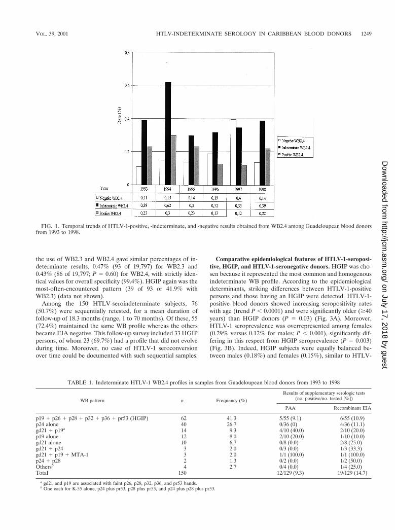
HTLV-1 serological results. Out of the 37,724 enrolledblood donors, 297 (0.79%) were RR in Abbott EIA. Of these,77 (25.9%) were HTLV-1 seropositive by WB2.4, yielding anoverall 0.20% seroprevalence (95% confidence interval [CI],0.16 to 0.26%), and 150 (50.5%) had an HTLV-1-seroindeter-minate WB2.4 pattern. This leads to an overall 0.40% preva-lence of HTLV-1-seroindeterminate donors (CI, 0.34 to0.47%), significantly higher than that of HTLV-1-positive do-nors (P , 0.0001). Finally, 70 samples (23.6%) were WB2.4seronegative. As shown in Fig. 1, the annual prevalence ratesfor HTLV-1-seroindeterminate results, obtained with WB2.4,were relatively stable during the studied period (except for a0.62% peak, of unknown origin, in 1994). They were alsosteadily and significantly higher than the HTLV-1-positiverates, especially for 1994, 1996, and 1997 (P 5 0.005, 0.02, and0.008, respectively).
As shown in Table 1 and Fig. 2, careful examination of the150 HTLV-indeterminate WB2.4 profiles allowed the identifi-cation of several different profile categories. The HGIP, exhib-iting reactivities to p19, p26, p28, p32, p36, and pr53, butlacking both p24 and Env bands, was the most frequent (62 of150 or 41.3%). Other indeterminate WB2.4 profiles were iden-tified, including those with isolated bands such as p24 (n 5 40),p19 (n 5 12), or gd21 (n 5 10). Furthermore, 20 specimensdisplayed reactivity to only one gag protein (p19 or p24) plusone env-encoded glycoprotein (i.e., gd21), either associated ornot with MTA-1. Among these 20 specimens, the most com-mon WB profile (n 5 14) exhibited reactivities to gd21 and p19associated with faint p26, p28, p32, p36, pr53 bands but lackingboth p24 and MTA-1 reactivities. This WB profile was closelyrelated to HGIP, except for the presence of the gd21 reactivity.
Among the 150 HTLV-indeterminate WB sera, 129 could beretested by complementary tests and the majority were scorednegative when screened by PAA or recombinant EIA, leadingan enhanced overall specificity of 90.7 or 85.3%, respectively(Table 1).
Considering the first 3-year screening period (1993 to 1995),
1248 ROUET ET AL. J. CLIN. MICROBIOL.
on July 17, 2018 by guesthttp://jcm
.asm.org/
Dow
nloaded from
the use of WB2.3 and WB2.4 gave similar percentages of in-determinate results, 0.47% (93 of 19,797) for WB2.3 and0.43% (86 of 19,797; P 5 0.60) for WB2.4, with strictly iden-tical values for overall specificity (99.4%). HGIP again was themost-often-encountered pattern (39 of 93 or 41.9% withWB2.3) (data not shown).
Among the 150 HTLV-seroindeterminate subjects, 76(50.7%) were sequentially retested, for a mean duration offollow-up of 18.3 months (range, 1 to 70 months). Of these, 55(72.4%) maintained the same WB profile whereas the othersbecame EIA negative. This follow-up survey included 33 HGIPpersons, of whom 23 (69.7%) had a profile that did not evolveduring time. Moreover, no case of HTLV-1 seroconversionover time could be documented with such sequential samples.
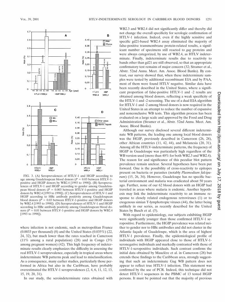
Comparative epidemiological features of HTLV-1-seroposi-tive, HGIP, and HTLV-1-seronegative donors. HGIP was cho-sen because it represented the most common and homogenousindeterminate WB profile. According to the epidemiologicaldeterminants, striking differences between HTLV-1-positivepersons and those having an HGIP were detected. HTLV-1-positive blood donors showed increasing seropositivity rateswith age (trend P , 0.0001) and were significantly older ($40years) than HGIP donors (P 5 0.03) (Fig. 3A). Moreover,HTLV-1 seroprevalence was overrepresented among females(0.29% versus 0.12% for males; P , 0.001), significantly dif-fering in this respect from HGIP seroprevalence (P 5 0.003)(Fig. 3B). Indeed, HGIP subjects were equally balanced be-tween males (0.18%) and females (0.15%), similar to HTLV-
FIG. 1. Temporal trends of HTLV-1-positive, -indeterminate, and -negative results obtained from WB2.4 among Guadeloupean blood donorsfrom 1993 to 1998.
TABLE 1. Indeterminate HTLV-1 WB2.4 profiles in samples from Guadeloupean blood donors from 1993 to 1998
WB pattern n Frequency (%)
Results of supplementary serologic tests(no. positive/no. tested [%])
PAA Recombinant EIA
p19 1 p26 1 p28 1 p32 1 p36 1 pr53 (HGIP) 62 41.3 5/55 (9.1) 6/55 (10.9)p24 alone 40 26.7 0/36 (0) 4/36 (11.1)gd21 1 p19a 14 9.3 4/10 (40.0) 2/10 (20.0)p19 alone 12 8.0 2/10 (20.0) 1/10 (10.0)gd21 alone 10 6.7 0/8 (0.0) 2/8 (25.0)gd21 1 p24 3 2.0 0/3 (0.0) 1/3 (33.3)gd21 1 p19 1 MTA-1 3 2.0 1/1 (100.0) 1/1 (100.0)p24 1 p28 2 1.3 0/2 (0.0) 1/2 (50.0)Othersb 4 2.7 0/4 (0.0) 1/4 (25.0)Total 150 12/129 (9.3) 19/129 (14.7)
a gd21 and p19 are associated with faint p26, p28, p32, p36, and pr53 bands.b One each for K-55 alone, p24 plus pr53, p28 plus pr53, and p24 plus p28 plus pr53.
VOL. 39, 2001 HTLV-INDETERMINATE SEROLOGY IN CARIBBEAN BLOOD DONORS 1249
on July 17, 2018 by guesthttp://jcm
.asm.org/
Dow
nloaded from
seronegative donors (P 5 0.58) (Fig. 3B). Furthermore,HTLV-1-seropositive donors were significantly more likely tobe positive (0.57%) than negative (0.16%) (P , 0.001) for HBcantibodies. This significantly differentiated them from HGIPdonors (P 5 0.03) (Fig. 3C). By contrast, the percentages ofHGIP persons positive (0.22%) and negative (0.16%) for HBcantibodies were similar to those for HTLV-seronegative do-nors (P 5 0.33) (Fig. 3C). Finally, the HTLV-1 seroprevalencewas clearly greater along the Atlantic facade of Guadeloupe(0.40%), an area of microendemicity, than in other areas(0.20%) (P 5 0.016), which was not the case for HGIP persons(P 5 0.01) (Fig. 3D). By contrast, no significant difference inthis geographic determinant between HGIP and HTLV-1-se-ronegative donors (0.08% for Atlantic facade versus 0.19% forother areas; P 5 0.13) could be detected.
Detection of HTLV-1 DNA sequences in the PBMCs by PCR.All the 43 studied DNA samples gave a positive result withprimers that amplify the b-globin gene. A positive PCR signalwas clearly detected in the PBMC DNA of 22 out of 24 HTLV-1-seropositive specimens with all the primer pairs, as well as inthe HTLV-1- and HTLV-2-positive control DNAs. However,for two HTLV-1-seropositive specimens having an optical den-sity ratio by EIA of .15 and exhibiting a peculiar WB profilewith strong env protein (gd21 and MTA-1) but very weak gagprotein (p19 and p24) antibody reactivities, PCR results werenegative. Furthermore, new PBMC DNAs were extracted for
these two persons and retested by PCR for HTLV-1, but theresults remained negative. No signal could be detected in thePBMC DNAs of 17 HTLV-1-seroindeterminate subjects, in-cluding 13 persons with an HGIP, 3 subjects with a gd21-plus-p19 pattern, and 1 person with an isolated p24 band. No signalwas also obtained from the DNA of the HTLV-1- and -2-seronegative specimen as well as for the control DNA-freetube. A sample (6802) exhibiting a faint gd21- and p19-positiveMTA-1 pattern but lacking p24 gave positive PCR results onlywith primer pairs amplifying the gag and tax genes. To avoid apossible lack of PCR sensitivity, we performed a nested PCR(one for the tax gene and the other for the long terminal repeatregion) as previously described (26) for the few samples withdiscordant results. Only the three HTLV-1-positive controlsand sample 6802 gave HTLV-1-positive results.
DISCUSSION
HTLV-1- and -2-seroindeterminate WB patterns are preva-lent worldwide, with rates fluctuating considerably accordingto countries. The present 0.4% seroindeterminate rate foundin Guadeloupe (French West Indies) appeared comparable tothose previously documented for blood donors in other WestIndian or South American countries, such as Martinique(0.50%) (6) and Brazil (0.63%) (38). This rate also appearedclearly higher than those found among donors from areas
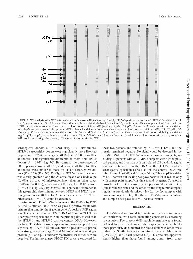
FIG. 2. WB analysis using WB2.4 from Genelabs Diagnostic Biotechnology. Lane 1, HTLV-1-positive control; lane 2, HTLV-2-positive control;lane 3, serum from one Guadeloupean blood donor with an isolated p24 band; lanes 4 and 5, sera from two Guadeloupean blood donors with anHGIP; lane 6, serum from one Guadeloupean blood donor exhibiting gd21 (weak), p19, p26, p28, p32, p36, and pr53 bands but without reactivitiesto both p24 and env-encoded glycoprotein MTA-1; lanes 7 and 8, sera from three Guadeloupean blood donors exhibiting gd21, p19, p26, p28, p32,p36, and pr53 bands but without reactivities to both p24 and MTA-1; lane 9, serum from one Guadeloupean blood donor exhibiting reactivitiesto gd21, p24, and p28, but without reactivities to both p19 and MTA-1; lane 10, serum from one Guadeloupean blood donor with a nearly completeWB profile but lacking p24 reactivity. This subject was positive in PCR.
1250 ROUET ET AL. J. CLIN. MICROBIOL.
on July 17, 2018 by guesthttp://jcm
.asm.org/
Dow
nloaded from
where infection is not endemic, such as metropolitan France(0.0033 per thousand) (8) and the United States (0.035%) (22,24, 32), but much lower than the rates reached in Cameroon(11% among a rural population) (28) and in Congo (3%among pregnant women) (42). This high frequency of indeter-minate results clearly emphasizes the difficulty in assessing thereal HTLV-1 seroprevalence, especially in tropical areas whereindeterminate WB patterns peak and lead to misclassification.As a consequence, many earlier studies, particularly those per-formed in Africa but also in the Caribbean, have probablyoverestimated the HTLV-1 seroprevalences (2, 4, 6, 11, 12, 13,15, 19, 20, 31).
In our study, the seroindeterminate rates obtained with
WB2.3 and WB2.4 did not significantly differ and thereby didnot change the overall specificity for serologic confirmation ofHTLV-1 infection. Indeed, even if the highly sensitive andspecific gd21-based WB2.4 assay eliminated the majority offalse-positive transmembrane protein-related results, a signif-icant number of specimens still reacted to gag proteins andwere always categorized, by use of WB2.4, as HTLV indeter-minate. Finally, indeterminate results due to reactivity tobands other than gd21 are still observed, so that an appropriateconfirmatory test remains of major concern (32; Stramer et al.,Abstr. 52nd Annu. Meet. Am. Assoc. Blood Banks). By con-trast, our survey showed that, when these indeterminate sam-ples were tested by additional recombinant EIA and by PAA,most of them were found HTLV negative. Similar data havebeen recently described in the United States, where a signifi-cant proportion of false-positive HTLV-1 and -2 results areobtained among blood donors, reflecting a weak specificity ofthe HTLV-1 and -2 screening. The use of a dual EIA algorithmfor HTLV-1 and -2 among blood donors is now required in theUnited States in an attempt to reduce the number of expansiveand nonconclusive WB tests. This algorithm process has beenevaluated on a large scale and approved by the Food and DrugAdministration (Stramer et al., Abstr. 52nd Annu. Meet. Am.Assoc. Blood Banks).
Although our survey disclosed several different indetermi-nate WB patterns, the leading one among local blood donorswas the HGIP, previously described in Cameroon (26, 28),other African countries (11, 42, 44), and Melanesia (20, 31).Among all the HTLV-indeterminate patterns, the frequency ofHGIP in Guadeloupe was particularly high regardless of theWB version used (more than 40% for both WB2.3 and WB2.4).The reason for and significance of this peculiar blot patternprevalence remain unclear. Several hypotheses have been putforward. One is the possibility of cross-reactivity to epitopespresent on bacteria or parasites (notably Plasmodium falcipa-rum) (15, 26, 34). However, Guadeloupe has no specific bac-terial environment and malaria was eradicated about 50 yearsago. Further, none of our 62 blood donors with an HGIP hadtraveled in areas where malaria is endemic. Another hypoth-esis may link the indeterminate reactivity to an immune re-sponse to closely related endogenous retroviruses (1) or toexogenous simian T-lymphotropic viruses (44), the latter beingunlikely in our series, as recently described for the UnitedStates by Busch et al. (5).
With regard to epidemiology, our subjects exhibiting HGIPwere significantly younger than those confirmed HTLV-1 se-ropositive. Furthermore, the HGIP prevalence was related nei-ther to gender nor to HBc antibodies and did not cluster in theAtlantic facade of Guadeloupe, which is the area of highestHTLV-1 prevalence. Finally, the epidemiological profile ofindividuals with HGIP appeared close to those of HTLV-1-seronegative individuals and markedly contrasted with those ofHTLV-1-seropositive individuals. Such contrast confirms theinitial data obtained by Mauclere et al. in Cameroon (28) butextends these findings to the Caribbean area, strongly suggest-ing that such an indeterminate Gag WB pattern does notappear to reflect true HTLV-1 infection. This statement wasconfirmed by the use of PCR. Indeed, this technique did notdetect HTLV-1 sequences in the PBMC of 13 tested HGIPpersons. It must be pointed out that the majority of previous
FIG. 3. (A) Seroprevalences of HTLV-1 and HGIP according toage among Guadeloupean blood donors (P 5 0.03 between HTLV-1-positive and HGIP donors by WB2.4 [1993 to 1998]). (B) Seropreva-lences of HTLV-1 and HGIP according to gender among Guadelou-pean blood donors (P 5 0.003 between HTLV-1-positive and HGIPdonors by WB2.4 [1993 to 1998]). (C) Seroprevalences of HTLV-1 andHGIP according to HBc antibody positivity among Guadeloupeanblood donors (P 5 0.03 between HTLV-1-positive and HGIP donorsby WB2.4 [1993 to 1998]). (D) Seroprevalences of HTLV-1 and HGIPaccording to HBc antibody positivity among Guadeloupean blood do-nors (P 5 0.01 between HTLV-1-positive and HGIP donors by WB2.4[1993 to 1998]).
VOL. 39, 2001 HTLV-INDETERMINATE SEROLOGY IN CARIBBEAN BLOOD DONORS 1251
on July 17, 2018 by guesthttp://jcm
.asm.org/
Dow
nloaded from
studies, performed in various areas, also failed to detectHTLV-1 proviral sequences, even by the use of highly con-served HTLV-1 and HTLV-2 primers on fresh or culturedPBMCs of those individuals presenting an HTLV indetermi-nate WB pattern (15, 18, 22, 26, 31, 44). However, a recentreport has described the amplification of an HTLV-1 tax se-quence from patients with neurological disease exhibiting anHGIP WB reactivity. This suggests that this seroindeterminateWB pattern might be associated in some rare cases with de-fective HTLV-1 strains or with a novel retrovirus having ho-mology with HTLV-1, or finally with slowly replicatingHTLV-1 (39, 47). In addition, it seems unlikely that the HGIPmay represent a delayed or slow seroconversion, because mostof our followed-up subjects did not show any evolution of theirWB profile over time and because the minority who did be-came EIA negative. However, we noticed that some of thelatter retained an HGIP, but with a significantly decreasedresponse to the Gag bands, likely reflecting a lower level ofantigenic stimulation. Finally, in our study, all seroindetermi-nate patterns do not correspond to an HTLV-1 seroconver-sion, contrary to a recent study carried out in Martinique,where 3 of 49 HTLV-seroindeterminate donors were reportedas being HTLV-1 seroconverters (6).
In conclusion, our data confirm that the stringent criteria forWB positivity proposed by the HTLV European ResearchNetwork (40) must be accurately carried out, especially forsamples originating from tropical areas. These criteria statethat, to be considered HTLV-1 positive, WB-tested sera mustreact with at least two native gag proteins, p19 and p24, inaddition to two recombinant env glycoproteins, gd21 andMTA-1. However, special attention must be paid to low-inten-sity signals: indeed, two “HTLV-1-seropositive” specimens inour study exhibiting a peculiar pattern with strong env proteinreactivities but very weak gag protein reactivities were PCRnegative. In such rare cases of faintly positive samples, it seemsnecessary to perform PCR in order to distinguish between trueand false HTLV-1 seropositivity. Conversely, one indetermi-nate sample in our study with the gd211 p191 p242 profilealong with MTA-1 reactivity was PCR positive. Similar resultshave been obtained in metropolitan France, where two inde-terminate samples with the same pattern were also PCR pos-itive (10). On the basis of these data, and by analogy withHTLV-2 seropositivity criteria, which required only threebands (i.e., gd21, p24, and K-55), we propose that HTLV-1seropositivity should be based on the presence of at least thethree reactivities gd21, p19, and MTA-1, even if p24 is lacking.By contrast, when both MTA-1 and p24, or the env proteinreactivities (such as HGIP) are lacking, our survey failed todetect, by PCR, evidence of HTLV-1 provirus in all cases.Healthy blood donors with such HGIP test results should bereassured that they are unlikely to be infected with HTLV-1 orHTLV-2.
ACKNOWLEDGMENTS
We thank Renaud Mahieux for critical review of this manuscript.We thank the Agence Nationale de Recherche contre le SIDA for
financial support.
REFERENCES
1. Banki, K., J. Maceda, E. Hurley, E. Ablonczy, D. H. Mattson, L. Szegedy, C.Hung, and A. Perl. 1992. Human T-cell lymphotropic virus (HTLV)-related
endogenous sequence, HRES-1, encodes a 28-kDa protein: a possible au-toantigen for HTLV-I gag-reactive autoantibodies. Proc. Natl. Acad. Sci.USA 89:1939–1943.
2. Bonis, J., P. M. Preux, L. Nzisabira, L. Letenneur, G. Muhirwa, T. Buzingo,A. Kamuragiye, C. Preux, E. Ngoga, M. Dumas, and F. Denis. 1994. HTLV-Iin Burundi (East Africa): lack of reactivity to the HTLV-I immunodominantenvelope epitope. J. Acquir. Immune Defic. Syndr. 7:1099–1100.
3. Brodine, S. K., E. M. Kaime, C. Roberts, R. P. Turnicky, and R. B. Lal. 1993.Simultaneous confirmation and differentiation of human T-lymphotropicvirus types I and II infection by modified Western blot containing recombi-nant envelope glycoproteins. Transfusion 33:925–929.
4. Busch, M. P., M. Laycock, S. H. Kleinman, J. W. Wages, Jr., M. Calabro,J. E. Kaplan, R. F. Khabbaz, and C. G. Hollingsworth. 1994. Accuracy ofsupplementary serologic testing for human T-lymphotropic virus types I andII in US blood donors. Retrovirus Epidemiology Donor Study. Blood 83:1143–1148.
5. Busch, M. P., W. M. Switzer, E. L. Murphy, R. Thomson, and W. Heneine.2000. Absence of evidence of infection with divergent primate T-lympho-tropic viruses in United States blood donors who have seroindeterminateHTLV test results. Transfusion 40:443–449.
6. Cesaire, R., O. Bera, H. Maier, A. Lezin, J. Martial, M. Ouka, B. Kerob-Bauchet, A. K. Ould Amar, and J. C. Vernant. 1999. Seroindeterminatepatterns and seroconversions to human T-lymphotropic virus type I positivityin blood donors from Martinique, French West Indies. Transfusion 39:1145–1149.
7. Cossen, C., S. Hagens, R. Fukuchi, B. Forghani, D. Gallo, and M. Ascher.1992. Comparison of six commercial human T-cell lymphotropic virus type I(HTLV-I) enzyme immunoassay kits for detection of antibody to HTLV-Iand -II. J. Clin. Microbiol. 30:724–725.
8. Courouce, A. M., J. Pillonel, J. M. Lemaire, M. Maniez, and J. B. Brunet.1993. Seroepidemiology of HTLV-I/II in universal screening of blood dona-tions in France. AIDS 7:841–847.
9. Cowan, E. P., G. J. Nemo, A. E. Williams, R. K. Alexander, A. Vallejo, I. K.Hewlett, R. B. Lal, C. S. Dezzutti, D. Gallahan, K. George, B. A. Pancake, D.Zucker-Franklin, P. R. McCurdy, and E. Tabor. 1999. Absence of humanT-lymphotropic virus type I tax sequences in a population of normal blooddonors in the Baltimore, MD/Washington, DC, area: results from a multi-center study. Transfusion 39:904–909.
10. Defer, C., J. Coste, F. Descamps, S. Voisin, J. M. Lemaire, M. Maniez, A. M.Courouce, and the Retrovirus Study Group of The French Society of BloodTransfusion. 1995. Contribution of polymerase chain reaction and radioim-munoprecipitation assay in the confirmation of human T-lymphotropic virusinfection in French blood donors. Transfusion 35:596–600.
11. Delaporte, E., M. Peeters, J. P. Durand, A. Dupont, D. Schrijvers, L. Bed-jabaga, C. Honore, S. Ossari, A. Trebucq, R. Josse, and M. Merlin. 1989.Seroepidemiological survey of HTLV-I infection among randomized popu-lations of western central African countries. J. Acquir. Immune Defic. Syndr.2:410–413.
12. de The, G., A. Gessain, L. Gazzolo, M. Robert-Guroff, G. Najberg, A.Calender, M. Peti, G. Brubaker, A. Bensliman, F. Fabry, et al. 1985. Com-parative seroepidemiology of HTLV-I and HTLV-III in the French WestIndies and some African countries. Cancer Res. 45:4633s–4636s.
13. Dumas, M., D. Houinato, M. Verdier, T. Zohoun, R. Josse, J. Bonis, I.Zohoun, A. Massougbodji, and F. Denis. 1991. Seroepidemiology of humanT-cell lymphotropic virus type I/II in Benin (West Africa). AIDS Res. Hum.Retroviruses 7:447–451.
14. Gessain, A., F. Barin, J. C. Vernant, O. Gout, L. Maurs, A. Calender, and G.de The. 1985. Antibodies to human T-lymphotropic virus type-I in patientswith tropical spastic paraparesis. Lancet ii:407–410.
15. Gessain, A., R. Mahieux, and G. de The. 1995. HTLV-I “indeterminate”Western blot patterns observed in sera from tropical regions: the situationrevisited. J. Acquir. Immune Defic. Syndr. Hum. Retrovirol. 9:316–318.
16. Hayes, C. G., J. P. Burans, and R. B. Oberst. 1991. Antibodies to human Tlymphotropic virus type I in a population from the Philippines: evidence forcross-reactivity with Plasmodium falciparum. J. Infect. Dis. 163:257–262.
17. Kawase, K., S. Katamine, R. Moriuchi, T. Miyamoto, K. Kubota, H. Iga-rashi, H. Doi, Y. Tsuji, T. Yamabe, and S. Hino. 1992. Maternal transmissionof HTLV-I other than through breast milk: discrepancy between the poly-merase chain reaction positivity of cord blood samples for HTLV-I and thesubsequent seropositivity of individuals. Jpn. J. Cancer Res. 83:968–977.
18. Khabbaz, R. F., W. Heneine, A. Grindon, T. M. Hartley, G. Shulman, and J.Kaplan. 1992. Indeterminate HTLV serologic results in U.S. blood donors:are they due to HTLV-I or HTLV-II? J. Acquir. Immune Defic. Syndr.5:400–404.
19. Lal, R., J. J. Lipka, S. K. H. Foung, K. G. Hadlock, G. R. Reyes, and W. P.Carney. 1993. Human T lymphotropic virus type I/II in Lake Lindu Valley,central Sulawesi, Indonesia. J. Acquir. Immune Defic. Syndr. 9:1067–1068.
20. Lal, R., D. L. Rudolph, V. Nerurkar, and R. Yanagihara. 1992. Humoralresponses to the immunodominant gag and env epitopes of Human T-Lymphotropic virus type I among Melanesians. Vir Immunol. 4:265–72.
21. Lal, R. B., S. Brodine, J. Kazura, E. Mbidde-Katonga, R. Yanagihara, and C.Roberts. 1992. Sensitivity and specificity of a recombinant transmembrane
1252 ROUET ET AL. J. CLIN. MICROBIOL.
on July 17, 2018 by guesthttp://jcm
.asm.org/
Dow
nloaded from
glycoprotein (rgp21)-spiked Western immunoblot for serological confirma-tion of human T-cell lymphotropic virus type I and type II infections. J. Clin.Microbiol. 30:296–299.
22. Lal, R. B., D. L. Rudoph, J. E. Coligan, S. K. Brodine, and C. R. Roberts.1992. Failure to detect evidence of human T-lymphotropic virus (HTLV)type I and type II in blood donors with isolated gag antibodies to HTLV-I/II.Blood 80:544–550.
23. Lipka, J. J., K. K. Y. Young, S. Y. Kwok, G. R. Reyes, J. J. Sninsky, andS. K. H. Foung. 1991. Significance of human T-lymphotropic virus type Iindeterminant serological findings among healthy individuals. Vox Sang.61:171–176.
24. Liu, H., M. Shah, S. L. Stramer, W. Chen, B. J. Weiblen, and E. L. Murphy.1999. Sensitivity and specificity of human T-lymphotropic virus (HTLV)types I and II polymerase chain reaction and several serologic assays inscreening a population with a high prevalence of HTLV-II. Transfusion39:1185–1193.
25. Mahieux, R., J. Pecon-Slattery, and A. Gessain. 1997. Molecular character-ization and phylogenetic analyses of a new, highly divergent simian T-celllymphotropic virus type 1 (STLV-1marc1) in Macaca arctoides. J. Virol.71:6253–6258.
26. Mahieux, R., P. Horal, P. Mauclere, O. Mercereau-Puijalon, M. Guillotte, L.Meertens, E. Murphy, and A. Gessain. 2000. Human T-cell lymphotropicvirus type-1 Gag indeterminate Western blot patterns in central Africa:relationship to Plasmodium falciparum infection. J. Clin. Microbiol. 38:4049–4057.
27. Manns, A., M. Hisada, and L. La Grenade. 1999. Human T-lymphotropicvirus type I infection. Lancet 353:1951–1958.
28. Mauclere, P., J. Y. Le Hesran, R. Mahieux, R. Salla, J. Mfoupouendoun,E. T. Abada, J. Millan, G. de The, and A. Gessain. 1997. Demographic,ethnic, and geographic differences between human T cell lymphotropic virus(HTLV) type I-seropositive carriers and persons with HTLV-I gag-indeter-minate Western blots in central Africa. J. Infect. Dis. 176:505–509.
29. Montplaisir, N., L. Valette, Y. Dezaphy, and C. Neisson-Vernant. 1989.Blood transfusion and HTLV1 infection in Martinique, p. 533–539. In G. C.Roman, J. C. Vernant, and M. Osame (ed.), HTLV-I and the nervoussystem. Liss, New York, N.Y.
30. Murphy, E. L., J. P. Figueroa, W. N. Gibbs, M. Holding-Cobham, B. Cran-ston, K. Malley, A. J. Bodner, S. S. Alexander, and W. A. Blattner. 1991.Human T-lymphotropic virus type I (HTLV-I) seroprevalence in Jamaica. I.Demographic determinants. Am. J. Epidemiol. 133:1114–1124.
31. Nerurkar, V. R., M. A. Miller, M. E. Leon-Monzon, A. B. Ajdukiewicz, C. L.Jenkins, R. C. Sanders, M. S. Godec, R. M. Garruto, and R. Yanagihara.1992. Failure to isolate human T cell lymphotropic virus type I and to detectvariant-specific genomic sequences by polymerase chain reaction in Melane-sians with indeterminate Western immunoblot. J. Gen. Virol. 73:1805–1810.
32. Ownby, H. E., J. J. Korelitz, M. P. Busch, A. E. Williams, S. H. Kleinman,R. O. Gilcher, P. Nourjah, and the Retrovirus Epidemiology Donor Study.1997. Loss of volunteer blood donors because of unconfirmed enzyme im-munoassay screening results. Transfusion 37:199–205.
33. Poiesz, B. J., F. W. Ruscetti, A. F. Gazdar, P. A. Bunn, J. D. Minna, and R. C.Gallo. 1980. Detection and isolation of type C retrovirus particles from freshand cultured lymphocytes of a patient with cutaneous T-cell lymphoma.Proc. Natl. Acad. Sci. USA 77:7415–7419.
34. Porter, K. R., J. Aguiar, A. Richards, B. Sandjaya, H. Ignatias, H. Hadipu-tranto, R. G. Ridley, B. Takacs, F. S. Wignall, S. L. Hoffman, and C. G.Hayes. 1998. Immune response against the Exp-1 protein of Plasmodiumfalciparum results in antibodies that cross-react with human T-cell lympho-
tropic virus type 1 proteins. Clin. Diagn. Lab. Immunol. 5:721–724.35. Rouet, F., C. Foucher, M. Rabier, I. Gawronski, D. Taverne, B. Chancerel, O.
Casman, and M. Strobel. 1999. Human T-lymphotropic virus type I(HTLV-I) among blood donors from Guadeloupe: donation, demographic,and biological characteristics. Transfusion 39:639–644.
36. Rouet, F., R. Rabier, C. Foucher, B. Chancerel, F. Agis, and M. Strobel.1999. Geographical clustering of human T-cell lymphotropic virus type I inGuadeloupe, an endemic Caribbean area. Int. J. Cancer 81:330–334.
37. Sabino, E. C., M. Zrein, C. P. Taborda, M. M. Otani, G. Ribeiro-Dos-Santos,and A. Saez-Alquezar. 1999. Evaluation of the INNO-LIA HTLV I/II assayfor confirmation of human T-cell leukemia virus-reactive sera in blood bankdonations. J. Clin. Microbiol. 37:1324–1328.
38. Segurado, A. A., C. M. Malaque, L. M. Sumita, C. S. Pannuti, and R. B. Lal.1997. Laboratory characterization of human T cell lymphotropic virus types1 (HTLV-1) and 2 (HTLV-2) infections in blood donors from Sao Paulo,Brazil. Am. J. Trop. Med. Hyg. 57:142–148.
39. Soldan, S. S., M. D. Graf, A. Waziri, A. N. Flerlage, S. M. Robinson, T.Kawanishi, T. P. Leist, T. J. Lehky, M. C. Levin, and S. Jacobson. 1999.HTLV-I/II seroindeterminate Western blot reactivity in a cohort of patientswith neurological disease. J. Infect. Dis. 180:685–694.
40. Taylor, G. P. 1996. The epidemiology of HTLV-I in Europe. J. Acquir.Immune Defic. Syndr. 13(Suppl. 1):S8–S14.
41. Touze, E., A. Gessain, O. Lyon-Caen, and O. Gout. 1996. Tropical spasticparaparesis/HTLV-I-associated myelopathy in Europe and in Africa: clinicaland epidemiologic aspects. J. Acquir. Immune Defic. Syndr. 13(Suppl. 1):S38–S45.
42. Tuppin, P., M. Makuwa, T. Guerma, M. M. Bazabana, J. C. Loukaka, D.Jeannel, P. M’Pele, and G. de The. 1996. Low HTLV-I/II seroprevalence inpregnant women in Congo and a geographic cluster of an HTLV-like inde-terminate Western blot pattern. J. Acquir. Immune Defic. Syndr. 11:105–107.
43. Uchiyama, T., J. Yodoi, K. Sagawa, K. Takatsuki, and H. Uchino. 1977.Adult T-cell leukemia: clinical and hematologic features of 16 cases. Blood50:481–492.
44. Vandamme, A. M., K. Van Laethem, H. F. Liu, M. Van Brussel, E. Delaporte,C. M. de Castro Costa, C. Fleischer, G. Taylor, U. Bertazzoni, J. Desmyter,and P. Goubau. 1997. Use of a generic polymerase chain reaction assaydetecting human T-lymphotropic virus (HTLV) types I, II and divergentsimian strains in the evaluation of individuals with indeterminate HTLVserology. J. Med. Virol. 52:1–7.
45. Varma, M., D. L. Rudolph, M. Knuchel, W. M. Switzer, K. G. Hadlock, M.Velligan, L. Chan, S. K. Foung, and R. B. Lal. 1995. Enhanced specificity oftruncated transmembrane protein for serologic confirmation of human T-cell lymphotropic virus type 1 (HTLV-1) and HTLV-2 infections by Westernblot (immunoblot) assay containing recombinant envelope glycoproteins.J. Clin. Microbiol. 33:3239–3244.
46. Wattel, E., M. Mariotti, F. Agis, E. Gordien, O. Prou, A. M. Courouce, P.Rouger, S. Wain-Hobson, I. S. Chen, and J. J. Lefrere. 1992. Human Tlymphotropic virus (HTLV) type I and II DNA amplification in HTLV-I/II-seropositive blood donors of the French West Indies. J. Infect. Dis. 165:369–372.
47. Waziri, A., S. S. Soldan, M. D. Graf, J. Nagle, and S. Jacobson. 2000.Characterization and sequencing of prototypic human T-lymphotropic virustype 1 (HTLV-1) from an HTLV-1/2 seroindeterminate patient. J. Virol.74:2178–2185.
48. Yamaguchi, K. 1994. Human T-lymphotropic virus type I in Japan. Lancet343:213–216.
VOL. 39, 2001 HTLV-INDETERMINATE SEROLOGY IN CARIBBEAN BLOOD DONORS 1253
on July 17, 2018 by guesthttp://jcm
.asm.org/
Dow
nloaded from