Embed Size (px)
Citation preview

Research ArticleSeroprevalence against Rickettsia and Borrelia Species inPatients with Uveitis: A Prospective Survey
Kim B. Madsen,1 Katarina Wallménius,2 Åke Fridman,1 Carl Påhlson,2
and Kenneth Nilsson2,3,4
1Section of Opthalmology, Falu Hospital, Falun, Sweden2Department of Medical Sciences, Section of Clinical Microbiology, Uppsala University, Uppsala, Sweden3Department of Medical Sciences, Section of Infectious Diseases, Uppsala University, Uppsala, Sweden4Centre of Clinical Research, Falu Hospital, Falun, Sweden
Correspondence should be addressed to Kenneth Nilsson; [email protected]
Received 11 June 2017; Revised 1 October 2017; Accepted 30 October 2017; Published 26 November 2017
Academic Editor: Miguel Cordero-Coma
Copyright © 2017 Kim B. Madsen et al. This is an open access article distributed under the Creative Commons Attribution License,which permits unrestricted use, distribution, and reproduction in any medium, provided the original work is properly cited.
Vector-borne diseases such as Lyme borreliosis and rickettsioses have been associated with ocular inflammation. Our aim was tostudy patients with diagnosed uveitis to evaluate serological signs of infection or exposure to these tick-borne agents. Forty-eightpatients were prospectively examined with serology together with medical records and a questionnaire concerning previousexposure, diseases, and treatments. Seven patients (14.6%) showed seroconversion to Rickettsia spp. between acute andconvalescent phase sera, which provides support for a positive Rickettsia diagnosis according to guidelines. The specificity wasconfirmed by Western blot. Additional 28 patients had stationary titres of which eight (16.6%) had 1 : 256 or higher titre in thefirst serum, and another 13 patients were seronegative. No epidemiological risk factor or marker could be identified. ForBorrelia, only three patients showed moderate IgG titres. A control group of 100 blood donors, 60 patients with rheumaticdisease, and 56 patients seeking medical care were tested of which 2.0–7.1% showed low anti-Rickettsia titres and 3.0–8.3%anti-Borrelia titres. The findings are indicative for an association between infection or exposure to Rickettsia spp. anduveitis with a seropositivity among patients with recurrent uveitis in concordance with the spread of rickettsial exposure ina tick-exposed population.
1. Introduction
The lifetime incidence of definite acute anterior uveitis(AAU), characterized by inflammation of the uvea, isapproximately 0.2% in the general population and 1% inthe histocompatibility antigen HLA-B27-positive population[1–3]. Classical signs of AAU are a red painful eye, photo-phobia, blurred vision, floaters, and sometimes loss ofperipheral vision [4, 5]. Chronic uveitis, on the other hand,may be symptomless in the initial stages but can ultimatelylead to permanent vision loss [4, 6–8].
The known aetiologies of uveitis fall into three main cat-egories: (a) infectious, including viruses and bacteria, wherethe latter may be a contributing factor in combination withHLA-B27, (b) systemic inflammatory disease, and (c) malig-nant and leukemic cells, lymphoma, and melanoma [8–10].
However, the aetiology often remains unknown and the lackof specific pathognomonic signs of certain infectious agentsis not uncommon [4, 11, 12].
Rickettsioses are systemic infections with symptomscaused by vasculitis due to the bacterial marked tropism forthe endothelial cells of small vessels. The Gram-negativeRickettsia bacteria are arthropod vector-borne and world-wide distributed [13, 14]. Ocular manifestations such asscotoma, floaters, redness, or decreased vision are reportedin Rickettsia infections but are usually self-limited and mayeasily be overlooked. Of the spotted fever Rickettsia (SFR),Rickettsia conorii and R. rickettsii have been reported as thecause of mainly posterior uveitis, with a chorioretinalinvolvement [15, 16].
In Sweden, R. helvetica, is, besides a single finding of R.sibirica, the only reported tick-transmitted SFR, occurring
HindawiJournal of OphthalmologyVolume 2017, Article ID 9247465, 10 pageshttps://doi.org/10.1155/2017/9247465

in approximately 5–10% of Ixodes ricinus ticks [17]. Previousserosurveys in Sweden have shown IgG antibodies to Rickett-sia spp. in 3.0–44.0% of tick-exposed subjects, compared to2–43.5% to Borrelia spp. [18–23]. Reported patients exposedto Rickettsia spp. have shown variable usually self-limitingunspecific symptoms such as flu-like fever, but sometimesmore severe symptoms like meningitis or facial paralysis[24–26]. Moreover, the louse-borne R. felis has beendetected in Sweden in patients with neurological symp-toms, but thus far, R. felis has not been reported in anyvector in Sweden [27].
The aim was to prospectively study patients presentingsymptoms of and being diagnosed with uveitis, regardlessof localization in the eye, to investigate if serological signsof Borrelia or Rickettsia infection or exposure are associatedwith acute or recurrent uveitis, for what would otherwise beconsidered a noninfectious uveitis.
2. Patients and Methods
2.1. Patients. A total of 48 patients diagnosed with uveitis,representing 1/3 of all patients with that diagnosis during2013 and 2014, at the Ophthalmological Clinic, Falun Hospi-tal, were after acceptance of informed consent included in thestudy, regardless of which part of the eye that was affected.Patients were sampled for two sera (S1-S2): sample 1 (S1)within the first 2 weeks of presentation and S2 up to 4–6weeks later. Some patients who were treated had additionaltitres drawn (S3-S4). All patients that fulfilled the selectioncriteria, comprising that the uveitis had to be primary (acute,recurrent, or chronic) and not a secondary complication tointraocular surgery, corneal infections, etc., were includedin the study. All patients were given a complete ocularexamination at every visit, that is, visual acuity, slit lampbiomicroscopy examination (including indirect slit lampbiomicroscopy of the posterior pole), and tonometry. Theuveitis was classified according to accepted practices(SUN working group definitions) as anterior, intermediate,posterior, or panuveitis.
All included patients were asked to answer a question-naire regarding prior tick or flea bite, frequent visits to for-est/rural areas, exposure to animals, past episodes of uveitis,rheumatic and autoimmune diseases, prior testing forHLA-B27, sarcoidosis, tuberculosis, inflammatory bowel dis-ease, and antibiotic treatment. Information concerning pre-vious and actual symptoms and diagnoses according to thequestions in the questionnaire, laboratory findings, and ini-tial treatment were obtained from the medical records whichverified or expanded the information given.
Treatment primarily consisted of topical steroids (dexa-methasone) and topical cycloplegic agents (cyclopentolate).Only a few patients required oral steroid treatment, andone patient was on a methotrexate regimen at the time oftesting, an option for controlling uveitis recurrence [28].
Antibiotic treatment in the form of doxycycline (100mgorally twice a day), for 14 days, was given to patients whohad high initial titres (>1 : 256) against Rickettsia spp. or con-firmed 4-fold rise in titre after the analysis of sample two(S2). Some of these patients were also followed with
additional serum samples (S3, S4) for the determination oftitre after completion of treatment. As a control group, fromthe same geographical area, sera from 100 healthy blooddonors were randomly chosen together with samples from60 patients diagnosed with rheumatic disease and 56 patientsrepresenting patients who had sought medical care for gen-eral medical reasons. The latter group represents patientswith scheduled visits for blood pressure controls and renewalof prescriptions, namely, patients that did not show any signof infection, and the blood samples were taken as part of theregular visit. Patients with rheumatic background were cho-sen as controls to examine their seroreactivity in general,although the reactivity might represent unspecific reactions,and if these patients differ from patients without rheumaticdisease or ongoing infection.
2.2. Serology (Immunofluorescence Assay). R. helvetica-infected Vero cells supplemented with 10% yolk sac solutionwere used as the bacterial antigen for an immunofluores-cence assay (IFA), as previously described [18, 19]. Accord-ing to the guidelines for the diagnosis of tick-bornebacterial diseases in Europe, IgG titers≥ 1 : 128 and/or IgMtiters≥ 1 : 64 and/or a fourfold increase in two sera within a2- to 4-week interval is considered indicative of infection byRickettsia spp. if homologous antigens are used in the IFAtest. The patients were divided into four groups based ontheir serologic results: group 1—a fourfold or greater rise inIgG titre between acute phase (S1) and convalescent phase(S2) sera was tested in parallel; group 2—single IgG endpointtitres of ≥1 : 256 in S1 or S2 were considered presumptive evi-dence of recent or current exposure; group 3—single IgGand/or IgM endpoint titres≥ 1 : 64 and <1 : 256, respectively,were regarded as supportive evidence indicative of either pastexposure or early response to exposure; and group4—titres< 1 : 64 were considered negative for IgG/IgM [26].Persisting IgM antibodies alone was interpreted as nonspe-cific cross-reactivity due to exposure to other organismsand autoimmune responses or possibly as a sign of a previousexposure. A fourfold increase in titre is, according to theguidelines, a criterion that provides a strong support for pos-itive Rickettsia spp. diagnosis along with epidemiological,clinical, laboratory, and bacteriological findings [29]. Aserum sample from a patient with proven endpoint IgG/IgM titres of 1 : 512/1 : 128, respectively, to R. helvetica wasused as a positive control and as a negative control of humanblood donor serum. When discrete structures that were mor-phologically compatible with Rickettsia were visible with ≥2+brightness, the sample was considered to be positive. Labora-tory evidence of current or previous infection with B. burg-dorferi (IgG/IgM) was based on the analysis of serumsamples using a commercial enzyme-linked immunosorbentassay (ELISA), according to the manufacturer’s instructions(Euroimmun AG (Aktiengesellschaft), Lübeck, Germany).For the 48 patients included in the study, only serum two(S2) was tested for Borrelia.
2.3. Western Blot (WB). Sera from seven of the IgG-positivepatients (pat. numbers 1–7, group 1) were diluted to titres1 : 200 and tested for WB with R. helvetica whole cell antigen
2 Journal of Ophthalmology

using Amersham WB system (GE Healthcare) in accordancewith the manufacture’s instructions. Serum from a patientpreviously proven to have a rickettsial infection with highantibody titres in IFA (IgG 1 : 128) was used as the positivecontrol. A serum from a healthy blood donor and the second-ary antibody alone served as the negative controls.
2.4. Statistical Analyses. Standard parametric statistics (confi-dence interval according to Fleiss with Yates correction) wereused for continuous variables, giving a mean+ 95% confi-dence interval (CI). Fisher’s exact test and chi-square test(χ2) were used to compare the proportions, and a p value <0.05 was considered statistically significant. Statistical analy-ses were conducted using Predictive Analytics Software(PASW®) Statistics 20.
3. Results
3.1. Patients. The age distribution was between 13 and 77years (mean age 53 years and median age 53.5 years) includ-ing 20 females and 28 males. Most patients had sought med-ical care within one week, usually one to two days aftersymptom onset. All sera were examined for the presence ofrickettsial antibodies, and all patients were analysed for Bor-relia spp. antibodies in serum S2. None of the patients under-went lumbar puncture. One of the included patients wasexcluded when it was discovered that the diagnosed uveitiswas secondary to cataract surgery as a result of corneal injury.This patient was tested negative for rickettsial antibodies.Five other patients dropped out of the study; they changedtheir decision to participate or failed to leave samples duringthe defined time period. Of the patients in the rheumatic con-trol group (median age 53, 19 men and 41 women), half wastested positive for rheumatoid factor and anticyclic citrulli-nated peptides (anti-CCP) and the other half showed thepresence of antinuclear antibodies (ANA) or ENA, that is,antibodies to extractable nuclear antigens. None of the 56patients (median age 56, 22 men and 34 women), represent-ing patients who had sought medical care for general medicalreasons showed any sign of infection and had normal valuesof C-reactive protein (<10mg/L).
3.2. Serology. The laboratory results and details from themedical record of each patient are summarized in Tables 1and 2. Of the 48 patients with uveitis, seven patients(14.6%) (numbers 1–7) showed a fourfold rise in IgG titrebetween S1 and S2 (Table 1). Of the remaining 41 patients,twelve patients (25%) (numbers 8–19) presented IgG/IgMtitres≥ 1 : 128 indicating a recent or current exposure, ofwhich eight had IgG titres≥ 1 : 256 in the first serum; sixteenpatients (33.3%) (numbers 19–35) had threshold titresbetween ≥1 : 64 and ≤1 : 128 as a result supportive of earlyresponse (IgM), past exposure, or nonspecific reactivity(IgM); and 13 patients (27.1%) (numbers 35–48) were sero-negative (<1 : 64). Of all the 48 patients analysed for conva-lescent sera (S2), three (numbers 1, 18, and 30) had IgGantibodies against Borrelia spp. in moderate titres and threepatients (numbers 16, 20, and 28) showed a slight seroreac-tivity against IgM for Borrelia spp. as well. Among the blood
donors, two of 100 were seropositive in IFA against Rickettsiaspp. with antibody titres at most 1 : 64; three of the 60 (5%)patients with rheumatoid arthritis and four of the 56 (7.1%)patients seeking general medical care presented IgG antibod-ies of at most 1 : 64 except for one case where the titre was1 : 128. The corresponding findings for Borrelia spp. in eachgroup were three (3%), five (8.3%), and four patients(7.1%), showing only low to moderate antibody levels ofIgG of which two individuals also had low IgM titres.
3.3. Western Blot.WB for patient numbers 1–7 showed a spe-cific response against lipopolysaccharide (LPS) and proteinantigens in the 110–150 kDa region for IgG to whole cell anti-gen of R. helvetica (Figure 1). Negative controls in the form ofserum from a healthy blood donor and IFA negative patientshowed no specific reactions.
3.4. Medical Records and Questionnaire.Of the 48 patients inthe study, 45 were diagnosed with uveitis engaging the ante-rior part of the eye, one with intermediate uveitis, and twopatients primarily had a posterior involvement of the inflam-mation (Table 2). Thirteen patients were diagnosed as acute,27 as recurrent acute, and eight as chronic uveitis in accor-dance with the criteria of the SUN Work Group [5].
Only patients with chronic uveitis had persistent inflam-mation after 3 months. In group 1 (pat. numbers 1–7), onlyone of seven patients (pat. number 5) had no prior historyof uveitis but developed a chronic uveitis that still remainedafter three months. Two patients had previously been diag-nosed with chronic uveitis, and the other four patients hadan acute relapse of a recurrent uveitis. In groups 2–4 (pat.numbers 8–48), fifteen patients had no prior history of uve-itis, and five patients were diagnosed with chronic uveitis ofwhich one was a posterior and another an intermediateuveitis. Thirteen patients had acute first time iritis. Theremaining 23 patients all had acute relapses of recurrentuveitis (Table 2).
The questionnaire and medical records showed that for17 of the patients a probable cause of uveitis had previouslybeen determined. These causes consisted of systemic inflam-matory disease, IBD, sarcoidosis, Fuchs heterochromic irido-cyclitis, and white dot syndrome. The remaining 31 causeswere judged as idiopathic (Table 2). The outcome in groups1–4 of the requested connections in the questionnaire is illus-trated in Table 2. No significant statistical differences in theprevalence of symptoms between group 1 and group 4could be demonstrated. Thirteen patients who previouslystated being bitten by a tick were evenly distributedbetween the groups.
4. Discussion
The present study shows that of 48 patients with primaryuveitis, 14.6% showed seroconversion with a fourfoldincrease in titre between two sera and another 16.6% initiallyhigh titres (≥1 : 256); together, 31.2% [CI 7.4–40.6] as sero-logical supportive evidence of an underlying rickettsialinfection or exposure might be associated to the ocularinflammation. In 7 patients (group 1), the specificity of the
3Journal of Ophthalmology
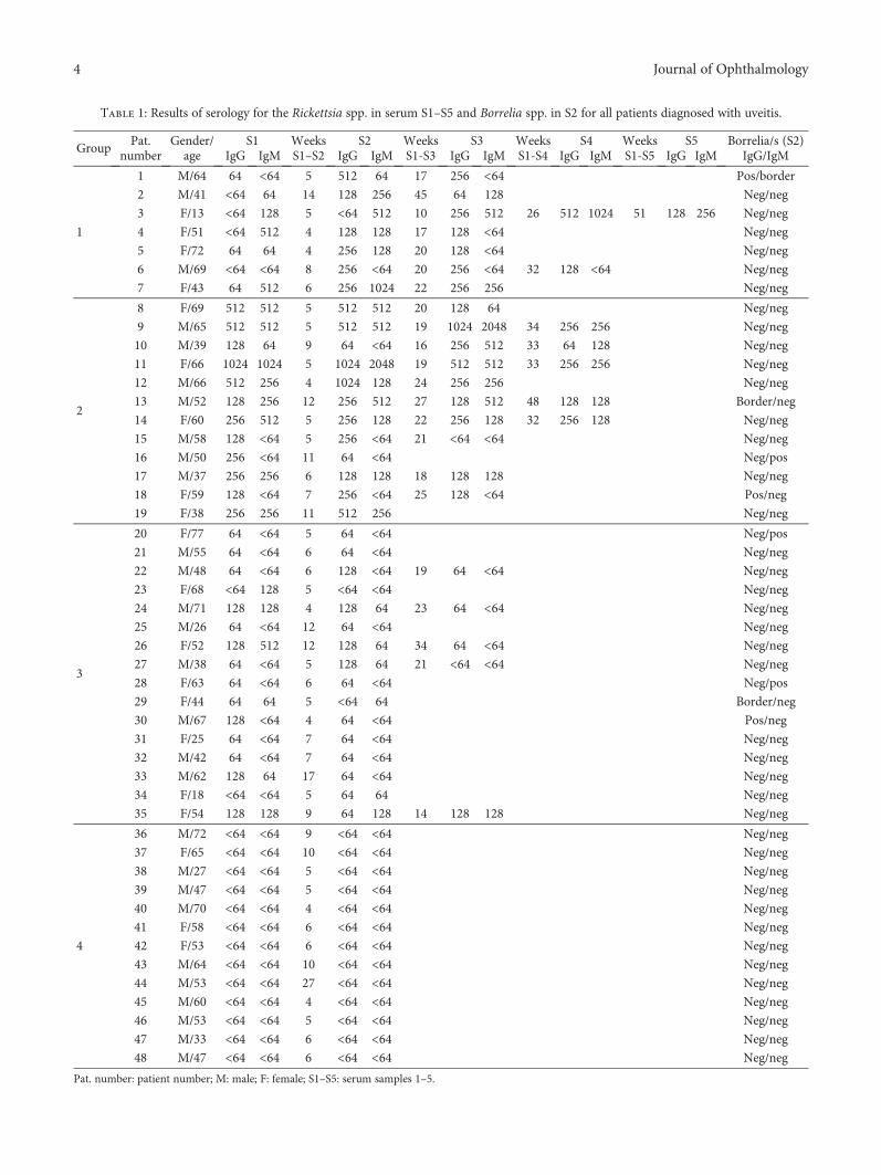
Table 1: Results of serology for the Rickettsia spp. in serum S1–S5 and Borrelia spp. in S2 for all patients diagnosed with uveitis.
GroupPat.
numberGender/age
S1 Weeks S2 Weeks S3 Weeks S4 Weeks S5 Borrelia/s (S2)IgG/IgMIgG IgM S1–S2 IgG IgM S1-S3 IgG IgM S1-S4 IgG IgM S1-S5 IgG IgM
1
1 M/64 64 <64 5 512 64 17 256 <64 Pos/border
2 M/41 <64 64 14 128 256 45 64 128 Neg/neg
3 F/13 <64 128 5 <64 512 10 256 512 26 512 1024 51 128 256 Neg/neg
4 F/51 <64 512 4 128 128 17 128 <64 Neg/neg
5 F/72 64 64 4 256 128 20 128 <64 Neg/neg
6 M/69 <64 <64 8 256 <64 20 256 <64 32 128 <64 Neg/neg
7 F/43 64 512 6 256 1024 22 256 256 Neg/neg
2
8 F/69 512 512 5 512 512 20 128 64 Neg/neg
9 M/65 512 512 5 512 512 19 1024 2048 34 256 256 Neg/neg
10 M/39 128 64 9 64 <64 16 256 512 33 64 128 Neg/neg
11 F/66 1024 1024 5 1024 2048 19 512 512 33 256 256 Neg/neg
12 M/66 512 256 4 1024 128 24 256 256 Neg/neg
13 M/52 128 256 12 256 512 27 128 512 48 128 128 Border/neg
14 F/60 256 512 5 256 128 22 256 128 32 256 128 Neg/neg
15 M/58 128 <64 5 256 <64 21 <64 <64 Neg/neg
16 M/50 256 <64 11 64 <64 Neg/pos
17 M/37 256 256 6 128 128 18 128 128 Neg/neg
18 F/59 128 <64 7 256 <64 25 128 <64 Pos/neg
19 F/38 256 256 11 512 256 Neg/neg
3
20 F/77 64 <64 5 64 <64 Neg/pos
21 M/55 64 <64 6 64 <64 Neg/neg
22 M/48 64 <64 6 128 <64 19 64 <64 Neg/neg
23 F/68 <64 128 5 <64 <64 Neg/neg
24 M/71 128 128 4 128 64 23 64 <64 Neg/neg
25 M/26 64 <64 12 64 <64 Neg/neg
26 F/52 128 512 12 128 64 34 64 <64 Neg/neg
27 M/38 64 <64 5 128 64 21 <64 <64 Neg/neg
28 F/63 64 <64 6 64 <64 Neg/pos
29 F/44 64 64 5 <64 64 Border/neg
30 M/67 128 <64 4 64 <64 Pos/neg
31 F/25 64 <64 7 64 <64 Neg/neg
32 M/42 64 <64 7 64 <64 Neg/neg
33 M/62 128 64 17 64 <64 Neg/neg
34 F/18 <64 <64 5 64 64 Neg/neg
35 F/54 128 128 9 64 128 14 128 128 Neg/neg
4
36 M/72 <64 <64 9 <64 <64 Neg/neg
37 F/65 <64 <64 10 <64 <64 Neg/neg
38 M/27 <64 <64 5 <64 <64 Neg/neg
39 M/47 <64 <64 5 <64 <64 Neg/neg
40 M/70 <64 <64 4 <64 <64 Neg/neg
41 F/58 <64 <64 6 <64 <64 Neg/neg
42 F/53 <64 <64 6 <64 <64 Neg/neg
43 M/64 <64 <64 10 <64 <64 Neg/neg
44 M/53 <64 <64 27 <64 <64 Neg/neg
45 M/60 <64 <64 4 <64 <64 Neg/neg
46 M/53 <64 <64 5 <64 <64 Neg/neg
47 M/33 <64 <64 6 <64 <64 Neg/neg
48 M/47 <64 <64 6 <64 <64 Neg/neg
Pat. number: patient number; M: male; F: female; S1–S5: serum samples 1–5.
4 Journal of Ophthalmology

Table2.:Sum
mationof
theou
tcom
efrom
medicalrecordsandqu
estion
naires
ingrou
ps1–4.
Group
Pat.
number
Inflam
edpart
oftheeye
Previou
suveitis
Possible
cause
uveitis
Acute/
chronic
Con
tactwith
furanim
als
Often
staying
intheforest
Kno
wn
tick
bite
Kno
wn
flea
bite
IBD
Rheum
atic
disease
TB
Sarcoido
sis
HLA
-B27
+
Antibiotic
treatm
entlast
mon
th
1
1ANT
YID
ACa
YY
NN
NN
ND
YN
2ANT
YID
AN
NN
NN
ND
NND
YN
3ANT
YRD
CN
NY
NN
YN
ND
ND
N
4ANT
YID
CCa
YN
YN
NN
ND
ND
T1
5ANT
NFU
CO
YN
NN
NN
NN
T2
6ANT
YMB
ACa+Do
YN
NN
YN
ND
ND
N
7ANT
YID
ACa
YY
NN
NN
ND
NN
2
8ANT
NIBD
AN
YN
YY
NN
ND
ND
N
9ANT
YMB
AN
NN
NN
YN
ND
ND
N
10ANT
YID
ACa
YN
NN
NN
ND
YN
11ANT
YID
ACa+Do
YN
NN
NN
NY
N
12ANT
YMB
AN
NN
NN
YN
ND
YN
13ANT
YRD
AN
YN
NN
YN
ND
ND
N
14ANT
YID
ACa+Do
YY
YN
NN
ND
YN
15ANT
NID
AN
YY
NN
NN
ND
YN
16ANT
YID
AN
YY
NN
NN
ND
YN
17ANT
YMB
AN
NY
YN
YN
ND
YN
18ANT
YID
AN
NN
NN
NN
ND
ND
N
19ANT
YID
CN
NN
NN
NN
NN
N
3
20ANT
NID
AN
NN
NN
NN
ND
ND
N
21ANT
YID
AN
YN
NN
NN
ND
NN
22ANT
NID
AN
YN
NN
NN
ND
ND
N
23ANT
YRD
AN
YY
NN
YN
NY
N
24ANT
YID
AN
YN
NN
NN
NY
N
25ANT
NS
AN
NN
NN
NN
YN
N
26ANT
NID
AN
NN
NN
NN
ND
ND
N
27ANT
NID
AN
NN
NN
NN
ND
NN
27ANT
YID
AN
YN
NN
NN
ND
NN
29ANT
YID
AN
YN
NN
NN
ND
YN
30ANT
NID
AN
YY
NN
NN
ND
YN
31ANT
NID
AN
YY
NN
NN
ND
ND
N
32ANT
NID
ACa
NY
YN
NN
ND
ND
N
33ANT
NMB
AN
YN
NN
YN
ND
ND
N
5Journal of Ophthalmology

Table2.:C
ontinu
ed.
Group
Pat.
number
Inflam
edpart
oftheeye
Previou
suveitis
Possible
cause
uveitis
Acute/
chronic
Con
tactwith
furanim
als
Often
staying
intheforest
Kno
wn
tick
bite
Kno
wn
flea
bite
IBD
Rheum
atic
disease
TB
Sarcoido
sis
HLA
-B27
+
Antibiotic
treatm
entlast
mon
th
34IN
TN
IDC
OY
YN
NN
NND
ND
N
35ANT
YID
AN
NN
NN
NN
NY
T3
4
36ANT
YID
CN
NN
NN
NN
ND
NN
37POST
YID
AD
YN
NN
NN
NN
N
38ANT
YID
AD
NN
NN
NN
ND
YN
39POST
NIBD
CCa
NN
NY
NN
NN
N
40ANT
NID
AN
YN
NN
NN
ND
ND
N
41ANT
YRD
AN
YN
YN
YN
ND
YN
42ANT
YRD
ACa
YN
NN
YN
ND
YN
43ANT
YRD
ACa
NN
NN
YN
ND
YN
44ANT
YID
AN
YY
YN
NN
ND
ND
N
45ANT
YID
AO
NN
NN
NN
ND
YN
46ANT
YRD
ADo
YY
NN
YN
ND
NN
47ANT
NID
AN
NN
NN
NN
ND
ND
N
48ANT
YFU
CDo+O
YN
NN
NN
ND
ND
N
ANT:anterior;POST
:posterior;INT:intermediate;Y
:yes;N
:no;
ID:idiop
athicdisease;RD:rheum
aticdisease;FU
:Fuchs
heterochromiciridocyclitis;IBD:infl
ammatoryboweldisease;MB:M
bBechterew
;S:
sarcoido
sis;A:acute;
C:chronic;
Ca:
cat;Do:
dog;
O:otherND:no
data;T1:
treatm
ent1(fenoxym
etylpenicillin);T2:
treatm
ent2(amoxicillin,bensylpenicillin,
cefotaxim,erytromtcin,moxifloxacin);T3:
treatm
ent3(m
ecillinam
,nitrofurantoin).
6 Journal of Ophthalmology

serological reaction was demonstrated by Western blot.According to the guidelines [29], the patients in group 1fulfill criteria that give support to a positive Rickettisa spp.diagnosis. After the initiation of antibiotic treatment, theantibody titres fell one to several steps from the highest mea-sured titre during the observation period, for five out of sevenof these patients. In group 2, most of the patients presentedan elevated titre (≥256) in S1, which in itself is a criterionfor current infection but, in accordance with guidelines, othercriteria need to be fulfilled to ensure a positive Rickettsiadiagnosis. The antibody levels in group 2 remained elevatedfor many of the patients during the observation period,despite ongoing treatment. Nine out of twelve of thesepatients were HLA-B27 positive or had a rheumatic disease,two patients were not tested for HLA-B27 or rheumaticdisease, and one was lacking signs of rheumatic disease.We therefore believe that the serological outcome in group2 is difficult to assess and may be a result of unspecificreactivity due to polyclonal activation. The findings ingroup 3 are judged to represent residual reactivity fromprevious exposure, and those in group 4 were completelyserologically negative.
Of the 19 serologically positive patients in groups 1 and 2,only three had no prior history of uveitis. The others had aprevious history of uveitis but were asymptomatic or hadan acute relapse at the study entry. None of these patientsdid present classic findings of rickettsial disease such as rashor eschar. A recent study however supports the assumptionthat infection with R. helvetica is often a subclinical diseasewith nonspecific symptoms likely leading to underestimationof human cases [20]. The majority of the patients did haveanterior uveitis, rather than the posterior manifestations thathave been reported previously in association with R. conoriiinfection, but the outcome may have been affected by the fact
that only one third of the total number of patients withuveitis was included in the study [15, 16]. The seroprevalenceamong patients with uveitis may seem high but is inaccordance with previously reported findings (3–44%) ofanti-Rickettsia spp antibodies among tick-exposed or Borre-lia-positive patients in Sweden. The corresponding seroprev-alence to Borrelia, in Swedish populations, is between 2% and26% up to 43.5% in a single study [18–23]. In southernEuropean countries, the documented seroprevalence toRickettsia spp. is between 4 and 37% from different areasup to 73.5% reported from an area in Spain [30–32]. As acomparison, the seroprevalence among healthy blood donorsin Sweden is previously found between 0.6 and 3% which canbe seen as a probable baseline [18–23]. Although most of thepatients with uveitis had not noticed any tick bite, the resultsprovide support that Rickettsia exposure is common in theuveitis group, and some of them did also develop serologicalevidence indicative of infection. The crucial issue is of courseif and how a Rickettsia spp. exposition affects the develop-ment of an uveitis; possible options are through a causalrelationship due to a primary infection, or as a trigger of animmunological response, or not at all. In order to answerthese questions, further research is needed.
Hypothetically, an ongoing active or sublinical infectionmight be a contributing mechanism of uveitis or a postinfec-tious immune-mediated phenomenon in the patients withIgG-positive titres that do not decline with time. Those withHLA-B27+ uveitis could also have reactive inflammationafter infection. It may also be possible that anterior uveitisis secondary to other molecular mimicry mechanisms thansecondary to rickettsial infection. None of the patients withuveitis had laboratory or clinical signs of infection with Lymeborreliosis or other known viruses or bacteria that can causeuveitis. The most interesting cohort for further studies to
Lane
110–150kDa
LPS
14
20
25
35
50
66
97
Mw P (h) A B C D E F G N (h)
Figure 1: Western blot analysis of IgG antibodies against R. helvetica whole cell antigen. Lane A–G demonstrates the lipopolysaccharideladders and specific reactions against R. helvetica proteins in the 110–150 kDa region for serum S2 and S3 (patient 3) for patients 1–7, intitres 1 : 200. Lane P(h) demonstrates specific proteins and the lipopolysaccharide (LPS) ladders reacting with a human antiserum from apatient diagnosed with rickettsial infection used as a positive control (P(h)). As a negative control, a serum from a healthy human blooddonor (N(h)) was used. Mw=molecular marker.
7Journal of Ophthalmology

evaluate a connection to rickettsial exposure seems to bethose who presented with acute primary or acute recurrentocular disease and had serological evidence of recent infec-tion or exposure. Performing rickettsia serology in allpatients with uveitis seems costly and unnecessary but ofvalue for the study of selected cohorts in order to betterunderstand the underlying causes. Early diagnosis andtreatment might also affect the disease progression andrecurrence rate in the short and long term. Methods fortesting other cell markers of immune activity than anti-body response, for example, interferon gamma productionwould also be desirable to distinguish immunity fromongoing infection. It is also possible that PCR on ocularfluid can be of value.
Intraocular inflammation has been reported as a mainmanifestation of R. conorii or Rickettsia spp. infection in ahandful of cases [15]. Besides iritis, also choroiditis, retini-tis, vasculitis, and vitreous manifestations have beenreported associated with rickettsial infections [15, 16, 33].However, the ocular involvement in rickettsial infectionsmay be subclinical and self-limited and therefore over-looked [15, 16]. Several other arthropod vector-borne dis-eases have been found associated with uveitis and otherocular manifestations including West Nile virus, denguefever, Rift Valley fever, and Chikungunya [33]. ConcerningLyme disease, ocular manifestations have been reportedbut are not considered being a common cause of uveitis[1, 34, 35]. In a previous retrospective study, anti-Borreliaantibodies were found in 10% of the patients, but nonehad Lyme borreliosis according to CDC criteria andshowed probably a serological response that mainly repre-sented a past infection [35, 36]. A similar outcome isshown in our study, in which 3 of the 48 patients showedlow IgG titres indicative of previous exposure to Lyme dis-ease and comparable to the findings in the control grouprepresenting the expected level of exposure.
About 1% of HLA-B27-positive patients develop AAU,and the risk is about ten times higher than that for anHLA-B27-negative person. HLA-B27-positive AAU is shownto be more severe, especially occurring in males, and proba-bly has a somewhat better ophthalmological prognosis butis more strongly associated with ankylosing spondylitis orreactive arthritis [37]. The majority, 12 of 19, HLA-B27-positive patients presenting AAU in this study were male,and seven also had an associated rheumatic disease. Thedistribution of patients with a medical history regardingprevious rheumatic disease or HLA-B27-positive andHLA-B27-negative patients was apparently dominant ingroup 2. No obvious risk factors or markers were identi-fied after reviewing the questionnaires, and no significantdifference in exposure was seen [15, 16, 37, 38]. It is wellknown in southern European countries that Rickettsiainfections might give ocular manifestations [15, 39]. Thecorresponding situation in Sweden has not been studiedbefore. However, the current study provides clear indica-tions of a relationship between rickettsia exposure anduveitis [29]. The study is a pilot study, where several issuesconcerning prevalence, treatment, and general guidelinesfor treatment need to be answered through further studies.
Among other things, the effect of antibiotics and its effecton the disease progression in the short and long termneeds to be evaluated. Doxycycline 100mg 1 × 2 is usuallystandard for treatment for 10–14 days, but longer treat-ment times of 8–10 weeks for ocular manifestations havebeen reported [33, 39, 40]. Because topical steroids weregiven as eye drops, it makes it more difficult to determinethe antibiotic effect. An early treatment for rickettsialinfections is generally of value, but the way in which anti-biotics should be used for suspected rickettsial infection inocular disease has to be further studied.
In conclusion, the current study reveals a possible associ-ation between uveitis and serological evidence of rickettsialexposure with a prevalence of seropositivity among patientswith recurrent uveitis in concordance with the spread ofrickettsial exposure in a tick-exposed population. However,further studies are required to understand the impact ofthese findings.
Ethical Approval
The study was reviewed and approved by the RegionalEthical Board in Uppsala University, Uppsala (Registrationno. 2013/103).
Conflicts of Interest
The authors declare that there is no conflict of interestsregarding the publications of this paper.
Acknowledgments
The study was financially supported by grants from theUppsala-Örebro-Regional Research Council (25021), theCenter for Clinical Research Dalarna (9028), and the Stiftel-sen Olle Engqvist Byggmästare (11877). The authors thankthe staff at the Section of Ophthalmology at Falu Hospitalfor their help with recruiting and testing the patients.
References
[1] A. Linssen, A. Rothova, H. A. Valkenburg et al., “The lifetimecumulative incidence of acute anterior uveitis in a normalpopulation and its relation to ankylosing spondylitis and histo-compatibility antigen HLA-B27,” Investigative Ophthalmology& Visual Science, vol. 32, no. 9, pp. 2568–2578, 1991.
[2] N. R. Acharya, V. M. Tham, E. Esterberg et al., “Incidence andprevalence of uveitis: results from the Pacific Ocular Inflam-mation Study,” JAMA Ophthalmology, vol. 131, no. 11,pp. 1405–1412, 2013.
[3] D. C. Gritz and I. G. Wong, “Incidence and prevalence ofuveitis in Northern California; the Northern California epi-demiology of uveitis study,” Ophthalmology, vol. 111, no. 3,pp. 491–500, 2004.
[4] American Academy of Ophtalmology (2010-2011). Section 9,Intraocular Inflammation and Uveitis, chapter 7-8, 10,American Academy of Ophthalmology, San Francisco, CA,USA, 2011.
[5] D. A. Jabs, R. B. Nussenblatt, J. T. Rosenbaum, andStandardization of Uveitis Nomenclature (SUN) Working
8 Journal of Ophthalmology

Group, “Standardization of uveitis nomenclature for reportingclinical data. Results of the First International Workshop,”American Journal of Ophthalmology, vol. 140, no. 3,pp. 509–516, 2005.
[6] M. S. Suttorp-Schulten and A. Rothova, “The possible impactof uveitis in blindness: a literature survey,” The British Journalof Ophthalmology, vol. 80, no. 9, pp. 844–848, 1996.
[7] B. Bodaghi, N. Cassoux, B. Wechsler et al., “Chronic severeuveitis: etiology and visual outcome in 927 patients from asingle center,” Medicine, vol. 80, no. 4, pp. 263–270, 2001.
[8] T. Barisani-Asenbauer, S. M. Maca, L. Mejdoubi,W. Emminger, K. Machold, and H. Auer, “Uveitis- a rare dis-ease often associated with systemic diseases and infections - asystematic review of 2619 patients,” Orphanet Journal of RareDiseases, vol. 29, no. 7, p. 57, 2012.
[9] M. C. Callegan, M. Engelbert, D. W. Parke 2nd, B. D. Jett, andM. S. Gilmore, “Bacterial endophtalmitis: epidemiology,therapeutics, and bacterium-host interactions,” ClinicalMicrobiology Reviews, vol. 15, no. 1, pp. 111–124, 2002.
[10] M. Drancourt, B. Bodaghi, H. Lepidi, P. LeHoang, andD. Raoult, “Intraocular detection of Bartonella henselae in apatient with HLA-B27 uveitis,” Journal of Clinical Microbiol-ogy, vol. 42, no. 4, pp. 1822–1825, 2004.
[11] A. Rothova, F. Kerkhoff, H. J. Hooft, and J. M. Ossewaarde,“Bartonella serology for patients with intraocular inflamma-tory disease,” Retina, vol. 18, no. 4, pp. 348–355, 1998.
[12] I. F. Gutteridge and A. J. Hall, “Acute anterior uveitis inprimary care,” Clinical and Experimental Optometry, vol. 90,no. 5, p. 390, 2007.
[13] D. H. Walker, G. A. Valbuena, and J. P. Olano, “Pathogenicmechanisms of diseases caused by Rickettsia,” Annals of theNew York Academy of Sciences, vol. 990, no. 1, pp. 1–11, 2003.
[14] P. Parola, C. D. Paddock, and D. Raoult, “Tick-borne rickett-sioses around the world: emerging diseases challenging oldconcepts,” Clinical Microbiology Reviews, vol. 18, no. 4,pp. 719–756, 2005.
[15] A. L. Agahan, J. Torres, G. Fuentes-Piez, H. Martinez-Osorio,A. Orduna, and M. Calonge, “Intraocular inflammation as themain manifestation of Rickettsia conorii infection,” ClinicalOpthalmology, vol. 5, pp. 1401–1407, 2011.
[16] M. Khairallah, A. Ladjimi, M. Chakroun et al., “Posteriorsegment manifestations of Rickettsia conorii infection,”Ophthalmology, vol. 111, no. 3, pp. 529–534, 2004.
[17] K. Wallménius, J. H. O. Pettersson, T. G. T. Jaenson, andN. Nilson, “Prevalence of Rickettsia spp., Anaplasma phagocy-tophilum and Coxiella burnetii in adult Ixodes ricinus from 29study areas in central and southern Sweden,” Ticks andTick-borne Diseases, vol. 3, no. 2, pp. 100–106, 2012.
[18] K. Elfving, A. Lindblom, and K. Nilsson, “Seroprevalence ofRickettsia spp. infection among tick-bitten patients and blooddonors in Sweden,” Scandinavian Journal of InfectiousDiseases, vol. 40, no. 1, pp. 74–77, 2008.
[19] A. Lindblom, K. Wallménius, M. Nordberg et al., “Seroreactiv-ity for spotted fever rickettsiae and co-infections with othertick-borne agents among habitants in central and southernSweden,” European Journal of Clinical Microbiology &Infectious Diseases, vol. 32, no. 3, pp. 317–323, 2013.
[20] A. Lindblom, K. Wallménius, J. Sjöwall et al., “Prevalence ofRickettsia spp. in ticks and serological and clinical outcomesin tick-bitten individuals in Sweden and on the Åland islands,”PLoS One, vol. 11, no. 11, article e0166653, 2016.
[21] J. Berglund and R. Eitrem, “Tick-borne borreliosis in thearchipelago of southern Sweden,” Scandinavian Journal ofInfectious Diseases, vol. 25, no. 1, pp. 67–72, 1993.
[22] S. A. Carlsson, H. Granlund, D. Nyman, and P. Wahlberg,“IgG seroprevalence of Lyme borreliosis in the population ofthe land islands in Finland,” Scandinavian Journal of InfectiousDiseases, vol. 30, no. 5, pp. 501–503, 1998.
[23] R. Gustafson, B. Svenungsson, A. Gardulf, G. Stiernstedt,and M. Forsgren, “Prevalence of tick-borne encephalitisand Lyme borreliosis in a defined Swedish population,”Scandinavian Journal of Infectious Diseases, vol. 22, no. 3,pp. 297–306, 1990.
[24] P. E. Fournier, C. Allombert, Y. Supputamongkol, G. Caruso,P. Brouqui, and D. Raoult, “Aneruptive fever associated withantibodies to Rickettsia helvetica in Europe and Thailand,”Journal of Clinical Microbiology, vol. 42, no. 2, pp. 816–818,2004.
[25] K. Nilsson, K. Elfving, and C. Påhlson, “Rickettsia helvetica inpatient with meningitis, 2006,” Emerging Infectious Diseases,vol. 16, no. 3, pp. 490–492, 2006.
[26] K. Nilsson, K. Wallménius, S. Hartwig, T. Norlander, andC. Påhlson, “Bell’s palsy and sudden deafness associated withRickettsia spp. infection in Sweden. A retrospective and pro-spective serological survey including PCR findings,” EuropeanJournal of Neurology, vol. 21, no. 2, pp. 206–214, 2014.
[27] A. Lindblom, K. Severinson, and K. Nilsson, “Rickettsia felisinfection in Sweden: report of two cases with subacutemeningitis and review of the literature,” Scandinavian Journalof Infectious Diseases, vol. 42, no. 11-12, pp. 906–909, 2010.
[28] S. Munoz-Fernandez, A. M. Garcia-Aparcio, M. V. Hidalgoet al., “Metotrexate: an option for preventing the recurrenceof acute anterior uveitis,” Eye, vol. 23, no. 5, pp. 1130–1133,2009.
[29] P. Brouqui, F. Bacellar, G. Baranton et al., “Guidelines for thediagnosis of tick-borne bacterial diseases in Europe,” ClinicalMicrobiology and Infection, vol. 10, no. 12, pp. 1108–1132,2004.
[30] E. Espelo, M. Andrés, J. Pérez et al., “Prevalence of antibodiesto Rickettsia conorii in human beings and dogs from Catalonia:a 20-year perspective,” Epidemiology and Infection, vol. 144,no. 09, pp. 1889–1894, 2016.
[31] M. Cinco, R. Luzzati, M. Mascioli, R. Floris, and P. Brouqui,“Serological evidence of Rickettsia infections in forestryrangers in north-eastern Italy,” Clinical Microbiology andInfection, vol. 12, no. 5, pp. 493–495, 2006.
[32] J. I. Herrero-Herrero, R. Ruiz-Beltran, A. M. Martin-Sánches,and E. J. Garcia, “Mediter-ranean spotted fever in Salamanca,Spain. Epidemiological study in patients and serosurveyinanimals and healthy human population,” Actra Tropica,vol. 46, no. 5-6, pp. 335–350, 1989.
[33] M. Khairallah, B. Jelliti, and S. Jenzeri, “Emergent infectiousuveitis,” Middel East African Journal of Ophthalmology,vol. 16, no. 4, pp. 225–238, 2009.
[34] G. W. Zaidman, “The ocular manifestations of Lyme disease,”International Ophthalmology Clinics, vol. 37, no. 2, pp. 13–28,1997.
[35] J. Breeveld, H. Kuiper, L. Spanjaard, L. Luyendijk, andA. Rothova, “Uveitis and Lyme borreliosis,” The BritishJournal of Ophthalmology, vol. 77, no. 8, pp. 480-481, 1993.
[36] F. Mackensen, S. Zimmermann, W. Alle et al., “Difficulties ofinterpreting Borrelia serology in patients with uveitis,” Ocular
9Journal of Ophthalmology
Immunology and Inflammation, vol. 19, no. 4, pp. 227–231,2011.
[37] T. E. Feltkamp, “HLA and uveitis,” International Ophtalmol-ogy, vol. 14, no. 5-6, pp. 327–333, 1990.
[38] M. Khairallah, R. Kahloun, S. Ben Yahia, B. Jelliti, andR. Messaoud, “New infectious etiologies for posterior uveitis,”Ophtalmic Research, vol. 49, no. 2, pp. 66–72, 2013.
[39] A. L. D. Agahan, G. Fuentes-Pàez, H. Martinez-Osorio,A. Orduna, and M. Calonge, “Intraocular inflammation asthe main manifestation of Rickettisa conorii infection,” ClinicalOpthalmology, vol. 5, pp. 1401–1407, 2011.
[40] D. Raoult and M. Drancourt, “Antimicrobial therapy ofrickettsial diseases,” Antimicrobial Agents and Chemotherapy,vol. 35, no. 12, pp. 2457–2462, 1991.
10 Journal of Ophthalmology

Submit your manuscripts athttps://www.hindawi.com
Stem CellsInternational
Hindawi Publishing Corporationhttp://www.hindawi.com Volume 2014
Hindawi Publishing Corporationhttp://www.hindawi.com Volume 2014
MEDIATORSINFLAMMATION
of
Hindawi Publishing Corporationhttp://www.hindawi.com Volume 2014
Behavioural Neurology
EndocrinologyInternational Journal of
Hindawi Publishing Corporationhttp://www.hindawi.com Volume 2014
Hindawi Publishing Corporationhttp://www.hindawi.com Volume 2014
Disease Markers
Hindawi Publishing Corporationhttp://www.hindawi.com Volume 2014
BioMed Research International
OncologyJournal of
Hindawi Publishing Corporationhttp://www.hindawi.com Volume 2014
Hindawi Publishing Corporationhttp://www.hindawi.com Volume 2014
Oxidative Medicine and Cellular Longevity
Hindawi Publishing Corporationhttp://www.hindawi.com Volume 2014
PPAR Research
The Scientific World JournalHindawi Publishing Corporation http://www.hindawi.com Volume 2014
Immunology ResearchHindawi Publishing Corporationhttp://www.hindawi.com Volume 2014
Journal of
ObesityJournal of
Hindawi Publishing Corporationhttp://www.hindawi.com Volume 2014
Hindawi Publishing Corporationhttp://www.hindawi.com Volume 2014
Computational and Mathematical Methods in Medicine
OphthalmologyJournal of
Hindawi Publishing Corporationhttp://www.hindawi.com Volume 2014
Diabetes ResearchJournal of
Hindawi Publishing Corporationhttp://www.hindawi.com Volume 2014
Hindawi Publishing Corporationhttp://www.hindawi.com Volume 2014
Research and TreatmentAIDS
Hindawi Publishing Corporationhttp://www.hindawi.com Volume 2014
Gastroenterology Research and Practice
Hindawi Publishing Corporationhttp://www.hindawi.com Volume 2014
Parkinson’s Disease
Evidence-Based Complementary and Alternative Medicine
Volume 2014Hindawi Publishing Corporationhttp://www.hindawi.com


![Research Article Seroprevalence of Borrelia-specific IgG and ......Conversely, cross-reactivity and low test specificity can produce false-positive results [9]. Lastly, geographical](https://img.pdfslide.net/doc/110x75/606dd86a82dcb173f02d14b5/research-article-seroprevalence-of-borrelia-specific-igg-and-conversely.jpg)