Embed Size (px)
Citation preview

SERUM BIOMARKERS FOR RISK OF CARDIOVASCU-LAR DISEASE IN PATIENTS ON HIGHLY ACTIVE AN-TIRETROVIRAL THERAPY IN HOMA-BAY COUNTY
REFERRAL HOSPITAL, KENYA
Student
KENNETH WEKE
H58/83086/2015
Department of Human Pathology, School of Medicine,
College of Health Sciences, University of Nairobi
Email: [email protected]
A Dissertation Submitted in Partial Fulfilment of the Award of Master of Science Degree in
Clinical Chemistry at the University of Nairobi

2
DECLARATION
Student
I KENNETH WEKE declare that this dissertation is my original work and has not been pre-
sented in any other institution of learning for the award of a degree or any other award.
Sign Date

3
Supervisors
We confirm that this dissertation was written by the above-named student and has been sub-
mitted with our approval as supervisors.
Dr. George Wandolo, MBChB, MSc. (Chemical Path)
Lecturer; Thematic Unit Clinical Chemistry
Department of Human Pathology,
School of Medicine, College of Health Sciences,
University of Nairobi, P.O. Box 19676-00202, Nairobi – Kenya
Sign Date
Prof. Angela Amayo, MBChB, MMed (Path), FCPath (ECSA)
Chairperson Department of Human Pathology, Lecturer; Thematic Unit Clinical Chemistry,
Department of Human Pathology,
School of Medicine, College of Health Sciences,
University of Nairobi, P.O. Box 19676-00202, Nairobi Kenya
Sign Date
Prof. Christine Kigondu, BSc, Ph.D.
Lecturer; Thematic Unit Clinical Chemistry
Department of Human Pathology,
School of Medicine, College of Health Sciences,
University of Nairobi, P.O. Box 19676-00202, Nairobi Kenya
Sign Date
Dr. Francis Ndiangui, MBChB, MMed (Path)
Pathologist, Kenyatta National Hospital (Biochemistry Laboratory),
P. O. Box 20723 – 00202, Nairobi- Kenya.
Sign Date

4
DEDICATION
To my supervisors who believed in me and immensely shaped my thoughts as I worked on
this project.

5
ACKNOWLEDGEMENT
This research would not have been feasible without the financial support and coaching that I
received from my sponsor, who chose to remain anonymous. I give my sincere gratitude to
my amazing supervisors. To Dr Wandolo, I am thankful for his unfailing guidance through-
out this research. To Prof. Amayo, she helped shape my thought process and was always
there to assist even at odd hours just to ensure I was on the right track. To Prof. Kigondu, she
was always ahead of me and her zest almost exceeded mine and she gave the much-needed
advice. Dr Ndiangui was ready to assist me whenever I needed his help.
My special appreciation to the individuals who consented to take part in the study.
Special thanks to Mr. Maina who took it upon himself to see the successful completion of
this work. I am indebted to the entire Thematic Unit Clinical Chemistry and colleagues in the
Department of Human Pathology for their unwavering encouragement. To my research assis-
tants; George Abila, Lilian, Mark and Ndubi, all I say is a big thank you.
I also want to thank Brian Khasimwa for the training he gave with SPSS program that ena-
bled me to analyse the research data by myself.
Much appreciation to my family for their relentless support and encouragement.
To the almighty God, I am thankful for the good health that He gave me throughout the re-
search period.

6
ABBREVIATIONS
AMI Acute myocardial infarction
ApoA-I Apolipoprotein A-I
ApoB Apolipoprotein B
ARV Antiretroviral
BMI Body mass index
CAD Coronary artery disease
CAM Cell adhesion molecule
CCC Comprehensive care clinic
CD Cluster differentiation
CRP C-reactive protein
CVD Cardiovascular disease
DAD Data collection on adverse events of anti-HIV drugs
DM Diabetes mellitus
EDTA Ethylenediaminetetraacetic acid
ELISA Enzyme-linked immunosorbent assay
HAART Highly active antiretroviral therapy
HbA1C Glycated hemoglobin
HDL High density lipoprotein
HIV/AIDS Human Immunodeficiency Virus/Acquired Immunodeficiency Syndrome
HLA Human leukocyte antigen
hsCRP high sensitivity C-reactive protein
ICAM-1 Intercellular cell adhesion molecule-1

7
IL-6 Interleukin 6
IQC Internal quality control
LDL Low density lipoprotein
Lp(a) Lipoprotein (a)
Lp-PLA2 Lipoprotein-associated phospholipase A2
MI Myocardial infarction
MPO Myeloperoxidase
NRTIs Nucleoside/nucleotide reverse transcriptase inhibitors
OxPL/ApoB Oxidised phospholipids on ApoB100-containing proteins
PI Protease inhibitor
RR Relative risk
SMART Strategies for management of anti-retroviral therapy study
T2DM Type 2 diabetes mellitus
TC Total cholesterol
TTT Transfusion transmissible infections
VCAM-1 Vascular cell adhesion molecule-1
VLDL Very low density lipoprotein

8
TABLE OF CONTENTS
DECLARATION .............................................................................................................2DEDICATION .................................................................................................................3ACKNOWLEDGEMENT ...............................................................................................5ABBREVIATIONS..........................................................................................................6TABLE OF CONTENTS .................................................................................................8LIST OF FIGURES........................................................................................................11LIST OF TABLES .........................................................................................................12ABSTRACT ...................................................................................................................13
1.INTRODUCTION ..........................................................................................................15
1.Background Information.................................................................................................16
2.LITERATURE REVIEW ...............................................................................................18
1.Epidemiology of CVD in HAART .................................................................................18
1.1.CVD RR for HAART Naïve Compared to HIV-uninfected Persons ..........................18
1.2.CVD RR for HAART Exposed Compared to HIV-uninfected Persons ......................19
1.3.CVD RR for HAART-exposed Compared to Treatment-naïve...................................19
2.Aetiopathogenesis of CVD in HAART-Naïve and Exposed..........................................19
2.1.HIV Infection ...............................................................................................................20
2.2.HAART........................................................................................................................22
2.3.Lipid and Metabolic Alterations ..................................................................................22
2.4.Inflammation in HAART-exposed ..............................................................................23
2.5.Effects of HAART on Endothelial Dysfunction and Damage.....................................23
2.6.Host/Traditional CVD Risk Factors.............................................................................24
2.7.Other factors.................................................................................................................25
3.Persistent Inflammation and Immune Activation ...........................................................26
4.Biochemical Markers of CVD ........................................................................................27
4.1.Lipoprotein and Lipid-related Markers........................................................................27
4.2.Lipoprotein (a) [Lp(a)].................................................................................................28
4.3.Oxidized Phospholipids on ApoB100-containing Proteins (OxPL/ApoB) .................28
4.4.Lipoprotein-associated Phospholipase A2 (Lp–PLA2) ...............................................29
4.5.Homocysteine ..............................................................................................................29

9
4.6.Glycated Hemoglobin A1C (HbA1C) .........................................................................29
4.7.High-Sensitivity C-Reactive Protein (hsCRP).............................................................30
4.8.Myeloperoxidase (MPO) .............................................................................................31
4.9.Soluble Vascular Cell Adhesion Molecule 1 (sVCAM-1) ..........................................31
5.Problem Statement ..........................................................................................................32
6.Rationale .........................................................................................................................32
7.Research Question ..........................................................................................................34
8.Objectives .......................................................................................................................34
8.1.Broad Objective ...........................................................................................................34
8.2.Specific Objectives ......................................................................................................34
3.METHODOLOGY .........................................................................................................34
1.Study Design...................................................................................................................35
2.Study Area ......................................................................................................................35
3.Study Population.............................................................................................................35
4.Selection Criteria ............................................................................................................35
4.1.Inclusion Criteria .........................................................................................................35
4.2.Exclusion Criteria ........................................................................................................36
5.Sample Size Determination.............................................................................................36
6.Sampling Method............................................................................................................37
7.Recruitment.....................................................................................................................37
8.Data Collection ...............................................................................................................37
9.Specimen Transportation and Storage ............................................................................38
10.Biochemical Analysis ...................................................................................................38
11.Quality Assurance.........................................................................................................38
12.Ethical Consideration....................................................................................................39
13.Data Management and Analysis ...................................................................................39
4.RESULTS .......................................................................................................................41
1.Introduction.....................................................................................................................41
2.Socio-Demographic characteristics of the study participants .........................................41
2.1.Gender .........................................................................................................................42
2.2.Age...............................................................................................................................43

10
2.3.Level of education........................................................................................................44
2.4.Marital status................................................................................................................45
2.5.Occupation ...................................................................................................................46
2.6.HAART duration .........................................................................................................47
3.Laboratory parameters for the study participants ...........................................................48
3.1.HbA1c ..........................................................................................................................49
3.2.Total Cholesterol..........................................................................................................50
3.3.LDL- Cholesterol .........................................................................................................51
3.4.HDL- Cholesterol.........................................................................................................51
3.5.Lp-PLA2 ......................................................................................................................51
3.6.MPO.............................................................................................................................52
5.DISCUSSION.................................................................................................................54
6.CONCLUSIONS AND RECOMMENDATIONS .........................................................58
1.Introduction.....................................................................................................................58
2.Conclusion ......................................................................................................................58
3.Recommendation ............................................................................................................59
4.Limitations ......................................................................................................................59
REFERENCES ..................................................................................................................60
APPENDICES ...................................................................................................................68
APPENDIX I (a): Informed Consent Explanation and Form.....................................68APPENDIX I (b): Fomu Ya Idhini.............................................................................71APPENDIX II (a): Study Questionnaire ....................................................................72APPENDIX II (b): Screening Questionare- Exclusion Criteria .................................75APPENDIX II (c): Utafiti Dodoso .............................................................................76APPENDIX II (d): Uchunguzi dodoso.......................................................................77APPENDIX III: Laboratory Methods/Principles .......................................................78APPENDIX IV: PROJECT WORK PLAN AND BUDGET MATRIX....................81

11
LIST OF FIGURES
1. Figure 2.2.1 Diagrammatic summary of the determining factors of CVD risk in HIV-
positive individuals, showing the interplay of the mechanisms involved in the development
of CVD
.……………………………………………………………….…….…………….…12
2. Figure 4.2.2 Age distribution of the participants on HAART in Homa-Bay County Referral
Hospital, Kenya .……………………………………………….…………………………29
3. Figure 4.2.3 Level of education of study participants on HAART in Homa-Bay County
Referral Hospital, Kenya ..……………………………………………………………….30
4. Figure 4.2.4 Marital status of the participants on HAART in Homa-Bay County Referral
Hospital, Kenya …………………………………………………………………………..31
5. Figure 4.2.5 Occupation of the participants on HAART in Homa-Bay County Referral
Hospital, Kenya …………………………………………………………………………..32
6. Figure 4.2.6 HAART duration among the participants on HAART in Homa-Bay County
Referral Hospital, Kenya …………………………………………………………………33
7. Figure 4.3.1 Median HbA1c concentration across different occupational status…………35
8. Figure 4.3.2 Median TC concentration across different occupational sta-
tus………………………………………………………………………………………….36

12
LIST OF TABLES
1. Table 4.2 Socio-demographic summary of the study subjects at Homa-Bay County Referral Hospi-
tal, Kenya………………………………………………………………………….……………….28
2. Table 4.3 Laboratory parameters for the study participants at Homa-Bay County Referral
Hospital, Kenya …………………………………………………………………………..34
3. Table 4.3.1 Association between HAART duration with gender and age at Homa-Bay
County Referral Hospital, Kenya…………………………………………………………38
4. Table 4.3.2 Association of lipids, glucose, lipoprotein-associated phospholipase 2 and
myeloperoxidase with gender, age and HAART duration at Homa-Bay County Referral
Hospital, Kenya ………………………………………………………………………….39

13
ABSTRACT
Background: Human Immunodeficiency Virus (HIV) continues as a major public health
problem both in developing as well as developed nations. The prevalence of HIV in Kenya as
determined by the National Aids Control Council in 2015 stands at six percent. However,
regional variation exists with Homa-Bay County ranking highest at 25.7 percent. With the
introduction of highly active antiretroviral therapy (HAART), a decline in morbidity and
mortality from HIV has been observed. Recent studies have showed that a strong association
exists between dysregulated concentrations of serum lipids and sugar, which are recognised
markers of cardiovascular disease, and HAART use. However, it remains unclear if the find-
ings of these studies would be observed at the Home-Bay County Referral Hospital. Thus,
this study intended to correlate the serum concentrations of these markers with the duration
of HAART use.
Research Question: Does prolonged use of HAART raise the levels of serum biomarkers for
risk of CVD among HIV-positive individuals receiving care in Homa Bay County referral
hospital?
Objective: To determine the correlation between levels of serum biomarkers for risk of CVD
with the duration of HAART use among HIV-positive individuals receiving care in Homa
Bay County referral hospital.
Specific Objectives:
To determine serum concentration of HbA1C, TC, LDL-C, HDL-C and MPO in indi-
viduals using HAART.
To calculate the proportion of the participants at low and high risk based on the the
levels of their serum HbA1C, TC, LDL-C, HDL-C and MPO.

14
To determine the correlation between HAART duration and increased levels of the
biomarkers for risk of CVD.
Study Design: This was a descriptive cross-sectional study
Study Area: Participants recruitment and data collection was done in the Comprehensive
Care Clinic at the Homa-Bay County Referral Hospital. The biochemical analysis was done
at the Kenyatta National Hospital.
Study Population: Study population consisted of HIV-positive males and females of ages
between 18 and 45 years, who had been on HAART for at least six months. The sample size
was 120.
Methodology: Systematic random sampling technique was used to collect blood samples
from participants after they gave consent to participate in the study. Four (4) ml non-fasting
blood samples was collected aseptically from the antecubital vein and was then processed for
biochemical analysis of Total-Cholesterol (TC), Low Density Lipoprotein- Cholesterol
(LDL-C), High Density Lipoprotein-Cholesterol (HDL-C), Glycated Haemoglobin A1c
(HbA1c), Lipoprotein-associated Phospholipase 2 (Lp-PLA2) and Myeloperoxidase (MPO).
At the same time, demographic data and medical history of the participants were collected by
use of a study questionnaire. Data entry was done in an excel spreadsheet. Descriptive statis-
tics (mean, median and standard deviation) were used to analyse continuous variables. Chi-
square test was used to test for the significance association between age and gender with
HAART duration. Logistic regression was then used to test for the independent association
between raised levels of the biomarkers with gender, age and HAART duration.
Results: Majority of the participants (77%) were married and 64.2% had attained primary
education. Most of the subjects (65%) were in self-employment. Majority of the participants

15
(67.5%) had been on HAART for more than sixty months. Most of the study participants had
TC, LDL-C, HDL-C, HbA1c, Lp-PLA2 and MPO levels within the reference interval. The
proportions with elevated levels above the reference interval were as follows; 14.2% (TC),
5.8% (LDL-C), 2.5% (HDL-C), 4.2% (HbA1c), 24.2% (Lp-PLA2) and 44.9% (MPO). Using
logistic regression analysis, no significance correlation between gender and raised laboratory
parameters was found. The same was reported for the different age categories except for 39 -
<45 years, which had significantly increased TC level; OR = 1.57 (CI: 0.14 – 17.29, p =
0.021). Raised levels of biomarkers were not significantly correlated to HAART duration ex-
cept for TC and Lp-PLA2 for HAART duration >60 months (OR = 1.62, CI: 0.28 – 9.43, p =
0.045 and OR = 1.65, CI: 0.43 – 6.39, p = 0.047 respectively).
Conclusion: Majority of the participants who had at least one derangement in the laboratory
parameters being abnormal had been on HAART for more than sixty months. Dysregulated
concentrations of the serum biomarkers were not significantly associated to gender. Age was
significantly associated to HAART duration. Serum concentrations of TC and Lp-PLA2
showed significant association between raised serum levels with the duration of HAART.

16
1.INTRODUCTION
1.Background Information
Highly active antiretroviral therapy (HAART) has become more available to individuals who
have Human immunodeficiency virus infection/Acquired Immunodeficiency Syndrome
(HIV/AIDS) worldwide. There is a growing concern that the metabolic dysregulation, in-
flammation and endothelial dysfunction that are associated with HIV and HAART may in-
crease cardiovascular risk and lead to cardiovascular diseases. In Kenya, Homa-Bay County
has the highest HIV prevalence, which is at 25.7 percent1,85. The use of HAART in this coun-
ty is also on the upper side. It would be expected that there would be an increased risk for
cardiovascular diseases. We, therefore, set out to describe the cardiovascular risk profile of
HIV-positive individuals receiving HAART in the Homa Bay County Referral Hospital in
Kenya.
The process leading to the deposition of leucocytes, lipids, calcium and other substances in
the intima of the artery and subsequently forming a plaque defines atherosclerosis. It usually
occurs in the medium to large arteries. With increased deposition, the plaques continue to
grow to large sizes that reduce blood flow through the artery significantly. Eventually, some
plaques may even rupture forming a thrombus that can move to smaller arteries and com-
pletely block them.
Research evidence points out that HIV infection and HAART increase the risk for atheroscle-
rosis2. The virus itself and HAART in the presence of other traditional risk factors can accel-
erate the progression of cardiovascular disease (CVD)2. Identification of biomarkers is useful
in the prediction of CVD risk among those on HAART. Biomarkers of metabolic dysregula-

17
tion, inflammation, and endothelial dysfunction according to the studies conducted, potential-
ly point the mechanisms by which HAART and HIV infection affect the cardiovascular sys-
tem.

18
2.LITERATURE REVIEW
1.Epidemiology of CVD in HAART
According to the World Health Organisation (WHO), an approximate number of 17.3 million
people die annually from CVD, which represents about 30% of all the global deaths. Islam et
al.3 conducted a meta-analysis, which reported an increased risk of CVD in HAART-exposed
and HAART-naïve when compared to HIV-uninfected people to be 1.61 and 2.00 relative
risk (RR) at 95% (1.43-1.83 and 1.7-2.37) confidence interval respectively. Clark et al.5 con-
ducted a study in South Africa, which involved 3641 study subjects and they reported 39%
incidence risk of CVD. In Western Kenya, Bloomfield et al.6 reported 7.4% prevalence of
hypertension and obesity of 7.4% in HIV-positive individuals. Studies have also been done
on novel markers of CVD where their levels have been observed to be raised in HAART use.
Ross et al.7, Cleveland, reported increased levels of Myeloperoxidase (MPO) in 861 subjects
on HAART. Another study done by Mangili et al.8, Boston, reported 75% abnormal level of
Lipoprotein-associated phospholipase A2 (Lp-PLA2) in 341 subjects.
1.1.CVD RR for HAART Naïve Compared to HIV-uninfected Persons
Lang et al.9conducted a study in France that reported a relative risk (RR) of myocardial in-
farction (MI) to be 1.5. Another study done in Denmark by Obel et al.10 reported a RR of is-
chemic heart disease for HIV-infected compared to uninfected individuals to be 1.39 and
2.12 for the pre-HAART and HAART duration respectively.

19
1.2.CVD RR for HAART Exposed Compared to HIV-uninfected Persons
Klein et al.11 carried out a study that compared 6702 HAART-exposed individuals with unin-
fected ones and approximated the MI RR to be 1.78 at 95% CI (1.43, 2.22). A study by Islam
et al.3 after adjusting traditional risk factors (blood pressure, diabetes, smoking, age, sex,
cholesterol and left ventricular hypertrophy) compared 80 HAART (PI-based) exposed with
256 uninfected individuals and estimated CVD RR to be 2.4 at 95% CI (1.69-3.46).
1.3.CVD RR for HAART-exposed Compared to Treatment-naïve
According to a meta-analysis by Islam et al.3 HAART-exposed has a 52% higher risk of
CVD than HAART-naïve. Aboud et al.12 reported a RR of CVD and CHD for HAART-
exposed and HAART-naïve after adjusting gender and age to be 1.13 and 1.02 respectively at
95% CI. Again, Islam et al.3 reported a RR of CVD to be 1.41 at 95% CI (1.2, 1.65; p<0.001)
for PI-based HAART compared to non-PI-based HAART.
2.Aetiopathogenesis of CVD in HAART-Naïve and Exposed
Understanding the cause of CVD in HAART-naïve and those on HAART is a phenomenon
that is difficult to understand. However, recent scientific explorations have demonstrated un-
derpinning evidence that links aetiopathogenesis of CVD in HAART-naïve and exposed to a
number of combined determinants13. The factors can be due to the inherent effects of the vi-
ral infection, the impact of HAART, a high prevalence of several traditional risk factors
among these population as well as the presence of other infections that often occur in HIV-
infected individuals especially hepatitis C virus13.

20
2.1.HIV Infection
A study by Strategies for Management of Anti-Retroviral Therapy Study (SMART)13
demonstrated HIV as a potential CVD risk in HIV-positive individuals. One-half of the study
population had a random assigning of HAART-interruption plan while the other had a con-
tinuous HAART plan. Patients assigned to the discontinuous plan showed an unexpected
cardiovascular outcomes compared to those who were on a continuous plan. Thus, the results
pointed out that HAART has a lower influence on CVD than uncontrolled HIV infection13.
Different proposed mechanisms attempt to explain the influence of HIV infection on CVD.
Some of the mechanisms explaining this occurrence include endothelial damage, persistent
immune activation and inflammation, higher oxidative stress, increased thrombotic activity
and indirect metabolic disorders13.
HIV infection causes activation of the immune response thereby activating various inflam-
matory pathways13. Subsequently, this leads to the release of cytokines as well as the endo-
thelial adhesion molecules expression, which then enhance adhesion and transmigration of
leukocytes15,2. There is a link between activation of the immune system, inflammation, and
endothelial dysfunction. Endothelial action by various cytokines often changes its functional-
ity11. The HIV itself also causes damage to endothelial cells directly by increasing its perme-
ability, which then promotes apoptosis and enhances the expression of adhesion molecules
including ICAM-1, VCAM-1, and E-selectin15’16.
Several studies show strong evidence that links HIV infection to immune activation as indi-
cated by markedly increased plasma levels of various activation markers on monocyte and
macrophages, which encompass sCD14, sCD163, and CD14+/CD16+ monocyte expansion17,

21
14. Additionally, these studies point that HIV infection has an association with an elevated
fraction of activated CD8 T-lymphocytes human leukocytes antigen (HLA)-DR+CD38+. The
role played by monocyte/macrophages is pivotal in the start and progression of atherosclero-
sis. Therefore, with altered endothelial functionality, the atherosclerotic plaque develops due
to increased apoptosis and expression of the adhesion molecules13. Consequently, macro-
phages phagocyte modify the lipoproteins in the atherosclerotic plaques, which then promote
chemotactic and proinflammatory cytokine and mediate cholesterol efflux from the arterial
wall18. One study reported elevated proportion of two principal types of monocytes, which
are CD14+ and CD16+ and have been marked as proinflammatory because of their higher
potency in the antigen presentation and increased expression of proinflammatory cytokines18.
Furthermore, the virus impairs the pathway of adenosine triphosphate-binding cassette trans-
porter A1 by acting via the viral protein Nef, which then inhibits cholesterol efflux from the
macrophages to HDL particles. Thus, promoting accumulation of foam macrophages in the
atherosclerotic plaques19. Increased thrombotic activity has also been linked with immune
activation and inflammation in which there are elevated levels of biomarkers including fi-
brinogen, D-dimer, and von Willebrand20.
HIV-induced oxidative stress blocks the DNA repair mechanisms; hence favouring the ac-
cumulation of oxidative lesions21. Other studies have exhibited the role that HIV infection
plays in the imbalance of lipid metabolism. Deranged lipid metabolism in HIV-positive indi-
viduals results from the decreasing levels of plasma HDL-C and apoA1 caused by the virus.
Additionally, HIV reduces the clearance of the LDL particles while increasing the levels of
triglycerides and VLDL-C thereby promoting atherogenesis21. Moreover, such a pattern de-
picts a high prevalence of proatherogenic small and dense LDL particles. Norata et al.22

22
pointed that in a proinflammatory state, the circulating HDL particles are usually functionally
less active and thus less atheroprotective, which limit their ability to carry out cholesterol ef-
flux.
2.2.HAART
Initially, the observed increased CVD risk in HIV-positive patients was ascribed to metabolic
derangement linked to HAART; this was particularly due to the impacts of viral protease in-
hibitors (PI)3. The initiation of PI in the clinical practice showed a coincidence with the first
cases of ischemic heart disease reported in patients with HIV23. Currier et al.24 conducted an
epidemiological study that confirmed the association between HAART and CVD risk. Addi-
tionally, D.A.D study (Data Collection on Adverse Events of Anti-HIV Drugs) was more
representative and demonstrated a substantial rise in acute myocardial infarction (AMI) inci-
dence after exposing HAART-naïve to HAART25. Mechanisms of HAART-mediated factors
are due to metabolic and plasma lipid changes, inflammation, and effects of HAART initia-
tion on endothelial dysfunction and damage.
2.3.Lipid and Metabolic Alterations
Leclercq and Blanc4 pointed that antiretroviral drugs cause metabolic imbalance, which pro-
motes the development of hypertriglyceridemia, hypercholesterolemia, and insulin resistance
as well as type 2 diabetes mellitus (T2DM). Such metabolic imbalance can occur singly or as
a part of other disorders including lipodystrophy and metabolic syndrome4. HIV patients
treated with nucleoside/nucleotide analog reverse transcription inhibitors (NRTIs) and PI
drugs have reported such metabolic alteration, which is typically an atherogenic pattern78.
While differences may exist between individual drugs within the PI class regarding lipid al-
23
tering effects, most studies have strongly linked PI treatment to CVD4. Even though the new
generation of these drugs has significantly reduced their lipid altering effects, their initiation
is often linked to increasing levels of plasma lipids.
2.4.Inflammation in HAART-exposed
There is a constant up-regulation of inflammatory markers in patients receiving HAART,
which could be due to a number of mechanisms including persistent low-level viral replica-
tion, drug toxicity as well as other factors. Madden et al.26 and Reingold et al.27 showed ele-
vated levels of fibrinogen in women as well as increased levels of hsCRP in men, all of
whom were on HAART26,27. Additionally, the two studies observed higher levels in those
patients on PI compared to the NRTI-based treatment regimen. Additionally, another study is
consistent with increased hsCRP levels in HIV-positive patients receiving HAART when
compared to the HAART-naïve counterpart28. Thus, these findings reinforce the significance
of metabolic and lipid alterations as key mediators of HAART-linked inflammation. Accord-
ing to DAD study, exposure to didanosine and abacavir showed an increased risk of AMI29.
2.5.Effects of HAART on Endothelial Dysfunction and Damage
There is a short-term improvement in endothelial dysfunction and damage caused by HIV
infection upon initiation of HAART. Wong et al.30 performed a longitudinal study, which
demonstrated decreased levels of markers of endothelial activation. Mechanisms leading to
PI-associated endothelial dysfunction are intricate; however, they may include insulin re-
sistance, increased oxidative stress, and lipoprotein alterations. Additionally, the PI-linked
endothelial damage shows variation with the individual drugs in the PI class. However, a
study by SMART showed net protective effects of HAART on CVD in patients receiving

24
HAART14. HAART suppresses viral replication thereby reducing immune activation, in-
flammation, and endothelial dysfunction. Thus, endothelial dysfunction and inflammatory
biomarkers are at lower levels in HAART-exposed than HAART-naïve patients. Other stud-
ies have also demonstrated that immune activation diminishes with the initiation of
HAART31. Further, the studies showed that HAART-treated patients had a slightly lower
fraction of pro-inflammatory CD14+/CD16 monocytes compared to HAART-naïve31.
According to SMART, HAART did not fully reverse endothelial dysfunction, immune acti-
vation, inflammation in HIV-positive patients14. The study involved two large populations
and demonstrated elevated plasma levels of hsCRP, D-dimer and IL-6 in HAART-exposed
patients compared to healthy subjects even after the suppression of viral replication32. The
study was consistent with one of the case-control in which the levels of hsCRP and sVCAM-
1 remained elevated in HIV patients even after HAART suppression as compared to healthy
individuals9. Additionally, Syed et al.33 demonstrated that HIV infection and HAART in-
creases the plasma levels of myeloperoxidase (MPO). The results from most of these investi-
gations indicate HAART as a key factor in reducing CVD in HIV patients receiving
HAART. However, HAART-patients maintain a state of immune activation, endothelial dys-
function, and inflammation, which explains the high prevalence of CVD in HIV-positive pa-
tients receiving HAART even with a stable suppression of HIV replication.
2.6.Host/Traditional CVD Risk Factors
Currier et al.24 demonstrated a high prevalence rate of some traditional CVD risk factors in-
cluding T2DM, smoking, and dyslipidaemia in HIV-infected individuals. Various cohort
studies have documented the significance of these cardiovascular risk factors and demon-

25
strated a robust linkage between CVD and HIV infection, which is stronger compared to
linking HAART to CVD21. However, as earlier stated, both HIV infection and HAART use
have the potential to induce diabetes and dyslipidemia9.
Lifestyle: Smoking and dietary habits. Joy et al.34 demonstrated that HIV patients had sub-
stantially greater consumption of trans-, saturated and total fat as well as cholesterol. They
attributed this to a compensatory mechanism for the lipoatrophy. Another study observed a
reduced incidence of dyslipidaemia in HAART-exposed patients upon initiation of low satu-
rated fat and hypo-caloric diet compared to subjects who never went a dietary intervention35.
Metabolic factors. As with the general population, HIV-positive individuals have similar
predisposing factors for developing diabetes, dyslipidaemia or metabolic syndrome in spite
of individual variation35. However, other factors coexist in HIV patients that include
HAART, HIV infection itself as well as other coinfections, which explain the high rate of
markedly abnormal lipid and carbohydrate metabolism in this population.
2.7.Other factors
Additional factors other than HIV infection and HAART also play a role in the development
of CAD. Some of these factors include the presence of chronic coinfections. As with the HIV
infection, the presence of chronic coinfections favours immune activation, endothelial dys-
function, inflammation and increased generation of reactive oxygen species36. There is a high
prevalence of subclinical atherosclerosis in the general population with chronic coinfection.
Atherosclerotic plaques, AMI, and stroke have been observed in HIV patients with chronic
coinfection37. Underlying infection with a herpes family of the virus has been demonstrated

26
to have atherogenic effects because of their immense contribution to inflammation and im-
mune activation.
Figure 2.2.1: Diagrammatic summary of the determining factors of CVD risk in HIV-positive
individuals, showing the interplay of the mechanisms involved in the development of CVD in
this population13.
3.Persistent Inflammation and Immune Activation
Various mechanisms explain the occurrence of persistent inflammation and immune activa-
tion even after the initiation of HAART. One factor is the homeostatic drive and time to initi-
ate HAART. Constant HIV replication often favours immune action and inflammation to the
point that it remains irreversible even after initiating HAART. Thus, starting HAART before

27
advancement to this point is essential in suppressing viral replication and hence reduced im-
mune/inflammatory responses. According to Burdo et al.31, an early initiation of HAART can
help reduce activation of monocytes and CD8 T-cells and reverse the condition to normal
levels. Additionally, residual viral replication that is below the detection level of frequently
used techniques has been demonstrated to promote persistent immune/inflammatory respons-
es.
HIV infection damages the intestinal mucosa thereby promoting bacterial products transloca-
tion, which then induces immune activation38. There are severe damages to the lymphoid tis-
sues linked to intestinal mucosa in acute HIV infection phases, which lead to a massive loss
of T-cell lymphocytes. Thus, a full recovery of this lymphoid tissue may be impossible,
which facilitates bacterial translocation.
4.Biochemical Markers of CVD
4.1.Lipoprotein and Lipid-related Markers
Lipoproteins function by transporting the hydrophobic cholesterol throughout the body.
Based on their density, which is linked to the abundance of either apolipoprotein A-I (ApoA-
I) or apolipoprotein B (ApoB), lipoproteins are different39. Therefore, according to their
varying densities, they are named as very low-density lipoprotein (VLDL), low-density lipo-
protein (LDL) and high-density lipoprotein (HDL) 79, 83, 84. There is a link between high lev-
els of cholesterol in ApoB-containing lipoprotein namely, VLDL and LDL with increased
risk of CVD. Several studies have documented that ApoA-I-containing lipoprotein, which is
HDL, shows an inverse relationship with CVD risk40,41. There is a disproportion of ApoB

28
present in VLDL and LDL, with almost 90% present in LDL but any measurement of non-
HDL or ApoB captures these atherogenic particles41.
4.2.Lipoprotein (a) [Lp(a)]
Lp(a) is an ApoB100 molecule synthesised in the liver and is covalently linked to Apo(a)42. It
has a similarity to both plasminogen and LDL since its Apo(a) component has a sequence of
Kringle IV repeats similar to plasminogen, a fibrinolytic proenzyme, and its ApoB100 compo-
nent resembles LDL43. Similar to LDL, it is a proatherogenic and contributes to the progres-
sion of atherosclerosis44,45,46 as well as the formation of foam-cell47. Some studies have
demonstrated it as an atherosclerosis-specific marker, but it has no relationship with the risk
of thrombosis48,49. Lp(a) correlates weakly with traditional cardiovascular risk factors includ-
ing triglycerides, non-HDL cholesterol, fibrinogen, and ApoB100, which points that it is an
independent risk factor for CVD82. According to a recommendation made in 2013 by a con-
sensus statement from the European Atherosclerosis Society, Lp(a) levels should be meas-
ured in patients with familial hypercholesterolemia49.
4.3.Oxidized Phospholipids on ApoB100-containing Proteins (OxPL/ApoB)
Oxidized lipids amplify the inflammatory response and have a central role in the progression
of atherosclerosis and arterial plaques50. Lp(a) transports OxPL/ApoB, which is an oxidation-
specific biomarker that circulates the plasma and deposit in the vascular wall thereby induc-
ing a local inflammation47.

29
4.4.Lipoprotein-associated Phospholipase A2 (Lp–PLA2)
Lp–PLA2 is a secretory phospholipase and a constituent of one of the fifteen groups, which
include phospholipase A2 enzyme superfamily51. While in circulation, 80% and 10% Lp–
PLA2 is LDL and HDL-bound respectively but negligible VLDL and Lp(a)-bound52,53. Ath-
erosclerotic cells and those involved in inflammation such as mast cells, T cells, and macro-
phages produce Lp–PLA2 and then cleave the oxidised phosphatidylcholine fraction of oxi-
dised LDL particles, thereby producing pro-inflammatory and proatherogenic oxidised lyso-
phosphatidylcholine and fatty acids, which then activate inflammatory pathways in the vas-
cular wall54. There is an increased upregulation of Lp–PLA2 in atherosclerotic plaques, and it
is associated with plaque rupture.
4.5.Homocysteine
Increased plasma levels of homocysteine have a positive relationship with various mecha-
nisms involved in the CVD risk that includes monocyte adhesion, endothelial cell dysfunc-
tion, and oxidation of LDL55. Its first identification as a potential CVD risk was in individu-
als with homocystinuria as a result of elevated levels of homocysteine, which some studies
have demonstrated a strong association with CVD risk56.
4.6.Glycated Hemoglobin A1C (HbA1C)
HbA1C is a long-term marker of the average concentration of blood glucose, and it is being
used for screening and management of diabetes mellitus (DM). Several studies have demon-
strated its strong association with CVD in HIV-positive individuals without diabetes melli-
tus57,58,59,60. Pai et al.58 and Sander et al.61 observed that HbA1C and CRP had a positive asso-

30
ciation in adults without DM for whom the combination of increased HbA1C and CRP levels
was linked to advanced early carotid atherosclerosis development and adverse events such as
stroke, myocardial infarction, and vascular death. Hyperglycaemia leads to the formation of
glycated end products, which induce the generation and secretion of inflammatory cytokines
and according to King et al.62,80,81, HbA1C levels can predict CRP levels in patients with es-
tablished DM. Therefore, a combined effect of HbA1C and CRP potentially increases the
CVD risk.
4.7.High-Sensitivity C-Reactive Protein (hsCRP)
CRP is an acute-phase reactant that is predominantly produced in the hepatocytes as a pen-
tamer of identical subunits in response to various cytokines63. It is a nonspecific marker of
inflammation, and one of the potent drivers of its production is interleukin (IL)-6, which is
released from activated leukocytes in response to trauma or an infection. IL-6 is also released
from vascular smooth muscle cells so as to respond to atherosclerosis. hsCRP directly binds
to the highly atherogenic oxidised LDL cholesterol64. The proatherogenic effects of hsCRP
involve complex mechanisms some of which include the facilitation of monocyte adhesion
and transmigration into the vascular wall, which is a critical early step in the process of ath-
erosclerosis65,66. hsCRP catalyses M1 macrophage polarisation, which is a trigger of pro-
inflammatory response in plaque deposition and subsequently leading to macrophage infiltra-
tion of both atherosclerotic lesions and adipose tissue67. Association of hsCRP with CVD is a
function of its strong correlation with traditional risk factors including visceral obesity, DM,
smoking, and markers of inflammation68.

31
4.8.Myeloperoxidase (MPO)
MPO is an enzyme associated with both oxidative stress and inflammation and is abundantly
expressed in the azurophilic granules of various leukocytes subspecies that include mono-
cytes and neutrophils69. Research has demonstrated that MPO and its corresponding oxidant
products including chlorotyrosine and nitrotyrosine have a key role in the progression of ath-
erosclerotic plaque and have been found at the site of plaque rupture70. According to Marathe
et al.70, MPO promotes several pathological events that include impaired nitric oxide bioa-
vailability and uptake of oxidized lipid by macrophages, which facilitate plaque formation
and subsequent rupture71.
4.9.Soluble Vascular Cell Adhesion Molecule 1 (sVCAM-1)
Cook-Mills et al.72 pointed out that one of the earliest events in the atherosclerotic process is
the binding of a leukocyte to cellular adhesion molecules (CAMs). Several studies have
demonstrated that increased expression of CAMs has an association with response to in-
flammatory cytokines that include IL-1, IL-4, interferon-gamma, tumour necrosis factor-
alpha, oxidised LDL, and lipopolysaccharide73,74. A number of pathologic and clinical studies
have demonstrated the role of sVCAM-1 in atherosclerotic plaque formation and its subse-
quent disruption73,74. sVCAM-1 are released from the surface of leukocytes and endothelial
cells after cytokine activation probably by proteolytic cleavage thereby rendering them
measurable in the plasma74.

32
5.Problem Statement
Kenya stands at position six among African countries with a high burden of HIV with over
one million people living with the virus1. Geographically, the epidemic is diverse in Kenya
with Homa Bay County having the highest prevalence at 25.7% and Wajir County at 0.2%.
Nationally, high HIV/AIDS burden in Kenya is responsible for an approximate 29% adult
deaths annually, 20% of maternal mortality and 15% of children below five years of age.
With the introduction of HAART coupled with the management of HIV disease, there has
been significant improvements and survival of HIV-positive individuals. As a result of such
improvements, there has been a constantly increasing population of individuals living with
the disease for several years. However, the high burden of illness, health care utilisation, as
well as premature death in individuals living with HIV, is now HIV infection and HAART-
related complications3. Some of these complications include an altered metabolic status that
comprises of dyslipidaemia and hyperglycaemia. With increased survival, these conditions
continue to contribute to other disorders that are life-threatening that are linked to atheroscle-
rotic cardiovascular risk4. The impacts of HAART on cardiovascular outcome among Homa
Bay County residents, for whom there is a high prevalence of new infection and access to
HAART is expanding, remain largely unknown.

33
6.Rationale
A meta-analysis of studies done in Western countries3 shows that HIV-infection itself and
HAART increase the risk of CVD. The current Kenya National HIV treatment guidelines aim
at the early initiation of ART in HIV patients. There is a paucity of local data on CVD risk in
patients on HAART, which would inform strategies to minimise the risk. This study sought
to provide data for Homa Bay County which has the highest national HIV prevalence with a
high population of patients on HAART who could, therefore, be at risk of HAART-
associated CVD.
The conventional methods used to predict CVD risk relied mostly on blood pressure and lipid
profile. Studies have shown that the mechanisms involved in the pathogenesis of atheroscle-
rotic plaque in HIV patients are complex; thus the conventional CVD risk assessment meth-
ods are unsatisfactory for this population. Therefore, it is imperative that assessment of CVD
risk in HIV patients adopt the use of novel biomarkers that show early events in atherosclero-
sis in addition to markers of metabolic dysregulation.

34
7.Research Question
Does prolonged use of HAART raise the levels of serum biomarkers for risk of CVD among
HIV-positive individuals receiving care in Homa Bay County referral hospital?
8.Objectives
8.1.Broad Objective
To determine the correlation between levels of serum biomarkers for risk of CVD with the
duration of HAART use among HIV-positive individuals receiving care in Homa Bay Coun-
ty referral hospital.
8.2.Specific Objectives
1. To determine serum concentration of HbA1C, TC, LDL-C, HDL-C and MPO in
HAART.
2. To calculate the proportion of the study subjects at low and high risk based on the the
levels of their serum HbA1C, TC, LDL-C, HDL-C and MPO.
3. To determine the correlation between HAART duration and increased levels of the
biomarkers for risk of CVD.

35
3.METHODOLOGY
1.Study Design
The study design was a descriptive cross-sectional study.
2.Study Area
The study was conducted in Homa-Bay County Referral Hospital and the clinical laboratories
of Kenyatta National Hospital. Homa-Bay County Referral Hospital receives many patients
from every part of the county. The hospital’s comprehensive care clinic (CCC) operates for
four days in a week. Close to fifty individuals on care attend the CCC in a single day of oper-
ation. Therefore, the study had sufficient participants within the scheduled time frame. The
management of the CCC offered us a room for the study. The room was used for storing the
study materials and taking of blood samples from individuals who met the inclusion criteria
and consented to participate in the investigation. HbA1c was done onsite. The facilities at the
KNH biochemistry and immunology laboratories were used to do analysis of TC, HDL-C,
LDL-C, MPO and Lp-PLA2.
3.Study Population
The study population consisted of HIV-positive individuals who were on HAART.
4.Selection Criteria
4.1.Inclusion Criteria
1. HIV-positive patients on HAART aged 18 to 45 years
2. Agree to consent

36
3. HAART duration of at least 6 months
4.2.Exclusion Criteria
1. Individuals with known history of myocardial infarction/stroke
2. Those with hypertension, diabetes, and dyslipidaemia prior to HAART
3. Pregnant women
4. Cigarette Smokers
5. Severely ill patients
6. Alcoholics
5.Sample Size Determination
The study used sample size calculation formula for quantitative cross-sectional study accord-
ing to Charan and Biswas75.
= ( / )Z1-α/2= is the standard normal variate, 1.96
SD= standard deviation of a variable taken from a previous/pilot study. This study used SD
for HDL-C of 66.64±27.94 mg/L, Nsagha et al. 2015, Cameroon76.
d= absolute error/precision set at ±5%
Substituting into the formula gave a sample size of 120

37
= (1.96) (27.94)5Sample size (n) = 120
6.Sampling Method
The study used a systematic random sampling with an interval of two.
7.Recruitment
The patients were recruited at Comprehensive Care Clinic (CCC) of Homa Bay County Re-
ferral Hospital after consenting to participate in the study. A screening questionnaire was
used to identify the individuals that met the inclusion criteria. HAART was defined as a mix-
ture of at least three classes of antiretroviral (ARV) drugs that include protease inhibitors
(PI), nucleoside reverse transcriptase inhibitors (NRTIs) and Non-nucleoside reverse tran-
scriptase inhibitors (NNRTs).
8.Data Collection
A structured questionnaire was used to collect demographic data and information on the
medical history of the study participants. For the specimen collection, 4 ml non-fasting ve-
nous blood was collected aseptically from the antecubital vein. 3 ml was put in a plain (red
top) tube, and 1 ml was put in an EDTA (purple) tube. Blood in the red top tube was allowed
to clot for 15 minutes at room temperature and then centrifuged for 10 minutes at 1,500xg in
a refrigerated centrifuge. 1.5 ml serum was aspirated and then apportioned into 0.5 ml ali-

38
quots. The samples were then frozen at -20°C until the biochemical analysis was done. 1 ml
of blood in the EDTA tube was used for point of care assay for HbA1C.
9.Specimen Transportation and Storage
The collected specimen was shipped from Homa Bay County Referral Hospital to the Bio-
chemistry Laboratory of Kenyatta National Hospital. Transportation was done by air. During
the transportation, cool box with ice packs were used to maintain low temperatures and keep
samples in a frozen state. Samples were then stored at -20°C until the time of analysis in
which they were allowed to thaw at room temperature.
10.Biochemical Analysis
The CobasTM chemistry analyser was used to analyse the serum concentration of lipids TC,
LDL-c, and HDL-c. HbA1c was analysed using the CloverA1CTM analyser. Lp-PLA2 and
MPO was analysed by quantitative sandwich enzyme immunoassay technique (BioTechne).
Using the US National Cholesterol Education Program guideline. Abnormal HbA1C was de-
fined as ≥6.5%.Abnormal lipid profile was defined as TC ≥ 5.18 mmol/L, LDL-c ≥ 3.37
mmol/L, HDL-c < 1.0 mmol/L. Abnormal Lp-PLA2 was defined as >167 ng/mL. Abnormal
MPO was defined as > 229 ng/mL.
11.Quality Assurance
Qualified personnel at the Homa Bay County Referral Hospital and KNH biochemistry and
immunology laboratories were involved in the study during specimen collection, handling,
and analysis.

39
Pre-analytical standardisation: blood specimen was collected aseptically and put in the cor-
rect tube. The samples were stored at the defined temperature before biochemical analysis
was done.
Analytical standardisation: Internal Quality Control (IQC) check was performed before doing
any analysis. The analysis was done as per the manufacturer’s protocol.
Post-analytical standardisation: the principal investigator made sure that the results were re-
ported correctly by use of participant identification number (ID).
12.Ethical Consideration
The principal investigator sought ethical approval from Kenyatta National Hospital / Univer-
sity of Nairobi (KNH/UON) Ethics and Research Committee. Permission to carry out the
study in Homa Bay County Referral Hospital was obtained from the county health direc-
tor/hospital’s administrator. Participation in the study was voluntary and all the information
gathered from the study subjects were kept confidential. The principal investigator was the
only person who had access to the gathered data. Patients only received their results through
the clinician for further diagnosis and treatment accordingly.
13.Data Management and Analysis
Data were entered into a Microsoft Excel spreadsheet and were kept inaccessible to an unau-
thorised person by use of passwords. For data entered in paperwork, safe and secure lockable
cabinets were used to keep them from access by an unauthorised person.The optical densities
for MPO and L-p-PLA2 were recorded and concentration calculated by creating a standard
curve using a GraphPad prism to generate a four parameter logistic (4-PL) curve-fit. The

40
IBM Statistical Package for Social Sciences (SPSS) version 23 was then used for data analy-
sis. Analysis of continuous variables was done using descriptive statistics (mean, mode, me-
dian, standard deviation and range). Chi-square test was used to test the significant associa-
tion between gender and age with HAART duration. Independent association between raised
levels of TC, LDL-C, HDL-C, HbA1c, Lp-PLA2 and MPO with gender, age and HAART
duration was tested in a logistic regression model.

41
4.RESULTS
1.Introduction
The chapter displays the findings of the study. One hundred and sixty five individuals attend-
ing the Comprehensive Care Clinic at the Homa Bay County Referral Hospital were ap-
proached to take part in the study. Majority (120) met inclusion criteria and consented to par-
ticipate in the study. Forty five declined to give consent indicating they did not want to give
blood samples.
2.Socio-Demographic characteristics of the study participants
A summary of the socio-demographic features of the 120 study subjects are shown in Table
4.2.1 below. HAART duration of those recruited was at least six months.

42
TABLE 4.2 SOCIO-DEMOGRAPHIC SUMMARY OF THE STUDY SUBJECTS AT HOMA BAY
COUNTY REFERRAL HOSPITAL, KENYA.
2.1.Gender
More females (77) than males (43) participated in the study; giving a female to male ratio of
1.8:1.

43
2.2.Age
The participants’ age ranged from 18 to 45 years with a mean of 34.34 (SD 6.55) years and
median of 35 years. Most of the participants (55.8%) were between 34 and 45 years of age as
shown in Figure 4.2.2.
FIGURE 4.2.2 AGE DISTRIBUTION OF THE PARTICIPANTS ON HAART IN HOMA-
BAY COUNTY REFERRAL HOSPITAL, KENYA.

44
2.3.Level of education
Only 2 of the participants (1.7%) indicated that they had not been to school. Most had at-
tained at least the primary level of education (55.8%), and 11.7% had obtained post-
secondary education. This is shown in Figure 4.2.3.
FIGURE 4.2.3 LEVEL OF EDUCATION OF STUDY PARTICIPANTS ON HAART IN
HOMA-BAY COUNTY REFERRAL HOSPITAL, KENYA.

45
2.4.Marital status
Majority of the subjects were married (66.7%). The widowed participants were 16% and they
were all females. Fifteen participants (12.5%) were single and twelve of them were females.
This is shown in Figure 4.2.4.
FIGURE 4.2.4 MARITAL STATUS OF THE PARTICIPANTS ON HAART IN HOMA-
BAY COUNTY REFERRAL HOSPITAL, KENYA.

46
2.5.Occupation
Most of the study subjects had some form of employment, the main one being self-
employment in 78% subjects. Only 22% participants indicated they were in formal employ-
ment, (Figure 4.2.5).
FIGURE 4.2.5 OCCUPATION OF THE PARTICIPANTS ON HAART IN HOMA-BAY
COUNTY REFERRAL HOSPITAL, KENYA.

47
2.6.HAART duration
Most of the study subjects had been on HAART for more than sixty months, which repre-
sented 67.5%. For those who had been on HAART for 6 – 35 months and 36 – 60 months
were 17% and 22% respectively, (Figure 4.2.6).
FIGURE 4.2.6 HAART DURATION AMONG THE PARTICIPANTS ON HAART IN
HOMA-BAY COUNTY REFERRAL HOSPITAL, KENYA.
48
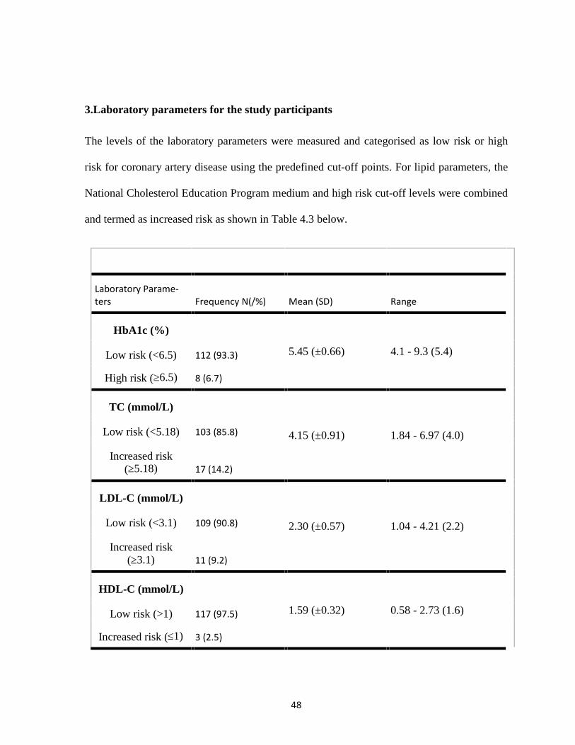
3.Laboratory parameters for the study participants
The levels of the laboratory parameters were measured and categorised as low risk or high
risk for coronary artery disease using the predefined cut-off points. For lipid parameters, the
National Cholesterol Education Program medium and high risk cut-off levels were combined
and termed as increased risk as shown in Table 4.3 below.
Laboratory Parame-ters Frequency N(/%) Mean (SD) Range
HbA1c (%)
5.45 (±0.66) 4.1 - 9.3 (5.4)Low risk (<6.5) 112 (93.3)
High risk (≥6.5) 8 (6.7)
TC (mmol/L)
4.15 (±0.91) 1.84 - 6.97 (4.0)Low risk (<5.18) 103 (85.8)
Increased risk(≥5.18) 17 (14.2)
LDL-C (mmol/L)
2.30 (±0.57) 1.04 - 4.21 (2.2)Low risk (<3.1) 109 (90.8)
Increased risk(≥3.1) 11 (9.2)
HDL-C (mmol/L)
1.59 (±0.32) 0.58 - 2.73 (1.6)Low risk (>1) 117 (97.5)
Increased risk (≤1) 3 (2.5)

49
Lp-PLA2 (ng/ml)
172.01 (±334.81) 0.3 - 1445.87 (52.52)Low risk (<167) 91 (75.8)
High risk (≥167) 29 (24.2)
*MPO (ng/ml)
259.84 (±191.65) 45.02 - 603.56 (208.94)Low risk (<229) 43 (55.1)
High risk (≥229) 35 (44.9)
N - Number; % -Percentage; SD -Standard deviation;*- had 78 subjects
TABLE 4.3 LABORATORY PARAMETERS FOR THE STUDY PARTICIPANTS AT HOMA BAY
COUNTY REFERRAL HOSPITAL, KENYA. (N=120).
3.1.HbA1c
The HbA1c for the study participants ranged from 4.1 to 9.3 with a mean HbA1c level of
5.449 % (±0.66) and median of 5.4%. It was found that 8 of the study participants (6.7%) had
an elevated level of HbA1c and they were classified as having high risk.
The median HbA1c levels demonstrated varied across the different occupational status for
both female and male. The salary-employed group exhibit a higher median.

50
3.2.Total Cholesterol
In this study, the total Cholesterol ranged from 1.84 to 6.97 mmol/L with a mean of 4.15
mmol/L (±0.91) and median 4.05 mmol/L. The number of participants having elevated TC
values was 17 (14.2%) and were classified as having increased risk.
The median TC concentration exhibited almost a similar pattern for both self-employed and
the unemployed groups.
FIGURE 4.3.1 MEDIAN HBA1C CONCENTRATION ACROSS DIFFERENT OCCUPA-
TIONAL STATUS

51
3.3.LDL- Cholesterol
The study found a mean LDL- Cholesterol level of 2.30 mmol/L (±0.57), median level of
2.22 mmol/L (range 1.04 to 4.21 mmol/L). Elevated HDL-C levels were found in 11 (9.2%)
of the study participants who were classified as having increased risk.
3.4.HDL- Cholesterol
The HDL-C in this study ranged from range 1.84 to 6.97 mmol/L with a mean HDL- Choles-
terol level of 4.15 mmol/L (±0.91) and median of 4.05 mmol/L. HDL-C was low in 17
(14.2%) of the study participants and they were classified as having increased risk.
FIGURE 4.3.2 MEDIAN TC CONCENTRATION ACROSS DIFFERENT OCCUPA-
TIONAL STATUS

52
3.5.Lp-PLA2
The mean value for Lp-PLA2 was found to be 172.01 ng/ml (±334.81) with a median of
52.52 ng/ml (range 0.3 to 1445.87 ng/ml). Subjects with an Elevated Lp-PLA2 values were
29 (24.2%).
3.6.MPO
The MPO levels in this study ranged from 45.02 to 603.56 ng/ml with a mean of 259.84
ng/ml (±191.65) and median of 208.94 ng/ml. Subjects with elevated MPO values were 35
(44.9%).
TABLE 4.3.1 ASSOCIATION BETWEEN HAART DURATION WITH GENDER AND AGE AT HOMA
BAY COUNTY REFERRAL HOSPITAL, KENYA (N = 120).
In a chi-square test, it was found out that the duration of HAART was not associated with
gender (p = 0.575). However, there was a significant association between age and HAART
duration (p = 0.007).

53

54
TABLE 4.3.2 ASSOCIATION OF LIPIDS, GLUCOSE, LIPOPROTEIN-ASSOCIATED PHOSPHOLIPASE
2 AND MYELOPEROXIDASE WITH GENDER, AGE AND HAART DURATION AT HOMA BAY
COUNTY REFERRAL HOSPITAL, KENYA. (N = 120).
Using logistic regression analysis, there was no significance association between gender and
raised laboratory parameters. The same was reported for the different age categories except
for 39 - <45 years, which significant association with increased TC level; OR = 1.57 (CI:
0.14 – 17.29, p = 0.021). No significance association was found between raised biomarkers
and HAART duration except for TC and Lp-PLA2 for HAART duration >60 months (OR =
1.62, CI: 0.28 – 9.43, p = 0.045 and OR = 1.65, CI: 0.43 – 6.39, p = 0.047 respectively).

55
5.DISCUSSION
The decrease in the mortality and morbidity of HIV infected individuals has been the out-
come of the widespread use of HAART. However, the concern now is the association of
HAART with the risk of cardiovascular disease. In this study, the levels of serum biomarkers
(TC, LDL-C, HbA1c, Lp-PLA2 and MPO) for risk of cardiovascular disease in patients re-
ceiving highly active antiretroviral therapy in Homa Bay county referral hospital was investi-
gated. Most of the participants (64.2%) were females giving a female to male ratio of 1.8:1,
which is in keeping with the Kenya AIDS Indicator Survey (KAIS) findings 85, which was at
1.6:1. The ratio is also comparable to what Tadewos et al.79 reported, which was 1.9:1. Ma-
jority of the study participants (28.3) were in the age brackets of 39 - <45 years. A proportion
of 77% were married and 64.2% had attained primary education. Majority (65%) were in
self-employment. Most of the subjects (67.5%) had been on HAART for more than sixty
months. The study found out that this finding departs from that of Abebe et al., 201478, Addis
Ababa, Ethiopia, which reported the highest proportion of subjects (59% of the 126 partici-
pants) had only been on HAART for 25 - 41 months.
In this study, majority of the participants had TC, LDL-C, HDL-C, HbA1c, Lp-PLA2 and
MPO levels within the reference interval. For the lipid and glycated haemoglobin, the pro-
portions with derangements in these biomarkers were as follows; 14.2% (TC), 5.8% (LDL-
C), 2.5% (HDL-C) and 4.2% (HbA1c). These percentages were found to be incomparable
from Tadewos et al., 201479, in Southern Ethiopia, who reported 31% (TC), 24% (LDL-C)
and 27% (HDL-C). This discrepancy can be explained by other confounding factors in
Tadewos et al.79 study such as smoking, which was included in the study. They compared the
lipid profiles for HAART and pre-HAART groups.

56
To the best knowledge of the researchers in this study, it is the first of its kind to assess
HbA1c as a marker of hyperglycaemia in this study population. Most of the studies80, 81 done
in this population mainly focused on evaluating the serum levels of fasting blood glucose. In
this study, cases of hyperglycaemia were at 4.2% in the 120 participants. This is comparable
percentage with findings of Abebe et al.78, Addis Ababa Ethiopia, which reported 7.9% in
126 study participants on HAART. However, there is a discrepancy between Mbunkah et al.,
201482 findings, South-West, Cameroon, which reported hyperglycaemia of 26.5% in the 241
study subjects, and this study. A possible explanation to this inconsistency is the effect of age
as a confounder, since the researchers included elderly participants of up to 70 years of age.
Old age is a risk factor to developing insulin resistance and needs to be controlled either at
the design stage or using a statistical analysis such as logistic regression model82.
The present investigation being an exploratory study, is the first of its kind to the best of the
investigators’ knowledge to assess MPO and Lp-PLA2 in this study population. As reported
by Mangili et al.8, and Ross et al.7, these two biomarkers predict the early events of the de-
velopment of cardiovascular diseases. Therefore, their inclusion in the study was to identify
those that were at risk of developing cardiovascular disease, which the already established
markers (TC, LDL-C, HDL-C and HbA1c) could have not captured. In this study, 24.2% of
the participants had raised levels above the reference interval of PLA2 of the 120 participants
that were studied. Whereas, out of the 78 subjects that were assessed for serum levels of
MPO, 44.9% had elevated values. Because of the exploratory nature of this study, there were
no previous investigation to compare the findings with.
Among the study participants, there was no significant association between elevated TC,
LDL-C, HbA1c, Lp-PLA2 and MPO levels with gender. This was found to be inconsistent

57
for the lipid profile in a study conducted in Thailand by Luatngoen.84 The difference can be
explained by the fact that the participants were not gender-matched in this study.
The present study also investigated the association between different age categories with
raised levels of TC, LDL-C, HbA1c, Lp-PLA2 and MPO. This was found to be insignificant
except for TC level for 39 - <45 age category (OR = 1.57, CI: 0.14 – 17.29, p = 0.021). The
finding was in agreement with Abebe et al.78 study, which also reported a significant associa-
tion between elevated TC with > 35 years (OR = 2.30, CI: 1.29 - 4.10, p = 0.005).
Abebe et al.78 found an association between HAART use and raised serum levels of TC,
HDL-C and LDL-C; OR = 2.99 (CI: 1.74 - 5.15), p<0.0001) and OR= 1.82 (CI: 1.06 - 1.12, p
= 0.02) for TC. In this study, using HAART duration for 6 - 35 months as the point of refer-
ence (base), no significance association between elevated laboratory parameters and 36 – 60
months HAART durations was found. However, the study showed a significant association
between raised TC and Lp-PLA2 levels to HAART duration of >60 months (OR = 1.62, CI:
0.28 – 9.43, p = 0.045 and OR = 1.65, CI: 0.43 – 6.39, p = 0.047 respectively) and this was
consistent with the findings of Abebe et al.78 However, for other component of lipid panel
(LDL-C and HDL-C), there was no significant association with prolonged HAART duration.
The findings of this study were consisted with that of Nsagha et al.,87 which reported a signif-
icant independent association between HAART duration of 42 months and more with raised
serum levels of TC (aOR = 2.26, 95 % CI: 1.16 – 4.42, p = 0.017).

58
6.CONCLUSIONS AND RECOMMENDATIONS
1.Introduction
This section details conclusion, recommendation and limitation of the study.
2.Conclusion
Majority of the participants who had at least one derangement in the laboratory parameters
being abnormal had been on HAART for more than sixty months. Dysregulated concentra-
tions of the serum biomarkers were not significantly associated to gender. Age was signifi-
cantly associated to HAART duration. Serum concentrations of TC and Lp-PLA2 showed
significant association between raised serum levels with the duration of HAART.

59
3.Recommendation
The researchers highly recommend that a validation study should be carried out to evaluate
and confirm the findings in a cohort study to aid in testing the clinical utility of both MPO
and Lp-PLA2. It is also recommended that HIV negative individuals should be included as
controls as well as anthropometrics parameters for the assessment of long-term effect of
HAART on well-controlled cohort conditions. Carrying out the study in different regions in
Kenya is also recommended.
4.Limitations
The absence of previous studies on metabolic abnormality in HIV-positive individu-
als on HAART in Homa Bay setting as well as lack of HIV negative and HAART-
naïve as controls to make comparison with, were deemed as potential limitations to
our study.
MPO was done only for 78 subjects due to financial constraints.

60
REFERENCES
1. National AIDS Control Council [Internet]. [cited 2016 May 18]. Available from:http://www.nacc.or.ke/
2. Palella FJ, Phair JP. Cardiovascular disease in HIV infection. Curr Opin HIV AIDS.2011 Jul;6(4):266–71.
3. Islam F, Wu J, Jansson J, Wilson D. Relative risk of cardiovascular disease among peo-ple living with HIV: a systematic review and meta-analysis. HIV Med. 2012 Sep1;13(8):453–68.
4. Leclercq P, Blanc M. [Metabolic abnormalities, lipodystrophy and cardiovascular riskin HIV-infected patients]. Rev Prat. 2006 May 15;56(9):987–94.
5. Clark SJ, Gómez-Olivé FX, Houle B, Thorogood M, Klipstein-Grobusch K, Angotti N,et al. Cardiometabolic disease risk and HIV status in rural South Africa: establishing abaseline. BMC Public Health. 2015;15:135.
6. Bloomfield GS, Hogan JW, Keter A, Sang E, Carter EJ, Velazquez EJ,et al. Hyperten-sion and Obesity as Cardiovascular Risk Factors among HIV Seropositive Patients inWestern Kenya. PLOS ONE. 2011 Jul 14;6(7):e22288.
7. Ross AC, Armentrout R, O’Riordan MA, Storer N, Rizk N, Harrill D, et al. EndothelialActivation Markers Are Linked to HIV Status and Are Independent of AntiretroviralTherapy and Lipoatrophy. J Acquir Immune Defic Syndr 1999. 2008 Dec15;49(5):499–506.
8. Mangili A, Ahmad R, Wolfert RL, Kuvin J, Polak JF, Karas RH, et al. Lipoprotein-Associated Phospholipase A2, a Novel Cardiovascular Inflammatory Marker, in HIV-Infected Patients. Clin Infect Dis Off Publ Infect Dis Soc Am. 2014 Mar 15;58(6):893–900.
9. Lang S, Mary-Krause M, Cotte L, Gilquin J, Partisani M, Simon A, et al. Increased riskof myocardial infarction in HIV-infected patients in France, relative to the general pop-ulation. AIDS Lond Engl. 2010 May 15;24(8):1228–30.
10. Obel N, Thomsen HF, Kronborg G, Larsen CS, Hildebrandt PR, Sørensen HT, et al.Ischemic heart disease in HIV-infected and HIV-uninfected individuals: a population-based cohort study. Clin Infect Dis Off Publ Infect Dis Soc Am. 2007 Jun15;44(12):1625–31.

61
11. Klein D, Hurley L, Silverberg M, Horberg M, Sidney S. Improved Cardiac DiseaseManagement, Risk for Myocardial Infarction Stabilizes. In: Conference on Retrovirusesand Opportunistic Infections [Internet]. Los Angeles, California; [cited 2016 Sep 28].Available from: http://www.natap.org/2007/CROI/croi_78.htm
12. Aboud M, Elgalib A, Pomeroy L, Panayiotakopoulos G, Skopelitis E, Kulasegaram R,et al. Cardiovascular risk evaluation and antiretroviral therapy effects in an HIV cohort:implications for clinical management: the CREATE 1 study. Int J Clin Pract. 2010Aug;64(9):1252–9.
13. Beltrán LM, Rubio-Navarro A, Amaro-Villalobos JM, Egido J, García-Puig J, MorenoJA. Influence of immune activation and inflammatory response on cardiovascular riskassociated with the human immunodeficiency virus. Vasc Health Risk Manag. 2015 Jan6;11:35–48.
14. Strategies for Management of Antiretroviral Therapy (SMART) Study Group, El-SadrWM, Lundgren JD, Neaton JD, Gordin F, Abrams D, et al. CD4+ count-guided inter-ruption of antiretroviral treatment. N Engl J Med. 2006 Nov 30;355(22):2283–96.
15. Hemkens LG, Bucher HC. HIV infection and cardiovascular disease. Eur Heart J. 2014Jan 9;eht528.
16. Mu H, Chai H, Lin PH, Yao Q, Chen C. Current Update on HIV-associated VascularDisease and Endothelial Dysfunction. World J Surg. 2007 Feb 1;31(4):632–43.
17. Paiardini M, Müller-Trutwin M. HIV-associated chronic immune activation. ImmunolRev. 2013 Jul;254(1):78–101.
18. Crowe SM, Westhorpe CLV, Mukhamedova N, Jaworowski A, Sviridov D, BukrinskyM. The macrophage: the intersection between HIV infection and atherosclerosis. J Leu-koc Biol. 2010 Apr;87(4):589–98.
19. Cui HL, Grant A, Mukhamedova N, Pushkarsky T, Jennelle L, Dubrovsky L, et al.HIV-1 Nef mobilizes lipid rafts in macrophages through a pathway that competes withABCA1-dependent cholesterol efflux. J Lipid Res. 2012 Apr 1;53(4):696–708.
20. Funderburg NT. Markers of coagulation and inflammation often remain elevated inART-treated HIV-infected patients. Curr Opin HIV AIDS. 2014 Jan;9(1):80–6.
21. Masiá M, Padilla S, Fernández M, Rodríguez C, Moreno A, Oteo JA, et al. OxidativeStress Predicts All-Cause Mortality in HIV-Infected Patients. PLoS ONE [Internet].
62
2016 Apr 25 [cited 2016 May 14];11(4). Available from:http://www.ncbi.nlm.nih.gov/pmc/articles/PMC4844170/
22. Norata GD, Pirillo A, Catapano AL. HDLs, immunity, and atherosclerosis. Curr OpinLipidol. 2011 Oct;22(5):410–6.
23. Bavinger C, Bendavid E, Niehaus K, Olshen RA, Olkin I, Sundaram V, et al. Risk ofCardiovascular Disease from Antiretroviral Therapy for HIV: A Systematic Review.PLOS ONE. 2013 Mar 26;8(3):e59551.
24. Currier JS, Lundgren JD, Carr A, Klein D, Sabin CA, Sax PE, et al. EpidemiologicalEvidence for Cardiovascular Disease in HIV-Infected Patients and Relationship toHighly Active Antiretroviral Therapy. Circulation. 2008 Aug 7;118(2):e29–35.
25. Baker JV, Henry WK, Neaton JD. The Consequences of HIV Infection and Antiretrovi-ral Therapy Use For Cardiovascular Disease Risk: Shifting Paradigms. Curr Opin HIVAIDS. 2009 May;4(3):176–82.
26. Madden E, Lee G, Kotler DP, Wanke C, Lewis CE, Tracy R, et al. Association of An-tiretroviral Therapy with Fibrinogen Levels in HIV Infection. AIDS Lond Engl. 2008Mar 30;22(6):707–15.
27. Reingold JS, Wanke C, Kotler DP, Lewis CE, Tracy R, Heymsfield S, et al. Associationof HIV Infection and HIV/HCV Coinfection With C-Reactive Protein Levels. J AcquirImmune Defic Syndr 1999. 2008 Jun 1;48(2):142–8.
28. Guimarães MMM, Greco DB, Figueiredo SM de, Fóscolo RB, Oliveira AR de, Macha-do LJ de C. High-sensitivity C-reactive protein levels in HIV-infected patients treatedor not with antiretroviral drugs and their correlation with factors related to cardiovascu-lar risk and HIV infection. Atherosclerosis. 2008 Dec 1;201(2):434–9.
29. Use of nucleoside reverse transcriptase inhibitors and risk of myocardial infarction inHIV-infected patients enrolled in the D:A:D study: a multi-cohort collaboration. Lancet.2008 Apr 26;371(9622):1417–26.
30. Wong G, Trevillyan JM, Fatou B, Cinel M, Weir JM, Hoy JF, et al. Plasma LipidomicProfiling of Treated HIV-Positive Individuals and the Implications for CardiovascularRisk Prediction. PLOS ONE. 2014 Apr 14;9(4):e94810.
31. Burdo TH, Lentz MR, Autissier P, Krishnan A, Halpern E, Letendre S, et al. SolubleCD163 Made by Monocyte/Macrophages Is a Novel Marker of HIV Activity in Early

63
and Chronic Infection Prior to and After Anti-retroviral Therapy. J Infect Dis. 2011 Jul1;204(1):154–63.
32. Neuhaus J, Jacobs DR, Baker JV, Calmy A, Duprez D, La Rosa A, et al. Markers of In-flammation, Coagulation and Renal Function Are Elevated in Adults with HIV Infec-tion. J Infect Dis. 2010 Jun 15;201(12):1788–95.
33. Syed SS, Balluz RS, Kabagambe EK, Meyer WA, Lukas S, Wilson CM, et al. Assess-ment of Biomarkers of Cardiovascular Risk Among HIV Type 1-Infected Adolescents:Role of Soluble Vascular Cell Adhesion Molecule As an Early Indicator of EndothelialInflammation. AIDS Res Hum Retroviruses. 2013 Mar;29(3):493–500.
34. Joy T, Keogh HM, Hadigan C, Lee H, Dolan SE, Fitch K, et al. Dietary Fat Intake andRelationship to Serum Lipid Levels Among HIV-Infected Subjects with Metabolic Ab-normalities in the Era of HAART. AIDS Lond Engl. 2007 Jul 31;21(12):1591–600.
35. Lazzaretti RK, Kuhmmer R, Sprinz E, Polanczyk CA, Ribeiro JP. Dietary InterventionPrevents Dyslipidemia Associated With Highly Active Antiretroviral Therapy in Hu-man Immunodeficiency Virus Type 1–Infected Individuals: A Randomized Trial. J AmColl Cardiol. 2012 Mar 13;59(11):979–88.
36. Beltrán LM, Hernández RM, Bernal RS de P, Morillo JSG, Egido J, Noval ML, et al.Reduced sTWEAK and Increased sCD163 Levels in HIV-Infected Patients: Modulationby Antiretroviral Treatment, HIV Replication and HCV Co-Infection. PLOS ONE.2014 Mar 4;9(3):e90541.
37. Gillis J, Smieja M, Cescon A, Rourke SB, Burchell AN, Cooper C, et al. Risk of cardi-ovascular disease associated with HCV and HBV coinfection among antiretroviral-treated HIV-infected individuals. Antivir Ther. 2014;19(3):309–17.
38. Klatt NR, Funderburg NT, Brenchley JM. Microbial translocation, immune activationand HIV disease. Trends Microbiol. 2013 Jan;21(1):6–13.
39. Jensen MK, Bertoia ML, Cahill LE, Agarwal I, Rimm EB, Mukamal KJ. Novel meta-bolic biomarkers of cardiovascular disease. Nat Rev Endocrinol. 2014 Nov;10(11):659–72.
40. Gordon T, Castelli WP, Hjortland MC, Kannel WB, Dawber TR. High density lipopro-tein as a protective factor against coronary heart disease. The Framingham Study. Am JMed. 1977 May;62(5):707–14.

64
41. McQueen MJ, Hawken S, Wang X, Ounpuu S, Sniderman A, Probstfield J, et al. Lipids,lipoproteins, and apolipoproteins as risk markers of myocardial infarction in 52 coun-tries (the INTERHEART study): a case-control study. The Lancet. 19;372(9634):224–33.
42. Anuurad E, Boffa MB, Koschinsky ML, Berglund L. Lipoprotein(a): A Unique RiskFactor for Cardiovascular Disease. Clin Lab Med. 2006 Dec;26(4):751–72.
43. Marcovina SM, Koschinsky ML. Lipoprotein(a) as a risk factor for coronary artery dis-ease. Am J Cardiol. 1998 Dec 17;82(12, Supplement 1):57U–66U.
44. Poon M, Zhang X, Dunsky KG, Taubman MB, Harpel PC. Apolipoprotein(a) InducesMonocyte Chemotactic Activity in Human Vascular Endothelial Cells. Circulation.1997 Oct 21;96(8):2514–9.
45. Nielsen LB, Grønholdt MLM, Schroeder TV, Stender S, Nordestgaard BG. In VivoTransfer of Lipoprotein(a) Into Human Atherosclerotic Carotid Arterial Intima. Arterio-scler Thromb Vasc Biol. 1997 Jan 5;17(5):905–11.
46. Helgadottir A, Gretarsdottir S, Thorleifsson G, Holm H, Patel RS, Gudnason T, et al.Apolipoprotein(a) Genetic Sequence Variants Associated With Systemic Atherosclero-sis and Coronary Atherosclerotic Burden But Not With Venous Thromboembolism. JAm Coll Cardiol. 2012 Aug 21;60(8):722–9.
47. Nielsen LB, Juul K, Nordestgaard BG. Increased Degradation of Lipoprotein(a) in Ath-erosclerotic Compared With Nonlesioned Aortic Intima–Inner Media of Rabbits In Vi-vo Evidence That Lipoprotein(a) May Contribute to Foam Cell Formation. ArteriosclerThromb Vasc Biol. 1998 Jan 4;18(4):641–9.
48. Danik JS, Buring JE, Chasman DI, Zee RYL, Ridker PM, Glynn RJ. Lipoprotein(a),polymorphisms in the LPA gene, and incident venous thromboembolism among 21 483women. J Thromb Haemost. 2013 Jan 1;11(1):205–8.
49. Nordestgaard BG, Chapman MJ, Humphries SE, Ginsberg HN, Masana L, DescampsOS, et al. Familial hypercholesterolaemia is underdiagnosed and undertreated in thegeneral population: guidance for clinicians to prevent coronary heart disease. Eur HeartJ. 2013 Dec 1;34(45):3478–90.
50. Taleb A, Witztum JL, Tsimikas S. Oxidized phospholipids on apoB-100-containing lip-oproteins: a biomarker predicting cardiovascular disease and cardiovascular events. Bi-omark Med. 2011 Oct 1;5(5):673–94.

65
51. Schaloske RH, Dennis EA. The phospholipase A2 superfamily and its group numberingsystem. Biochim Biophys Acta BBA - Mol Cell Biol Lipids. 2006 Nov;1761(11):1246–59.
52. Asano K, Okamoto S, Fukunaga K, Shiomi T, Mori T, Iwata M, et al. CellularSource(s) of Platelet-Activating-Factor Acetylhydrolase Activity in Plasma. BiochemBiophys Res Commun. 1999 Aug 2;261(2):511–4.
53. Tsimihodimos V, Karabina S-AP, Tambaki AP, Bairaktari E, Goudevenos JA, Chap-man MJ, et al. Atorvastatin Preferentially Reduces LDL-Associated Platelet-ActivatingFactor Acetylhydrolase Activity in Dyslipidemias of Type IIA and Type IIB. Arterio-scler Thromb Vasc Biol. 2002 Jan 2;22(2):306–11.
54. Zalewski A, Macphee C. Role of Lipoprotein-Associated Phospholipase A2 in Athero-sclerosis Biology, Epidemiology, and Possible Therapeutic Target. Arterioscler ThrombVasc Biol. 2005 Jan 5;25(5):923–31.
55. Splaver A, Lamas GA, Hennekens CH. Homocysteine and cardiovascular disease: bio-logical mechanisms, observational epidemiology, and the need for randomized trials.Am Heart J. 2004 Jul;148(1):34–40.
56. McCully KS. Homocysteine Metabolism, Atherosclerosis, and Diseases of Aging.Compr Physiol. 2015 Jan;6(1):471–505.
57. Selvin E, Steffes MW, Zhu H, Matsushita K, Wagenknecht L, Pankow J, et al. GlycatedHemoglobin, Diabetes, and Cardiovascular Risk in Nondiabetic Adults. N Engl J Med.2010 Mar 4;362(9):800–11.
58. Pai JK, Cahill LE, Hu FB, Rexrode KM, Manson JE, Rimm EB. Hemoglobin A1c IsAssociated With Increased Risk of Incident Coronary Heart Disease Among ApparentlyHealthy, Nondiabetic Men and Women. J Am Heart Assoc. 2013 Apr 24;2(2):e000077.
59. Sarwar N, Aspelund T, Eiriksdottir G, Gobin R, Seshasai SRK, Forouhi NG, et al.Markers of Dysglycaemia and Risk of Coronary Heart Disease in People without Diabe-tes: Reykjavik Prospective Study and Systematic Review. PLOS Med. 2010 May25;7(5):e1000278.
60. Adams RJ, Appleton SL, Hill CL, Wilson DH, Taylor AW, Chittleborough CR, et al.Independent Association of HbA1c and Incident Cardiovascular Disease in PeopleWithout Diabetes. Obesity. 2009 Mar 1;17(3):559–63.

66
61. Sander D, Schulze-Horn C, Bickel H, Gnahn H, Bartels E, Conrad B. Combined Effectsof Hemoglobin A1c and C-Reactive Protein on the Progression of Subclinical CarotidAtherosclerosis The INVADE Study. Stroke. 2006 Jan 2;37(2):351–7.
62. King DE, Mainous AG, Buchanan TA, Pearson WS. C-Reactive Protein and GlycemicControl in Adults With Diabetes. Diabetes Care. 2003 May 1;26(5):1535–9.
63. Mora S, Musunuru K, Blumenthal RS. The Clinical Utility of High-Sensitivity C-Reactive Protein in Cardiovascular Disease and the Potential Implication of JUPITERon Current Practice Guidelines. Clin Chem. 2009 Jan 2;55(2):219–28.
64. Libby P. Inflammation in atherosclerosis. Nature. 2002 Dec 19;420(6917):868–74.
65. Tavora F, Cresswell N, Li L, Ripple M, Burke A. Immunolocalisation of fibrin in coro-nary atherosclerosis: implications for necrotic core development. Pathology (Phila).2010 Jan;42(1):15–22.
66. Libby P, Nahrendorf M, Pittet MJ, Swirski FK. Diversity of Denizens of the Athero-sclerotic Plaque Not All Monocytes Are Created Equal. Circulation. 2008 Jun24;117(25):3168–70.
67. Kones R. Primary prevention of coronary heart disease: integration of new data, evolv-ing views, revised goals, and role of rosuvastatin in management. A comprehensivesurvey. Drug Des Devel Ther. 2011 Jun 13;5:325–80.
68. Yousuf O, Mohanty BD, Martin SS, Joshi PH, Blaha MJ, Nasir K, et al. High-Sensitivity C-Reactive Protein and Cardiovascular DiseaseA Resolute Belief or an Elu-sive Link? J Am Coll Cardiol. 2013 Jul 30;62(5):397–408.
69. Klebanoff SJ. Myeloperoxidase: friend and foe. J Leukoc Biol. 2005 Jan 5;77(5):598–625.
70. Marathe GK, Pandit C, Lakshmikanth CL, Chaithra VH, Jacob SP, D’Souza CJM. Tohydrolyze or not to hydrolyze: the dilemma of platelet-activating factor acetylhydrolase.J Lipid Res. 2014 Sep 1;55(9):1847–54.
71. Schindhelm RK, Zwan LP van der, Teerlink T, Scheffer PG. Myeloperoxidase: A Use-ful Biomarker for Cardiovascular Disease Risk Stratification? Clin Chem. 2009 Jan8;55(8):1462–70.
67
72. Cook-Mills JM, Marchese ME, Abdala-Valencia H. Vascular Cell Adhesion Molecule-1 Expression and Signaling During Disease: Regulation by Reactive Oxygen Speciesand Antioxidants. Antioxid Redox Signal. 2011 Sep 15;15(6):1607–38.
73. Yang X-F, Yin Y, Wang H. Vascular Inflammation And Atherogenesis Are ActivatedVia Receptors For Pamps And Suppressed By Regulatory T Cells. Drug Discov TodayTher Strateg. 2008;5(2):125–42.
74. Al Gadban MM, German J, Truman J-P, Soodavar F, Riemer EC, Twal WO, et al. Lackof nitric oxide synthases increases lipoprotein immune complex deposition in the aortaand elevates plasma sphingolipid levels in lupus. Cell Immunol. 2012 Mar;276(1–2):42–51.
75. Charan J, Biswas T. How to Calculate Sample Size for Different Study Designs in Med-ical Research? Indian J Psychol Med. 2013;35(2):121–6.
76. Nsagha DS, Assob JCN, Njunda AL, Tanue EA, Kibu OD, Ayima CW, et al. Risk Fac-tors of Cardiovascular Diseases in HIV/AIDS Patients on HAART. Open AIDS J. 2015Oct 20;9:51–9.
77. CLSI. Defining, Establishing, and Verifying Reference Intervals in the Clinical Labora-tory; Approved Guideline—Third Edition. CLSI document C28-A3. Wayne, PA: Clini-cal and Laboratory Standards Institute; 2008.
78. Abebe M, Kinde S, Belay G, Gebreegziabxier A, Challa F, Gebeyehu T, et al. An-tiretroviral treatment associated hyperglycemia and dyslipidemia among HIV infectedpatients at Burayu Health Center, Addis Ababa, Ethiopia: a cross-sectional comparativestudy. BMC Res Notes. 2014 Jun 21;7:380.
79. Tadewos A, Addis Z, Ambachew H, Banerjee S. Prevalence of dyslipidemia amongHIV-infected patients using first-line highly active antiretroviral therapy in SouthernEthiopia: a cross-sectional comparative group study. AIDS Res Ther. 2012 Oct 25;9:31.
80. Hejazi N, Rajikan R, Kwok Choong CL, Sahar S. Metabolic abnormalities in adult HIVinfected population on antiretroviral medication in Malaysia: a cross-sectional survey.BMC Public Health. 2013 Aug 15;13:758.
81. Guillen MA, Mejia FA, Villena J, Turin CG, Carcamo CP, Ticse R. Insulin resistanceby homeostasis model assessment in HIV-infected patients on highly active antiretrovi-ral therapy: cross-sectional study. Diabetol Metab Syndr. 2015 May 30;7:49.

68
82. Mbunkah HA, Meriki HD, Kukwah AT, Nfor O, Nkuo-Akenji T. Prevalence of meta-bolic syndrome in human immunodeficiency virus - infected patients from the South-West region of Cameroon, using the adult treatment panel III criteria. Diabetol MetabSyndr. 2014 Aug 25;6:92.
83. Nsagha DS, Weledji EP, Assob NJC, Njunda LA, Tanue EA, kibu OD, et al. Highlyactive antiretroviral therapy and dyslipidemia in people living with HIV/AIDS in FakoDivision, South West Region of Cameroon. BMC Cardiovasc Disord [Internet]. 2015Aug 28 [cited 2017 Jul 16];15. Available from:http://www.ncbi.nlm.nih.gov/pmc/articles/PMC4552364/
84. Luatngoen J. HAART - Associated Hyperlipidemia and Hyperglycemia in HIV Patients,Surin Hospital. http://thailand.digitaljournals.org/index.php/KKMJ/issue/archive [Inter-net]. 2010 Feb 22 [cited 2017 Aug 23]; Available from:http://imsear.hellis.org/handle/123456789/131630
85. Kenya Aids Indicator Survey (KAIS) 2012 . [Cited 2017 Aug 23].
APPENDICES
APPENDIX I (a): Informed Consent Explanation and Form
Introduction and Research Objective
My name is Kenneth Weke a postgraduate student pursuing a Master of Science degree inclinical chemistry at the department of human pathology, University of Nairobi. I am carry-ing out a study to determine biomarkers for risk of cardiovascular disease (CVD) amongHIV-positive individuals on highly active antiretroviral therapy (HAART) in Homa-BayCounty Referral Hospital, Kenya using facilities at the Kenyatta National Hospital Biochem-istry Laboratory.
Benefits and Risks of the Study to You
Potential Benefits:
You will benefit by having your serum markers for risk of CVD determined, which willguide the clinician on the best treatment strategy for managing your health.
Potential Risks
There will be a minimal risk and discomfort, which will be a slight pain from the blood-drawing site.
Procedure

69
If you agree to take part in the study, then you will be asked to fill a questionnaire, whichwill take a maximum of 30 minutes to complete. I will ask a number of questions, and I willnote down the responses that you will give. I will then collect a sample of blood of a volumeof 4ml, which will be taken to the laboratory for analysis for biomarkers for risk of cardio-vascular disease.
Confidentiality
Names will not be required in the study since you will be identified by a study number. Thequestionnaire will be kept in lockable cabinets, and I will be the only person to access it.Questionnaires will be kept for one year then destroyed. Any information given to us willremain confidential and will be for your benefit. You will get your results in the usual man-ner during your subsequent visit.
Withdrawal from StudyYour participation in this study is voluntary. You may refuse to participate or withdraw atany time without losing the benefits to which you are entitled in this clinic.I …………………………………………………………. after reading and being explainedthe study purpose, do hereby give informed consent to participate in the study and I am fullyaware of the benefits and risks. I have not been forced to take part in this study in any way. Iunderstand that participation in this study is completely voluntary and that I may withdrawfrom it at any time and without loss of any benefit or quality of management to which I amentitled. I am fully aware that the results of this study will be used for scientific purposes andmay be published.Participant’s Signature: ……….…………………. Date……………………………..Investigator’s Signature………………....…………Date ……………………………..Contact Information
If you have any question regarding the study, please contact:1. Principal Investigator
Kenneth Weke,University of Nairobi, Department of Human PathologyP.O BOX 19676-00202 NairobiMobile number +254715237396.
2. SupervisorProfessor Angela AmayoChairperson Department of Human Pathology, Lecturer; Thematic Unit ClinicalChemistry, Department of Human Pathology,University of Nairobi, P.O. Box 19676-00202, Nairobi Kenya

70
Mobile phone: +254733617678.3. Ethical Concern
The Secretary,KNH/UoN ERC,P.O Box20723, KNH, NairobiTel+254-020-2726300-9 Ext44355

71
APPENDIX I (b): Fomu Ya Idhini
Kuanzishwa na Lengo la Utafiti
Jina langu ni Kenneth Weke mwanafunzi wa chuo kikuu cha Nairobi idara ya pathologia yawanadamu. Nafanya shahada ya uzamili katika somo ya sayansi uwanja wa klinikol kemia.Nataka kuchunguza kiwango cha viashiria vinavyoweza hatarisha kupata ugonjwa wa moyona mishipa kwa wale ambao wanatumia madawa ya kupunguza idadi ya virusi vya ukimwikatika hospitali ya rufaa ya jimbo la Homa-Bay. Nitatumia vifaa vya uchambuzi vinavyopatikana kataki hospital ya kitaifa ya Kenyatta.
Faida na Madhara ya Utafiti huu Kwako
Faida
Utafaidika kwa kujua viwango vya viashiria ambavyo vinawezafanya upate ugonjwa wamoyo na mishipa na hii itaongoza daktari jinsi atakavyofanya kulinda afya yako.
Uwezekano wa Hatari
Kutakua na hatari ingawa kidogo ambayo itatokana na uchungu utahisi kwenye pahali damuutakaotolowe.
Utaratibu
Ukikubali kushiriki katika utafiti huu, basi nitakuuliza ujaze hojaji nitakayokupa na hii ita-chukua muda wa dakika kumi. Nitakuuliza maswali ambayo majibu yao nitaandika chini.Baada ya hayo nitatoa damu kiwango cha 4 ml kutoka mshipa kwenye mkono wako.Nitapeleka damu kwa maabara ili nichunguze kiwango cha viashiria vya kutaharishi mtu ku-pata ungonjwa wa moyo na mishipa.
Siri
Majina hayaitajiki katika utafiti na utatambuliwa na namba ya utafiti. Stakabadhi zilizo namaswali zitawekwa chini ya ufunguo na kufuli na mpelelezi mkuu ataweza kupata hudumahiyo. Maswali yatawekwa kwa mwaka mmoja kisha kuharibiwa. Taarifa yoyote utatupeaitabaki siri na itakuwa kwa faida yako. Wewe utapata matokeo yako kwa njia ya kawaidawakati wa ziara yako ijayo.
Kujitoa kutoka kwa utafiti

72
Kushiriki katika utafiti huu itakuwa ni kwa hiari yako na ni sehemu ya tathmini yako marakwa mara na unaweza kujiondoa wakati wowote bila kupoteza faida ambayo una haki katikataasisi hii.
FOMU YA IDHINI:
Mimi ................................................................... baada ya kusoma na kuelezewa madhumuniya utafiti huu hili kutoa ridhaa ya kushiriki katika uchunguzi kikamilifu na ufahamu wa faidana hatari. Si kushinikizwa kushiriki katika utafiti huu kwa njia yoyote. Mimi naelewa yakwamba ushiriki katika utafiti huu ni kwa hiari yangu kabisa na kwamba naweza kujiondoawakati wowote, bila hasara ya faida yoyote au ubora wa usimamizi. Mimi nimefahamukikamilifu kwamba matokeo ya utafiti huu yatatumika kwa ajili ya madhumuni ya kisayansina yanaweza kuchapishwa.
Sahihi ya mshiriki .......... ..................... ………Tarehe ................................. ..
Sahihi ya mkuu wa uchunguzi ..................... ….Tarehe ................................. ..
Mawasiliano ya Habari
Kama una swali lolote kuhusu utafiti tafadhali wasiliana na:
1. Mchunguzi
Kenneth Weke, Chuo Kikuu cha Nairobi, S.L.P 19676-00,202 Nairobi
Nambari ya simu +254715237396.
2. Msimamizi
Profesa Angela Amayo
Chuo Kikuu cha Nairobi, S.L.P 19676-00,202 Nairobi
Nambari ya simu +254733617678.
3. Maadili
The Secretary, KNH/UoN ERC,
P.O Box20723, KNH, Nairobi
Tel+254-020-2726300-9 Ext44355
APPENDIX II (a): Study Questionnaire

73
Project Title: Serum Biomarkers for Risk of Cardiovascular Disease in Patients on Highly
Active Antiretroviral Therapy in Homa-Bay County Referral Hospital, Kenya
Study Number…………………….
A. DEMOGRAPHIC DATA
1. Gender Male Female
2. Age……………………………… (Years).
3. What is your level of education?
None Primary Secondary Post-secondary
4. What is your marital status?
Married Single Divorced Widowed
5. What is your occupation?
Salary employed Self-employed Unemployed
B. MEDICAL HISTORY
1. Which year and month were you diagnosed with HIV?
YEAR MONTH

74
2. Which year and month were you put on HAART?
YEAR MONTH
3. HAART duration
6-35 months 36-60 months >60 months
C. LABORATORY: BIOCHEMICAL ANALYSIS
Serum biomarker Result Units Score
(N/H)
TC mmol/L
LDL-C mmol/L
HDL-C mmol/L
HbA1C %
MPO U/ml
Lp-PLA2 ng/ml
D.

75
APPENDIX II (b): Screening Questionare- Exclusion Criteria
1. Age between 18 to <45 years YES NO
2. History of myocardial infarction/stroke YES NO
3. Are you on treatment for raised blood pressure? YES NO
4. Are you on treatment for raised blood sugar? YES NO
5. Are you on lipid lowering treatment? YES NO
6. Do you smoke cigarette? YES NO
7. HAART for less than 6 months? YES NO

76
APPENDIX II (c): Utafiti Dodoso
Lengo la Utafiti: Uchunguzi wa kiwango cha viashiria vinavyoweza hatarisha kupata
ugonjwa wa moyo na mishipa kwa wale ambao wanatumia madawa ya kupunguza idadi ya
virusi vya ukimwi katika hospitali ya rufaa ya jimbo la Homa-Bay
Nambari ya usajili…………………….
Jaza kwa kuweka alama ya kwa jibu sahihi
I. MSHIRIKI
6. Jinsia Mme Mke
7. Umri……………………………….
8. Kiwango cha elimu
Hakuna Mshingi Upili Zaidi ya upili
9. Hali ya ndoa?
Katika ndoa Binafsi Talaka Mjane
10. Uvamizi?
Ajiriwa Mwnabiashara Bila kazi
II. HISTORIA YA MATIBABU
4. Ni tarehe gani ulipimwa na ukapatwa na virusi vya ukimwi?
MWAKA MWEZI

77
5. Ni tarehe gani ulianzishwa kwa madawa ya kupunguza idadi ya virusi vya ukimwi?
MWAKA MWEZI
6. Muda wa kutumia madawa ya kupunguza idadi ya virusi vya ukimwi
Miezi 6-35 Miezi 36-60 Miezi Zaidi ya 60
APPENDIX II (d): Uchunguzi dodoso
8. Umri wa 18 to <45 NDIO LA
9. Historia ya ungonjwa wa moyo? NDIO LA
10. Matibabu ya ungonjwa wa sukari? NDIO LA
11. Matibabu ya shinikizo la damu? NDIO LA
12. Matibabu ya kupunguza mafuta kwa mwili? NDIO LA
13. Unavuta sigara? NDIO LA
14. Matibabu ya HAART chini ya miezi 6? NDIO LA

78
APPENDIX III: Laboratory Methods/Principles
MPO (Bio-Techne Europe Ltd.)
This assay employs the quantitative sandwich enzyme immunoassay technique. A monoclo-nal antibody specific for human MPO has been pre-coated onto a microplate. Standards andsamples are pipetted into the wells, and any MPO present is bound by the immobilized anti-body. After washing away any unbound substances, an enzyme-linked polyclonal antibodyspecific for human MPO is added to the wells. Following a wash to remove any unbound an-tibody-enzyme reagent, a MPO bound in the initial step. The colour development is stoppedand the intensity of the colour is measured.
LP-PLA2 (Bio-Techne Europe Ltd.)
This assay employs the quantitative sandwich enzyme immunoassay technique. A monoclo-nal antibody specific for human PLA2G7 has been pre-coated onto a microplate. Standardsand samples are pipetted into the wells, and any PLA2G7 present is bound by the immobi-lized antibody. After washing away any unbound substances, an enzyme-linked polyclonalantibody specific for human PLA2G7 is added to the wells. Following a wash to remove anyunbound antibody-enzyme reagent, a substrate solution is added to the wells and color devel-ops in proportion to the amount of PLA2G7 bound in the initial step. The color developmentis stopped and the intensity of the color is measured.
LIPID PANEL (Roche Cobas-cobas.com)
The erythrocytes of the capillary or venous blood sample are separated from the plasma bycentrifugation. In the next step, the plasma sample is diluted with phosphate buffer. The HDLtest uses a precipitation method with Mg2+ and phosphotungstic acid as a precipitant reagent.The components except for HDL-cholesterol are precipitated and removed. The Cobas sys-tem determines total cholesterol and HDL-cholesterol by an enzymatic method. Cholesterolesters in the sample are hydrolyzed to cholesterol and fatty acids. Cholesterol and NAD+generate cholestenone and NADH in the presence of cholesterol dehydrogenase. WST8 isreduced to formazan dye by diaphorase and NADH through an oxidation-reduction reaction.The color intensity of formazan is measured at a specific wavelength of 460 nm and is direct-ly proportional to the concentration of HDL-cholesterol and total cholesterol in the sample.

79
The triglycerides test is an enzymatic method. Triglycerides in the sample are hydrolyzed toglycerol and fatty acids by lipoprotein lipase. Glycerol and NAD+ generate dihydroxyace-tone and NADH in the presence of glycerol dehydrogenase. WST8 is reduced to formazandye by diaphorase and NADH through an oxidation-reduction reaction. The color intensity ofthe formazan is proportional to triglyceride concentration and calculated by measuring at awavelength of 460 nm.
Low-density lipoprotein (calculated)
Where the concentration of triglycerides is < 400 mg/dL (4.52 mmol/L), the LDL cholesterolis calculated using the Friedewald formula.
LDL = TC - HDL – TG/5 (measured in mg/dL).8 Where the concentration of triglycerides is≥ 400 mg/dL (4.52 mmol/L), the calculated LDL-cholesterol is not reported. The formula isalso not valid for non-fasting patients and patients with Type III hyperlipoproteinemia(dysbetalipoproteinemia).
Total Cholesterol/HDL ratio and Non-high-density lipoprotein
The Cobas b 101 instrument calculates the TC/HDL ratio as well as the non-HDL cholesterol(TC - HDL) from the measured values. Where the measured values data are not available, theTC/HDL ratio or non-HDL-cholesterol values are not reported.
HBA1C (CLOVER A1C ™)
The CLOVER A1c™ Self system is a fully automated boronate affinity assay for the determi-nation of the percentage of Hemoglobin A1c (HbA1c %) in whole blood(capillary and ve-nous). The Test Cartridge is composed of a cartridge and a reagent pack containing the rea-gents necessary for the determination of hemoglobin A1c. A cartridge should be inserted intothe analyzer cartridge holder first and later assembled with a reagent pack which was appliedwith 4ul blood sample at the sample collecting area. A reagent pack is composed of reactionsolution and washing solution. The reaction solution contains hemolyzing agent which lysesthe red blood cells, and boronate resin which binds to the cis-diols of glycated hemoglobin.The washing solution cleans the nonspecific glycated haemoglobin (except HbA1c)

80
The blood sample (4uL) is collected at the collection area of the reagent pack; then the rea-gent pack is inserted into the cartridge, where the blood is instantly lysed releasing the hemo-globin and the boronate resin binding the glycated hemoglobin. The assembled cartridge isinserted into the CLOVER A1c™ Self Analyzer and rotated so that the sample mixture isplaced in the measurement zone of the cartridge, where the amount of total hemoglobin in theblood sample is photometrically the diffused reflectance of the optical sensor composed ofLED (Light Emitting Diode) andPD (PhotoDiode)
Then, the assembled cartridge is rotated so that the washing solution washes outnong-lycatedhemoglobin from the blood sample, thus the amount of glycatedhaemoglobincan bephotometrically measured. The ratio of glycated Hemoglobin with respect to total hemoglo-bin in the blood sample is calculated.
Where ‘HbA1c’ and ‘Total Hemoglobin’ are the signal obtained from the CLOVER A1c™
Self-system, ‘A’ and ‘B’ are the slope and intercept factor to correct the value for DCCT (Di-abetes Control and Complication Trial) calibration.

81
APPENDIX IV: PROJECT WORK PLAN AND BUDGET MATRIX
Project Title:Serum Biomarkers for Risk of Cardiovascular Disease in Patients on HighlyActive Antiretroviral Therapy in Homa-Bay County Referral Hospital, Kenya
Sample Size: 120
Activity Date Person/s Responsi-
ble
Item/s Cost
(KES)
Budget
Source
Journey to
Homa-Bay
3/5/2017 PI Fare 1,000 PI
Journey back
to Nairobi
4/5/2017 PI Fare 1,000 PI
Paper work 5/5/2017 PI Stationery, printing,
photocopy
5,000 PI
Purchase of
sample col-
lection prod-
ucts
5/5/2017 PI a) Vacutainer 2@ 800 per 100box
1,600 PI
b) Micro-vial 2 @ 800per 100 box
1,600
c) Syring-es and nee-dles3 @ 200per 100 box
600

82
d) Pipettetips 2 @ 1000per 100 box
2,000
e) 1 litrespirit
1,000
f) 1 rollcotton wool
500
g) Gloves4 @ 500
2,000
Purchase of
HbA1C Kit
8/5/2017 PI HbA1C kit 90,000 PI
sJourney to
Homa-Bay
for data col-
lection
12/5/2017 PI Fare 1,000 PI
Data collec-
tion
15/5/2017
to
26/5/2017
PI and Research as-
sistant
Assistance fee 20,000 PI
Sample 29/5/2017 PI and courier offi- a. Ship-ping fee
1,000 PI

83
transportation
from Homa-
Bay to KNH
cials b. Dry icebox
2,000 PI
Ordering of
ELISA kits +
TC, LDL-C,
HDL-C rea-
gents
15/5/2017 PI MPO ELISA Kit
1@45,000
45,000 Well-
wisher/s
Lp-PLA2 ELISA kit
2@45,000
90,000
TC, LDL-C, HDL-C 40,000 PI
Clearing of
the Consign-
ment at the
Airport
12 June,
2017
PI Port and duties charg-
es
55,000 PI
Sample anal-
ysis
23/6/2017
to
26/6/2017
PI and Lab technol-
ogist/scientist
Nil Nil Nil

84
Data analysis 5/7/2017
to
7/7/2017
PI Nil Nil Nil
Presentation
of results
12/7/2017 PI + audience (aca-
demic staff and
postgraduate stu-
dents)
Nil Nil Nil
Manuscript
submission
28/7/2017 PI Publication fee 10,000 PI
Compiling
the book and
printing for
external ex-
amination
14/7/2017 PI and UoN printing
office
Book printing and
binding fee 5 copies
@2,000
10,000 PI
TOTAL 415,300

85

86