Embed Size (px)
Citation preview

Serum CXCL9 Levels Are Associated with TumorProgression and Treatment Outcome in Patients withNasopharyngeal CarcinomaLi-Jen Hsin1., Huang-Kai Kao2., I-How Chen1,6*, Ngan-Ming Tsang3, Cheng-Lung Hsu4, Shiau-Chin Liu1,
Yu-Sun Chang5, Kai-Ping Chang1,5,6*
1 Department of Otolaryngology-Head & Neck Surgery, Chang Gung Memorial Hospital at Lin-Kou, Tao-Yuan, Taiwan, 2 Department of Plastic & Reconstructive Surgery,
Chang Gung Memorial Hospital at Lin-Kou, Tao-Yuan, Taiwan, 3 Department of Radiation Oncology, Chang Gung Memorial Hospital at Lin-Kou, Tao-Yuan, Taiwan,
4 Department of Internal Medicine, Division of Hematology & Oncology, Chang Gung Memorial Hospital at Lin-Kou, Tao-Yuan, Taiwan, 5 Molecular Medicine Research
Center, Chang Gung University, Tao-Yuan, Taiwan, 6 College of Medicine, Chang Gung University, Tao-Yuan, Taiwan
Abstract
Objectives: The aim of this cohort study was to examine the role of the chemokine (C-X-C motif) ligand 9 (CXCL9) onnasopharyngeal carcinoma (NPC).
Materials & Methods: Sera from 205 NPC patients and 231 healthy individuals, and 86 NPC tumor samples were enrolled.CXCL9 expression in tissue samples was analyzed by quantitative real-time PCR and immunohistochemistry. CXCL9 serumconcentrations were measured by enzyme-linked immunosorbent assay.
Results: CXCL9 expression was significantly higher in tumors than in normal epithelium. CXCL9 serum concentrations werealso significantly higher in NPC patients compared to those in healthy individuals (516.86617.6 vs. 170.76375.0 pg/mL,P,0.0001). Serum CXCL9 levels were significantly higher in NPC patients with higher tumor stages, nodal stages, and overallstages (P,0.001, P = 0.001, and P,0.001, respectively). We found a statistically significant correlation between theconcentrations of CXCL9 and EBV DNA load in the NPC patients (Spearman’s correlation analysis; r = 0.473, P,0.001; 95%confidence interval, 0.346–0.582). Moreover, NPC patients with higher CXCL9 levels (.290 pg/mL, median) beforetreatment had worse prognoses for overall survival and disease-free survival (P = 0.045 and P = 0.008, respectively).Multivariate logistic regression analyses also indicated that higher CXCL9 serum levels were an independent prognosticfactor for disease-free survival (P = 0.015).
Conclusion: Our study demonstrated that CXCL9 is associated with tumor burden and aggressiveness of NPC tumors andthe serum level of this ligand may be useful as a prognostic indicator.
Citation: Hsin L-J, Kao H-K, Chen I-H, Tsang N-M, Hsu C-L, et al. (2013) Serum CXCL9 Levels Are Associated with Tumor Progression and Treatment Outcome inPatients with Nasopharyngeal Carcinoma. PLoS ONE 8(11): e80052. doi:10.1371/journal.pone.0080052
Editor: Maria G. Masucci, Karolinska Institutet, Sweden
Received May 12, 2013; Accepted September 29, 2013; Published November 21, 2013
Copyright: � 2013 Hsin et al. This is an open-access article distributed under the terms of the Creative Commons Attribution License, which permits unrestricteduse, distribution, and reproduction in any medium, provided the original author and source are credited.
Funding: This study was supported by the grant (NSC99-2314-B-182A-051-MY3) from the National Science Council, the grant (DOH99-TD-C-111-006) from theDepartment of Health, and by grants (CMRPG32090, CMRPG360213, CMRPG381113, CMRPG390643) from Chang Gung University and Chang Gung MemorialHospital, Taiwan. The funders had no role in study design, data collection and analysis, decision to publish, or preparation of the manuscript.
Competing Interests: The authors have declared that no competing interests exist.
* E-mail: [email protected] (IC); [email protected] (KC)
. These authors contributed equally to this work.
Introduction
Nasopharyngeal carcinoma (NPC) is a rare head and neck
malignancy worldwide, except for certain endemic areas in
Southeast Asia including southern China, Hong Kong, and
Taiwan [1]. Except for the distinct geographic and ethnic
distribution, Epstein-Barr virus (EBV) is also known to be
closely related to the carcinogenesis of NPC [2]. Therefore,
various EBV-derived markers, such as EBV-specific viral capsid
antigen (VCA) IgA and circulating cell-free EBV DNA, have
been discovered as tumor markers for NPC [3,4]. Despite the
advances of radiotherapy and chemotherapy, some patients in
the endemic areas still fail locoregionally or distantly under
contemporary treatment. Thus the development of novel tumor
markers stratifying the treatment outcomes might contribute to
better prediction of prognosis and more insights with regard to
the mechanisms of treatment failure.
In addition to EBV-related tumor markers, the role of
chemokines in the NPC tumor cells has also been being studied
[5,6]. Chemokines represent a large family of proteins character-
ized by structural homologies based on conserved cysteine
residues; four families of chemokines have been described
according to the relative position of the conserved cysteine
residues: CC, CXC, XC, and CX3C [7]. These molecules can
work as tumor growth factors that relate to proliferation and
angiogenesis, promoting tumor cell chemotaxis and contributing
to tumor progression and even distant metastasis [8,9]. We
PLOS ONE | www.plosone.org 1 November 2013 | Volume 8 | Issue 11 | e80052

speculated that levels of the tumor-related cytokines would also be
dysregulated in the tumor microenvironment and/or systemic
circulation and associated with the growth of NPC. In a previous
study, we have used the multiplex suspension array system, a high-
throughput proteomic platform, to measure 48 different cyto-
kines/chemokines simultaneously and discovered that circulating
plasma chemokine (C-X-C motif) ligand 9 (CXCL9) levels were
significantly elevated in the patients with oral cavity squamous cell
carcinoma and nasopharyngeal carcinoma [10,11]. However, the
clinical roles and applications of CXCL9 serum levels in NPC
patients still remain unclear.
In the current study, we initially confirmed that CXCL9
overexpression in NPC tumors by both quantitative real-time RT-
PCR and immunohistochemistry. Next, the enzyme-linked
immunosorbent (ELISA) method was used to measure the CXCL9
serum levels and evaluated the potential clinicopathologic
relevance and role as a prognostic indicator in our NPC patients.
The correlation between CXCL9 serum levels and EBV DNA
load was also analyzed to clarify its relationship with the NPC
tumor burden. Finally, we examined the survival analyses with
CXCL9 serum levels and several possible prognostic factors to
determine the potential role of CXCL9 serum levels as a
prognostic indicator for NPC.
Materials & Methods
Patients, Clinical Staging Protocol, Oncologic Treatment,and Clinical Outcome Assessment
This study was approved by the ethics committee of Chang
Gung Memorial Hospital – Linkou Medical Center. All subjects
signed an Institutional Review Board-approved informed consent
prior to study participation. The serum samples were obtained
from 205 consecutively enrolled and newly identified patients with
NPC tumors and 231 healthy individuals seen in Chang Gung
Memorial Hospital, Linkou, from August 2003 to November
2009. The tumor tissue samples for immunohistochemical analyses
were obtained from 86 patients with untreated NPC tumors
during the same period (Table 1). The inclusion and exclusion
criteria in the study were detailed in our previous report [11–13].
The TNM stage was defined according to the 2002 cancer staging
system revised by the American Joint Committee on Cancer
(AJCC) [14]. All NPC patients had been biopsy-proven and had
undergone routine check-ups including head and neck magnetic
resonance imaging, chest x-rays, abdominal ultrasonographies and
bone scans, before treatment and every 6 months after treatment,
according to the standard protocol. Fluorodeoxyglucose-positron
emission tomography (FDG-PET) scans were performed in all
untreated NPC patients to confirm the initial tumor stage. The
control subjects were all volunteers undergoing routine health
examinations or individuals presenting with otolaryngological-
related, non-neoplastic diseases. Patients and controls with
histories of malignant disease were excluded from the study.
Blood samples were collected before treatment and during regular
follow-ups.
All patients enrolled in the prospective cohort had been treated
with definitive radiotherapy (cumulative dose of external beam
radiotherapy §64.8 Gy). According to our current NPC
treatment protocol, all patients whose tumor stages were equal
to or exceeded 3 received additional cisplatin-based concurrent
chemoradiotherapy in the Department of Radiation Oncology at
CGMH [15,16]. Patients who were diagnosed with distant
metastatic disease at presentation (M1 stage), and/or who had
undergone previous treatment at other institutes were excluded
from the present study. Patients were followed-up at 3-month
interval during the first 3 years after therapy and at 6-month
interval thereafter.
Quantitative Real-time RT-PCRSixteen paired NPC tumor and pericancerous normal tissues
were homogenized in liquid nitrogen with a mortar and pestle,
incubated with RNAzol B reagent (Tel-Test, Friendwood, TX),
and total RNA was extracted according to the manufacturer’s
protocol. The RNA was further purified using an RNeasy cleanup
kit (Qiagen, Valencia, CA) according to the manufacturer’s
protocol. First-strand cDNA was synthesized from 5 mg of total
RNA and then mixed with a reaction mixture consisting of
commercially available primers (CXCL9, Hs00171065_m1 and
normalization control GAPDH, Hs99999905_m1; Assay-on-De-
mand, Applied Biosystems, Foster City, CA), RNase-free water,
and TaqMan Universal PCR Master Mix. Quantitative real-time
RT-PCR was performed and analyzed using a 7900 HT Sequence
Detection System and SDS version 2 (Applied Biosystems, Foster
City, CA), respectively. All experiments were repeated in triplicate,
and the mean fold-change was calculated for each sample.
Detection of the Presence of EBV LMP-1 Gene in theSpecimens
We chose to amplify the regions in the EBV latent membrane
protein-1 (LMP-1) gene as EBV detection markers in NPC
samples. The procedures of DNA extraction from formalin-fixed
paraffin-embedded samples, PCR amplification, and gel electro-
phoresis were described as we reported previously [17]. For PCR
amplification, oligonucleotide primers for detecting the LMP-1
gene (sense BN1:59-AGC GAC TCT GCT GGA AAT GAT-39 or
antisense BN2:59-TGA TTA GCT AAG GCA TTC CCA-39)
were used. Negative control samples containing water were always
processed in parallel to the patient samples. DNA from the B95.8
cell line was employed as the EBV positive control.
Table 1. Characteristics of NPC patients and control subjects.
Characteristics Patient numberControl serumnumber
Tumor tissues Serum samples
Gender
Male 56 142 187
Female 30 63 24
Age (years)
mean6SD 47.2612.4 48.7613.6 49.2614.5
Histological typea
UC 49 123
NKC 35 77
SCC 2 5
Overall pathological stage
Stage I 5 13
Stage II 23 46
Stage III 31 74
Stage IV 27 72
Total 86 205 231
aUC, undifferentiated carcinoma; NKC, non-keratinizing carcinoma; SCC,squamous cell carcinoma.doi:10.1371/journal.pone.0080052.t001
CXCL9 Serum Levels in Patients with NPC
PLOS ONE | www.plosone.org 2 November 2013 | Volume 8 | Issue 11 | e80052
ImmunohistochemistryConsecutive slide-mounted NPC sections were first treated with
proteinase K at room temperature for 15 minutes. Endogenous
peroxidase activity was inhibited by incubating with 3% H2O2
(DAKO, Glostrup, Denmark). Nonspecific binding was blocked
with Antibody Diluent and Background Reducing Component
(DAKO, Glostrup, Denmark). Sections were then incubated with
anti-CXCL9 (1:20; R&D Systems, Minneapolis, MN) and anti-
LMP1 (1:20; DAKO, Glostrup, Denmark) antibodies at room
temperature for 1 hour. After a washing step, a HRP-conjugated
secondary antibody was added and sections were incubated at
room temperature for 20 minutes. Tissue sections were then
treated with DAB reagent (DAKO, Glostrup, Denmark); 3,39-
diaminobenzidine tetrahydrochloride was used as a chromogen.
Images of the stained slides were obtained using a ScanScope CT
automated slide-scanning system (Aperio Technologies, Vista,
CA). Expressions of CXCL9 and LMP-1 were scored using a
combined scoring method that accounted for both the staining
intensity and the percentage of stained cells [11,12,18]. Strong,
moderate, weak, and negative staining intensities were scored as 3,
2, 1, and 0, respectively. For each of the intensity scores, the
percentage of cells that stained at that specific level was visually
estimated. The resulting combined score was calculated as the sum
of the percentage of stained cells multiplied by the intensity scores.
All specimens were independently evaluated by a pathologist
(Liang Y.) without prior knowledge of the clinical origin of the
specimen.
ELISA MeasurementCXCL9 levels in the tested samples were determined using the
ELISA kit QuantikineH for human CXCL9 (R&D Systems
Minneapolis, MN). Human recombinant CXCL9 was used as
the standard. Briefly, 100 ml of serum samples or standard were
added to microtiter plates coated with a murine monoclonal
antibody against human CXCL9 and incubated for 2 hours at
room temperature. The plates were then washed three times with
wash buffer, a horseradish peroxidase-conjugated polyclonal
antibody was added to the wells, and the plates were incubated
for 2 hours at room temperature. The plates were then washed,
and hydrogen peroxide and tetramethylbenzidine were added for
color development at room temperature for 30 minutes. The
reaction was stopped by addition of 2 N sulfuric acid, and the
color intensity in each well was measured as the optical density
using a microplate reader set to 450 nm. Each experiment was
performed in duplicate.
Quantitation of Plasma EBV DNA LoadDNA extraction and quantitation of the EBV DNA load in
plasma were performed according to the previously described
protocols [4,12].
Statistical AnalysisAll statistical data are expressed as mean 6 SD. A Wilcoxon test
was used to examine significance. Spearman’s (nonparametric)
rank correlation was calculated to measure the correlation
between the CXCL9 serum levels and plasma EBV DNA load
in NPC patients. All NPC patients received follow-up evaluations
at our outpatient clinic until June 2012 or until their death.
Kaplan-Meier plots were used for survival analysis, with statistical
significance measured by the log-rank test. Multivariate regression
analyses were used to define specific risk factors for overall survival
and disease-specific survival. A P-value ,0.05 was considered
statistically significant. All statistical analyses were performed using
SAS software version 9.1 (SAS Institute Inc., Cary, NC).
Results
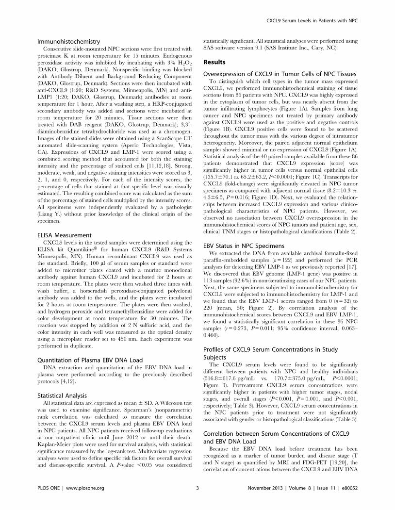
Overexpression of CXCL9 in Tumor Cells of NPC TissuesTo distinguish which cell types in the tumor mass expressed
CXCL9, we performed immunohistochemical staining of tissue
sections from 86 patients with NPC. CXCL9 was highly expressed
in the cytoplasm of tumor cells, but was nearly absent from the
tumor infiltrating lymphocytes (Figure 1A). Samples from lung
cancer and NPC specimens not treated by primary antibody
against CXCL9 were used as the positive and negative controls
(Figure 1B). CXCL9 positive cells were found to be scattered
throughout the tumor mass with the various degree of intratumor
heterogeneity. Moreover, the paired adjacent normal epithelium
samples showed minimal or no expression of CXCL9 (Figure 1A).
Statistical analysis of the 40 paired samples available from these 86
patients demonstrated that CXCL9 expression (score) was
significantly higher in tumor cells versus normal epithelial cells
(135.7670.1 vs. 65.2663.2, P,0.0001; Figure 1C). Transcripts for
CXCL9 (fold-change) were significantly elevated in NPC tumor
specimens as compared with adjacent normal tissue (8.2610.3 vs.
4.366.5, P = 0.016; Figure 1D). Next, we evaluated the relation-
ships between increased CXCL9 expression and various clinico-
pathological characteristics of NPC patients. However, we
observed no association between CXCL9 overexpression in the
immunohistochemical scores of NPC tumors and patient age, sex,
clinical TNM stages or histopathological classifications (Table 2).
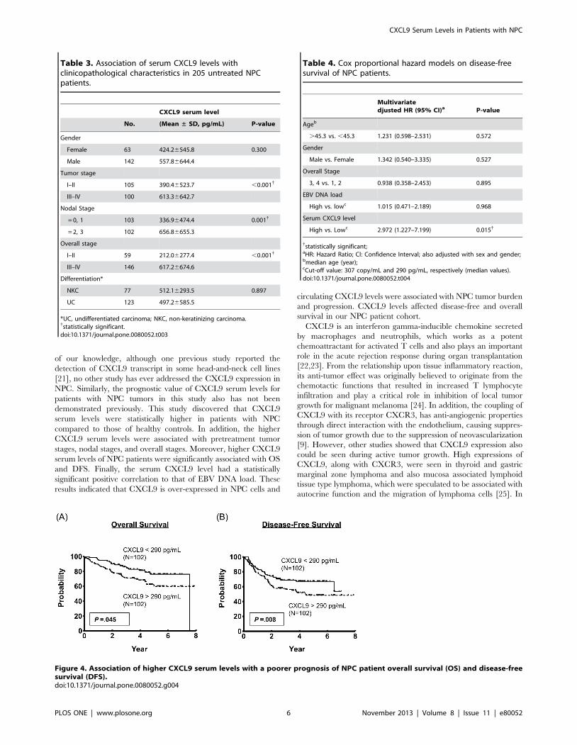
EBV Status in NPC SpecimensWe extracted the DNA from available archival formalin-fixed
paraffin-embedded samples (n = 122) and performed the PCR
analyses for detecting EBV LMP-1 as we previously reported [17].
We discovered that EBV genome (LMP-1 gene) was positive in
113 samples (92.6%) in non-keratinzing cases of our NPC patients.
Next, the same specimens subjected to immunohistochemistry for
CXCL9 were subjected to immunohistochemistry for LMP-1 and
we found that the EBV LMP-1 scores ranged from 0 (n = 32) to
220 (mean, 50; Figure 2). By correlation analysis of the
immunohistochemical scores between CXCL9 and EBV LMP-1,
we found a statistically significant correlation in these 86 NPC
samples (r = 0.273, P = 0.011; 95% confidence interval, 0.063–
0.460).
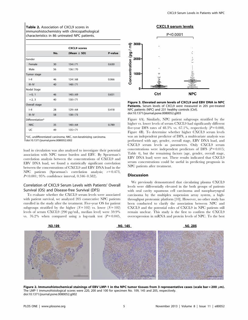
Profiles of CXCL9 Serum Concentrations in StudySubjects
The CXCL9 serum levels were found to be significantly
different between patients with NPC and healthy individuals
(516.86617.6 pg/mL vs. 170.76375.0 pg/mL, P,0.0001;
Figure 3). Pretreatment CXCL9 serum concentrations were
significantly higher in patients with higher tumor stages, nodal
stages, and overall stages (P,0.001, P = 0.001, and P,0.001,
respectively; Table 3). However, CXCL9 serum concentrations in
the NPC patients prior to treatment were not significantly
associated with gender or histopathological classifications (Table 3).
Correlation between Serum Concentrations of CXCL9and EBV DNA Load
Because the EBV DNA load before treatment has been
recognized as a marker of tumor burden and disease stage (T
and N stage) as quantified by MRI and FDG-PET [19,20], the
correlation of concentrations between the CXCL9 and EBV DNA
CXCL9 Serum Levels in Patients with NPC
PLOS ONE | www.plosone.org 3 November 2013 | Volume 8 | Issue 11 | e80052
Figure 1. Overexpression of CXCL9 in NPC tissues. (A) Immunohistochemical staining of CXCL9 in paired pericancerous adjacent normal (AN)and tumor tissues from two representative cases (scale bar: 100 mm). Brown signals indicate the CXCL9 expression. Images in the box (left panel,200x) were enlarged and shown in the right panel (400x). (B) Positive and negative controls for CXCL9 staining. (C) Box and whisker plots showing theimmunohistochemical staining scores of CXCL9 in 40 paired AN and tumor tissues. (D) Box and whisker plots showing CXCL9 mRNA transcript levelsin the 16 paired pericancerous AN and tumor tissues, as assessed by quantitative real-time RT-PCR. CXCL9 was highly overexpressed in both analysesfor NPC tissues. Box, the range of the middle 50% of CXCL9 level; line inside box, median; whiskers, minimal to maximal levels.doi:10.1371/journal.pone.0080052.g001
CXCL9 Serum Levels in Patients with NPC
PLOS ONE | www.plosone.org 4 November 2013 | Volume 8 | Issue 11 | e80052
load in circulation was also analyzed to investigate their potential
association with NPC tumor burden and EBV. By Spearman’s
correlation analysis between the concentrations of CXCL9 and
EBV DNA load, we found a statistically significant correlation
between the concentrations of CXCL9 and EBV DNA load in the
NPC patients (Spearman’s correlation analysis; r = 0.473,
P,0.001; 95% confidence interval, 0.346–0.582).
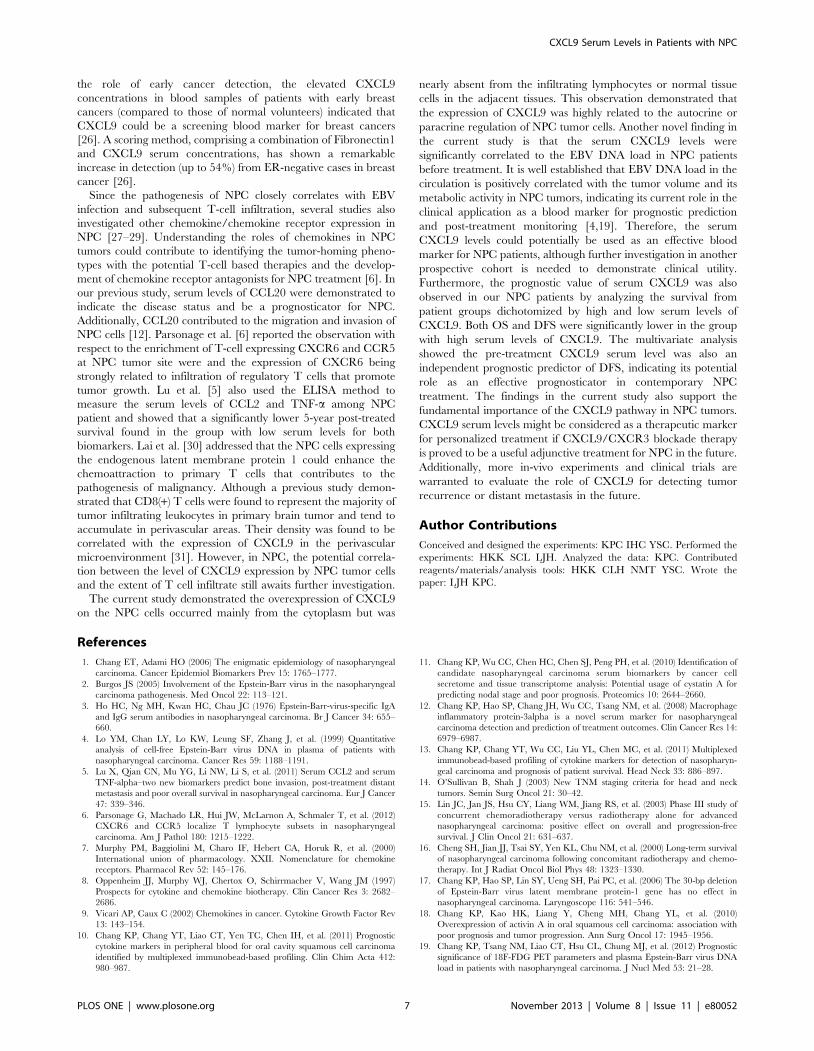
Correlation of CXCL9 Serum Levels with Patients’ OverallSurvival (OS) and Disease-free Survival (DFS)
To evaluate whether the CXCL9 serum levels were associated
with patient survival, we analyzed 205 consecutive NPC patients
enrolled in the study after the treatment. Five-year OS for patient
subgroups stratified by the higher (N = 102) vs. lower (N = 102)
levels of serum CXCL9 (290 pg/mL, median level) were 59.9%
vs. 76.2% when compared using a log-rank test (P = 0.045,
Figure 4A). Similarly, NPC patient subgroups stratified by the
higher vs. lower levels of serum CXCL9 had significantly different
five-year DFS rates of 48.3% vs. 67.1%, respectively (P = 0.008,
Figure 4B). To determine whether higher CXCL9 serum levels
was an independent predictor of DFS, a multivariate analysis was
performed with age, gender, overall stage, EBV DNA load, and
CXCL9 serum levels as parameters. Only CXCL9 serum
concentrations were independent predictors of DFS (P = 0.015;
Table 4), but the remaining factors (age, gender, overall stage,
EBV DNA load) were not. These results indicated that CXCL9
serum concentrations could be useful in predicting prognosis in
NPC patients after treatment.
Discussion
We previously demonstrated that circulating plasma CXCL9
levels were differentially elevated in the both groups of patients
with oral cavity squamous cell carcinoma and nasopharyngeal
carcinoma by the multiplex suspension array system, a high-
throughput proteomic platform [10]. However, no other study has
been conducted to clarify the association between NPC and
CXCL9 and the potential roles of CXCL9 in NPC patients still
remain unclear. This study is the first to confirm the CXCL9
overexpression in mRNA and protein levels of NPC. To the best
Table 2. Association of CXCL9 scores inimmunohistochemistry with clinicopathologicalcharacteristics in 86 untreated NPC patients.
CXCL9 scores
No. (Mean ± SD) P-value
Gender
Female 30 134671 0.630
Male 56 136670
Tumor stage
I–II 46 124668 0.066
III–IV 40 148671
Nodal Stage
= 0, 1 46 140669 0.651
= 2, 3 40 130671
Overall stage
I–II 28 129664 0.418
III–IV 58 138673
Differentiation*
NKC 35 140664 0.789
UC 49 135671
*UC, undifferentiated carcinoma; NKC, non-keratinizing carcinoma.doi:10.1371/journal.pone.0080052.t002
Figure 2. Immunohistochemical stainings of EBV LMP-1 in the NPC tumor tissues from 3 representative cases (scale bar = 200 mm).The LMP-1 immunohistological scores were 220, 200 and 100 for specimen No. 109, 145 and 205, respectively.doi:10.1371/journal.pone.0080052.g002
Figure 3. Elevated serum levels of CXCL9 and EBV DNA in NPCPatients. Serum levels of CXCL9 were measured in 205 pre-treatedNPC patients (NPC) and 231 healthy controls (Ctrl).doi:10.1371/journal.pone.0080052.g003
CXCL9 Serum Levels in Patients with NPC
PLOS ONE | www.plosone.org 5 November 2013 | Volume 8 | Issue 11 | e80052
of our knowledge, although one previous study reported the
detection of CXCL9 transcript in some head-and-neck cell lines
[21], no other study has ever addressed the CXCL9 expression in
NPC. Similarly, the prognostic value of CXCL9 serum levels for
patients with NPC tumors in this study also has not been
demonstrated previously. This study discovered that CXCL9
serum levels were statistically higher in patients with NPC
compared to those of healthy controls. In addition, the higher
CXCL9 serum levels were associated with pretreatment tumor
stages, nodal stages, and overall stages. Moreover, higher CXCL9
serum levels of NPC patients were significantly associated with OS
and DFS. Finally, the serum CXCL9 level had a statistically
significant positive correlation to that of EBV DNA load. These
results indicated that CXCL9 is over-expressed in NPC cells and
circulating CXCL9 levels were associated with NPC tumor burden
and progression. CXCL9 levels affected disease-free and overall
survival in our NPC patient cohort.
CXCL9 is an interferon gamma-inducible chemokine secreted
by macrophages and neutrophils, which works as a potent
chemoattractant for activated T cells and also plays an important
role in the acute rejection response during organ transplantation
[22,23]. From the relationship upon tissue inflammatory reaction,
its anti-tumor effect was originally believed to originate from the
chemotactic functions that resulted in increased T lymphocyte
infiltration and play a critical role in inhibition of local tumor
growth for malignant melanoma [24]. In addition, the coupling of
CXCL9 with its receptor CXCR3, has anti-angiogenic properties
through direct interaction with the endothelium, causing suppres-
sion of tumor growth due to the suppression of neovascularization
[9]. However, other studies showed that CXCL9 expression also
could be seen during active tumor growth. High expressions of
CXCL9, along with CXCR3, were seen in thyroid and gastric
marginal zone lymphoma and also mucosa associated lymphoid
tissue type lymphoma, which were speculated to be associated with
autocrine function and the migration of lymphoma cells [25]. In
Table 3. Association of serum CXCL9 levels withclinicopathological characteristics in 205 untreated NPCpatients.
CXCL9 serum level
No. (Mean ± SD, pg/mL) P-value
Gender
Female 63 424.26545.8 0.300
Male 142 557.86644.4
Tumor stage
I–II 105 390.46523.7 ,0.001{
III–IV 100 613.36642.7
Nodal Stage
= 0, 1 103 336.96474.4 0.001{
= 2, 3 102 656.86655.3
Overall stage
I–II 59 212.06277.4 ,0.001{
III–IV 146 617.26674.6
Differentiation*
NKC 77 512.16293.5 0.897
UC 123 497.26585.5
*UC, undifferentiated carcinoma; NKC, non-keratinizing carcinoma.{statistically significant.doi:10.1371/journal.pone.0080052.t003
Figure 4. Association of higher CXCL9 serum levels with a poorer prognosis of NPC patient overall survival (OS) and disease-freesurvival (DFS).doi:10.1371/journal.pone.0080052.g004
Table 4. Cox proportional hazard models on disease-freesurvival of NPC patients.
Multivariatedjusted HR (95% CI)a P-value
Ageb
.45.3 vs. ,45.3 1.231 (0.598–2.531) 0.572
Gender
Male vs. Female 1.342 (0.540–3.335) 0.527
Overall Stage
3, 4 vs. 1, 2 0.938 (0.358–2.453) 0.895
EBV DNA load
High vs. lowc 1.015 (0.471–2.189) 0.968
Serum CXCL9 level
High vs. Lowc 2.972 (1.227–7.199) 0.015{
{statistically significant;aHR: Hazard Ratio; CI: Confidence Interval; also adjusted with sex and gender;bmedian age (year);cCut-off value: 307 copy/mL and 290 pg/mL, respectively (median values).doi:10.1371/journal.pone.0080052.t004
CXCL9 Serum Levels in Patients with NPC
PLOS ONE | www.plosone.org 6 November 2013 | Volume 8 | Issue 11 | e80052
the role of early cancer detection, the elevated CXCL9
concentrations in blood samples of patients with early breast
cancers (compared to those of normal volunteers) indicated that
CXCL9 could be a screening blood marker for breast cancers
[26]. A scoring method, comprising a combination of Fibronectin1
and CXCL9 serum concentrations, has shown a remarkable
increase in detection (up to 54%) from ER-negative cases in breast
cancer [26].
Since the pathogenesis of NPC closely correlates with EBV
infection and subsequent T-cell infiltration, several studies also
investigated other chemokine/chemokine receptor expression in
NPC [27–29]. Understanding the roles of chemokines in NPC
tumors could contribute to identifying the tumor-homing pheno-
types with the potential T-cell based therapies and the develop-
ment of chemokine receptor antagonists for NPC treatment [6]. In
our previous study, serum levels of CCL20 were demonstrated to
indicate the disease status and be a prognosticator for NPC.
Additionally, CCL20 contributed to the migration and invasion of
NPC cells [12]. Parsonage et al. [6] reported the observation with
respect to the enrichment of T-cell expressing CXCR6 and CCR5
at NPC tumor site were and the expression of CXCR6 being
strongly related to infiltration of regulatory T cells that promote
tumor growth. Lu et al. [5] also used the ELISA method to
measure the serum levels of CCL2 and TNF-a among NPC
patient and showed that a significantly lower 5-year post-treated
survival found in the group with low serum levels for both
biomarkers. Lai et al. [30] addressed that the NPC cells expressing
the endogenous latent membrane protein 1 could enhance the
chemoattraction to primary T cells that contributes to the
pathogenesis of malignancy. Although a previous study demon-
strated that CD8(+) T cells were found to represent the majority of
tumor infiltrating leukocytes in primary brain tumor and tend to
accumulate in perivascular areas. Their density was found to be
correlated with the expression of CXCL9 in the perivascular
microenvironment [31]. However, in NPC, the potential correla-
tion between the level of CXCL9 expression by NPC tumor cells
and the extent of T cell infiltrate still awaits further investigation.
The current study demonstrated the overexpression of CXCL9
on the NPC cells occurred mainly from the cytoplasm but was
nearly absent from the infiltrating lymphocytes or normal tissue
cells in the adjacent tissues. This observation demonstrated that
the expression of CXCL9 was highly related to the autocrine or
paracrine regulation of NPC tumor cells. Another novel finding in
the current study is that the serum CXCL9 levels were
significantly correlated to the EBV DNA load in NPC patients
before treatment. It is well established that EBV DNA load in the
circulation is positively correlated with the tumor volume and its
metabolic activity in NPC tumors, indicating its current role in the
clinical application as a blood marker for prognostic prediction
and post-treatment monitoring [4,19]. Therefore, the serum
CXCL9 levels could potentially be used as an effective blood
marker for NPC patients, although further investigation in another
prospective cohort is needed to demonstrate clinical utility.
Furthermore, the prognostic value of serum CXCL9 was also
observed in our NPC patients by analyzing the survival from
patient groups dichotomized by high and low serum levels of
CXCL9. Both OS and DFS were significantly lower in the group
with high serum levels of CXCL9. The multivariate analysis
showed the pre-treatment CXCL9 serum level was also an
independent prognostic predictor of DFS, indicating its potential
role as an effective prognosticator in contemporary NPC
treatment. The findings in the current study also support the
fundamental importance of the CXCL9 pathway in NPC tumors.
CXCL9 serum levels might be considered as a therapeutic marker
for personalized treatment if CXCL9/CXCR3 blockade therapy
is proved to be a useful adjunctive treatment for NPC in the future.
Additionally, more in-vivo experiments and clinical trials are
warranted to evaluate the role of CXCL9 for detecting tumor
recurrence or distant metastasis in the future.
Author Contributions
Conceived and designed the experiments: KPC IHC YSC. Performed the
experiments: HKK SCL LJH. Analyzed the data: KPC. Contributed
reagents/materials/analysis tools: HKK CLH NMT YSC. Wrote the
paper: LJH KPC.
References
1. Chang ET, Adami HO (2006) The enigmatic epidemiology of nasopharyngeal
carcinoma. Cancer Epidemiol Biomarkers Prev 15: 1765–1777.
2. Burgos JS (2005) Involvement of the Epstein-Barr virus in the nasopharyngeal
carcinoma pathogenesis. Med Oncol 22: 113–121.
3. Ho HC, Ng MH, Kwan HC, Chau JC (1976) Epstein-Barr-virus-specific IgA
and IgG serum antibodies in nasopharyngeal carcinoma. Br J Cancer 34: 655–
660.
4. Lo YM, Chan LY, Lo KW, Leung SF, Zhang J, et al. (1999) Quantitative
analysis of cell-free Epstein-Barr virus DNA in plasma of patients with
nasopharyngeal carcinoma. Cancer Res 59: 1188–1191.
5. Lu X, Qian CN, Mu YG, Li NW, Li S, et al. (2011) Serum CCL2 and serum
TNF-alpha–two new biomarkers predict bone invasion, post-treatment distant
metastasis and poor overall survival in nasopharyngeal carcinoma. Eur J Cancer
47: 339–346.
6. Parsonage G, Machado LR, Hui JW, McLarnon A, Schmaler T, et al. (2012)
CXCR6 and CCR5 localize T lymphocyte subsets in nasopharyngeal
carcinoma. Am J Pathol 180: 1215–1222.
7. Murphy PM, Baggiolini M, Charo IF, Hebert CA, Horuk R, et al. (2000)
International union of pharmacology. XXII. Nomenclature for chemokine
receptors. Pharmacol Rev 52: 145–176.
8. Oppenheim JJ, Murphy WJ, Chertox O, Schirrmacher V, Wang JM (1997)
Prospects for cytokine and chemokine biotherapy. Clin Cancer Res 3: 2682–
2686.
9. Vicari AP, Caux C (2002) Chemokines in cancer. Cytokine Growth Factor Rev
13: 143–154.
10. Chang KP, Chang YT, Liao CT, Yen TC, Chen IH, et al. (2011) Prognostic
cytokine markers in peripheral blood for oral cavity squamous cell carcinoma
identified by multiplexed immunobead-based profiling. Clin Chim Acta 412:
980–987.
11. Chang KP, Wu CC, Chen HC, Chen SJ, Peng PH, et al. (2010) Identification of
candidate nasopharyngeal carcinoma serum biomarkers by cancer cell
secretome and tissue transcriptome analysis: Potential usage of cystatin A for
predicting nodal stage and poor prognosis. Proteomics 10: 2644–2660.
12. Chang KP, Hao SP, Chang JH, Wu CC, Tsang NM, et al. (2008) Macrophage
inflammatory protein-3alpha is a novel serum marker for nasopharyngeal
carcinoma detection and prediction of treatment outcomes. Clin Cancer Res 14:
6979–6987.
13. Chang KP, Chang YT, Wu CC, Liu YL, Chen MC, et al. (2011) Multiplexed
immunobead-based profiling of cytokine markers for detection of nasopharyn-
geal carcinoma and prognosis of patient survival. Head Neck 33: 886–897.
14. O’Sullivan B, Shah J (2003) New TNM staging criteria for head and neck
tumors. Semin Surg Oncol 21: 30–42.
15. Lin JC, Jan JS, Hsu CY, Liang WM, Jiang RS, et al. (2003) Phase III study of
concurrent chemoradiotherapy versus radiotherapy alone for advanced
nasopharyngeal carcinoma: positive effect on overall and progression-free
survival. J Clin Oncol 21: 631–637.
16. Cheng SH, Jian JJ, Tsai SY, Yen KL, Chu NM, et al. (2000) Long-term survival
of nasopharyngeal carcinoma following concomitant radiotherapy and chemo-
therapy. Int J Radiat Oncol Biol Phys 48: 1323–1330.
17. Chang KP, Hao SP, Lin SY, Ueng SH, Pai PC, et al. (2006) The 30-bp deletion
of Epstein-Barr virus latent membrane protein-1 gene has no effect in
nasopharyngeal carcinoma. Laryngoscope 116: 541–546.
18. Chang KP, Kao HK, Liang Y, Cheng MH, Chang YL, et al. (2010)
Overexpression of activin A in oral squamous cell carcinoma: association with
poor prognosis and tumor progression. Ann Surg Oncol 17: 1945–1956.
19. Chang KP, Tsang NM, Liao CT, Hsu CL, Chung MJ, et al. (2012) Prognostic
significance of 18F-FDG PET parameters and plasma Epstein-Barr virus DNA
load in patients with nasopharyngeal carcinoma. J Nucl Med 53: 21–28.
CXCL9 Serum Levels in Patients with NPC
PLOS ONE | www.plosone.org 7 November 2013 | Volume 8 | Issue 11 | e80052

20. Ma BB, King A, Lo YM, Yau YY, Zee B, et al. (2006) Relationship between
pretreatment level of plasma Epstein-Barr virus DNA, tumor burden, andmetabolic activity in advanced nasopharyngeal carcinoma. Int J Radiat Oncol
Biol Phys 66: 714–720.
21. Wolff HA, Rolke D, Rave-Frank M, Schirmer M, Eicheler W, et al. (2011)Analysis of chemokine and chemokine receptor expression in squamous cell
carcinoma of the head and neck (SCCHN) cell lines. Radiat Environ Biophys 50:145–154.
22. Whiting D, Hsieh G, Yun JJ, Banerji A, Yao W, et al. (2004) Chemokine
monokine induced by IFN-gamma/CXC chemokine ligand 9 stimulates Tlymphocyte proliferation and effector cytokine production. J Immunol 172:
7417–7424.23. Miura M, Morita K, Kobayashi H, Hamilton TA, Burdick MD, et al. (2001)
Monokine induced by IFN-gamma is a dominant factor directing T cells intomurine cardiac allografts during acute rejection. J Immunol 167: 3494–3504.
24. Kunz M, Toksoy A, Goebeler M, Engelhardt E, Brocker E, et al. (1999) Strong
expression of the lymphoattractant C-X-C chemokine Mig is associated withheavy infiltration of T cells in human malignant melanoma. J Pathol 189: 552–
558.25. Ohshima K, Suefuji H, Karube K, Hamasaki M, Hatano B, et al. (2003)
Expression of chemokine receptor CXCR3 and its ligand, mig, in gastric and
thyroid marginal zone lymphomas. Possible migration and autocrine mecha-nism. Leuk Lymphoma 44: 329–336.
26. Ruiz-Garcia E, Scott V, Machavoine C, Bidart JM, Lacroix L, et al. (2010) Gene
expression profiling identifies Fibronectin 1 and CXCL9 as candidate
biomarkers for breast cancer screening. Br J Cancer 102: 462–468.
27. Ou DL, Chen CL, Lin SB, Hsu CH, Lin LI (2006) Chemokine receptor
expression profiles in nasopharyngeal carcinoma and their association with
metastasis and radiotherapy. J Pathol 210: 363–373.
28. Hu J, Deng X, Bian X, Li G, Tong Y, et al. (2005) The expression of functional
chemokine receptor CXCR4 is associated with the metastatic potential of
human nasopharyngeal carcinoma. Clin Cancer Res 11: 4658–4665.
29. Tang KF, Tan SY, Chan SH, Chong SM, Loh KS, et al. (2001) A distinct
expression of CC chemokines by macrophages in nasopharyngeal carcinoma:
implication for the intense tumor infiltration by T lymphocytes and
macrophages. Hum Pathol 32: 42–49.
30. Lai HC, Hsiao JR, Chen CW, Wu SY, Lee CH, et al. (2010) Endogenous latent
membrane protein 1 in Epstein-Barr virus-infected nasopharyngeal carcinoma
cells attracts T lymphocytes through upregulation of multiple chemokines.
Virology 405: 464–473.
31. Venetz D, Ponzoni M, Schiraldi M, Ferreri AJ, Bertoni F, et al. (2010)
Perivascular expression of CXCL9 and CXCL12 in primary central nervous
system lymphoma: T-cell infiltration and positioning of malignant B cells.
Int J Cancer 127: 2300–2312.
CXCL9 Serum Levels in Patients with NPC
PLOS ONE | www.plosone.org 8 November 2013 | Volume 8 | Issue 11 | e80052


![Research Article Prognostic Significance of Serum Free Light … · 2019. 7. 31. · the progression of MGUS [ ], solitary plasmacytoma [ ], and smoldering myeloma [ ]intomultiplemyeloma](https://img.pdfslide.net/doc/110x75/60b139df8dfefb1baa01f551/research-article-prognostic-significance-of-serum-free-light-2019-7-31-the.jpg)