Embed Size (px)
Citation preview
Bowles et al. Patient Safety in Surgery (2015) 9:22 DOI 10.1186/s13037-015-0061-x
CASE REPORT Open Access
Severe mycosis as a rare infection after a cornauger injury of the hand: a case reportRichard J Bowles1, Justin J Mitchell1, Connie Price2 and Kyros Ipaktchi3*
Abstract
Mucormycosis is a rare but serious infection that can be seen in immunocompetent individuals who experiencetraumatic injury. The authors report a case in a 28 year-old man who sustained a mangling hand injury in a cornaugur accident. After initial aggressive debridement ongoing tissue necrosis was seen, and in subsequent biopsiesinvasive mucormycosis was diagnosed. The patient was successfully managed with immediate surgical debridementand antifungal medication and showed no sign of infection at six-month follow-up.
Keywords: Agricultural injuries, Mangled hand, Mucormycosis, Zygomycosis
BackgroundMucormycosis, formerly known as zygomycosis, is a rareinfection caused by fungal species that are commonlyfound in soil and decaying organic matter. Thoughmucormycosis is more likely in immunocompromisedindividuals, traumatic injury may allow penetration ofthe fungus through the mucocutaneous barrier andaggressive infection may result. Typical for this infectionis the “angioinvasive” spreading of the disease processwhich can result in a risk for loss of limb and life if notrapidly controlled. Identification and treatment of thisserious infection is important as the mortality rate asso-ciated with cutaneous mucormycosis has been shown tobe as high as 31% [1].In this case report we describe an immunocompetent
patient involved in an agricultural accident who sus-tained a mangled left hand that was complicated by asubsequent mycotic infection.
Case presentationA 28-year-old right-hand-dominant man was airlifted forlimb salvage to the level 1 trauma center where theinvestigators practice after sustaining an agricultural ac-cident in which his left hand was pulled into a cornaugur. The mangling injury of his hand included frac-tures of his index through ring fingers with extensive
* Correspondence: [email protected] of Orthopedic Surgery, Denver Health Medical Center, 777Bannock St, Denver, CO 80204, USAFull list of author information is available at the end of the article
© 2015 Bowles et al.; licensee BioMed CentralCommons Attribution License (http://creativecreproduction in any medium, provided the orDedication waiver (http://creativecommons.orunless otherwise stated.
lacerations and exposed flexor tendons. There was a par-tial degloving injury to his left palm (Figures 1 and 2).The patient was otherwise stable and had no additionalinjuries. Upon arrival he was taken emergently to theoperating room, where he underwent irrigation and de-bridement, primary fusion with Kirschner wires of thedestroyed PIP joints in his left index, long, and ring fin-gers, a split-thickness skin-graft to his left palm and longfinger from a left forearm donor site, and initiation of IVantibiotics, administering vancomycin and ampicillin/sulbactam. He recovered uneventfully and was dis-charged home four days later without additional anti-biotic therapy.At his first follow-up appointment two weeks post-
operatively, he showed no sign of systemic infection butwas noted to have an extensive necrosis involving partof his left long finger and the palmar skin graft (Figure 3).Surgical debridement including amputation of the longfinger was scheduled and he underwent a filet flap clos-ure of the second webspace after long finger ablation.Tissue samples revealed both bacterial and mold growth,and the Infectious Disease team was consulted. Due tothe suspicion for an invasive mucor mycosis he wastaken the next morning for additional irrigation and de-bridement, and lipid-complex amphotericin B was addedto his antibacterial medications. Cultures were speciatedto coagulase-negative Staphylococcus, two different spe-cies of Pseudomonas, and mycotic species includingMucor and Aspergillus fumigates. He recovered well andwas later discharged home on hospital day five with a
. This is an Open Access article distributed under the terms of the Creativeommons.org/licenses/by/4.0), which permits unrestricted use, distribution, andiginal work is properly credited. The Creative Commons Public Domaing/publicdomain/zero/1.0/) applies to the data made available in this article,
Figure 1 Preoperative situation demonstrating extensive soft tissue laceration by corn grinding machinery (left). Postoperative view afterirrigation and debridement with partial soft tissue closure (right).
Bowles et al. Patient Safety in Surgery (2015) 9:22 Page 2 of 5
Hohn catheter and an outpatient antibiotic regimenplanned for four total weeks that included vancomycin 1g intravenously twice daily, Zosyn 12 g continuous infu-sion daily, and Posaconazole suspension 400 mg (10 mL)orally twice daily.He continued to show improvement at his follow-up
appointments with both Orthopaedic Surgery and Infec-tious Disease. Approximately one week after dischargehis AFB cultures taken intraoperatively turned positiveby probe for Mycobacterium tuberculosis complex, whichstood in contrast to a negative PPD test that had beendocumented within the previous two years. At this clinicvisit a new PPD was placed and later found to be
Figure 2 Preoperative radiographs showing proximal interphalangial jointphalanx fracture of the thumb.
negative when read in his home town. A chest radio-graph also showed no sign of TB infection. Ultimatelythe positive AFB was attributed to an environmentalmycobacterial contaminant.His weekly labs drawn by a home health agency were
otherwise unremarkable until approximately three weeksafter his discharge. At that time he was found to be neu-tropenic with a white blood cell count of 3100 (absoluteneutrophil count 120), and he was admitted to the hos-pital in his hometown where all of his antibiotics andantifungals were held. He was discharged several dayslater after recovery of his white blood cell count withoutsign of infection or any further complication. Though
fracture dislocations of left index, long, and ring fingers and proximal
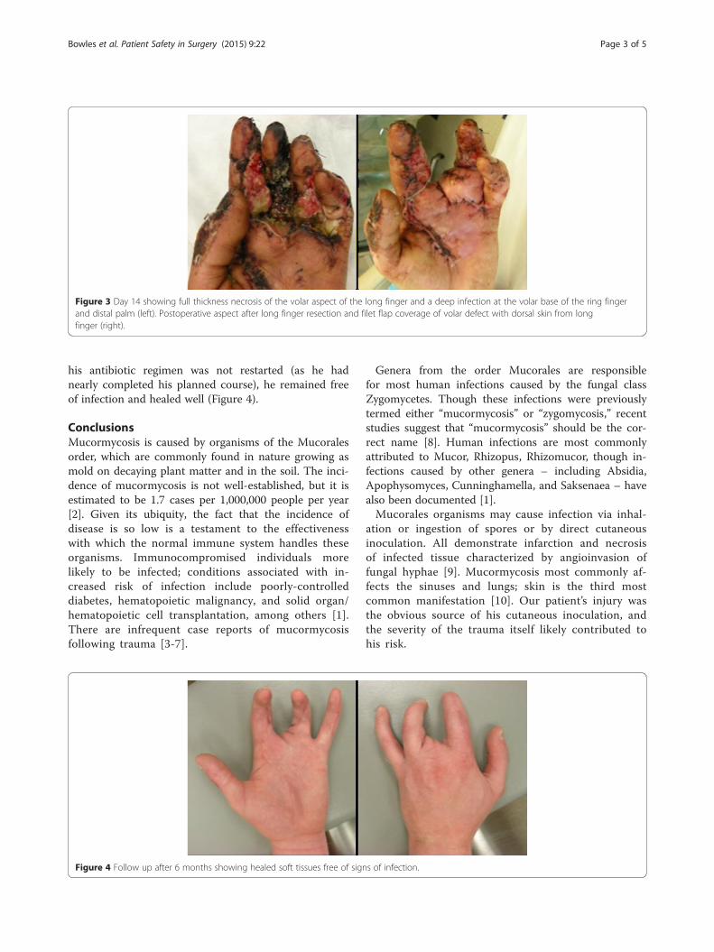
Figure 3 Day 14 showing full thickness necrosis of the volar aspect of the long finger and a deep infection at the volar base of the ring fingerand distal palm (left). Postoperative aspect after long finger resection and filet flap coverage of volar defect with dorsal skin from longfinger (right).
Bowles et al. Patient Safety in Surgery (2015) 9:22 Page 3 of 5
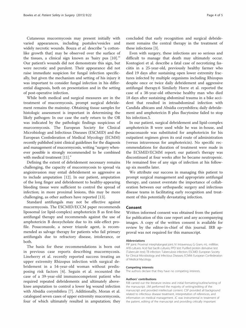
his antibiotic regimen was not restarted (as he hadnearly completed his planned course), he remained freeof infection and healed well (Figure 4).
ConclusionsMucormycosis is caused by organisms of the Mucoralesorder, which are commonly found in nature growing asmold on decaying plant matter and in the soil. The inci-dence of mucormycosis is not well-established, but it isestimated to be 1.7 cases per 1,000,000 people per year[2]. Given its ubiquity, the fact that the incidence ofdisease is so low is a testament to the effectivenesswith which the normal immune system handles theseorganisms. Immunocompromised individuals morelikely to be infected; conditions associated with in-creased risk of infection include poorly-controlleddiabetes, hematopoietic malignancy, and solid organ/hematopoietic cell transplantation, among others [1].There are infrequent case reports of mucormycosisfollowing trauma [3-7].
Figure 4 Follow up after 6 months showing healed soft tissues free of sign
Genera from the order Mucorales are responsiblefor most human infections caused by the fungal classZygomycetes. Though these infections were previouslytermed either “mucormycosis” or “zygomycosis,” recentstudies suggest that “mucormycosis” should be the cor-rect name [8]. Human infections are most commonlyattributed to Mucor, Rhizopus, Rhizomucor, though in-fections caused by other genera – including Absidia,Apophysomyces, Cunninghamella, and Saksenaea – havealso been documented [1].Mucorales organisms may cause infection via inhal-
ation or ingestion of spores or by direct cutaneousinoculation. All demonstrate infarction and necrosisof infected tissue characterized by angioinvasion offungal hyphae [9]. Mucormycosis most commonly af-fects the sinuses and lungs; skin is the third mostcommon manifestation [10]. Our patient’s injury wasthe obvious source of his cutaneous inoculation, andthe severity of the trauma itself likely contributed tohis risk.
s of infection.
Bowles et al. Patient Safety in Surgery (2015) 9:22 Page 4 of 5
Cutaneous mucormycosis may present initially withvaried appearances, including pustules/vesicles andwidely necrotic wounds. Bouza et al. describe “a cotton-like growth that] may be observed over the surface ofthe tissues, a clinical sign known as ‘hairy pus [10].’”Our patient’s wounds did not demonstrate this sign, butwere necrotic and purulent. Their appearance did notraise immediate suspicion for fungal infection specific-ally, but given the mechanism and setting of his injury itwas important to consider fungal infection in his differ-ential diagnosis, both on presentation and in the settingof post-operative infection.While both medical and surgical measures are in the
treatment of mucormycosis, prompt surgical debride-ment remains the mainstay. Obtaining tissue samples forhistologic assessment is important in determining thelikely pathogen: In our case the early return to the ORwas indicated by the pathologic findings suspicious ofmurcormycosis. The European Society for ClinicalMicrobiology and Infectious Diseases (ESCMID) and theEuropean Confederation of Medical Mycology (ECMM)recently published joint clinical guidelines for the diagnosisand management of mucormycosis, writing “surgery when-ever possible is strongly recommended to be combinedwith medical treatment [11].”Defining the extent of debridement necessary remains
challenging, the capacity of mucormycosis to spread viaangioinvasion may entail debridement so aggressive asto include amputation [12]. In our patient, amputationof the long finger and debridement to healthy-appearingbleeding tissue were sufficient to control the spread ofinfection; in more proximal lesions, this may be morechallenging, as other authors have reported [13].Standard antifungals may not be effective against
mucormycosis. The ESCMID/ECCM paper recommendsliposomal (or lipid-complex) amphotericin B as first-lineantifungal therapy and recommends against the use ofamphotericin B deoxycholate due to its side-effect pro-file. Posaconazole, a newer triazole agent, is recom-mended as salvage therapy for patients who fail primaryantifungals due to refractory disease, intolerance, orboth.The basis for these recommendations is born out
in previous case reports describing mucormycosis.Lineberry et al. recently reported success treating anupper extremity Rhizopus infection with surgical de-bridement in a 54-year-old woman without predis-posing risk factors [4]. Seguin et al. recounted thecase of a 39-year-old immunocompetent patient whorequired repeated debridements and ultimately above-knee amputation to control a lower leg wound infectionwith Absidia corymbifera [7]. Additionally, Moran et al.catalogued seven cases of upper extremity mucormycosis,four of which ultimately resulted in amputation; they
concluded that early recognition and surgical debride-ment remains the central therapy in the treatment ofthese infections [3].Even with surgery, these infections are so serious and
difficult to manage that death may ultimately occur.Kontogiori et al. describe a fatal case of necrotizing fas-ciitis in a 25-year-old, previously healthy farmer whodied 19 days after sustaining open lower extremity frac-tures infected by multiple organisms including Rhizopusdespite once or twice daily debridement and aggressiveantifungal therapy.6 Similarly Horre et al. reported thecase of a 38-year-old otherwise healthy man who died18 days after sustaining abdominal trauma in a bike acci-dent that resulted in intraabdominal infection withCandida albicans and Absidia corymbifera; daily debride-ment and amphotericin B plus flucytosine failed to stophis infection.5.In our patient, surgical debridement and lipid-complex
amphotericin B were used while he was in-house, andposaconazole was substituted for amphotericin for hisoutpatient regimen given its oral route of administration(versus intravenous for amphotericin). No specific rec-ommendations for duration of treatment were made inthe ECSMID/ECMM report; our patient’s therapy wasdiscontinued at four weeks after he became neutropenic.He remained free of any sign of infection at his follow-up six months later.We attribute our success in managing this patient to
prompt surgical management and appropriate antifungaltherapy, and cannot overstate the importance of collab-oration between our orthopaedic surgery and infectiousdisease teams in facilitating early recognition and treat-ment of this potentially devastating infection.
ConsentWritten informed consent was obtained from the patientfor publication of this case report and any accompanyingimages. A copy of the written consent is available forreview by the editor-in-chief of this journal. IRB ap-proval was not required for this manuscript.
AbbreviationsPIP joint: Proximal interphalangeal joint; IV: Intravenous; G: Gram; mL: milliliter;AFB cultures: Acid fast bacilli cultures; PPD test: Purified protein derivative test(Tuberculin test); TB infection: Tuberculosis infection; ESCMID: European Societyfor Clinical Microbiology and Infectious Diseases; ECMM: European Confederationof Medical Mycology.
Competing interestsThe authors declare that they have no competing interests.
Authors’ contributionsRJB carried out the literature review and initial formatting/outline/writing ofthe manuscript. JJM performed the majority of writing/editing of themanuscript and provided intellectual content. CSP provided all backgroundrelated to infectious disease treatment, interpretation of references, andinformation on medical management. JC was instrumental in treatment ofthe patient, editing of the manuscript and providing critically important
Bowles et al. Patient Safety in Surgery (2015) 9:22 Page 5 of 5
imaging and intellectual content. KI is the senior author, provided the caseand idea and oversaw all writing, editing, and compilation of the manuscript.All authors read and approved the final manuscript.
Author details1Department of Orthopaedic Surgery, University of Colorado School ofMedicine, 12631 E. 17th Avenue, Aurora, CO 80045, USA. 2Department ofMedicine, Infectious Diseases, Denver Health Medical Center, 777 BannockStreet, Denver, CO 80204, USA. 3Department of Orthopedic Surgery, DenverHealth Medical Center, 777 Bannock St, Denver, CO 80204, USA.
Received: 10 November 2014 Accepted: 15 April 2015
References1. Roden MM, Zaoutis TE, Buchanan WL, Knudsen TA, Sarkisova TA,
Schaufele RL, et al. Epidemiology and outcome of zygomycosis: areview of 929 reported cases. Clin Infect Dis. 2005;41(5):634–53.
2. Rees JR, Pinner RW, Hajjeh RA, Brandt ME, Reingold AL. The epidemiologicalfeatures of invasive mycotic infections in the San Francisco Bay area,1992-1993: results of population-based laboratory active surveillance.Clin Infect Dis. 1998;27(5):1138–47.
3. Moran SL, Strickland J, Shin AY. Upper-extremity mucormycosis infections inimmunocompetent patients. J Hand Surg Am. 2006;31(7):1201–5.
4. Lineberry KD, Boettcher AK, Blount AL, Burgess SD. Cutaneousmucormycosis of the upper extremity in an immunocompetent host: casereport. J Hand Surg Am. 2012;37(4):787–91.
5. Horre R, Jovanic B, Herff S, Marklein G, Zhou H, Heinze I, et al. Woundinfection due to Absidia corymbifera and Candida albicans with fataloutcome. Med Mycol. 2004;42(4):373–8.
6. Kontogiorgi M, Floros I, Koroneos A, Vamvouka C, Paniara O, Roussos C, et al.Fatal post-traumatic zygomycosis in an immunocompetent young patient.J Med Microbiol. 2007;56(Pt 9):1243–5.
7. Seguin P, Musellec H, Le Gall F, Chevrier S, Le Bouquin V, Malledant Y.Post-traumatic course complicated by cutaneous infection with Absidiacorymbifera. Eur J Clin Microbiol Infect Dis. 1999;18(10):737–9.
8. Kwon-Chung KJ. Taxonomy of fungi causing mucormycosis andentomophthoramycosis (zygomycosis) and nomenclature of the disease:molecular mycologic perspectives. Clin Infect Dis. 2012;54 Suppl 1:S8–15.
9. Petrikkos G, Skiada A, Lortholary O, Roilides E, Walsh TJ, Kontoyiannis DP.Epidemiology and clinical manifestations of mucormycosis. Clin Infect Dis.2012;54 Suppl 1:S23–34.
10. Bouza E, Muñoz P, Guinea J. Mucormycosis: an emerging disease? ClinMicrobiol Infect. 2006;12:7–23.
11. Cornely OA, Arikan-Akdagli S, Dannaoui E, Groll AH, Lagrou K, Chakrabarti A, et al.ESCMID and ECMM Joint Clinical Guidelines for the Diagnosis and Managementof Mucormycosis 2013. Clin Microbiol Infect. 2014.
12. Anaya DA, Dellinger EP. Necrotizing soft-tissue infection: diagnosis andmanagement. Clin Infect Dis. 2007;44(5):705–10.
13. Koonce RC, Price CS, Sutton DA, Wickes BL, Montero PN, Morgan SJ.Lower-extremity zygomycosis in a patient with traumatic injuries, A casereport. J Bone Joint Surg Am. 2009;91(3):686–92.
Submit your next manuscript to BioMed Centraland take full advantage of:
• Convenient online submission
• Thorough peer review
• No space constraints or color figure charges
• Immediate publication on acceptance
• Inclusion in PubMed, CAS, Scopus and Google Scholar
• Research which is freely available for redistribution
Submit your manuscript at www.biomedcentral.com/submit


![[Micro] opportunistic mycosis](https://img.pdfslide.net/doc/110x75/55d6fc6bbb61ebfa2a8b47ec/micro-opportunistic-mycosis.jpg)