Embed Size (px)
Citation preview

‘Sewage fungus’ A field and microscopic guide
Tim Geatches1, Julie Gething2 and Graham Rutt2
1 Environment Agency 2 Cyfoeth Naturiol Cymru / Natural Resources Wales
Version 3 March 2014

Contents 4 Introduction
5 References
6-7 Main sewage fungus taxa and growth forms
8 Assessment methodology
9 Examples of sewage fungus densities
10 Sewage fungus recording sheet
Main sewage fungus taxa
12-13 Sphaerotilus natans
14-15 Beggiatoa alba
16-17 Zoogloeal bacteria
18-19 Fusarium aquaeductuum
20-21 Geotrichum candidum
22-23 Leptomitus lacteus
24-25 Carchesium polypinum
Other sewage fungus taxa
27 Flexibacter spp.
28-29 Thiothrix II
30 Achyla spp.
31 Flavobacterium spp.
Taxa commonly mistaken for sewage fungus
33 Didymosphenia spp.
34 Leptothrix ochracea
3

Introduction • Mainly heterotrophic micro-organisms
• Sensitive indicators of organic pollution
• Matrix of filamentous bacteria, fungi and/or stalked protozoa
• Can be present either as an almost pure monoculture of one species or
as a mixed growth of several species
• Growth form varies due to species type and severity of organic pollution
• Species present can indicate type of organic pollution
• Colour varies widely from white to brown or pink
• Can be used to identify organic pollution sources several kilometres
away
• Assessment below substrate particularly useful as lack of competition
with autotrophic organisms
• Microscopic examination required to confirm identification
4
References Curds CR, Gates MA and Roberts DMcL (1983) British and Other Freshwater Ciliated Protozoa: Part II, Synopsis of the British Fauna No. 23. Curtis EJC (1969) Sewage fungus: its nature and effects, Water Research, Vol. 3, 289-311. Eikelbloom DH (2000) Activated Sludge Information Systems www.asissludge.com Gray NF (1982) A key to the major slime-forming organisms of ‘sewage fungus’, J. life Sci. R. Dubl. Soc. 4, 97-102. Gray NF (1985) Heterotrophic slimes in flowing waters, Biol. Rev., 60, 499- 548. Tomlinson TG and Williams IL (1975) Fungi. In Ecological Aspects of Used Water Treatment. I. The Organisms and their Ecology, pp 93-152. Van Veen WL, Mulder EG and Deinema MH (1978) The Sphaerotilus- Leptothrix Group of Bacteria, Microbiological Reviews, 329-356.
5
Main sewage fungus taxa and growth forms
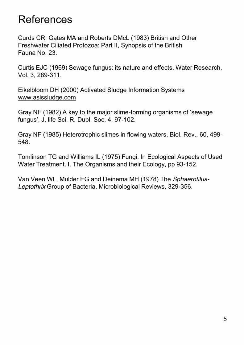
Sphaerotilus natans Filamentous bacterium Slimy fronds Zoogloeal bacterium Ill defined taxonomically Jelly-like gelatinous mass Beggiatoa alba Filamentous bacterium Thin white film
The taxa below are the main sewage fungus organisms found in freshwaters and typical growth forms. One or more taxa can be present in sewage fungus outbreaks and microscopic examination is required to confirm identification. Further information on growth forms, environmental conditions and key identification features can be found on pages 12 to 25.
6

Main sewage fungus taxa and growth forms
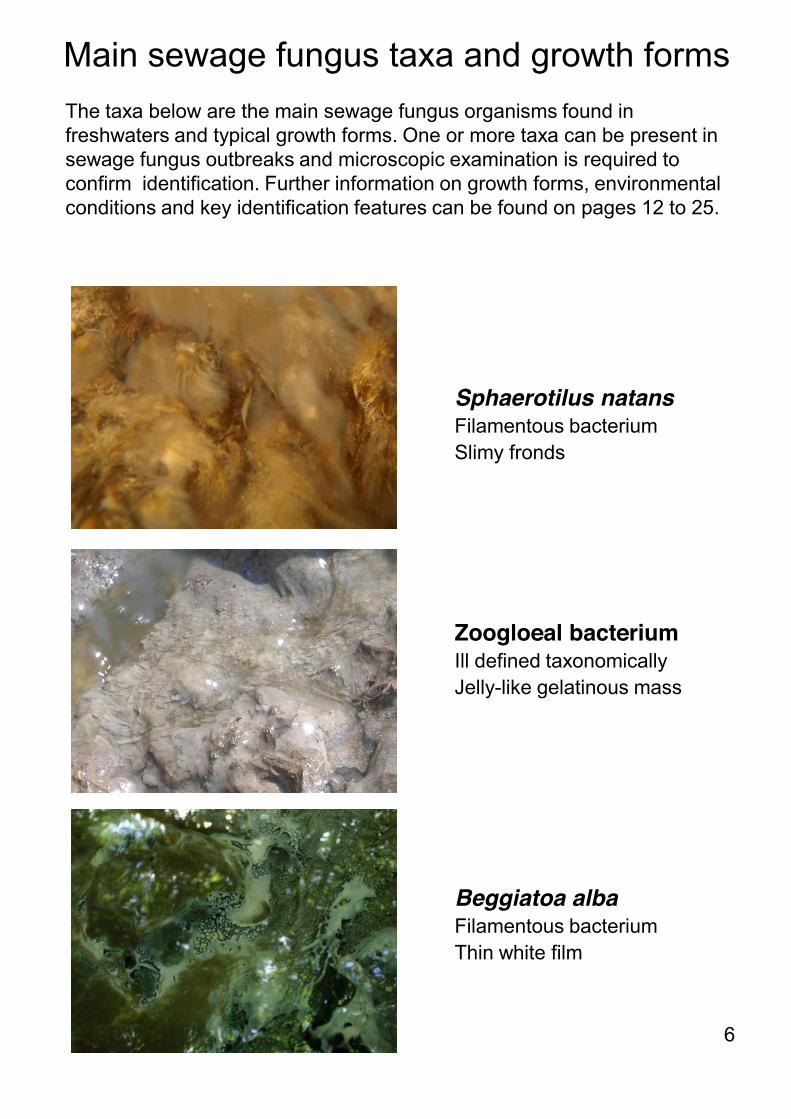
Fusarium aquaeductuum Filamentous fungus Imparts pink or red colouration Geotrichum candidum Filamentous fungus Soft texture loosely following contours of stones Leptomitus lacteus Filamentous fungus Overlapping cotton wool-like streamers Carchesium polypinum Stalked protozoan Short 2-3mm tufts
7
Assessment methodology Record cover and density above and below substrate as follows: Cover None Local - <30% Widespread - 30 – 60% Extensive - >60%
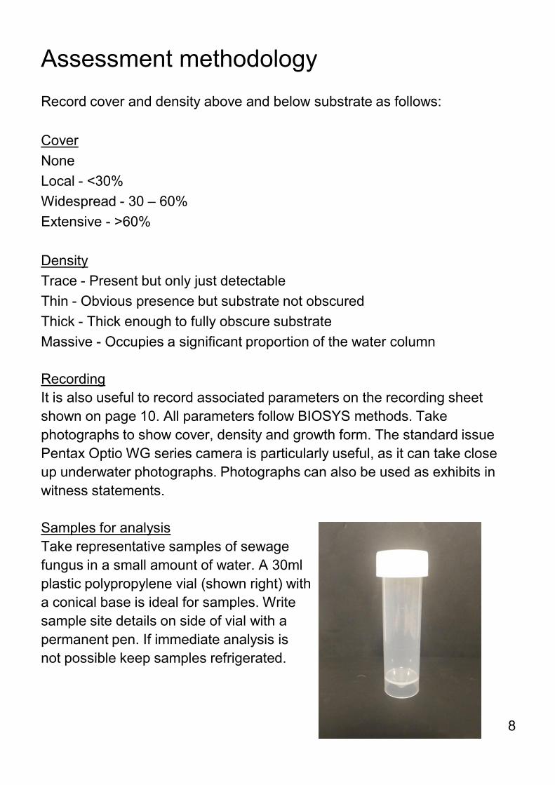
Density Trace - Present but only just detectable Thin - Obvious presence but substrate not obscured Thick - Thick enough to fully obscure substrate Massive - Occupies a significant proportion of the water column Recording It is also useful to record associated parameters on the recording sheet shown on page 10. All parameters follow BIOSYS methods. Take photographs to show cover, density and growth form. The standard issue Pentax Optio WG series camera is particularly useful, as it can take close up underwater photographs. Photographs can also be used as exhibits in witness statements. Samples for analysis Take representative samples of sewage fungus in a small amount of water. A 30ml plastic polypropylene vial (shown right) with a conical base is ideal for samples. Write sample site details on side of vial with a permanent pen. If immediate analysis is not possible keep samples refrigerated.
8
Examples of sewage fungus densities
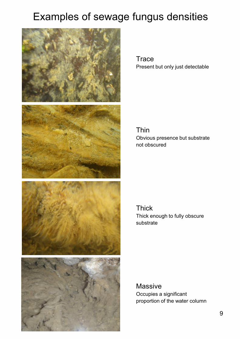
Trace Present but only just detectable
Thin Obvious presence but substrate not obscured
Thick Thick enough to fully obscure substrate
Massive Occupies a significant proportion of the water column
9

Sewage fungus recording sheet
10
Water Body: Site ID: Date: Time:
Site Name: NGR (GPS):
Photos: Yes / No Sampler:
TURBIDITY: Tick box ODOUR: Tick box SEWAGE LITTER: Tick box GENERAL COMMENTS:
OVERLAYING SILT COVER: Tick box for cover and density OCHRE: Tick box for cover and density
SEWAGE FUNGUS ABOVE STONES: Tick box for presence and density SEWAGE FUNGUS BELOW STONES: Tick box for presence and density
SEWAGE FUNGUS ABOVE STONES: Field comments SEWAGE FUNGUS BELOW STONES: Field comments
Lab use only:
Analyst: Laboratory: Date: Time:
SEWAGE FUNGUS ABOVE STONES: Lab analysis SEWAGE FUNGUS BELOW STONES: Lab analysis
THIN: Obvious presence but substrate not obscuredTHICK: Thick enough to fully obscure the substrateMASSIVE: Occupies a signif icant proportion of the w ater column
TRACE: Present but only just detectableLOCAL: Occasional patches: <30% of area
THIN: Obvious presence but substrate not obscured
WIDESPREAD: 30 - 60% of area
THICK: Thick enough to fully obscure the substrate
EXTENSIVE: >60%MASSIVE: Occupies a signif icant proportion of the w ater column
GROSS: >60% of possible area
WIDESPREAD: 30 - 60% of the possible area
LOCAL: <30% of possible area
NONE: No litter present
NONE: None presentNONE: None present TRACE: Present but only just detectable
TRACE: Just detectable by eyeLOCAL: Occasional patches: <30% of area
THIN: Obvious presence but f ine details of substrate not obscured
LOCAL: Occasional patches: <30% of areaWIDESPREAD: 30 - 60% of area
EXTENSIVE: >60%
NONE: No discernible odour
WIDESPREAD: 30 - 60% of area
THICK: Coats stones and obscures f ine details of substrate
EXTENSIVE: >60%MASSIVE: Fills interstices betw een gravel sized particles
CLEAR: Water not visibly turbidSLIGHT : Visible turbidity but no effect on light penetrationMODERATE: Signif icant effect on light penetrationHIGH: Visibility limited to 10cm depth
SLIGHT: Odour detectable within the channel
STRONG: Odour obvious within the channel or noticeable away from it
EXTENSIVE: >60%MASSIVE: Fills interstices betw een gravel sized particles
NONE: None presentNONE: None present TRACE: Just detectable by eyeLOCAL: Occasional patches: <30% of area
THIN: Obvious presence but f ine details of substrate not obscured
WIDESPREAD: 30 - 60% of area
THICK: Coats stones and obscures f ine details of substrate

Main sewage fungus taxa
11
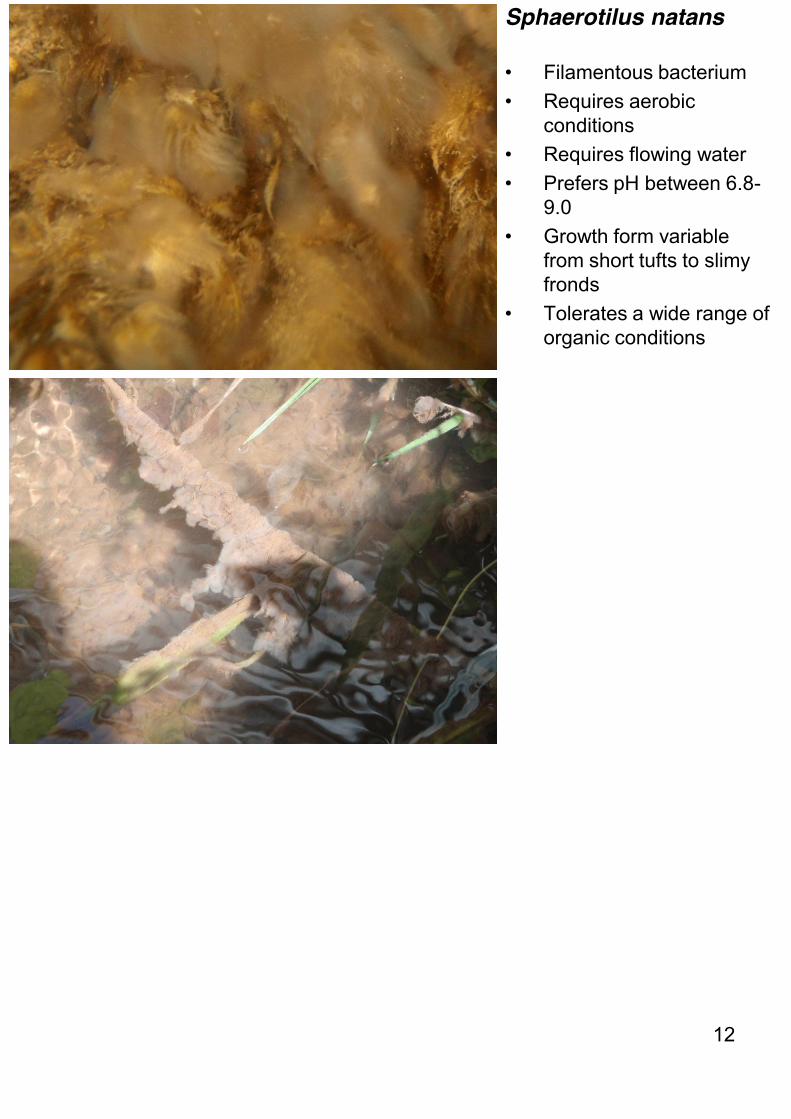
Sphaerotilus natans
• Filamentous bacterium • Requires aerobic
conditions • Requires flowing water • Prefers pH between 6.8-
9.0 • Growth form variable
from short tufts to slimy fronds
• Tolerates a wide range of organic conditions
12
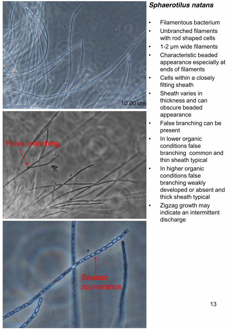
Sphaerotilus natans
• Filamentous bacterium • Unbranched filaments
with rod shaped cells • 1-2 µm wide filaments • Characteristic beaded
appearance especially at ends of filaments
• Cells within a closely fitting sheath
• Sheath varies in thickness and can obscure beaded appearance
• False branching can be present
• In lower organic conditions false branching common and thin sheath typical
• In higher organic conditions false branching weakly developed or absent and thick sheath typical
• Zigzag growth may indicate an intermittent discharge
False branching
Beaded appearance
13

Beggiatoa alba
• Filamentous bacterium • Forms a thin white film on
surface of substrate • Prefers low dissolved
oxygen • Prevalent in high organic
conditions • Oxidises hydrogen
sulphide • Characteristic of slow
flowing waters • Can be found as a mono-
culture in faster flowing waters where it forms very long fine filaments
• Tolerant of saline conditions
Anoxic sediment
White film
14
Long fine filaments

Beggiatoa alba
• Filamentous bacterium • Unbranched filaments
with rod shaped cells • 3-4 µm wide filaments • Motile filaments • Sulphur granules stored
within cells increasing in number with age
Granular appearance
15
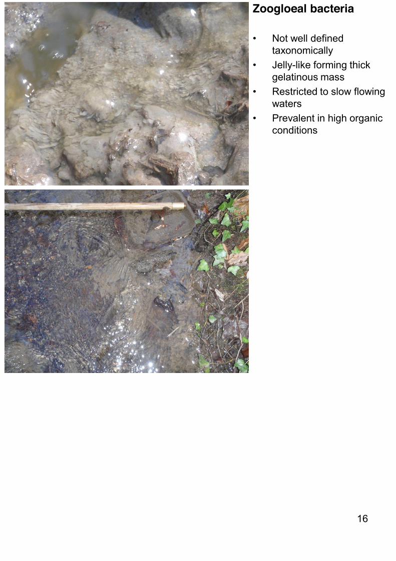
Zoogloeal bacteria
• Not well defined taxonomically
• Jelly-like forming thick gelatinous mass
• Restricted to slow flowing waters
• Prevalent in high organic conditions
16

Zoogloeal bacteria
• Not well defined taxonomically
• Cells embedded in a gelatinous matrix
• Forms lobed and unlobed spherical masses
17
Unlobed spherical mass
Cells
Lobed spherical masses
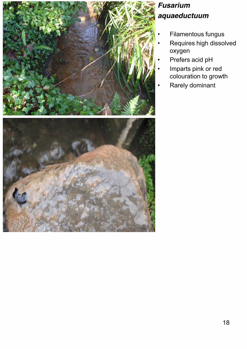
Fusarium aquaeductuum
• Filamentous fungus • Requires high dissolved
oxygen • Prefers acid pH • Imparts pink or red
colouration to growth • Rarely dominant
18
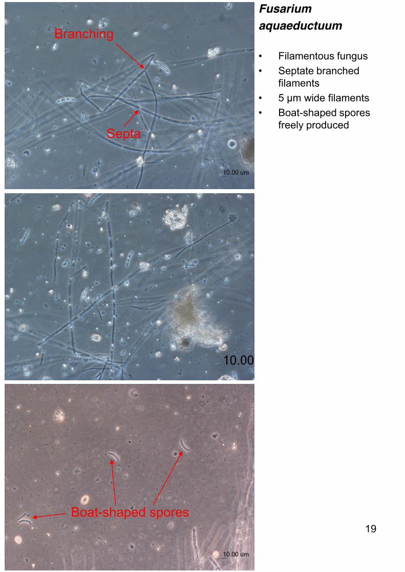
Fusarium aquaeductuum
• Filamentous fungus • Septate branched
filaments • 5 µm wide filaments • Boat-shaped spores
freely produced
Boat-shaped spores
Branching
Septa
19
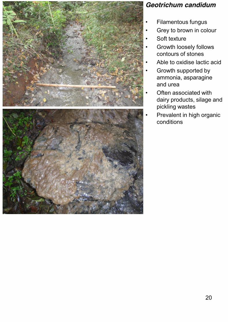
Geotrichum candidum
• Filamentous fungus • Grey to brown in colour • Soft texture • Growth loosely follows
contours of stones • Able to oxidise lactic acid • Growth supported by
ammonia, asparagine and urea
• Often associated with dairy products, silage and pickling wastes
• Prevalent in high organic conditions
20

Geotrichum candidum
• Filamentous fungus • Septate branched
filaments • 5-10 µm wide filaments • Dichotomous branching • Brick-shaped
arthrospores may be present
Dichotomous branching
Septa
21
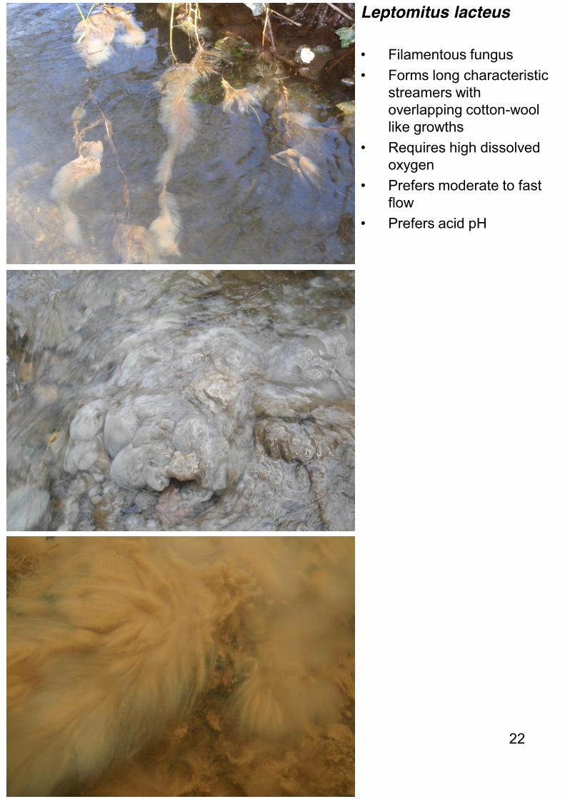
Leptomitus lacteus • Filamentous fungus • Forms long characteristic
streamers with overlapping cotton-wool like growths
• Requires high dissolved oxygen
• Prefers moderate to fast flow
• Prefers acid pH
22

Leptomitus lacteus
• Filamentous fungus • Non-septate coarse
branching filaments • 8-15 µm wide filaments • Characteristic
constrictions at intervals with spherical cellulin plugs
• Cellulin plugs block the constrictions and prevent cytoplasm flowing away from the growing apices of the filaments
Spherical cellulin plugs
23
Constriction
Branching
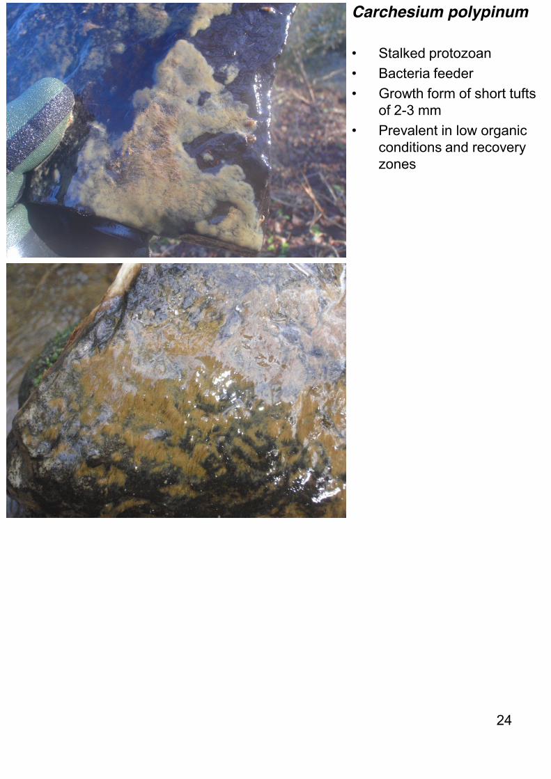
Carchesium polypinum
• Stalked protozoan • Bacteria feeder • Growth form of short tufts
of 2-3 mm • Prevalent in low organic
conditions and recovery zones
24

Carchesium polypinum
• Stalked protozoan • Inverted bell-shaped
zooids at ends of stalks • Branched stalks • Zooids can become
separated from stalks, especially in degraded samples
• Stalks can contract independent of each other
• Stalks contract spirally • Stalk with discontinuous
myoneme • Sinuous myoneme • Zooids 100-125 µm long • Zooid peristomial lip
bulges out • Zooid with C-shaped
macronucleus • Zooid with smooth
surface
25 Sinuous myoneme
Peristomial lip bulges out
Bell-shaped zooid
Branched stalk
Discontinuous myoneme

Other sewage fungus taxa
26

Flexibacter spp.
• Filamentous bacterium • Unbranched filaments • Motile with whole filament
bending and flexing • No sulphur granules in
cells • Rarely dominant
27

Thiothrix II • Gammaproteobacteria • Uses low molecular
carbon sources (short-chain fatty acids and alcohols) as well as reduced sulphur compounds
• Does not grow in anoxic conditions
• Found associated with cows bedded on waste gypsum in West Wales
28

Thiothrix II • Straight or bowed
filaments • 1 um wide filaments • False branching and
rosette formation • Sulphur granules evident • No obvious septa • Not motile • Filaments do not taper
29 Rosette
Sulphur granules

Achyla spp.
• Filamentous phycomycete fungus
• Similar to Leptomitus but wider filaments and no spherical cellulin plugs
• Tends to grow near source of silage effluent with which it is closely associated
30
Flavobacterium spp.
• Filamentous bacterium • Unbranched filaments • 0.5-1 µm wide filaments
lying parallel to each other
• Filaments lying close together separated by 10-12µm
• Individual cells 0.5-1 µm wide by 10-50µm long
• Occasionally forming pink or yellow growths
• Requires organic nitrogen source for growth
• Rarely dominant
31

Taxa commonly mistaken for sewage fungus
32
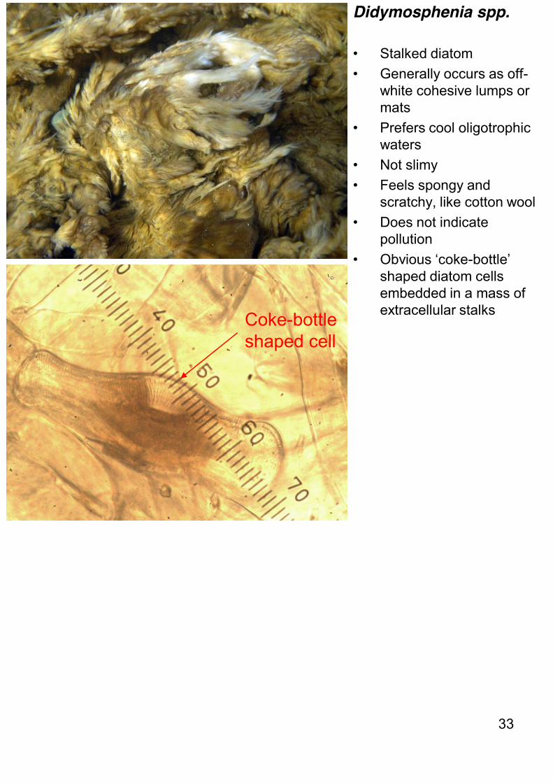
Didymosphenia spp. • Stalked diatom • Generally occurs as off-
white cohesive lumps or mats
• Prefers cool oligotrophic waters
• Not slimy • Feels spongy and
scratchy, like cotton wool • Does not indicate
pollution • Obvious ‘coke-bottle’
shaped diatom cells embedded in a mass of extracellular stalks
33
Coke-bottle shaped cell

Leptothrix ochracea
• Filamentous iron bacterium
• 2-3 µm wide filaments • Grows in slow flowing
waters high in ferrous iron and low in organic matter
• Requires ferrous (iron) salts, oxygen and carbon dioxide for growth
• Growth of bacterium results in accumulation and sedimentation of orange-brown ferric hydroxide in sheath surrounding filament
• Sheath has smooth surface
• Growth results in many empty sheaths
34
Smooth surface
Sheath