Embed Size (px)
Citation preview
Sex-chromosome dosage effects on gene expressionin humansArmin Raznahana,1, Neelroop N. Parikshakb,c, Vijay Chandrand, Jonathan D. Blumenthala, Liv S. Clasena,Aaron F. Alexander-Blocha, Andrew R. Zinne,f, Danny Wangsag, Jasen Wiseh, Declan G. M. Murphyi, Patrick F. Boltoni,Thomas Riedg, Judith Rossj, Jay N. Gieddk, and Daniel H. Geschwindb,c
aDevelopmental Neurogenomics Unit, National Institute of Mental Health, National Institutes of Health, Bethesda, MD 20892; bNeurogenetics Program,Department of Neurology, David Geffen School of Medicine, University of California, Los Angeles, CA 90095; cCenter for Autism Research and Treatment,Semel Institute, David Geffen School of Medicine, University of California, Los Angeles, CA 90095; dDepartment of Pediatrics, School of Medicine, Universityof Florida, Gainesville, FL 32610; eMcDermott Center for Human Growth and Development, University of Texas Southwestern Medical School, Dallas, TX75390; fDepartment of Internal Medicine, University of Texas Southwestern Medical School, Dallas, TX 75390; gGenetics Branch, Center for Cancer Research,National Cancer Institute, National Institutes of Health, Bethesda, MD 20892; hQiagen, Frederick, MD 21703; iInstitute of Psychiatry, Psychology andNeuroscience, King’s College London, University of London, London WC1B 5DN, United Kingdom; jDepartment of Pediatrics, Thomas Jefferson University,Philadelphia, PA 19107; and kDepartment of Psychiatry, University of California, San Diego, La Jolla, CA 92093
Edited by James A. Birchler, University of Missouri, Columbia, MO, and approved May 29, 2018 (received for review February 17, 2018)
A fundamental question in the biology of sex differences has eludeddirect study in humans: How does sex-chromosome dosage (SCD)shape genome function? To address this, we developed a systematicmap of SCD effects on gene function by analyzing genome-wideexpression data in humans with diverse sex-chromosome aneu-ploidies (XO, XXX, XXY, XYY, and XXYY). For sex chromosomes, wedemonstrate a pattern of obligate dosage sensitivity among evolu-tionarily preserved X-Y homologs and update prevailing theoreticalmodels for SCD compensation by detecting X-linked genes thatincrease expression with decreasing X- and/or Y-chromosome dosage.We further show that SCD-sensitive sex-chromosome genes regulatespecific coexpression networks of SCD-sensitive autosomal genes withcritical cellular functions and a demonstrable potential to mediatepreviously documented SCD effects on disease. These gene coex-pression results converge with analysis of transcription factor bindingsite enrichment and measures of gene expression in murine knockoutmodels to spotlight the dosage-sensitive X-linked transcription factorZFX as a key mediator of SCD effects on wider genome expression.Our findings characterize the effects of SCD broadly across thegenome, with potential implications for human phenotypic variation.
sex chromosomes | X-inactivation | sex differences | Turner syndrome |Klinefelter syndrome
Disparity in sex-chromosome dosage (SCD) is fundamental tothe biological definition of sex in almost all eutherian
mammals: Females carry two X chromosomes, while males carryan X and a Y chromosome. The presence of the Y-linked SRYgene determines a testicular gonadal phenotype, while its absenceallows development of ovaries (1, 2). Sexual differentiation of thegonads leads to hormonal sex differences that have traditionallybeen considered the major proximal cause for extragonadal phe-notypic sex differences. However, diverse studies, including recentwork in transgenic mice that uncouple Y chromosome and go-nadal status, have revealed direct SCD effects on several sex-biased metabolic, immune, and neurological phenotypes (3).These findings together with reports of widespread tran-
scriptomic differences between preimplantation XY and XXembryos (4) suggest that SCD has gene-regulatory effects inde-pendently of gonadal status. However, the genome-wide conse-quences of SCD remain poorly understood, especially in humans,where experimental dissociation of SCD and gonadal status is notpossible. Understanding these regulatory effects is critical forclarifying the biological underpinnings of phenotypic sex differ-ences and the clinical features of sex-chromosome aneuploidy(SCA) [e.g., Turner (XO) and Klinefelter (XXY) syndrome (5)],which both involve altered risk for several common autoimmunedisorders (ADs) and neurodevelopmental disorders (e.g., systemiclupus erythematosus and autism spectrum disorders) (6, 7). Here,we explore the genome-wide consequences of SCD through
comparative transcriptomic analyses among humans across arange of dosages including typical XX and XY karyotypes as wellas several rare SCA syndromes associated with 1, 3, 4, or 5 copiesof the sex chromosomes. We harness these diverse karyotypesto dissect the architecture of dosage compensation among sex-chromosome genes and to systematically map the regulatory ef-fects of SCD on autosomal gene expression in humans. Theseresearch goals also inform more general questions regarding theeffects of aneuploidy on genome function. In particular, the widerange of chromosome-dosage variation in SCAs can help determineif previously reported inverse effects of aneuploidy on gene ex-pression in maize and Drosophila (i.e., negative cis and trans effectsof supernumerary chromosomes) (8, 9) also operate in humans.We model SCD effects using gene-expression profiles in a total
of 471 lymphoblastoid cell lines (LCLs) from (i) a core sample of68 participants (12 XO, 10 XX, 9 XXX, 10 XY, 8 XXY, 10 XYY,and 9 XXYY) yielding for each sample genome-wide expressiondata for 19,984 autosomal and 894 sex-chromosome genes usingthe Illumina oligonucleotide BeadArray platform (Methods, SIAppendix, Text S1, and Dataset S1), and (ii) an independent set ofvalidation/replication samples from 403 participants (6 XO, 146XX, 22 XXX, 145 XY, 33 XXY, 16 XYY, 17 XXYY, 8 XXXY,
Significance
Sex-chromosome dosage (SCD) effects on human gene expressionare central to the biology of sex differences and sex-chromosomeaneuploidy syndromes but are challenging to study given thecosegregation of SCD and gonadal status. We address this ob-stacle by systematically modeling SCD effects on genome-wideexpression data from a large and rare cohort of individuals withdiverse SCDs (XO, XX, XXX, XXXX, XY, XXY, XYY, XXYY, andXXXXY). Our findings update current models of sex-chromosomebiology by (i) pinpointing a core set of X- and Y-linked genes withobligate SCD sensitivity, (ii) discovering several noncanonicalmodes of X-chromosome dosage compensation, and (iii) dissectingcomplex regulatory effects of X-chromosome dosage on largeautosomal gene networks with key roles in cellular functioning.
Author contributions: A.R., J.D.B., L.S.C., J.N.G., and D.H.G. designed research; A.R. andD.H.G. performed research; A.R., N.N.P., V.C., A.F.A.-B., A.R.Z., D.W., J.W., D.G.M.M., P.F.B.,T.R., and J.R. contributed new reagents/analytic tools; A.R. analyzed data; and A.R., V.C.,J.W., and D.H.G. wrote the paper.
The authors declare no conflict of interest.
This article is a PNAS Direct Submission.
Published under the PNAS license.1To whom correspondence should be addressed. Email: [email protected].
This article contains supporting information online at www.pnas.org/lookup/suppl/doi:10.1073/pnas.1802889115/-/DCSupplemental.
Published online June 26, 2018.
7398–7403 | PNAS | July 10, 2018 | vol. 115 | no. 28 www.pnas.org/cgi/doi/10.1073/pnas.1802889115
Dow
nloa
ded
by g
uest
on
Apr
il 26
, 202
0
and 10 XXXXY) with qRT-PCRmeasures of expression for genesof interest identified in our core sample (Methods, SI Appendix,Text S2, and Dataset S1). All SCA samples were from individualswith nonmosaic aneuploidy.
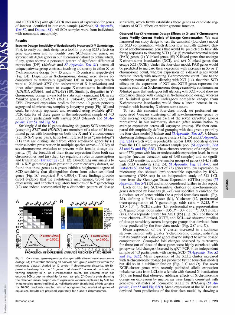
ResultsExtreme Dosage Sensitivity of Evolutionarily Preserved X-Y Gametologs.First, to verify our study design as a tool for probing SCD effects ongene expression and to identify core SCD-sensitive genes, wescreened all 20,878 genes in our microarray dataset to define which,if any, genes showed a persistent pattern of significant differentialexpression (DE) (Methods and SI Appendix, Text S1) across allunique pairwise group contrasts involving a disparity in either X- orY-chromosome dosage (n = 15 and n = 16 contrasts, respectively)(Fig. 1A). Disparities in X-chromosome dosage were always ac-companied by statistically significant DE in four genes, whichwere all X-linked: XIST (the orchestrator of X inactivation) andthree other genes known to escape X-chromosome inactivation(HDHD1, KDM6A, and EIF1AX) (10). Similarly, disparities in Y-chromosome dosage always led to statistically significant DE in sixY-linked genes: CYorf15B, DDX3Y, TMSB4Y, USP9Y, UTY, andZFY. Observed expression profiles for these 10 genes perfectlysegregated all microarray samples by karyotype group (Fig. 1B) andcould be robustly replicated and extended using available qRT-PCR data for of these genes in the independent sample of 403LCLs from participants with varying SCD (Methods and SI Ap-pendix, Text S1 and Fig. S1).Strikingly, 8 of the 10 genes showing obligatory SCD sensitivity
(excepting XIST and HDHD1) are members of a class of 16 sex-linked genes with homologs on both the X and Y chromosomes(i.e., 16 X-Y gene pairs, henceforth referred to as “gametologs”)(11) that are distinguished from other sex-linked genes by (i)their selective preservation in multiple species across ∼300 My ofsex-chromosome evolution to prevent male–female dosage dis-parity, (ii) the breadth of their tissue expression from both sexchromosomes, and (iii) their key regulatory roles in transcriptionand translation (Dataset S2) (11, 12). Broadening our analysis toall 14 X-Y gametolog pairs present in our microarray dataset, wefound that these genes as a group exhibit a heightened degree ofSCD sensitivity that distinguishes them from other sex-linkedgenes (Fig. 1C, empirical P < 0.0001). These findings providedirect evidence that the evolutionary maintenance, broad tissueexpressivity, and enriched regulatory functions of X-Y gametologs(12) are indeed accompanied by a distinctive pattern of dosage
sensitivity, which firmly establishes these genes as candidate reg-ulators of SCD effects on wider genome function.
Observed Sex-Chromosome Dosage Effects on X- and Y-ChromosomeGenes Modify Current Models of Dosage Compensation. We nextharnessed our study design to test the canonical (four-class) modelfor SCD compensation, which defines four mutually exclusive clas-ses of sex-chromosome genes that would be predicted to have dif-fering responses to changing SCD (13): (i) pseudoautosomal region(PAR) genes, (ii) Y-linked genes, (iii) X-linked genes that undergoX-chromosome inactivation (XCI), and (iv) X-linked genes thatescape XCI (XCIE). Under the four-class model, PAR genes wouldbe predicted to increase their expression with increases in X- or Ychromosome count, whereas expression of Y-linked genes wouldincrease linearly with mounting Y-chromosome count. Due to thenonbinary nature of gene silencing with XCI (14), theorized SCDeffects on the expression of XCI and XCIE genes represent theextreme ends of an X-chromosome dosage-sensitivity continuum: anX-linked gene that undergoes full silencing with XCI would show noexpression change with changes in X-chromosome dosage, whereasan X-linked gene that undergoes complete escape fromX-chromosome inactivation would show a linear increase in ex-pression with increasing X-chromosome count.To test this canonical four-class model, we performed un-
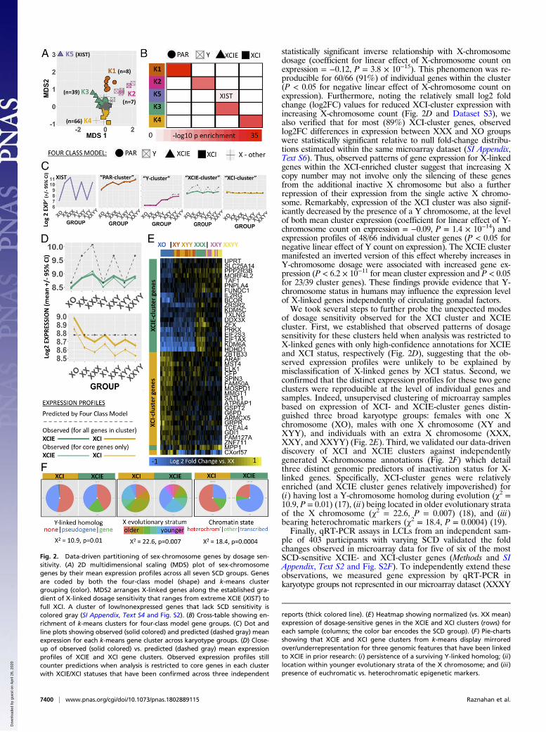
supervised k-means clustering of all sex-chromosome genes bytheir average expression in each of the seven karyotype groupsrepresented in our microarray dataset (after normalization byaverage expression across all karyotype groups). We then com-pared this empirically defined grouping with that given a priori bythe four-class model (Methods and SI Appendix, Text S3). k-Meansclustering distinguished six gene clusters (Fig. 2A and SI Appendix,Fig. S2A) which were reproducible across 1,000 bootstrap drawsfrom the LCL microarray dataset sample pool (SI Appendix, TextS3 and S4 and Fig. S2B). These clusters consisted of a single largeset of 773 genes with low or undetectable expression levels in mostsamples (median detection rate of 4/68 samples) and no signifi-cant SCD sensitivity, and five smaller groups of genes (k1–k5) withhigh SCD sensitivity (Fig. 2A, SI Appendix, Fig. S2 A–C, andDatasets S3 and S4). The set of 773 genes with low expression bymicroarray also showed low/undetectable expression by RNA-sequencing (RNA-seq) in an independent study of 343 LCLlines from the Genotype-Tissue Expression (GTEx) project (SIAppendix, Text S4) (15) and is not considered further in this report.Each of the five SCD-sensitive clusters of sex-chromosome
genes detected by k-means (k1–k5) was specifically enriched fora distinct set of genes within the a priori four-class model (Fig.2B), defining a PAR cluster (k1), Y cluster (k2, preferentialoverrepresentation of Y gametologs: odds ratio = 5,213, P =1.3 × 10⁻¹⁵), XCIE cluster (k3, preferential overrepresentationof X gametologs: odds ratio = 335, P = 3.4 × 10⁻¹¹), XCI cluster(k4), and a separate cluster for XIST (k5) (Fig. 2B). For three ofthese clusters—Y-linked, XCIE, and XCI—we observed profilesof dosage sensitivity across karyotype groups that deviated fromthose predicted by the four-class model (Fig. 2 C and D).Mean expression of the Y cluster increased in a sublinear
stepwise fashion with greater Y-chromosome dosage, indicatingthat its constituent Y-linked genes may be subject to active dosagecompensation. Groupwise fold changes observed by microarrayfor three out of three of these genes were highly correlated withgroupwise fold changes observed by qRT-PCR in an independentsample of 403 participants with varying SCD (SI Appendix, Text S2and Fig. S2E). Mean expression of the XCIE cluster increasedwith X-chromosome dosage (as predicted by the four-class model)but did so in a sublinear fashion (Fig. 2 C and D). For sevenXCIE-cluster genes with recently published allelic expressionimbalance data from LCLs in a female with skewed X-inactivation(16), we found that observed sublinear effects of X-chromosomedosage on expression by microarray were largely consistent withgene-level estimates of incomplete XCIE by RNA-seq (SI Ap-pendix, Text S5 and Fig. S2D). Mean expression of the XCI clusterdeviated from predictions of the four-class model by showing a
Fig. 1. Consistent gene-expression changes with altered sex-chromosomedosage. (A) Cross-table showing all pairwise SCD group contrasts within themicroarray dataset shaded by X- and/or Y-chromosome disparity. (B) Ex-pression heatmap for the 10 genes that show DE across all contrasts in-volving disparity in X- or Y-chromosome count. The column color barencodes SCD group membership for each sample. (C) Density plots showingthe observed mean proportion of expression variance explained by SCD for14 gametolog genes (red line) vs. null distribution (black line) of this variablefor 10,000 randomly sampled sets of nongametolog sex-linked genes ofequal size. Results are provided separately for X and Y chromosomes.
Raznahan et al. PNAS | July 10, 2018 | vol. 115 | no. 28 | 7399
GEN
ETICS
Dow
nloa
ded
by g
uest
on
Apr
il 26
, 202
0
statistically significant inverse relationship with X-chromosomedosage (coefficient for linear effect of X-chromosome count onexpression = −0.12, P = 3.8 × 10⁻¹⁵). This phenomenon was re-producible for 60/66 (91%) of individual genes within the cluster(P < 0.05 for negative linear effect of X-chromosome count onexpression). Furthermore, noting the relatively small log2 foldchange (log2FC) values for reduced XCI-cluster expression withincreasing X-chromosome count (Fig. 2D and Dataset S3), wealso verified that for most (89%) XCI-cluster genes, observedlog2FC differences in expression between XXX and XO groupswere statistically significant relative to null fold-change distribu-tions estimated within the same microarray dataset (SI Appendix,Text S6). Thus, observed patterns of gene expression for X-linkedgenes within the XCI-enriched cluster suggest that increasing Xcopy number may not involve only the silencing of these genesfrom the additional inactive X chromosome but also a furtherrepression of their expression from the single active X chromo-some. Remarkably, expression of the XCI cluster was also signif-icantly decreased by the presence of a Y chromosome, at the levelof both mean cluster expression (coefficient for linear effect of Y-chromosome count on expression = −0.09, P = 1.4 × 10⁻¹⁴) andexpression profiles of 48/66 individual cluster genes (P < 0.05 fornegative linear effect of Y count on expression). The XCIE clustermanifested an inverted version of this effect whereby increases inY-chromosome dosage were associated with increased gene ex-pression (P < 6.2 × 10⁻¹¹ for mean cluster expression and P < 0.05for 23/39 cluster genes). These findings provide evidence that Y-chromosome status in humans may influence the expression levelof X-linked genes independently of circulating gonadal factors.We took several steps to further probe the unexpected modes
of dosage sensitivity observed for the XCI cluster and XCIEcluster. First, we established that observed patterns of dosagesensitivity for these clusters held when analysis was restricted toX-linked genes with only high-confidence annotations for XCIEand XCI status, respectively (Fig. 2D), suggesting that the ob-served expression profiles were unlikely to be explained bymisclassification of X-linked genes by XCI status. Second, weconfirmed that the distinct expression profiles for these two geneclusters were reproducible at the level of individual genes andsamples. Indeed, unsupervised clustering of microarray samplesbased on expression of XCI- and XCIE-cluster genes distin-guished three broad karyotype groups: females with one Xchromosome (XO), males with one X chromosome (XY andXYY), and individuals with an extra X chromosome (XXX,XXY, and XXYY) (Fig. 2E). Third, we validated our data-drivendiscovery of XCI and XCIE clusters against independentlygenerated X-chromosome annotations (Fig. 2F) which detailthree distinct genomic predictors of inactivation status for X-linked genes. Specifically, XCI-cluster genes were relativelyenriched (and XCIE cluster genes relatively impoverished) for(i) having lost a Y-chromosome homolog during evolution (χ2 =10.9, P = 0.01) (17), (ii) being located in older evolutionary strataof the X chromosome (χ2 = 22.6, P = 0.007) (18), and (iii)bearing heterochromatic markers (χ2 = 18.4, P = 0.0004) (19).Finally, qRT-PCR assays in LCLs from an independent sam-
ple of 403 participants with varying SCD validated the foldchanges observed in microarray data for five of six of the mostSCD-sensitive XCIE- and XCI-cluster genes (Methods and SIAppendix, Text S2 and Fig. S2F). To independently extend theseobservations, we measured gene expression by qRT-PCR inkaryotype groups not represented in our microarray dataset (XXXY
Fig. 2. Data-driven partitioning of sex-chromosome genes by dosage sen-sitivity. (A) 2D multidimensional scaling (MDS) plot of sex-chromosomegenes by their mean expression profiles across all seven SCD groups. Genesare coded by both the four-class model (shape) and k-means clustergrouping (color). MDS2 arranges X-linked genes along the established gra-dient of X-linked dosage sensitivity that ranges from extreme XCIE (XIST) tofull XCI. A cluster of low/nonexpressed genes that lack SCD sensitivity iscolored gray (SI Appendix, Text S4 and Fig. S2). (B) Cross-table showing en-richment of k-means clusters for four-class model gene groups. (C) Dot andline plots showing observed (solid colored) and predicted (dashed gray) meanexpression for each k-means gene cluster across karyotype groups. (D) Close-up of observed (solid colored) vs. predicted (dashed gray) mean expressionprofiles of XCIE and XCI gene clusters. Observed expression profiles stillcounter predictions when analysis is restricted to core genes in each clusterwith XCIE/XCI statuses that have been confirmed across three independent
reports (thick colored line). (E) Heatmap showing normalized (vs. XX mean)expression of dosage-sensitive genes in the XCIE and XCI clusters (rows) foreach sample (columns; the color bar encodes the SCD group). (F) Pie-chartsshowing that XCIE and XCI gene clusters from k-means display mirroredover/underrepresentation for three genomic features that have been linkedto XCIE in prior research: (i) persistence of a surviving Y-linked homolog; (ii)location within younger evolutionary strata of the X chromosome; and (iii)presence of euchromatic vs. heterochromatic epigenetic markers.
7400 | www.pnas.org/cgi/doi/10.1073/pnas.1802889115 Raznahan et al.
Dow
nloa
ded
by g
uest
on
Apr
il 26
, 202
0
and XXXXY) (Methods and SI Appendix, Text S1) and were able toconfirm reduction in expression with greater X-chromosome dosagefor two of three XCI-cluster genes, NGFRAP1 and CXorf57 (SIAppendix, Fig. S2G,) and Y-chromosome dosage effects upon ex-pression for four of six X-linked genes from the XCI and XCIEclusters (down-regulation: NGFRAP1 and CXorf57; up-regulation:PIM2, and PRKX) (SI Appendix, Fig. S2H). Taken together, thesefindings update the canonical four-class model of SCD compensationfor specific Y-linked and X-linked genes and expand the list ofX-linked genes capable of mediating wider phenotypic consequencesof SCD variation. The existence of X-linked genes that decreaseexpression with increasing X-chromosome count also indicates thatpreviously documented inverse effects of aneuploidy on gene ex-pression in maize and Drosophila (8, 9) are also seen in humans.
Context-Specific Disruption of Autosomal Expression by Sex-Chromosome Aneuploidy. We next leveraged the diverse SCAsrepresented in our study to assess how SCD variation shapes ex-pression on a genome-wide scale. By counting the total number ofdifferentially expressed genes (DEGs) (Methods) in each SCAgroup relative to its respective euploid control (i.e., XO and XXXcompared with XX; XXY, XYY, and XXYY compared with XY),we detected order-of-magnitude differences in DEG count amongSCAs across a range of log2FC cutoffs (SI Appendix, Fig. S3 A andB). We observed an order-of-magnitude increase in DEG countwith X-chromosome supernumeracy in males vs. females, which,although previously undescribed, is congruent with the more se-vere phenotypic consequences of X-supernumeracy in males vs.females (20). Overall, increasing the dosage of the sex chromo-some associated with the sex of an individual (i.e., X in femalesand Y in males) had a far smaller effect than other types of SCDchange. Moreover, the ∼20 DEGs seen in XXX contrastedwith >1,200 DEGs in XO, revealing a profoundly asymmetricimpact of X-chromosome loss vs. gain on the transcriptome offemale LCLs, which echoes the asymmetric phenotypic severity ofX-chromosome loss (Turner syndrome) vs. gain (XXX) syn-dromes in females (6). The large number of DEGS in XO in-cluded a similar proportion with increases vs. decreases inexpression (e.g., 782 XO > XX, 605 XO < XX). Autosomal genesaccounted for >75% of all DEGs in females with X-monosomy(XO) and males with X-supernumeracy (XXY and XXYY)but <30% DEGs in all other SCD groups (Methods and SI Ap-pendix, Fig. S3C). These results reveal that SCD changes varywidely in their capacity to disrupt genome function and indicatethat differential involvement of autosomal genes is central to thisvariation. Moreover, SCA differences in LCL DEG count broadlyrecapitulate SCA differences in phenotypic severity.
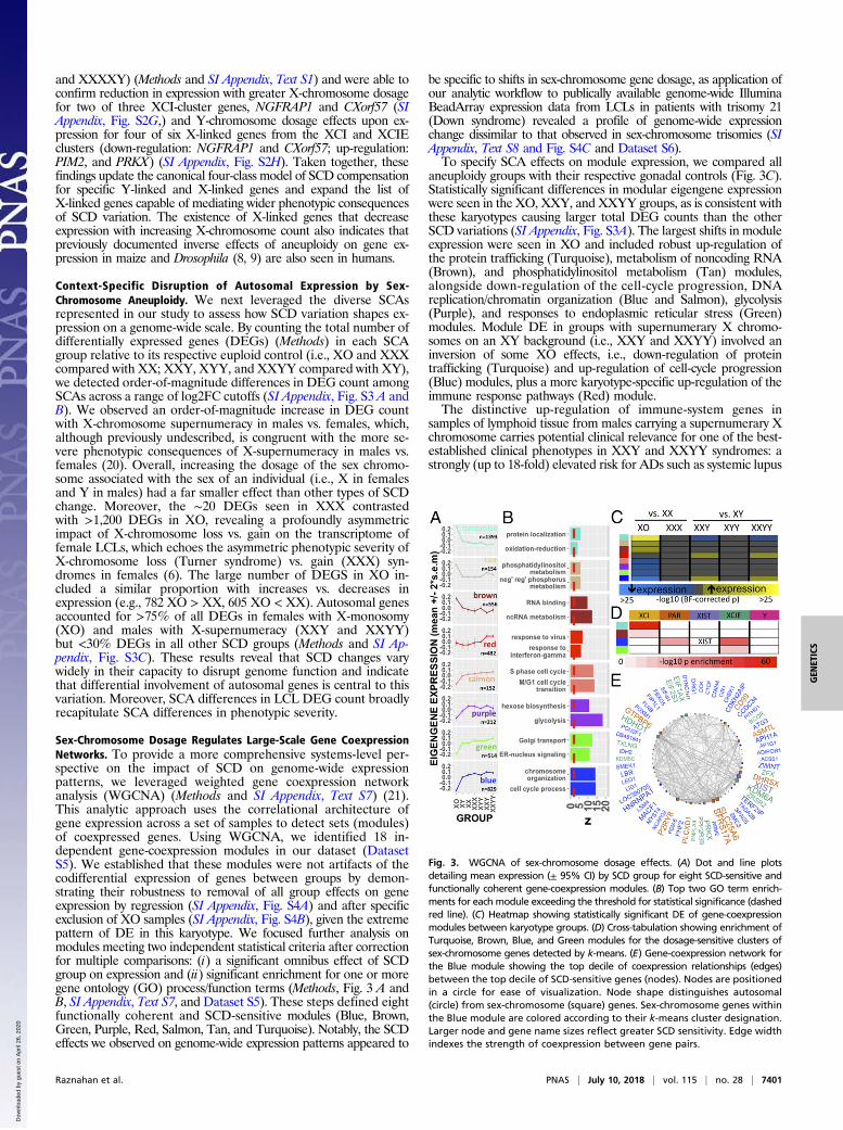
Sex-Chromosome Dosage Regulates Large-Scale Gene CoexpressionNetworks. To provide a more comprehensive systems-level per-spective on the impact of SCD on genome-wide expressionpatterns, we leveraged weighted gene coexpression networkanalysis (WGCNA) (Methods and SI Appendix, Text S7) (21).This analytic approach uses the correlational architecture ofgene expression across a set of samples to detect sets (modules)of coexpressed genes. Using WGCNA, we identified 18 in-dependent gene-coexpression modules in our dataset (DatasetS5). We established that these modules were not artifacts of thecodifferential expression of genes between groups by demon-strating their robustness to removal of all group effects on geneexpression by regression (SI Appendix, Fig. S4A) and after specificexclusion of XO samples (SI Appendix, Fig. S4B), given the extremepattern of DE in this karyotype. We focused further analysis onmodules meeting two independent statistical criteria after correctionfor multiple comparisons: (i) a significant omnibus effect of SCDgroup on expression and (ii) significant enrichment for one or moregene ontology (GO) process/function terms (Methods, Fig. 3 A andB, SI Appendix, Text S7, and Dataset S5). These steps defined eightfunctionally coherent and SCD-sensitive modules (Blue, Brown,Green, Purple, Red, Salmon, Tan, and Turquoise). Notably, the SCDeffects we observed on genome-wide expression patterns appeared to
be specific to shifts in sex-chromosome gene dosage, as application ofour analytic workflow to publically available genome-wide IlluminaBeadArray expression data from LCLs in patients with trisomy 21(Down syndrome) revealed a profile of genome-wide expressionchange dissimilar to that observed in sex-chromosome trisomies (SIAppendix, Text S8 and Fig. S4C and Dataset S6).To specify SCA effects on module expression, we compared all
aneuploidy groups with their respective gonadal controls (Fig. 3C).Statistically significant differences in modular eigengene expressionwere seen in the XO, XXY, and XXYY groups, as is consistent withthese karyotypes causing larger total DEG counts than the otherSCD variations (SI Appendix, Fig. S3A). The largest shifts in moduleexpression were seen in XO and included robust up-regulation ofthe protein trafficking (Turquoise), metabolism of noncoding RNA(Brown), and phosphatidylinositol metabolism (Tan) modules,alongside down-regulation of the cell-cycle progression, DNAreplication/chromatin organization (Blue and Salmon), glycolysis(Purple), and responses to endoplasmic reticular stress (Green)modules. Module DE in groups with supernumerary X chromo-somes on an XY background (i.e., XXY and XXYY) involved aninversion of some XO effects, i.e., down-regulation of proteintrafficking (Turquoise) and up-regulation of cell-cycle progression(Blue) modules, plus a more karyotype-specific up-regulation of theimmune response pathways (Red) module.The distinctive up-regulation of immune-system genes in
samples of lymphoid tissue from males carrying a supernumerary Xchromosome carries potential clinical relevance for one of the best-established clinical phenotypes in XXY and XXYY syndromes: astrongly (up to 18-fold) elevated risk for ADs such as systemic lupus
Fig. 3. WGCNA of sex-chromosome dosage effects. (A) Dot and line plotsdetailing mean expression (± 95% CI) by SCD group for eight SCD-sensitive andfunctionally coherent gene-coexpression modules. (B) Top two GO term enrich-ments for each module exceeding the threshold for statistical significance (dashedred line). (C) Heatmap showing statistically significant DE of gene-coexpressionmodules between karyotype groups. (D) Cross-tabulation showing enrichment ofTurquoise, Brown, Blue, and Green modules for the dosage-sensitive clusters ofsex-chromosome genes detected by k-means. (E) Gene-coexpression network forthe Blue module showing the top decile of coexpression relationships (edges)between the top decile of SCD-sensitive genes (nodes). Nodes are positionedin a circle for ease of visualization. Node shape distinguishes autosomal(circle) from sex-chromosome (square) genes. Sex-chromosome genes withinthe Blue module are colored according to their k-means cluster designation.Larger node and gene name sizes reflect greater SCD sensitivity. Edge widthindexes the strength of coexpression between gene pairs.
Raznahan et al. PNAS | July 10, 2018 | vol. 115 | no. 28 | 7401
GEN
ETICS
Dow
nloa
ded
by g
uest
on
Apr
il 26
, 202
0
erythematosus, Sjögren syndrome, and diabetes mellitus (7). Infurther support of this interpretation, we found the Red module tobe significantly enriched (P = 0.01 by Fisher’s test and P = 0.01 bygene set permutation) for a set of known AD-risk genes compiledfrom multiple large-scale genome-wide association studies (GWAS)(Methods and SI Appendix, Text S7). The two GWAS-implicatedAD-risk genes showing strongest connectivity within the Redmodule and up-regulation in males bearing an extra X chro-mosome were CLECL1 and ELF1, indicating that these twogenes should be prioritized for further study in mechanisms ofrisk for heightened autoimmunity in XXY and XXYY males.Collectively, these results represent a systems-level charac-terization of SCD effects on genome function and provideconvergent evidence that increased risk for AD risk in XXYand XXYY syndromes may be due to an up-regulation ofimmune pathways by supernumerary X chromosomes in malelymphoid cells.To test for evidence of coordination between the expression
changes in sex-chromosome genes imparted by SCD (Fig. 2) andthe genome-wide transcriptomic variations detected throughWGCNA (Fig. 3A), we asked if any SCD-sensitive gene-coexpressionmodules were enriched for one or more of the five SCD-sensitiveclusters of sex-chromosome genes defined by k-means analysis (Fig.2). FourWGCNAmodules, all composed of >95% autosomal genes,showed such enrichment (Fig. 3D). XCI-cluster genes were enrichedwithin Turquoise and Brown coexpression modules, indicating thatthe inverse cis effects of X-chromosome dosage on the expression ofXCI-cluster genes (Fig. 2) are closely coordinated with inverse transeffects of X-chromosome dosage on autosomal expression (Fig. 3 Aand C). Conversely, XCIE-cluster genes were enriched within theGreen and Blue coexpression modules, with the Blue module beingfurther distinguished by an additional enrichment in PAR-clustergenes and inclusion of XIST (Fig. 3D). We generated a network vi-sualization to examine more closely SCD-sensitive genes and gene-coexpression relationships within the Blue module (Methods andFig. 3E). This network highlights the high SCD sensitivity of XIST,select PAR genes (SLC25A6 and SFRS17A), and multiple X-linkedgenes from X-Y gametolog pairs [EIF1AX, KDM6A (UTX), ZFX,and PRKX] and shows that these genes are closely coexpressed withmultiple SCD-sensitive autosomal genes including ZWINT,TERF2IP, and CDKN2AIP.Our detection of highly organized coexpression relationships be-
tween SCD-sensitive sex-linked and autosomal genes hints at specificregulatory effects of dosage-sensitive sex-chromosome genes in me-diating the genome-wide effects of SCD variation. To test this and toelucidate potential regulatory mechanisms, we performed an un-biased transcription factor binding site (TFBS) enrichment analysisof genes within the Blue, Green, Turquoise, and Brown WGCNAmodules (Methods and SI Appendix, Text S9). This analysis con-verged on a single transcription factor—ZFX, encoded by theX-linked member of an X-Y gametolog pair—as the only SCD-sensitive transcription factor (see SI Appendix, Fig. S1 for qRT-PCR validation) showing significant TFBS enrichment in one ormore modules. Remarkably, the gene ZFX was itself part of the Bluemodule, and ZFX-binding sites were enriched not only among Blueand Green module genes (increasing in expression with increasingX-chromosome dose) but also among Brown module genes that aredown-regulated as X-chromosome dose increases (SI Appendix, Fig.S4D). To directly test if changes in ZFX expression are sufficient tomodify the expression of Blue, Green, or Brown module genes inimmortalized lymphocytes, we harnessed existing gene-expressiondata from murine T-lymphoblastic leukemia cells with and withoutZFX knockout (Gene Expression Omnibus GSE43020) (22). Thesedata revealed that genes down-regulated by ZFX knockout in micehave human homologs that are specifically and significantly over-represented in Blue (e.g., PARP16 and TCEA1; odds ratio = 2.4, P =0.0005, Fisher’s test) and Green (e.g., BAG3 and CCDC117; oddsratio = 2.4, P = 0.005, Fisher’s test) modules (P > 0.1 for each of theother six WGCNA modules), providing experimental validation ofour hypothesized regulatory role for ZFX.
DiscussionIn conclusion, our study, which systematically examined gene-expression data from 471 individuals representing nine different sex-chromosome karyotypes, yields several insights into sex-chromosomebiology with consequences for basic and clinical science. First, ourdiscovery and validation of X-linked genes that are up-regulated byreducing X-chromosome count (so that their expression is elevatedin XO vs. XX, for example) runs counter to dominant models of sex-chromosome dosage compensation in mammals. This noncanonicalmode of SCD sensitivity modifies current thinking regarding whichsubsets of X-chromosome genes could contribute to sex- and SCA-biased phenotypes (23). An inverse effect of X-chromosome dosageon X-chromosome gene expression could reflect repression of X-linked genes on the active X-chromosome by PAR or X-linked genesthat are expressed from inactive X chromosomes or by stoichio-metric imbalances (see below) between the products of PAR/X-linked and autosomal genes. Alternatively, inverse effects ofX-chromosome dosage may reflect changes in nuclear heterochro-matin dosage that would follow from varying numbers of inactivatedX-chromosomes. The potential for heterochromatin-mediated con-sequences of SCD variation has been documented in mice, Dro-sophila, and humans (24, 25). Our findings also modify classic modelsof sex-chromosome biology by identifying X-linked genes that vary intheir expression as a function of Y-chromosome dosage, indicat-ing that the phenotypic effects of normative and aneuploid varia-tions in Y-chromosome dose could theoretically be mediated byaltered expression of X-linked genes. Moreover, the discovery ofY-chromosome dosage effects on X-linked gene expression providesroutes for competition between maternally and paternally inheritedgenes beyond the previously described mechanisms of parental im-printing and genomic conflict, with consequences for our mecha-nistic understanding of sex-biased evolution and disease (26).Beyond their theoretical implications, our data help pinpoint
specific genes that are likely to play key roles in mediating SCDeffects on wider genome function. Specifically, we establish that adistinctive group of sex-linked genes notable for their evolutionarypreservation as X-Y gametolog pairs across multiple species and thebreadth of their tissue expression in humans (12) are further dis-tinguished from other sex-linked genes by their exquisite sensitivity toSCD and exceptionally close coexpression with SCD-sensitive au-tosomal genes. These results add critical evidence in support of theidea that X-Y gametologs play a key role in mediating SCD effectson wider genome function. In convergent support of this idea, weshow that (i) multiple SCD-sensitive modules of coexpressed auto-somal genes are enriched with TFBS for an X-linked transcriptionfactor from the highly dosage-sensitive ZFX-ZFY gametolog pair and(ii) ZFX deletion causes targeted gene-expression changes in suchmodules. Inclusion of ZFX in a coexpression module (Blue) withenriched annotations for chromatin organization and cell-cyclepathways is especially striking given the rich bodies of experimen-tal data which have independently identified ZFX as a key regulatorof cellular renewal and maintenance (27).Gene-coexpression analysis also reveals the diverse domains of
cellular function that are sensitive to SCD, spanning cell-cycleregulation, protein trafficking, and energy metabolism. Many ofthese processes are known to be sensitive to shifts in cellular kar-yotype more generally (28), although we find several which appearto be specific to shifts in SCD, as they are not induced by trisomy ofchromosome 21. Furthermore, gene-coexpression analysis of SCDeffects dissects out specific immune activation pathways that are up-regulated by supernumerary X-chromosomes in males and enrichedfor genes known to confer risk for ADs that are overrepresentedamong males bearing an extra X chromosome. Thus, we report acoordinated genomic response to SCD that could potentially ex-plain observed patterns of disease risk in SCA. Finally, coordinatedgenomic responses to SCA also feature extensive trans-acting in-verse effects of SCD on autosomal expression. Such inverse transeffects of aneuploidy have been well described in model systemssuch as maize and Drosophila but remain relatively undocumentedin humans (8, 9). Inverse trans effects in these model systems havebeen linked to specific regulatory genes on aneuploidy chromosomes
7402 | www.pnas.org/cgi/doi/10.1073/pnas.1802889115 Raznahan et al.
Dow
nloa
ded
by g
uest
on
Apr
il 26
, 202
0
and altered stoichiometry between the products of these genes andpartner molecules encoded by nonaneuploid chromosomes (29),suggesting that similar processes may underlie our observations inhumans. Collectively, our findings help refine current models of sex-chromosome biology and advance our understanding of genomicpathways through which sex chromosomes can shape phenotypicvariation in health and sex-chromosome aneuploidy.
Materials and MethodsAcquisition and Preparation of Biosamples. RNA was extracted by standardmethods (Qiagen) from LCLs from 471 participants recruited through studies ofSCA at the NIH Health Intramural Research Program and Thomas Jefferson Uni-versity (Dataset S1) (30). Sixty-eight participants provided RNA samples formicroarray analyses (12 XO, 10 XX, 9 XXX, 10 XY, 8 XXY, 10 XYY, and 9 XXYY),and 403 participants provided RNA samples for a separate qRT-PCR validation/extension study (6 XO, 146 XX, 22 XXX, 145 XY, 33 XXY, 16 XYY, 17 XXYY,8 XXXY, and 10 XXXXY). The microarray and qRT-PCR samples were fully in-dependent of each other (biological replicates), with the exception of four XOparticipants in themicroarray studywho each also provided a separate LCL samplefor the qRT-PCR study. All participants with X/Y aneuploidy were nonmosaic basedon visualization of at least 50 metaphase spreads in peripheral blood. Stability ofkaryotype across LCL derivation was confirmed by chromosome FISH in allmembers of a randomly selected subset of nine LCL samples representing each ofthe four supernumerary SCA groups included in our microarray analysis. Theresearch protocol was approved by the institutional review board at theNational Institute of Mental Health, and informed consent or assent wasobtained from all children who participated in the study, as well as consentfrom their parents if the child was under the legal age of majority.
Microarray Data Preparation, Differential Expression Analysis, Annotation, andProbe Selection. Gene expression was profiled using the Illumina HT-12 v4 Ex-pression BeadChip Kit (Illumina, Inc.). Preprocessing and annotation ofmicroarraydata (SI Appendix, Text S1) resulted in high-quality measures of expression for19,984 autosomal and 894 sex-chromosome genes in each of 68 independentsamples from seven different karyotype groups. For each SCD group contrast,differentially expressed genes survived correction for multiple comparison across allprobes, with q (the expected proportion of falsely rejected nulls) set at 0.05, andshowed a log2FC >0.26. This log2 cutoff was selected empirically, by defining thelog2 fold increment associated with the greatest drop in DEG count for each SCAgroup and averaging these thresholds all five groups (SI Appendix, Fig. S3).
Testing the Four-Class Model. All 894 sex-chromosome genes with microarrayexpression datawere uniquely assigned to PAR, Y-linked, XCIE, and XCI classesusing known PAR boundaries and a consensus classification of X-linked genesby X-inactivation/escape status (SI Appendix, Text S3) (10). These a priori genegroupings were compared with groups defined by k-means clustering ofgenes by their profile of mean expression across SCD groups. k-Means clus-tering defined five clusters of expressed genes with SCD sensitivity and a re-mainder cluster of genes with low or undetectable expression levels across allsamples (SI Appendix, Text S3 and Fig. S2A). k-Means cluster stability wasassessed using bootstrap methods (SI Appendix, Text S4 and S5), and clusteroverlaps with four-class model groupings were assessed by two-tailed Fisher’stests. General linear models were used to estimate X- and Y-chromosomedosage effects on mean expression of each k-means gene cluster (SI Appen-dix, Text S3). χ2 tests were used to compare k-means clustering of X-linkedgenes with independently published X-chromosome annotations for (i)chromatin state (19), (ii) evolutionary strata (18), and (iii) presence vs. absenceof a surviving Y-chromosome homolog (SI Appendix, Text S3) (10).
Comparison of DEG Count and Genomic Distribution Across SCA Groups. TotalDEG counts were compared across SCD groups across a range of log2FCcutoffs as described above and are reported in SI Appendix, Fig. S3 A and B.Observed DEG counts across four genomic regions—autosomal, PAR,Y-linked, and X-linked—were compared with the background distribution oftotal gene counts across these regions using the prop.test function in R.
qRT-PCR Validation of Differentially Expressed Genes in Microarray. For se-lected genes showing significant DE between karyotype groups in our coresample, we used qRT-PCR measures of gene expression (Fluidigm) to validateand extent observed fold changes in an independent sample of 403 partic-ipants representing all the karyotypes in our core sample plus two additionalSCAs: XXXY and XXXXY (SI Appendix, Text S2, Dataset S1).
WGCNA. Gene-coexpression modules were generated and assessed for re-producibility using the R package WGCNA (SI Appendix, Text S7). Modular GOterm enrichments were assessed using GO elite (31) and Gorilla (32). Fisher’s exacttest and resampling methods were used to assess modular enrichments for AD-risk genes from a reference catalog of GWAS findings (https://www.ebi.ac.uk/gwas/). See SI Appendix, Text S8 for details of TFBS enrichment analysis and ex-perimental validation for the hypothesized regulatory role of ZFX assessed usinggene-expression data from lymphocytes in a murine ZFX-knockout model (22).
1. Hughes JF, Rozen S (2012) Genomics and genetics of human and primate y chromo-somes. Annu Rev Genomics Hum Genet 13:83–108.
2. Bachtrog D, et al.; Tree of Sex Consortium (2014) Sex determination: Why so manyways of doing it? PLoS Biol 12:e1001899.
3. Arnold AP (2012) The end of gonad-centric sex determination in mammals. TrendsGenet 28:55–61.
4. Bermejo-Alvarez P, Rizos D, Rath D, Lonergan P, Gutierrez-Adan A (2010) Sex de-termines the expression level of one third of the actively expressed genes in bovineblastocysts. Proc Natl Acad Sci USA 107:3394–3399.
5. Belling K, et al. (2017) Klinefelter syndrome comorbidities linked to increased X chromo-some gene dosage and altered protein interactome activity. HumMol Genet 26:1219–1229.
6. Hong DS, Reiss AL (2014) Cognitive and neurological aspects of sex chromosomeaneuploidies. Lancet Neurol 13:306–318.
7. Seminog OO, Seminog AB, Yeates D, Goldacre MJ (2015) Associations betweenKlinefelter’s syndrome and autoimmune diseases: English national record linkagestudies. Autoimmunity 48:125–128.
8. Guo M, Birchler JA (1994) Trans-acting dosage effects on the expression of modelgene systems in maize aneuploids. Science 266:1999–2002.
9. Sun L, et al. (2013) Dosage compensation and inverse effects in triple X metafemalesof Drosophila. Proc Natl Acad Sci USA 110:7383–7388.
10. Balaton BP, Cotton AM, Brown CJ (2015) Derivation of consensus inactivation statusfor X-linked genes from genome-wide studies. Biol Sex Differ 6:35.
11. Skaletsky H, et al. (2003) The male-specific region of the human Y chromosome is amosaic of discrete sequence classes. Nature 423:825–837.
12. Bellott DW, et al. (2014) Mammalian Y chromosomes retain widely expressed dosage-sensitive regulators. Nature 508:494–499, and erratum (2014) 514:126.
13. Deng X, Berletch JB, Nguyen DK, Disteche CM (2014) X chromosome regulation: Diversepatterns in development, tissues and disease. Nat Rev Genet 15:367–378.
14. Carrel L, Willard HF (2005) X-inactivation profile reveals extensive variability in X-linkedgene expression in females. Nature 434:400–404.
15. GTEx Consortium (2015) Human genomics. The Genotype-tissue expression (GTEx)pilot analysis: Multitissue gene regulation in humans. Science 348:648–660.
16. Tukiainen T, et al.; GTEx Consortium; Laboratory, Data Analysis &Coordinating Center(LDACC)—Analysis Working Group; Statistical Methods groups—Analysis Working Group;Enhancing GTEx (eGTEx) groups; NIH Common Fund; NIH/NCI; NIH/NHGRI; NIH/NIMH; NIH/NIDA; Biospecimen Collection Source Site—NDRI; Biospecimen Collection Source Site—RPCI; Biospecimen Core Resource—VARI; Brain Bank Repository—University of Miami
Brain Endowment Bank; Leidos Biomedical—Project Management; ELSI Study; GenomeBrowser Data Integration &Visualization—EBI; Genome Browser Data Integration &Vi-sualization—UCSC Genomics Institute, University of California Santa Cruz (2017) Land-scape of X chromosome inactivation across human tissues. Nature 550:244–248.
17. Wilson Sayres MA, Makova KD (2013) Gene survival and death on the humanY chromosome. Mol Biol Evol 30:781–787.
18. Pandey RS, Wilson Sayres MA, Azad RK (2013) Detecting evolutionary strata on thehuman x chromosome in the absence of gametologous y-linked sequences. GenomeBiol Evol 5:1863–1871.
19. Ernst J, et al. (2011) Mapping and analysis of chromatin state dynamics in nine humancell types. Nature 473:43–49.
20. Aksglaede L, Juul A (2013) Testicular function and fertility in men with Klinefeltersyndrome: A review. Eur J Endocrinol 168:R67–R76.
21. Parikshak NN, Gandal MJ, Geschwind DH (2015) Systems biology and gene networksin neurodevelopmental and neurodegenerative disorders. Nat Rev Genet 16:441–458.
22. Weisberg SP, et al. (2014) ZFX controls propagation and prevents differentiation ofacute T-lymphoblastic and myeloid leukemia. Cell Rep 6:528–540.
23. Disteche CM (2016) Dosage compensation of the sex chromosomes and autosomes.Semin Cell Dev Biol 56:9–18.
24. Wijchers PJ, Festenstein RJ (2011) Epigenetic regulation of autosomal gene expressionby sex chromosomes. Trends Genet 27:132–140.
25. Trolle C, et al. (2016) Widespread DNA hypomethylation and differential gene ex-pression in Turner syndrome. Sci Rep 6:34220.
26. Cocquet J, et al. (2012) A genetic basis for a postmeiotic X versus Y chromosomeintragenomic conflict in the mouse. PLoS Genet 8:e1002900.
27. Galan-Caridad JM, et al. (2007) Zfx controls the self-renewal of embryonic and he-matopoietic stem cells. Cell 129:345–357.
28. Sheltzer JM, Torres EM, Dunham MJ, Amon A (2012) Transcriptional consequences ofaneuploidy. Proc Natl Acad Sci USA 109:12644–12649.
29. Birchler JA, Veitia RA (2012) Gene balance hypothesis: Connecting issues of dosagesensitivity across biological disciplines. Proc Natl Acad Sci USA 109:14746–14753.
30. Zinn AR, et al. (2007) A Turner syndrome neurocognitive phenotype maps to Xp22.3.Behav Brain Funct 3:24.
31. Zambon AC, et al. (2012) GO-elite: A flexible solution for pathway and ontology over-representation. Bioinformatics 28:2209–2210.
32. Eden E, Navon R, Steinfeld I, Lipson D, Yakhini Z (2009) GOrilla: A tool for discovery andvisualization of enriched GO terms in ranked gene lists. BMC Bioinformatics 10:48.
Raznahan et al. PNAS | July 10, 2018 | vol. 115 | no. 28 | 7403
GEN
ETICS
Dow
nloa
ded
by g
uest
on
Apr
il 26
, 202
0