Embed Size (px)
Citation preview
Mansoura J. Forens. Med. Clin. Toxicol., Vol. 29, No. 1, Jan. 2021
51
Sex Determination by Metric Assessment of Mastoid Triangle Using Multidetector Computed tomography: Egyptian Study Manar A Helmy 1*, Mona Elbeshbeshi 2, Basma Gadelhak 3
ABSTRACT
KEYWORDS Mastoid triangle area; Forensic anthropology; Sex determination.
Estimation of sex is the first step in the evaluation of unknown human remains. The study aimed to establish a reliable and accurate method for sex determination in Egyptian individuals through metric analysis of the mastoid triangle using three-dimensional reconstruction techniques of computed tomography images, drive a cutoff point that would be useful in the estimation of sex with the calculation of sensitivity and specificity of this cutoff point. In this retrospective observational analytical study, the sample was composed of 132 CT images of skull recruited from Mansoura University Hospital, Radiology Department. The three limbs and angles of the mastoid triangle (formed by asterion, porion and mastoid apex) were measured on both sides. The area of the triangle was calculated by Heron's formula on the right side and left side individually then the sum of them was considered as the total area. Statistical analysis showed a significant difference between both sexes in the triangle limbs, area and the porion-mastoid apex- asterion angle. The receiver operating characteristic (ROC) curve analysis was performed and the best variable was the total area providing 69% accuracy. Univariate discriminant analysis showed cross-validated classification accuracies 69.3%, 80.3% and 75% for males, females, and combined respectively. The multivariate discriminant analysis demonstrated reduced classification percentages. Based on the results, a reference table was developed to be applied for the sex estimation of Egyptian individuals. In conclusion, the mastoid area is sexually dimorphic in Egyptian skulls and can be used as a relatively good sex discriminator.
Introduction
Human skeletal remains identification is considered of major importance in forensic,
_________________________________________ (1) Forensic Medicine and Clinical Toxicology Department,
Mansoura Faculty of Medicine, Mansoura University, Mansoura, Egypt.
(2) Administration of Medical Affairs, Mansoura University, Mansoura, Egypt..
(3) Diagnostic Radiology Department, Mansoura Faculty of Medicine, Mansoura University, Mansoura, Egypt. Phone: 00201223997218 Email: [email protected].
*Corresponding Author: Phone: 00201004996882. Email: [email protected]. ORCID: 0000-0001-6075-8655. Phone: 00201224250801. Email: [email protected].
Phone: 00201223997218 Email: [email protected].
medicolegal, and bioarcheological conditions (Jain et al., 2013). Sex identification is the first step to be done using either metric or morphological parameters and based on it subsequently age and stature can be identified as they are based on sex (Petaros et al., 2015). Pelvis and skull provided the most accurate measures for sex discrimination with the skull providing 92% reliability when used alone (Petaros et al., 2015; Yilmaz et al., 2015). However, these measures are population-specific and each community must develop his own anthropometric standards and update it from time to time for reliability (Saini and Saini, 2016).
Helmy et al.
Mansoura J. Forens. Med. Clin. Toxicol., Vol. 29, No. 1, Jan. 2021
52
In certain times, the examined corpus shows excessive fragmentation as in explosions, mutilation by animals, or extensive antemortem injury and this necessitates age and sex identification from isolated fragmentary remains and would be ideal if these remains are resistant to all mentioned conditions. The mastoid region involving the mastoid process is a craniofacial feature that was identified as high-quality traits for sex discrimination. Its protected anatomical position and compact structure, it is highly resistant to physical damage and usually remains intact even in old and damaged skulls (Williams and Rogers, 2006; Kanchan et al., 2013). In 2003, a metric method for sex discrimination in Brazilian skulls was developed. It used measurements from a triangle formed by three craniometric points (porion, mastoid apex and asterion) located on the lateral aspect of the skull in the mastoid region (de Paiva and Segre, 2003). This triangle was later described as the mastoid triangle (Kemkes and Göbel, 2006; Madadin et al., 2015). Later, numerous researchers have studied this area in their populations to obtain their discriminant function (Kramer et al., 2018).
The study aimed to establish a reliable and accurate method for sex determination in Egyptian individuals through metric analysis of the mastoid triangle using three-dimensional reconstruction techniques of computed tomography images, drive a cutoff point that would be useful in the estimation of sex with the calculation of sensitivity and specificity of this cutoff point. Subjects and Methods: Sample size calculation:
A pilot study was conducted on 40 subjects (20 males and 20 females) to determine the sample size required for the
study. Subjects were randomly selected from images stored on archiving and communication system (PACS) after application of exclusion criteria. A sample size of 66 subjects for both male and female groups was determined to provide 80% power for a two-tailed t-test at the level of 5% significance (Sample size calculated using G power 3.1.9.2 software). Subjects:
A retrospective observational study was conducted on CT images of 132 adult Egyptian patients presented to Mansoura University Hospitals during the period between July 2018 to March 2019. Images were recruited from patients’ records at PACS. Egyptian nationality was confirmed from the hospital information system. All participants have given consent to do the radiological examination for different diagnostic reasons. The work has been approved by the Institutional Review Board (R/19.09.602).
Any CT images for non-Egyptian individuals were excluded. Besides, the exclusion was performed for any person showed findings compatible with chronic otitis media and/or mastoiditis on CT (soft tissue mass found in the middle ear cleft or the mastoid with or without bone erosion of ossicles or surrounding temporal bones, opacification of multiple mastoid air cells, or demineralization of trabeculae with coalescence). Also, patients with congenitally absent sinuses or presented with trauma to the mastoid process were excluded. Method:
High-resolution CT imaging was performed with a 128-slice multidetector-row CT scanner (Philips Medical system). The following imaging parameters were used: Slice thickness 1cm, 0.5 second rotation time, 0.5-1 pitch factor, 250mAs, 120KV, 512x512
Helmy et al.
Mansoura J. Forens. Med. Clin. Toxicol., Vol. 29, No. 1, Jan. 2021
53
matrix, bony algorithm and 0.5cm reconstruction thickness.
The Digital Imaging and Communications in Medicine (DICOM) files were retrieved from the archive system and transferred to the Philips workstation for review. The CT images were re-evaluated in all three planes (axial, sagittal and coronal).
Two radiologists with 10 years of practical experience were called to assess the CT scans for the following mastoid measurements on both sides, the inter-observer variability was calculated and the mean value of the two measures was taken. Area was calculated in centimeter square (cm2), angles in degrees, and other measurements in centimeter (cm).
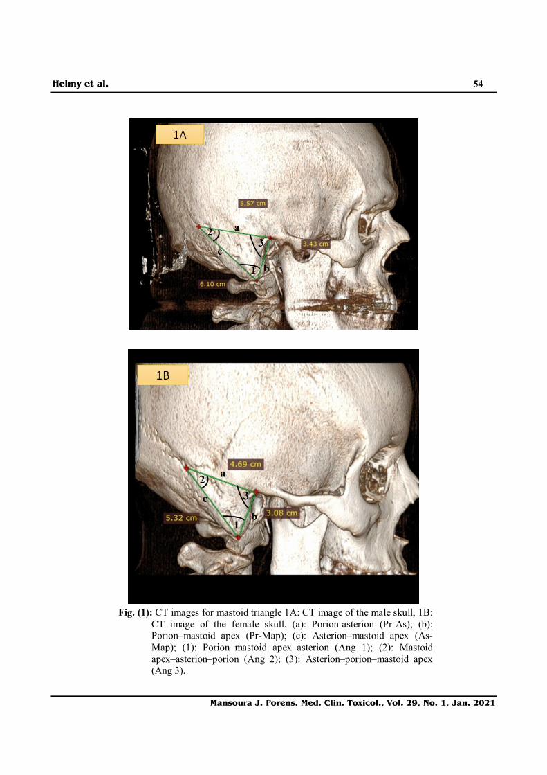
Landmarks used in the study measurement are (Figure 1A):
1. Asterion (As): “the junction of the lambdoid, the occipitomastoid, and the parieto-mastoid suture”.
2. Porion (Pr): “the superior surface of the external auditory meatus”.
3. Mastoid apex (Map): “the lowest craniometric point at the mastoid process”.
The following measures were recorded on both the right and left side (Figure 1B)
1. Porion-asterion (Pr-As): “the distance between the porion and the asterion”.
2. Porion–mastoid apex (Pr-Map): “the distance between the porion and the mastoid apex”.
3. Asterion–mastoid apex (As-Map): “the distance between the asterion and the mastoid apex”.
4. Mastoid triangle area: the summation of the area calculated on both side using Heron’s formula (Kemkes and Göbel, 2006):
Area =
s= a, b and c are the lengths of the triangle sides, a is Pr-As, b is Pr-Map and c is As-Map. 5. Porion–mastoid apex–asterion (Ang 1):
“the angle presented between the porion, the mastoid apex, and the asterion”.
6. Mastoid apex–asterion–porion (Ang 2): “the angle presented between the mastoid apex, the asterion, and the porion”.
7. Asterion–porion–mastoid apex (Ang 3): “the angle presented between the asterion, the porion, and the mastoid apex”.
Data were analyzed using the Statistical Package of Social Science (SPSS) program for Windows (Standard version 10). Continuous variables were presented as mean ± SD (standard deviation). The two groups were compared with the Student t-test while paired groups and inter-observer variability compared by paired t-test. The receiver operating characteristic (ROC) curve was obtained for sex classification using continuous variables. Sensitivity and specificity at the different cut-off points were tested by the ROC curve. Direct discriminant function analysis was done by univariate and multivariate methods. The sectioning point was calculated by adding the two group centroids and dividing by two to discriminate between males and females. A leave-one-out classification was performed to measure the effectiveness and thus cross-validated the accuracy of assignments to either males or females. For all above mentioned statistical tests done, the threshold of significance is fixed at 5% level (p-value).
Helmy et al.
Mansoura J. Forens. Med. Clin. Toxicol., Vol. 29, No. 1, Jan. 2021
54
Fig. (1): CT images for mastoid triangle 1A: CT image of the male skull, 1B:
CT image of the female skull. (a): Porion-asterion (Pr-As); (b): Porion–mastoid apex (Pr-Map); (c): Asterion–mastoid apex (As-Map); (1): Porion–mastoid apex–asterion (Ang 1); (2): Mastoid apex–asterion–porion (Ang 2); (3): Asterion–porion–mastoid apex (Ang 3).
Helmy et al.
Mansoura J. Forens. Med. Clin. Toxicol., Vol. 29, No. 1, Jan. 2021
55
Results:
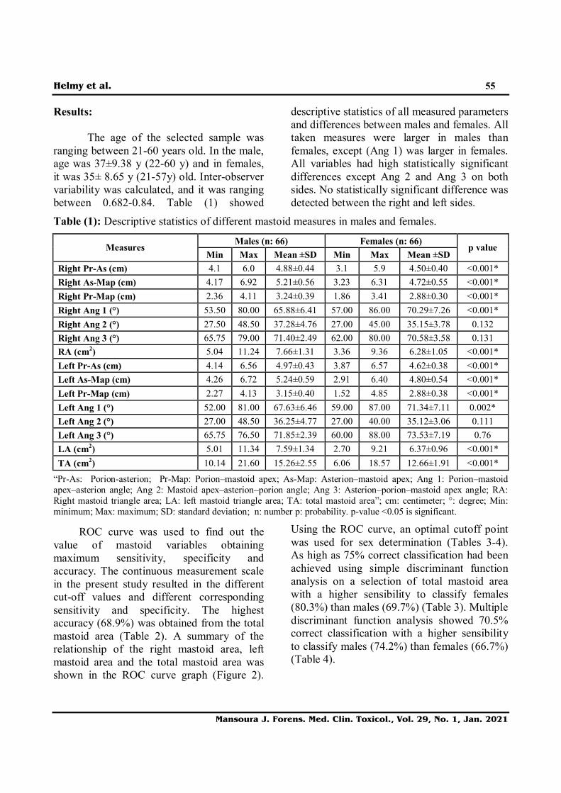
The age of the selected sample was ranging between 21-60 years old. In the male, age was 37±9.38 y (22-60 y) and in females, it was 35± 8.65 y (21-57y) old. Inter-observer variability was calculated, and it was ranging between 0.682-0.84. Table (1) showed
descriptive statistics of all measured parameters and differences between males and females. All taken measures were larger in males than females, except (Ang 1) was larger in females. All variables had high statistically significant differences except Ang 2 and Ang 3 on both sides. No statistically significant difference was detected between the right and left sides.
Table (1): Descriptive statistics of different mastoid measures in males and females.
Males (n: 66) Females (n: 66) Measures
Min Max Mean ±SD Min Max Mean ±SD p value
Right Pr-As (cm) 4.1 6.0 4.88±0.44 3.1 5.9 4.50±0.40 <0.001* Right As-Map (cm) 4.17 6.92 5.21±0.56 3.23 6.31 4.72±0.55 <0.001* Right Pr-Map (cm) 2.36 4.11 3.24±0.39 1.86 3.41 2.88±0.30 <0.001* Right Ang 1 (°) 53.50 80.00 65.88±6.41 57.00 86.00 70.29±7.26 <0.001* Right Ang 2 (°) 27.50 48.50 37.28±4.76 27.00 45.00 35.15±3.78 0.132 Right Ang 3 (°) 65.75 79.00 71.40±2.49 62.00 80.00 70.58±3.58 0.131 RA (cm2) 5.04 11.24 7.66±1.31 3.36 9.36 6.28±1.05 <0.001* Left Pr-As (cm) 4.14 6.56 4.97±0.43 3.87 6.57 4.62±0.38 <0.001* Left As-Map (cm) 4.26 6.72 5.24±0.59 2.91 6.40 4.80±0.54 <0.001* Left Pr-Map (cm) 2.27 4.13 3.15±0.40 1.52 4.85 2.88±0.38 <0.001* Left Ang 1 (°) 52.00 81.00 67.63±6.46 59.00 87.00 71.34±7.11 0.002* Left Ang 2 (°) 27.00 48.50 36.25±4.77 27.00 40.00 35.12±3.06 0.111 Left Ang 3 (°) 65.75 76.50 71.85±2.39 60.00 88.00 73.53±7.19 0.76 LA (cm2) 5.01 11.34 7.59±1.34 2.70 9.21 6.37±0.96 <0.001* TA (cm2) 10.14 21.60 15.26±2.55 6.06 18.57 12.66±1.91 <0.001*
“Pr-As: Porion-asterion; Pr-Map: Porion–mastoid apex; As-Map: Asterion–mastoid apex; Ang 1: Porion–mastoid apex–asterion angle; Ang 2: Mastoid apex–asterion–porion angle; Ang 3: Asterion–porion–mastoid apex angle; RA: Right mastoid triangle area; LA: left mastoid triangle area; TA: total mastoid area”; cm: centimeter; °: degree; Min: minimum; Max: maximum; SD: standard deviation; n: number p: probability. p-value <0.05 is significant.
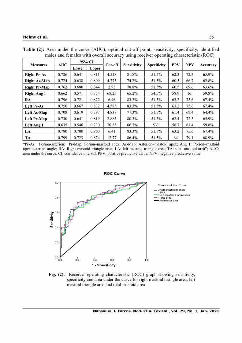
ROC curve was used to find out the value of mastoid variables obtaining maximum sensitivity, specificity and accuracy. The continuous measurement scale in the present study resulted in the different cut-off values and different corresponding sensitivity and specificity. The highest accuracy (68.9%) was obtained from the total mastoid area (Table 2). A summary of the relationship of the right mastoid area, left mastoid area and the total mastoid area was shown in the ROC curve graph (Figure 2).
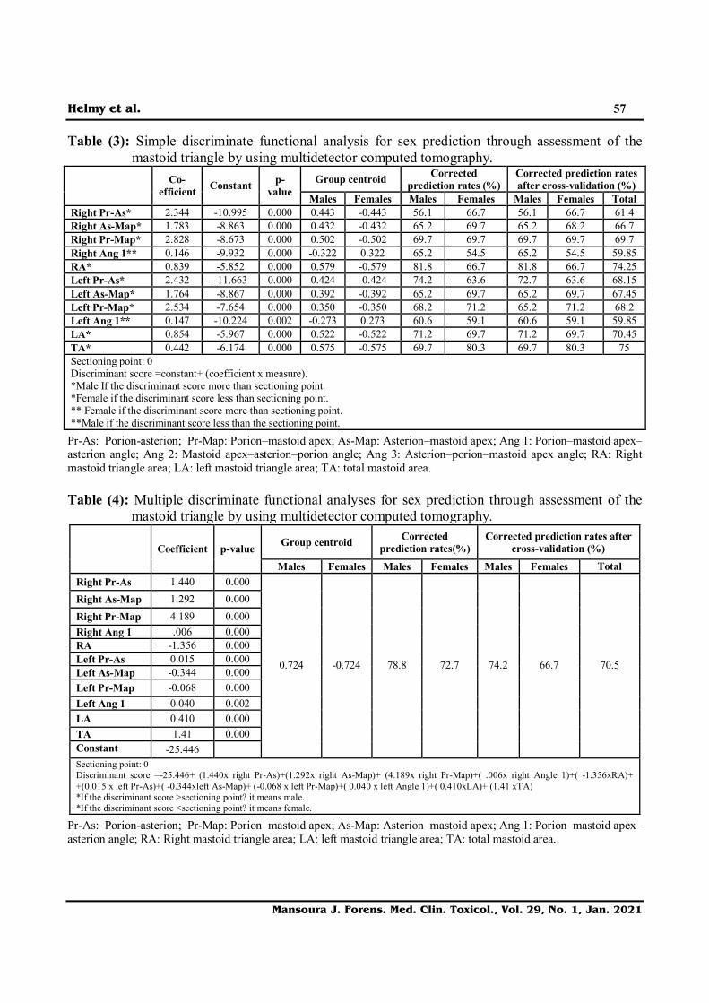
Using the ROC curve, an optimal cutoff point was used for sex determination (Tables 3-4). As high as 75% correct classification had been achieved using simple discriminant function analysis on a selection of total mastoid area with a higher sensibility to classify females (80.3%) than males (69.7%) (Table 3). Multiple discriminant function analysis showed 70.5% correct classification with a higher sensibility to classify males (74.2%) than females (66.7%) (Table 4).
Helmy et al.
Mansoura J. Forens. Med. Clin. Toxicol., Vol. 29, No. 1, Jan. 2021
56
Table (2): Area under the curve (AUC), optimal cut-off point, sensitivity, specificity, identified males and females with overall accuracy using receiver operating characteristic (ROC).
95% CI Measures AUC
Lower Upper Cut-off Sensitivity Specificity PPV NPV Accuracy
Right Pr-As 0.726 0.641 0.811 4.518 81.8% 51.5% 62.3 72.3 65.9% Right As-Map 0.724 0.638 0.809 4.775 74.2% 51.5% 60.5 66.7 62.8% Right Pr-Map 0.762 0.680 0.844 2.93 78.8% 51.5% 60.5 69.6 63.6% Right Ang 1 0.662 0.571 0.754 68.25 65.2% 54.5% 58.9 61 59.8% RA 0.796 0.721 0.872 6.46 83.3% 51.5% 63.2 75.6 67.4% Left Pr-As 0.750 0.667 0.832 4.585 83.3% 51.5% 63.2 75.6 67.4% Left As-Map 0.708 0.619 0.797 4.837 77.3% 51.5% 61.4 69.4 64.4% Left Pr-Map 0.730 0.641 0.819 2.885 80.3% 51.5% 62.4 72.3 65.9% Left Ang 1 0.635 0.540 0.730 70.25 66.7% 53% 58.7 61.4 59.8% LA 0.780 0.700 0.860 6.41 83.3% 51.5% 63.2 75.6 67.4% TA 0.799 0.723 0.874 12.77 86.4% 51.5% 64 79.1 68.9%
“Pr-As: Porion-asterion; Pr-Map: Porion–mastoid apex; As-Map: Asterion–mastoid apex; Ang 1: Porion–mastoid apex–asterion angle; RA: Right mastoid triangle area; LA: left mastoid triangle area; TA: total mastoid area”; AUC: area under the curve, CI: confidence interval, PPV: positive predictive value, NPV: negative predictive value
Fig. (2): Receiver operating characteristic (ROC) graph showing sensitivity,
specificity and area under the curve for right mastoid triangle area, left mastoid triangle area and total mastoid area
Helmy et al.
Mansoura J. Forens. Med. Clin. Toxicol., Vol. 29, No. 1, Jan. 2021
57
Table (3): Simple discriminate functional analysis for sex prediction through assessment of the mastoid triangle by using multidetector computed tomography.
Group centroid Corrected prediction rates (%)
Corrected prediction rates after cross-validation (%) Co-
efficient Constant p-value Males Females Males Females Males Females Total
Right Pr-As* 2.344 -10.995 0.000 0.443 -0.443 56.1 66.7 56.1 66.7 61.4 Right As-Map* 1.783 -8.863 0.000 0.432 -0.432 65.2 69.7 65.2 68.2 66.7 Right Pr-Map* 2.828 -8.673 0.000 0.502 -0.502 69.7 69.7 69.7 69.7 69.7 Right Ang 1** 0.146 -9.932 0.000 -0.322 0.322 65.2 54.5 65.2 54.5 59.85 RA* 0.839 -5.852 0.000 0.579 -0.579 81.8 66.7 81.8 66.7 74.25 Left Pr-As* 2.432 -11.663 0.000 0.424 -0.424 74.2 63.6 72.7 63.6 68.15 Left As-Map* 1.764 -8.867 0.000 0.392 -0.392 65.2 69.7 65.2 69.7 67.45 Left Pr-Map* 2.534 -7.654 0.000 0.350 -0.350 68.2 71.2 65.2 71.2 68.2 Left Ang 1** 0.147 -10.224 0.002 -0.273 0.273 60.6 59.1 60.6 59.1 59.85 LA* 0.854 -5.967 0.000 0.522 -0.522 71.2 69.7 71.2 69.7 70.45 TA* 0.442 -6.174 0.000 0.575 -0.575 69.7 80.3 69.7 80.3 75 Sectioning point: 0 Discriminant score =constant+ (coefficient x measure). *Male If the discriminant score more than sectioning point. *Female if the discriminant score less than sectioning point. ** Female if the discriminant score more than sectioning point. **Male if the discriminant score less than the sectioning point.
Pr-As: Porion-asterion; Pr-Map: Porion–mastoid apex; As-Map: Asterion–mastoid apex; Ang 1: Porion–mastoid apex–asterion angle; Ang 2: Mastoid apex–asterion–porion angle; Ang 3: Asterion–porion–mastoid apex angle; RA: Right mastoid triangle area; LA: left mastoid triangle area; TA: total mastoid area. Table (4): Multiple discriminate functional analyses for sex prediction through assessment of the
mastoid triangle by using multidetector computed tomography.
Group centroid Corrected prediction rates(%)
Corrected prediction rates after cross-validation (%)
Coefficient p-value
Males Females Males Females Males Females Total Right Pr-As 1.440 0.000 Right As-Map 1.292 0.000 Right Pr-Map 4.189 0.000 Right Ang 1 .006 0.000 RA -1.356 0.000 Left Pr-As 0.015 0.000 Left As-Map -0.344 0.000 Left Pr-Map -0.068 0.000 Left Ang 1 0.040 0.002 LA 0.410 0.000 TA 1.41 0.000 Constant -25.446
0.724 -0.724 78.8 72.7 74.2 66.7 70.5
Sectioning point: 0 Discriminant score =-25.446+ (1.440x right Pr-As)+(1.292x right As-Map)+ (4.189x right Pr-Map)+( .006x right Angle 1)+( -1.356xRA)+ +(0.015 x left Pr-As)+( -0.344xleft As-Map)+ (-0.068 x left Pr-Map)+( 0.040 x left Angle 1)+( 0.410xLA)+ (1.41 xTA) *If the discriminant score >sectioning point? it means male. *If the discriminant score <sectioning point? it means female.
Pr-As: Porion-asterion; Pr-Map: Porion–mastoid apex; As-Map: Asterion–mastoid apex; Ang 1: Porion–mastoid apex–asterion angle; RA: Right mastoid triangle area; LA: left mastoid triangle area; TA: total mastoid area.
Helmy et al.
Mansoura J. Forens. Med. Clin. Toxicol., Vol. 29, No. 1, Jan. 2021
58
Discussion:
The mastoid process is one of sexually dimorphic bone with greater size in males. The greater size in males can be contributed to stronger muscle action that is attached to relatively larger areas on males compared with females, exerting forces that affect the development of its size and shape, specifically by pulling the temporal bone downward. The other suggested factor for morphological differences between sexes and among individuals is variable growth patterns of the mastoid cells, which create the hollow interior spaces of the mastoid process (Petaros et al., 2015; Yilmaz et al., 2015). In the current study, the mastoid process was selected for the need of development of methods for sex determination in the fragmentary remains and the mastoid region involving the mastoid process is a craniofacial feature that is highly resistant, thus the studied method can be applied when either a fragmented skull is obtained or when only the mastoid region of the skull is obtained in isolation. Also, multidetector CT was selected for metric assessment of sexual dimorphism in the mastoid area owing to its sensitive and solid method in measuring all mastoid dimensions (Kanchan et al., 2013; Yilmaz et al., 2015).
According to our knowledge, the present study is the first one to assess the mastoid triangle area in Egyptian individuals. Measures in the right and left mastoid triangle were taken separately and there was a difference in measures that was statistically insignificant. This is similar to the study performed on 100 Indian cranium in which the mean value for the three limbs of the triangle on the right side was smaller than the left side (Jain et al., 2013). This asymmetry between both sides makes it better to discriminate sex using the summation of the
right and left mastoid areas (de Paiva and Segre, 2003).
As regards the estimated limbs' length, the nearest measures to the Egyptians were that of Saudi Arabia individuals, in which As-Map, Pr-As, Pr-Map were 5.2, 4.4 and 3.2 cm in males, and 4.8, 4.2 and 2.9 cm in Saudi Arabia females (Madadin et al., 2015). In this study, the longest limb was As-Map followed by Pr-As then Pr-Map is the shortest limb in both sides and both sexes. This was similar to a study performed in Saudi Arabia, Turkish, Malaysian CT images (Ibrahim et al., 2018; Madadin et al., 2015; Yilmaz et al., 2015) and German, Portuguese and Indian skulls (Kemkes and Göbel, 2006; Kanchan et al., 2013), but this sequence was different in Nigerian skulls (Jaja et al., 2013).
On comparing both sexes, all limbs were longer in males with a significant difference between both sexes. This is similar to many previous studies (Kemkes and Göbel, 2006; Madadin et al., 2015; Yilmaz et al., 2015; Ibrahim et al., 2018) but against the findings of Kanchan et al. (2013) who did not observe statistically significant sex differences in As- Map in 118 South Indian skulls and that of Jaja et al. (2013) who observed sex difference only in As-Map in Nigerian skulls.
For the measured angles, in a study by Yilmaz et al. (2015) all male measures were higher than females except angle 3 which was smaller in males. This was unlike the current study in which both angles 1 and 2 were smaller in males than females. Besides, Ang 1 did not show a statistically significant difference, unlike our study in which there was a statistically significant difference between both sexes in Ang 1. Although Ang 1 was significantly different between both sexes, it had poor accuracy for sex identification.
Regarding the mastoid area, in this study, the mean value for total area in males was 15.26 cm2 and in females was 12.66 cm2 with

Helmy et al.
Mansoura J. Forens. Med. Clin. Toxicol., Vol. 29, No. 1, Jan. 2021
59
an overlap in the area between both sexes. These values were close to results on Brazilian, German, Portuguese (de Paiva and Segre, 2003), smaller than that of Thai individuals (Manoonpol and Plakornkul, 2012) and larger than that of Nigerian skulls (Jaja et al., 2013).
The ROC curve analysis, simple and multiple discriminate functional analyses were then performed for all obtained measures. The overall accuracy of the three limbs of the triangle and the calculated areas was ranging between 64-69 % with the highest AUC (0.799) and prediction accuracy of 69% is the total mastoid area. As high as 75% correct classification has been achieved using discriminant function analysis with a selection of total mastoid area with a higher sensibility to classify females (80%) than males (70%).
Other studies had also analyzed the classification accuracy and prediction rate of the mastoid process and showed wide variations. Regarding the Egyptian population, the study performed for metric assessment of mastoid measures was performed on 80 CT images using different measures and it showed prediction accuracy ranging between 60-75% (Allam and Allam, 2016). For other populations, the correct classification was around 69% using the three limbs of the triangle and the total area in study performed on 206 CT in Saudi Arabia individuals (Madadin et al., 2015), 58% using the mastoid area in 97 German skulls, 66% in 100 Portuguese skulls (Kemkes and Göbel, 2006), 64% using Pr-Map in study on 81 Brazilian skulls (Suazo Galdames et al., 2008), 75% using the total mastoid area in study performed on 60 microtomography images from dried skulls of Brazilian individuals (Kramer et al., 2018), 66.7% on using the Pr-As length in 48 Japanese skulls (Nagaoka et al., 2008), ranging between 70-75% using the three limbs of the triangle in
138 North Indian skulls (Saini et al., 2012), ranging between 45-80% using the three limbs of the triangle in 100 Indian skulls with the best prediction rate recorded from right As-Map and Pr-Map achieving 80% accuracy, and the highest reported prediction accuracy was 84.4% using the total mastoid area in 388 CT images of Malaysian individuals (Ibrahim et al., 2018).
The multivariate discriminant analysis showed less accuracy compared to some of the single variables. Some authors encourage the combination of variables since it may increase methodological accuracy. However, in the present study, the combinations of variables showed inferior results compared to single variables similar to some previous studies (Madadin et al., 2015; Kramer et al., 2018).
The difference in measures between different populations and the accuracy for sex prediction from different measures can be explained by population heterogenicity in cranial size and shape (Manoonpol and Plakornkul, 2012), asterion location vary with the progression of age in a population-specific manner in many anatomical and clinical/neurological studies (Uz et al., 2001; Mwachaka et al., 2010). This variation is attributed to environment, age, and migratory processes occurring during the ethnographic formation of a given population. Another source of variation in results is the methodological difference related to instrumentation that can add a source for variability (Kramer et al., 2018). Conclusion:
In conclusion, this study demonstrated that the total mastoid triangle area had relatively good sexual dimorphism in Egyptian individuals and can be considered a useful methodology in predicting sex in cases of fragmented crania. Based on the ROC curve
Helmy et al.
Mansoura J. Forens. Med. Clin. Toxicol., Vol. 29, No. 1, Jan. 2021
60
analysis, univariate and multivariate discriminate analysis, a reference table was developed to be applied for the sex determination in Egyptian individuals from the mastoid triangle area. The limitation of the current study is the relatively small sample size, especially for Ang 2 and 3, further researches on a larger scale are recommended to study their role in sex determination. Conflicts of interest statement:
The authors certify that they have NO affiliations with or involvement in any organization or entity with any financial interest or non-financial in the subject matter or materials discussed in this manuscript. References: Allam, F.A.F.A.B. and Allam, M.F.A.B.
(2016): "Sex discrimination of mastoid process by anthropometric measurements using multidetector computed tomography in Egyptian adult population". Egyptian Journal of Forensic Sciences, 6(4): 361–369. https://doi.org/10.1016/j.ejfs.2016.05.001
de Paiva, L.A.S. and Segre, M. (2003): "Sexing the human skull through the mastoid process". Revista Do Hospital Das Clínicas, 58(1): 15–20. https://doi.org/10.1590/s0041-87812003000100004
Ibrahim, A.; Alias, A.; Shafie, M. S.; et al. (2018): "Osteometric estimation of sex from the mastoid triangle in Malaysian population". Asian Journal of Pharmaceutical and Clinical Research, 11(7): 303–307. https://doi.org/10.22159/ajpcr.2018.v11i7.25986
Jain, D.; Jasuja, O.P. and Nath, S. (2013): "Sex determination of human crania
using Mastoid triangle and Opisthion-Bimastoid triangle". Journal of Forensic and Legal Medicine, 20(4): 255–259. https://doi.org/10.1016/j.jflm.2012.09.020
Jaja, B.N.R.; Ajua, C.O. and Didia, B.C. (2013): "Mastoid triangle for sex determination in adult Nigerian population: A validation study". Journal of Forensic Sciences, 58(6): 1575–1578. https://doi.org/10.1111/1556-4029.12299
Kanchan, T.; Gupta, A. and Krishan, K. (2013): "Estimation of sex from mastoid triangle - A craniometric analysis". Journal of Forensic and Legal Medicine, 20(7): 855–860. https://doi.org/10.1016/j.jflm.2013.06.016
Kemkes, A. and Göbel, T. (2006): "Metric assessment of the “mastoid triangle” for sex determination: A validation study". Journal of Forensic Sciences, 51(5): 985–989. https://doi.org/10.1111/j.1556-4029. 2006. 00232.x
Kramer, N.A.; Lopez-Capp, T.T.; Michel-Crosato, E.; et al. (2018): "Sex estimation from the mastoid process using Micro-CT among Brazilians: Discriminant analysis and ROC curve analysis". Journal of Forensic Radiology and Imaging, 14, 1–7. https://doi.org/10.1016/j.jofri.2018.05.003
Madadin, M.; Menezes, R. G.; Al Dhafeeri, O.; et al. (2015): "Evaluation of the mastoid triangle for determining sexual dimorphism: A Saudi population-based study". Forensic Science International, 254, 244.e1-244.e4. https://doi.org/10.1016/j.forsciint.2015.06.019
Manoonpol, C. and Plakornkul, V. (2012): "Sex determination using mastoid process measurement in Thais". Journal of the Medical Association of Thailand, 95(3): 423–429.
Helmy et al.
Mansoura J. Forens. Med. Clin. Toxicol., Vol. 29, No. 1, Jan. 2021
61
Mwachaka, P.M.; Hassanali, J. and Odula, P.O. (2010): "Anatomic position of the asterion in Kenyans for posterolateral surgical approaches to cranial cavity". Clinical Anatomy, 23(1): 30–33. https://doi.org/10.1002/ca.20888
Nagaoka, T.; Shizushima, A.; Sawada, J.; et al. (2008): "Sex determination using mastoid process measurements: Standards for Japanese human skeletons of the medieval and early modern periods". Anthropological Science, 116(2): 105–113. https://doi.org/10.1537/ase.070605
Petaros, A.; Sholts, S. B.; Slaus, M.; et al. (2015): "Evaluating sexual dimorphism in the human mastoid process: A viewpoint on the methodology". Clinical Anatomy, 28(5): 593–601. https://doi.org/10.1002/ca.22545
Saini, R. and Saini, V. (2016): "Sexual Dimorphism of North Indian Crania and its Forensic Application". Journal of Forensic Medicine and Legal Affairs, 01(02): 1–6. https://doi.org/10.19104/jfml.2016.108
Saini, V.; Srivastava, R.; Rai, R. K.; et al. (2012): "Sex Estimation from the Mastoid Process Among North Indians". Journal of Forensic Sciences, 57(2): 434–439.
https://doi.org/10.1111/j.1556-4029. 2011. 01966.x
Galdames, I.C.; Matamala, D.A.; et al. (2008): "Sex Determination Using Mastoid Process Measurements in Brazilian Skulls". International Journal of Morphology, 26(4): 941–944. https://doi.org/10.4067/s0717-95022008000400025
Uz, A.; Ugur, H. C. and Tekdemir, I. (2001): "Is the asterion a reliable landmark for the lateral approach to posterior fossa?". Journal of Clinical Neuroscience, 8(2): 146–147. https://doi.org/10.1054/jocn.2000.0798
Williams, B.A. and Rogers, T.L. (2006): "Evaluating the accuracy and precision of cranial morphological traits for sex determination". Journal of Forensic Sciences, 51(4): 729–735. https://doi.org/10.1111/j.1556-4029.2006.00177.x
Yilmaz, M.T.; Yüzbasioglu, N.; Cicekcibasi, A.E.; et al. (2015): "The evaluation of morphometry of the mastoid process using multidetector computed tomography in a living population". Journal of Craniofacial Surgery, 26(1): 259–263. https://doi.org/10.1097/SCS.0000000000001216.
Helmy et al.
Mansoura J. Forens. Med. Clin. Toxicol., Vol. 29, No. 1, Jan. 2021
62
قسم الطب الشرعي والسموم الإكلینیكیة، كلیة الطب جامعة المنصورة١
الإدارة الطبیة، جامعة المنصورة٢ قسم الأشعة التشخیصیة، كلیة الطب جامعة المنصورة٣
ا یم الرف ي تقی ة تقدیر الجنس ھو الخطوة الأولى ف شریة المجھول ري . ت الب ل المت ى التحلی دف الدراسة إل تھ. لمثلث الخشاء باستخدام التصویر المقطعي المحوسب متعدد الكواشف للتمییز بین الجنسین في الجماجم المصریة
ث الخشاء ١٣٢العینة مكونة من ة لمثل ا الثلاث اس الأطراف والزوای ا قی م منھ یتكون ( صورة مقطعیة للجمجمة تورون، وخشاء من قمة ة، ب انبین ) نجمی ى كلا الج ى . عل رون عل ن خلال صیغة ھی ث م ساحة المثل م حساب م ت
ة ساحة الإجمالی ل . الجانب الأیمن والجانب الأیسر بشكل فردي، ثم تم اعتبار مجموعھما على أنھ الم ر التحلی أظھة قم الإحصائي فرق بین الجنسین في ة وزاوی ة والمنطق ورون والخشاء قیاسات الأطراف المثلثی م إجراء . ة الب ت
ى ل منحن ة ROC تحلی وفر دق ي ت ة الت ساحة الإجمالی و الم ر ھ ان أفضل متغی ایز . ٪٦٩وك ل التم ر التحلی اظھر دق ة أحادي المتغی اث ومجتمع ذكور والإن صالبة لل والي ٧٥٪ و٨٠٫٣٪ و٦٩٫٣ة تصنیف مت ى الت أظھر . ٪ عل
ى . تحلیل التمایز متعدد المتغیرات نسب تصنیف منخفضة ھ عل دول مرجعي لتطبیق م وضع ج ائج ت ى النت اءً عل بنن . تقدیر جنس الأفراد المصریین ز للج ن استخدامھا كممی سبیا ویخلص البحث الي ان منطقة الخشاء یمك د ن س جی
.في الجماجم المصریة