Embed Size (px)
Citation preview
Shaw Mark R, Kan P, Kan-van Limburg Stirum B, Schwarz M (2021) The remarkable
biology of a new species of Gelis Thunberg, 1827 (Ichneumonidae, Phygadeuontinae), a
solitary endoparasitoid of fresh eggs of Timarcha (Coleoptera, Chrysomelidae). Journal of
Hymenoptera Research 82: 161–186.
https://doi.org/10.3897/jhr.82.64657
Deposited on: 12th October 2021
NMS Repository – Research publications by staff of the National Museums Scotland
https://nms.iro.bl.uk

The remarkable biology of a new species of Gelis Thunberg, 1827 (Ichneumonidae, Phygadeuontinae),
a solitary endoparasitoid of fresh eggs of Timarcha (Coleoptera, Chrysomelidae)
Mark R. Shaw1, Pieter Kan2, Brigitte Kan-van Limburg Stirum2, Martin Schwarz3
1 Honorary Research Associate, National Museums of Scotland, Chambers Street, Edinburgh EH1 1JF, UK 2 295 Chemin de la Croix, Quartier la Ferrage du Ray, 83830 Callas, France 3 Eben 21, A-4202 Kirchschlag, Austria
Corresponding author: Mark R. Shaw ([email protected])
Academic editor: Gavin Broad | Received 19 February 2021 | Accepted 29 March 2021 | Published 29 April 2021
http://zoobank.org/852712E6-BBE4-4791-B0C8-DD76D2B4CE3C
Citation: Shaw MR, Kan P, Kan-van Limburg Stirum B, Schwarz M (2021) The remarkable biology of a new species of Gelis Thunberg, 1827 (Ichneumonidae, Phygadeuontinae), a solitary endoparasitoid of fresh eggs of Timarcha (Coleoptera, Chrysomelidae). Journal of Hymenoptera Research 82: 161–186. https://doi.org/10.3897/jhr.82.64657
AbstractA new species, Gelis timarchae Schwarz, Shaw & Kan, is figured and described from specimens reared as a solitary endoparasitoid of fresh eggs of Timarcha nicaeensis in the south of France. Oviposition behaviour of the adult parasitoid, directly into the host cytoplasm, is described and links to videos are given. This appears to be the first record of any ichneumonid developing as an endoparasitoid of an insect egg, and it is a major departure from hitherto known ectoparasitoid (or spiders’ egg-predation) behaviour in the genus Gelis. Fluid from the host egg issuing from the base of the parasitoid’s ovipositor early in the oviposi-tion process is interpreted as a necessary reduction of hydrostatic pressure before the parasitoid egg can be forced down the ovipositor. The egg and first instar larva are figured; the latter is caudate, with the caudal appendage very unusual in being bifurcate. The complex phenology and diapause of the parasitoid were investigated partly experimentally; it is broadly bivoltine with a prepupal diapause in summer, but extra generations and prolonged diapause were both also seen.
KeywordsCocoon, diapause, egg, first instar larva, France, Gelis timarchae, oviposition, plastic phenology
JHR 82: 161–186 (2021)
doi: 10.3897/jhr.82.64657
https://jhr.pensoft.net
Copyright Mark R. Shaw et al. This is an open access article distributed under the terms of the Creative Commons Attribution License (CC BY 4.0), which permits unrestricted use, distribution, and reproduction in any medium, provided the original author and source are credited.
RESEARCH ARTICLE
Mark R. Shaw et al. / Journal of Hymenoptera Research 82: 161–186 (2021)162
Introduction
The phygadeuontine genus Gelis Thunberg exhibits a wide range of morphological forms, its various species being fully winged, brachypterous or apterous, sometimes with plasticity (as investigated by Salt 1952) and often with sexual dimorphism in these respects. The European fauna of 142 species (Fauna Europaea does not take ac-count of species described by Schwarz (2016)) has been comprehensively treated taxo-nomically, principally by Horstmann (1986) and Schwarz (1995, 1998, 2002, 2016), and is now reasonably well known. Biologically the vast majority of species are either solitary ectoparasitoid idiobionts of the content of holometabolous insect cocoons or cocoon-like structures (e.g. Muesebeck and Dohanian 1927; Salt 1952; Harvey 2008), or successive egg predators within spider egg sacs and then often developing gregari-ously. Host information is known for a number of species (see also Schwarz and Shaw 1999), demonstrating not only that particular species show absolute fidelity either to spider egg sacs or to other hosts but also that, within the last category especially, host ranges vary from being extremely specialised to remarkably catholic and then often involving several orders of insects. However, none has been reported to be an endo-parasitoid, and the host property that has seemed almost universal is that the adult parasitoid oviposits through a structure that is at least partly silken.
In this paper we describe a remarkable and quite profound departure from the nor-mal biology of Gelis species: one that oviposits directly into the newly-laid large egg of another insect, in which the whole of the parasitoid’s larval development takes place as a solitary endoparasitoid in the cytoplasm. The Gelis species proved to be undescribed, and it is described below.
Materials and methods
Host biology and study site
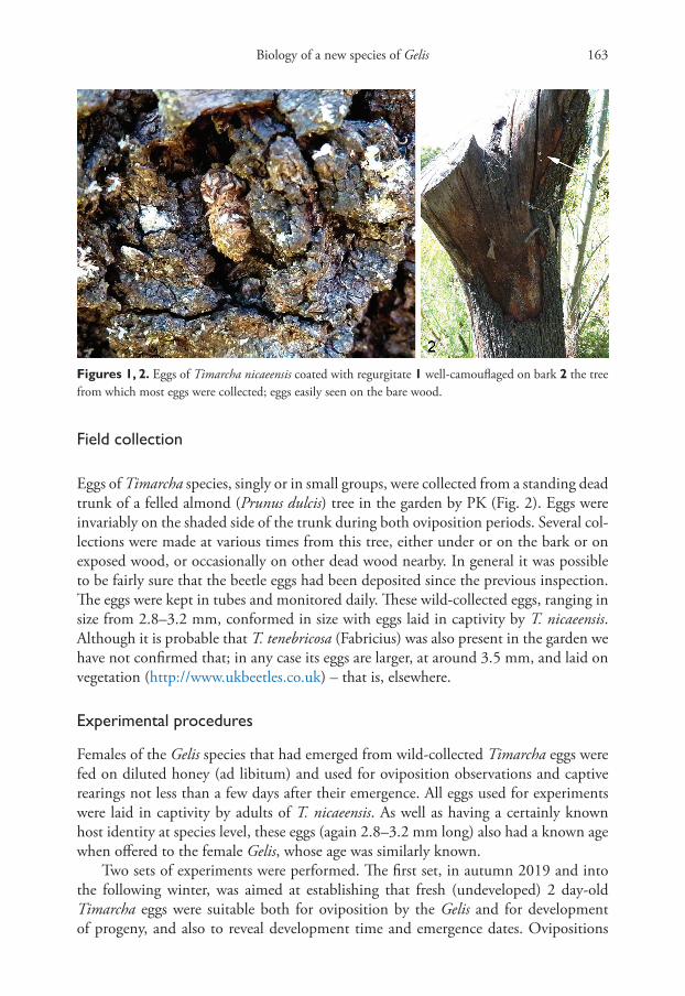
The chrysomelid beetle genus Timarcha Latreille comprises large flightless species, most-ly feeding as larvae on the leaves and, especially in the case of adults, also stems of Galium (Rubiaceae) or related plants. At our study site, a large wild garden at Callas in the southern French Departément of Var, T. nicaeensis Villa was abundant, and all material collected appeared to belong to this species (three males and three females det. R. M. Lyszkowski). Timarcha species are long-lived as adults, leading to rather complex phenol-ogy (Chevin 1985, 1986, 1994). The univoltine Timarcha nicaeensis has two main ovipo-sition periods, prolonged in each case: in autumn (eggs that will diapause, laid by adults that emerged that summer) and again in early spring to early summer (eggs laid by the same females that have overwintered). Adults from both oviposition periods result more or less simultaneously in late summer (Chevin 1986). The eggs are laid solitarily, or more often in small batches, and coated with small fragments of hard plant tissue regurgitated by the adult female (Selman 1994) (Fig. 1). A video of the life-history of T. nicaeensis is in preparation and will be available on YouTube via the Filming VarWild website.
Biology of a new species of Gelis 163
Field collection
Eggs of Timarcha species, singly or in small groups, were collected from a standing dead trunk of a felled almond (Prunus dulcis) tree in the garden by PK (Fig. 2). Eggs were invariably on the shaded side of the trunk during both oviposition periods. Several col-lections were made at various times from this tree, either under or on the bark or on exposed wood, or occasionally on other dead wood nearby. In general it was possible to be fairly sure that the beetle eggs had been deposited since the previous inspection. The eggs were kept in tubes and monitored daily. These wild-collected eggs, ranging in size from 2.8–3.2 mm, conformed in size with eggs laid in captivity by T. nicaeensis. Although it is probable that T. tenebricosa (Fabricius) was also present in the garden we have not confirmed that; in any case its eggs are larger, at around 3.5 mm, and laid on vegetation (http://www.ukbeetles.co.uk) – that is, elsewhere.
Experimental procedures
Females of the Gelis species that had emerged from wild-collected Timarcha eggs were fed on diluted honey (ad libitum) and used for oviposition observations and captive rearings not less than a few days after their emergence. All eggs used for experiments were laid in captivity by adults of T. nicaeensis. As well as having a certainly known host identity at species level, these eggs (again 2.8–3.2 mm long) also had a known age when offered to the female Gelis, whose age was similarly known.
Two sets of experiments were performed. The first set, in autumn 2019 and into the following winter, was aimed at establishing that fresh (undeveloped) 2 day-old Timarcha eggs were suitable both for oviposition by the Gelis and for development of progeny, and also to reveal development time and emergence dates. Ovipositions
Figures 1, 2. Eggs of Timarcha nicaeensis coated with regurgitate 1 well-camouflaged on bark 2 the tree from which most eggs were collected; eggs easily seen on the bare wood.
Mark R. Shaw et al. / Journal of Hymenoptera Research 82: 161–186 (2021)164
were conducted indoors, in a 14.5 cm diameter Petri dish or, since the female Gelis is apterous, in some cases on a piece of almond bark in the open to facilitate filming. The description of behaviour is taken largely from these filmed events and based on observations at various times involving three virgin females, each of which successfully oviposited into 1–3 eggs in a session. Because egg-limitation was likely to be a behav-iour-changing issue, observations subsequent to the last successful oviposition by a female in a session are not included in the account of behaviour. Host eggs were offered in small tight batches (1–7 eggs), on the pieces of dead almond bark on which they had been laid in captivity, and the light coverings of minute fragments of regurgitated plant material were not removed. Most of the experimentally parasitised eggs were kept until the parasitoid emerged, but concurrently three 2 day-old T. nicaeensis eggs in which a Gelis egg had been deposited were immediately preserved in 70% ethanol in a tube which was placed in a freezer at -27 °C until being sent for dissection in Edinburgh.
The second set of experiments were conducted in Edinburgh in October and No-vember 2020, using T. nicaeensis eggs recently laid in captivity in Callas that were by then around a week to 20 days old, and five mated females that had emerged in Septem-ber and October from wild-collected Timarcha eggs collected in January, April and early June. Mating occurred almost immediately when a female, on the day of her emergence, was added to a slightly older (fed) male in a 5 cm covered Petri dish. After only cursory courtship the unions (male aligned on top of female) typically lasted for about 20 sec-onds. Once the host eggs reached Edinburgh they were generally kept under outdoor temperatures, but some were brought indoors to accelerate development. Single host eggs were offered in 7.5 × 2.5 cm corked glass tubes. Eggs that had received a Gelis egg were kept under outdoor temperature conditions (mostly well below 10 °C) and dissect-ed at various times over the following month to check development of their contents. Variously treated unparasitized eggs were also dissected to investigate development.
Dissections
Some of the wild-collected host eggs that had not produced a Timarcha larva or parasi-toid for a prolonged period, including through summer, were opened in August 2020 in Edinburgh to assess their content. These and other partially cleaned host eggs were dissected, either lightly embedded in plasticine or in a drop of water on a microscope slide, using a razor blade, fine forceps and pins under a Wild M5A binocular micro-scope with Volpi ring illumination.
Photography
Films were taken with a Canon XL2 with a 20× zoom, XL 5.4–108 mm lens, supple-mented when appropriate with the addition of a Canon 72 mm close-up 500D lens. For macro a Canon EF 100 mm 1:2:8 with an EF Canon XL adaptor was used. The footage was recorded on mini DV tapes of 60 minutes. For still pictures of living speci-mens a Lumix HD Panasonic DMC-TZ10 was used. Morphological photos of dead
Biology of a new species of Gelis 165
mounted specimens of the new species, G. brevis (Bridgman) and G. proximus (Förster) were taken using a Nikon AZ100M and stacked with NIS-Elements Microscope Im-aging. Dissections of host eggs and parasitoid cocoons were photographed as single shots down one arm of a Wild M5A binocular microscope with Volpi ring illumina-tion using a Canon PowerShot S110. For the preparation of Fig. 35, by István Mikó, heads stored in ethanol were dissected and transferred to glycerol, then imaged with a Nikon A1R-HD CLSM using two laser excitation wavelengths, 407 and 487 nm with emission ranges defined using the A1-DUS spectral detector, 430–480, 500–560 and 570–630 nm, with assignation of pseudo-colours reflecting the fluorescence spectra, and the files were created using FIJI (Schindelin et al. 2012).
Depositories of cited specimens
MSC Martin Schwarz personal collection, Kirchschlag near Linz;MNCN Museo Nacional de Ciencias Naturales, Madrid;NMS National Museums of Scotland, Edinburgh;NHMUK Natural History Museum, London;ZSM Zoologische Staatssammlung, München.
Results
Description of new species
Gelis timarchae Schwarz, Shaw & Kan, sp. nov.http://zoobank.org/5E4196E0-6427-45F9-AA1C-6B540D70C1CCFigs 3–13 (type series); Figs 14–18 (additional material)
Material examined. Holotype (♀): “France: Var, Callas, La Ferrage du Ray ex Tima-rcha nicaeensis egg under loose Prunus dulcis bark coll. 15.4.2019 em. 26.10.2019. P. Kan 660.” (National Museums of Scotland (NMS), Edinburgh). Paratypes (14 ♀, 15 ♂) same location, solitarily ex eggs of Timarcha nicaeensis, various collection and emergence dates in 2019 and 2020, including rearings in culture (see Tables 1 and 2, where all paratypes are recorded) (NMS, NHMUK, MSC).
Additional, non-paratype, material. Germany: Sachsen, Kyffhäuser Gebirge, Kattenhof, 450 m, 15.iv.1914, leg. Petry (1♀; ZSM); Ebelsbach (1♀; ZSM). Spain: El Coll, 7.iv.1895 (1♀; MNCN).
It is intended that a CO1 barcode sequence will be obtained from one of the para-types (783 in Table 1) and deposited in GenBank.
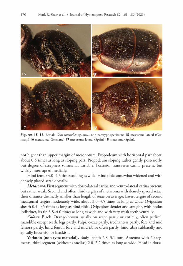
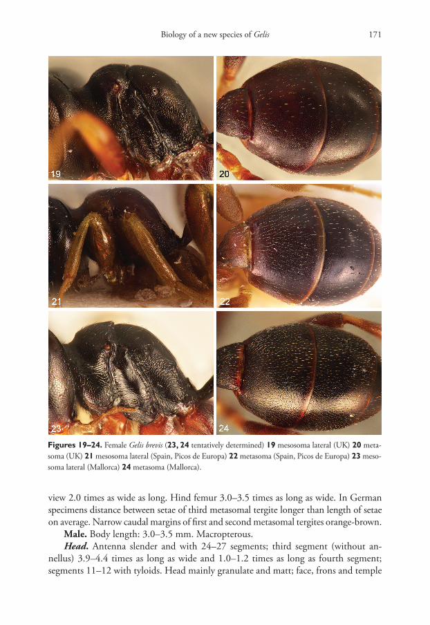
Diagnosis and remarks. In the female sex this species is most similar to Gelis bre-vis (Figs 19–24, which include a doubtfully determined specimen) and, because both species are variable with somewhat overlapping characters, they are not always easily distinguished. In G. timarchae the propodeum is less steeply sloping caudally on average;
Mark R. Shaw et al. / Journal of Hymenoptera Research 82: 161–186 (2021)166
and the second and often third tergites of the metasoma have dense, closely spaced setae with the distance between setae distinctly smaller than the length of setae, while in G. brevis the distance between setae varies from wider to somewhat smaller than the length of setae. In studied specimens of G. brevis from UK (including the type of Pezomachus brevis Bridgman, 1883) and Germany the metasoma has widely spaced setae, though in material from southern Europe the setae are much closer, but still less densely spaced than in southern European material of G. timarchae. The two specimens of G. timarchae seen from Germany have (especially) third tergites with setae distinctly more widely spaced (distance between setae longer on average than length of setae) than in material from southern France and Spain. From this we conclude that in G. brevis, and less dis-tinctly so in G. timarchae, the density of pilosity of the metasoma increases from north to south, as is also the case in western European G. proximus (Schwarz 2002). Further, in both G. timarchae and G. brevis the head is a little more transverse in larger specimens. Because the reared series is so constant and slightly different from other specimens that
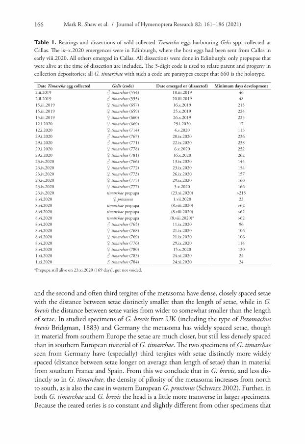
Table 1. Rearings and dissections of wild-collected Timarcha eggs harbouring Gelis spp. collected at Callas. The ix–x.2020 emergences were in Edinburgh, where the host eggs had been sent from Callas in early viii.2020. All others emerged in Callas. All dissections were done in Edinburgh: only prepupae that were alive at the time of dissection are included. The 3-digit code is used to relate parent and progeny in collection depositories; all G. timarchae with such a code are paratypes except that 660 is the holotype.
Date Timarcha egg collected Gelis (code) Date emerged or (dissected) Minimum days development2.ii.2019 ♂ timarchae (554) 18.iii.2019 462.ii.2019 ♂ timarchae (555) 20.iii.2019 4815.iii.2019 ♀ timarchae (657) 16.x.2019 21515.iii.2019 ♀ timarchae (659) 25.x.2019 22415.iii.2019 ♀ timarchae (660) 26.x.2019 22512.i.2020 ♀ timarchae (669) 29.i.2020 1712.i.2020 ♀ timarchae (714) 4.v.2020 11329.i.2020 ♂ timarchae (767) 20.ix.2020 23629.i.2020 ♂ timarchae (771) 22.ix.2020 23829.i.2020 ♀ timarchae (778) 6.x.2020 25229.i.2020 ♀ timarchae (781) 16.x.2020 26223.iv.2020 ♂ timarchae (766) 13.ix.2020 14423.iv.2020 ♀ timarchae (772) 23.ix.2020 15423.iv.2020 ♀ timarchae (773) 26.ix.2020 15723.iv.2020 ♀ timarchae (775) 29.ix.2020 16023.iv.2020 ♀ timarchae (777) 5.x.2020 16623.iv.2020 timarchae prepupa (23.xi.2020) >2158.vi.2020 ♀ proximus 1.vii.2020 238.vi.2020 timarchae prepupa (8.viii.2020) >628.vi.2020 timarchae prepupa (8.viii.2020) >628.vi.2020 timarchae prepupa (8.viii.2020)* >628.vi.2020 ♂ timarchae (765) 11.ix.2020 968.vi.2020 ♀ timarchae (768) 21.ix.2020 1068.vi.2020 ♀ timarchae (769) 21.ix.2020 1068.vi.2020 ♀ timarchae (776) 29.ix.2020 1148.vi.2020 ♀ timarchae (780) 15.x.2020 1301.xi.2020 ♂ timarchae (783) 24.xi.2020 241.xi.2020 ♂ timarchae (784) 24.xi.2020 24
*Prepupa still alive on 23.xi.2020 (169 days), gut not voided.

Biology of a new species of Gelis 167
we regard as G. timarchae, we choose to restrict the type series of G. timarchae to the material originating from T. nicaeensis eggs at Callas. Gelis fossae Schwarz, known only from north-western Africa, also resembles G. timarchae, but G. fossae has a deep furrow between the mesonotum and propodeum, and the hind tibia without setae dorsally. The males of G. brevis and G. fossae are unknown, but the male of Gelis rotundiventris (Först-er) resembles that of G. timarchae in many characters (e.g. propodeum evenly sloping from anterior margin, long first metasomal segment, colour). However, G. rotundiven-tris has the mesoscutum with distinct notaulus, fore wing with 2m-cu nearly straight, propodeum medially distinctly granulate and matt, head behind the eyes weakly nar-rowed, and second and third tergites of metasoma with widely spaced setae.
Description. Female. Body length: 3.2–3.7 mm. Apterous.Head. Antenna moderately thick and with 21–23 segments; third segment (with-
out annellus) 2.3–2.7 times as long as wide and 0.8–0.9 times as long as fourth segment. Head mainly granulate and matt; face, frons and temple with very fine and scattered punctation. Clypeus strongly convex, only granulate above and with few distinct punc-tures placed more or less in a transverse row medially, lower part of clypeus smooth and lustrous. Clypeus with ventral margin depressed and evenly rounded, without tooth. Mandible with teeth of equal length. Malar space without a distinct furrow, but with a line of very fine granulation. Malar space 0.8–1.1 times as long as basal width of mandible. Genal carina joining hypostomal carina behind mandibular base. Head in dorsal view 1.9 times as wide as long. Head behind eyes short and strongly narrowed or sometimes moderately narrowed.
Mesosoma mainly granulate and weakly matt, but partly lustrous (e.g. meso-pleuron). Mesonotum more or less fused with pronotum, short and 0.5–0.6 times as long as wide, without distinct depression medially. Scutellum very short and not distinctly separated from mesoscutum. Metanotum absent dorsally. Mesonotum and propodeum dorsally with moderately spaced setae. Furrow between mesonotum and propodeum of normal size, not unusually deep. Mesosternum ventrally very short and much shorter than diameter of antenna. In profile upper margin of propodeum
Table 2. Rearings of Gelis timarchae sp. nov. from cultured 2 day-old Timarcha nicaeensis eggs parasitized under observation, except for 679 in which the age of the egg is consequently also unknown. All parent Gelis females were virgin. The 3-digit codes are used to relate parent and progeny in specimen depositories; all recorded adults are paratypes.
Ovipositing ♀ Date parasitized ♂ Emerged date (code) Date dissected = live prepupa Days since oviposition659 23.x.2019 – 23.xi.2019 (32)657 26.x.2019 – 8.viii.2020 (288)657 26.x.2019 – 8.viii.2020 (288)657 26.x.2019 22.ix.2020 (770) 333659 27.x.2019 16–26.xii.2019 (666) – 51–61659 27.x.2019 6.i.2020 (667) – 71659 27.x.2019 7.i.2020 (668) – 72660 1.xi.2019 29.iv.2020 (708) – 180669 3.ii.2020 12.iv.2020 (685) – 70669 6–18.ii.2020 15.iii.2020 (679) – 27–39
Mark R. Shaw et al. / Journal of Hymenoptera Research 82: 161–186 (2021)168
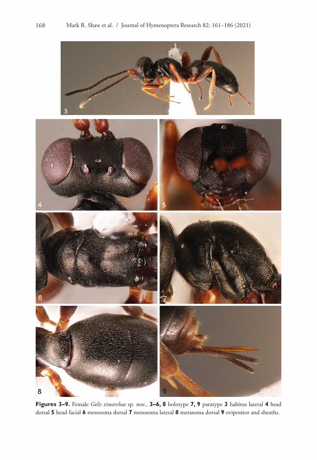
Figures 3–9. Female Gelis timarchae sp. nov., 3–6, 8 holotype 7, 9 paratype 3 habitus lateral 4 head dorsal 5 head facial 6 mesosoma dorsal 7 mesosoma lateral 8 metasoma dorsal 9 ovipositor and sheaths.

Biology of a new species of Gelis 169
Figures 10–14. 10–13 male Gelis timarchae sp. nov., paratype 10 habitus lateral 11 head dorsal 12 head facial 13 mesosoma lateral 14 non-paratype female G. timarchae, head dorsal (Germany).
Mark R. Shaw et al. / Journal of Hymenoptera Research 82: 161–186 (2021)170
not higher than upper margin of mesonotum. Propodeum with horizontal part short, about 0.5 times as long as sloping part. Propodeum sloping rather gently posteriorly, but degree of steepness somewhat variable. Posterior transverse carina present, but widely interrupted medially.
Hind femur 4.0–4.3 times as long as wide. Hind tibia somewhat widened and with densely placed setae dorsally.
Metasoma. First segment with dorso-lateral carina and ventro-lateral carina present, but rather weak. Second and often third tergites of metasoma with densely spaced setae, their distance distinctly smaller than length of setae on average. Laterotergite of second metasomal tergite moderately wide, about 3.0–3.5 times as long as wide. Ovipositor sheath 0.4–0.5 times as long as hind tibia. Ovipositor slender and straight, with nodus indistinct, its tip 3.8–4.0 times as long as wide and with very weak teeth ventrally.
Colour. Black. Orange-brown usually on scape partly or entirely, often pedicel, mandible except teeth, legs partly. Palpi, coxae partly, trochanters partly, fore and mid femora partly, hind femur, fore and mid tibiae often partly, hind tibia subbasally and apically brownish or blackish.
Variaton (non-type material). Body length 2.8–3.1 mm. Antenna with 20 seg-ments; third segment (without annellus) 2.0–2.2 times as long as wide. Head in dorsal
Figures 15–18. Female Gelis timarchae sp. nov., non-paratype specimens 15 mesosoma lateral (Ger-many) 16 metasoma (Germany) 17 mesosoma lateral (Spain) 18 metasoma (Spain).
Biology of a new species of Gelis 171
view 2.0 times as wide as long. Hind femur 3.0–3.5 times as long as wide. In German specimens distance between setae of third metasomal tergite longer than length of setae on average. Narrow caudal margins of first and second metasomal tergites orange-brown.
Male. Body length: 3.0–3.5 mm. Macropterous.Head. Antenna slender and with 24–27 segments; third segment (without an-
nellus) 3.9–4.4 times as long as wide and 1.0–1.2 times as long as fourth segment; segments 11–12 with tyloids. Head mainly granulate and matt; face, frons and temple
Figures 19–24. Female Gelis brevis (23, 24 tentatively determined) 19 mesosoma lateral (UK) 20 meta-soma (UK) 21 mesosoma lateral (Spain, Picos de Europa) 22 metasoma (Spain, Picos de Europa) 23 meso-soma lateral (Mallorca) 24 metasoma (Mallorca).
Mark R. Shaw et al. / Journal of Hymenoptera Research 82: 161–186 (2021)172
with very fine and scattered punctation or without distinct punctation. Clypeus strongly convex, only granulate above and with few distinct punctures placed more or less in a transverse row medially, lower part of clypeus smooth and lustrous. Clypeus with ventral margin depressed and evenly rounded, without tooth. Mandible with teeth of equal length. Malar space without a distinct furrow, but with a line of very fine granulation. Malar space 0.4–0.5 times as long as basal width of mandible. Genal carina joining hypostomal carina behind mandibular base. Ocelli large. Distance be-tween eye and lateral ocellus (OOL) 0.7–0.9 times diameter of lateral ocellus. Head behind eyes moderately short and moderately narrowed.
Mesosoma. Mesoscutum with fine granulation and matt, with very fine and hardly recognisable punctation and with densely spaced setae. Mesopleuron weakly granulate and lustrous with scattered punctation. Metapleuron granulate and matt. Propodeum evenly sloping from anterior margin. Propodeum mainly granulate and matt and partly with rugosity (mainly anteriorly); area superomedia and area postica lustrous and with shallow sculpture. Pleural carina, posterior transverse carina laterally and lateromedian longitudinal carina distinct, but the latter usually very fine anteriorly; other carinae of propodeum absent.
Hind femur 5.1–5.8 times as long as wide. Hind tibia weakly widened.Fore wing with pterostigma large and triangular. Marginal cell with RS beyond
areolet nearly straight but distally weakly bent. Areolet open with 3rs-m absent. 2m-cu rather long, distinctly sinuate and with two widely separated bullae.
Metasoma with first segment rather long and slender, dorso-lateral and ventro-lateral carinae present but rather weak, latero-median carina short and present close to spiracle. Second and third tergites of metasoma with densely spaced setae.
Colour. Black. Tegula often brown or orange brown. Orange brown are sometimes postpetiole posteriorly, second tergite of metasoma usually entirely or more rarely only partly, third tergite partly or more rarely entirely, rarely fourth tergite anteriorly, some-times coxae partly, trochantelli partly, trochanters entirely or partly, femora partly or entirely, fore and mid tibiae entirely or partly, hind tibia partly, fore and mid tarsi often partly. Hind tibia narrowly black basally, often brown subbasally and apically. Tarsi mainly brown. Fore wing with pterostigma blackish and only narrowly white basally. Mandible partly reddish. Palpi varying from mainly orange brown to blackish.
Etymology. The name refers to the host genus, meaning “of Timarcha”.
Natural rearings
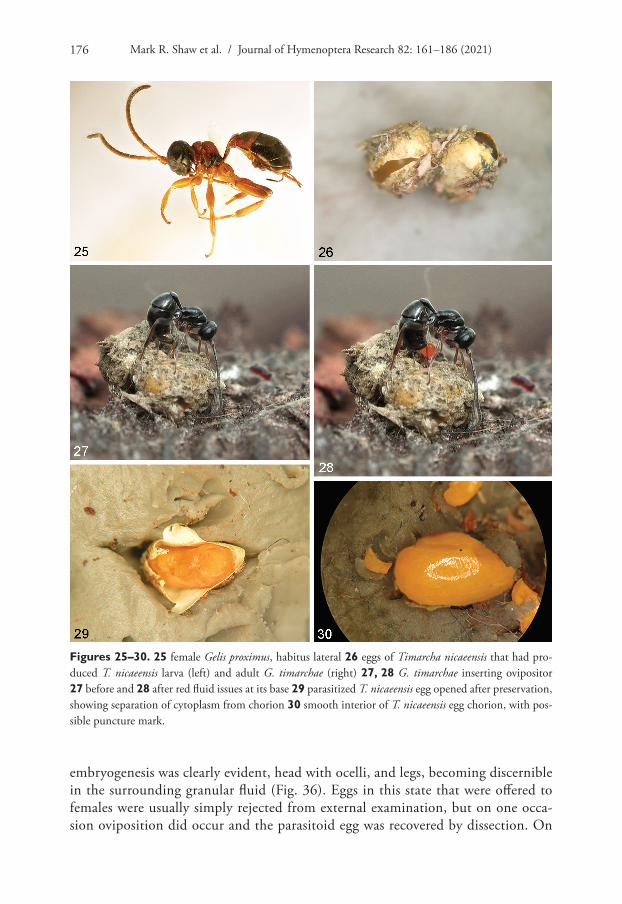
Table 1 records the individuals of Gelis species that were reared from wild-collected Timarcha eggs. While most were clearly G. timarchae sp. nov., a single female was obvi-ously different and proved to be a rather stout and slightly malformed specimen of Gelis proximus (Fig. 25). This specimen emerged on 1 July from an egg collected on 8 June 2020 that had not been found on 23 April, and is clearly not in phenological alignment with the others; it was evidently a pseudohyperparasitoid, as is further discussed below.
Several of the Gelis timarchae emerged from small batches of eggs from which at least one Timarcha larva also hatched, showing that (in the wild, as in our experiments
Biology of a new species of Gelis 173
below) not all eggs in a batch were necessarily parasitized when found by a female G. timarchae. First instar Timarcha larvae hatch through a slit in the chorion, whereas G. timarchae adults chew a roughly circular hole in the host egg, making it easy to as-sess the past history of most empty eggs (Fig. 26).
Many other eggs were collected at various times, some well camouflaged on the bark (Fig. 1); others more easily seen on exposed smooth wood (Fig. 2). A few had already produced a parasitoid (presumably G. timarchae); some had already produced Timarcha larvae, or subsequently did so; and many collected in late spring 2020 were destined to produce G. timarchae adults in the autumn (see later). Because of the way we dealt with the eggs (dissecting some), and especially the unstructured and variable collecting intensity (also, potentially before key events had happened) as well as there being a small proportion of dead or empty host eggs whose fate was not easy to score, it is not possible to present results quantitatively or to give a reliable estimate of percent-age parasitism, either overall or for particular generations. However, a small proportion (about 8%) of eggs had or subsequently shrivelled up without producing anything; although it is possible that some had been predated, the same outcome was seen in a similar proportion (more in some batches) of eggs laid in captivity. From around 75 eggs collected from the wild overall, about 40% had been parasitized (we believe ex-clusively by G. timarchae), which is a minimum estimate of the sampled population’s fate had we not intervened. The approximately 40% that produced Timarcha larvae is a maximum estimate as some might have become parasitized if they had been left in situ. It is not known whether T. nicaeensis oviposits in other situations; if it does so it may suffer a different rate of parasitism there.
Experimental exposures and rearings
In the first set of experiments (in late 2019 and early 2020) eggs obtained in captivity from T. nicaeensis were offered to females of G. timarchae under two regimes: (i) full observation for the entire period of interaction, and (ii) without continuous observa-tion. Somewhat older host eggs oviposited into (by mated females) in the second set of experiments (autumn 2020) were not kept for rearing, but were dissected at various times to assess development (see later).(i) Full observation, autumn 2019All the females used in these oviposition experiments were virgin, and the all-male experimental progeny demonstrated that this particular Gelis is a normal haplodiploid. The outcomes, including phenology, of the experimental rearings in which oviposition was under full observation are recorded in Table 2.(ii) Not observed for the full period of exposure, spring 2020Twenty-five T. nicaeensis eggs laid in captivity between 3–15 February 2020 were ex-posed to a virgin female G. timarchae from 6–18 February. During March 2020 14 host larvae hatched and a single male of G. timarchae emerged on 15 March 2020, the remaining eggs eventually shrivelling up. Although the date of oviposition is not known, the development time to adult parasitoid emergence was not greater than 39 days, shorter than for any other experimental rearing (Table 2).
Mark R. Shaw et al. / Journal of Hymenoptera Research 82: 161–186 (2021)174
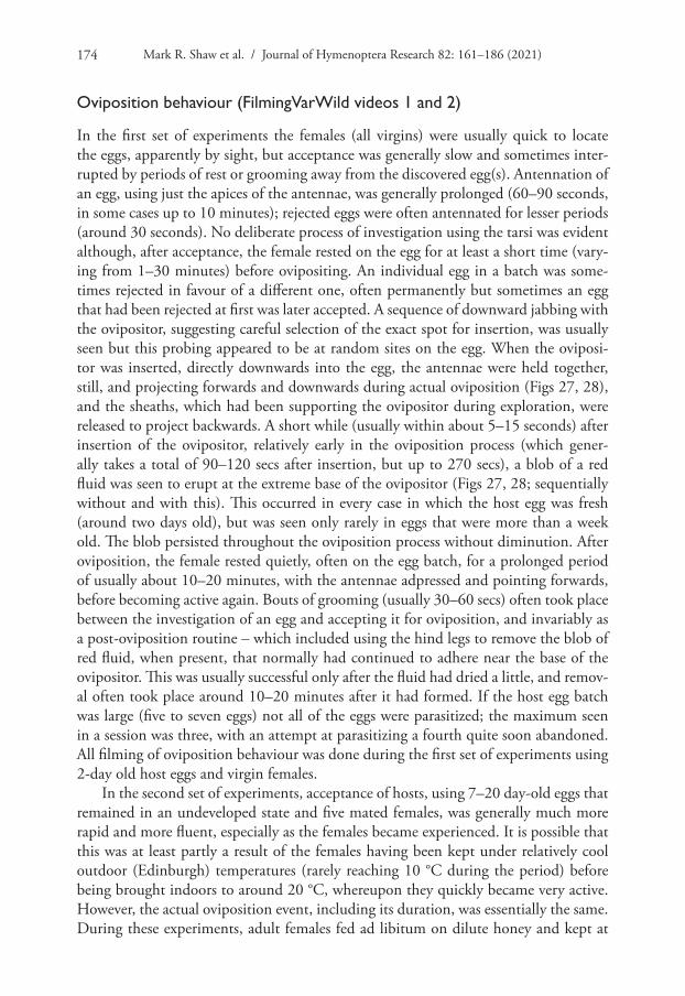
Oviposition behaviour (FilmingVarWild videos 1 and 2)
In the first set of experiments the females (all virgins) were usually quick to locate the eggs, apparently by sight, but acceptance was generally slow and sometimes inter-rupted by periods of rest or grooming away from the discovered egg(s). Antennation of an egg, using just the apices of the antennae, was generally prolonged (60–90 seconds, in some cases up to 10 minutes); rejected eggs were often antennated for lesser periods (around 30 seconds). No deliberate process of investigation using the tarsi was evident although, after acceptance, the female rested on the egg for at least a short time (vary-ing from 1–30 minutes) before ovipositing. An individual egg in a batch was some-times rejected in favour of a different one, often permanently but sometimes an egg that had been rejected at first was later accepted. A sequence of downward jabbing with the ovipositor, suggesting careful selection of the exact spot for insertion, was usually seen but this probing appeared to be at random sites on the egg. When the oviposi-tor was inserted, directly downwards into the egg, the antennae were held together, still, and projecting forwards and downwards during actual oviposition (Figs 27, 28), and the sheaths, which had been supporting the ovipositor during exploration, were released to project backwards. A short while (usually within about 5–15 seconds) after insertion of the ovipositor, relatively early in the oviposition process (which gener-ally takes a total of 90–120 secs after insertion, but up to 270 secs), a blob of a red fluid was seen to erupt at the extreme base of the ovipositor (Figs 27, 28; sequentially without and with this). This occurred in every case in which the host egg was fresh (around two days old), but was seen only rarely in eggs that were more than a week old. The blob persisted throughout the oviposition process without diminution. After oviposition, the female rested quietly, often on the egg batch, for a prolonged period of usually about 10–20 minutes, with the antennae adpressed and pointing forwards, before becoming active again. Bouts of grooming (usually 30–60 secs) often took place between the investigation of an egg and accepting it for oviposition, and invariably as a post-oviposition routine – which included using the hind legs to remove the blob of red fluid, when present, that normally had continued to adhere near the base of the ovipositor. This was usually successful only after the fluid had dried a little, and remov-al often took place around 10–20 minutes after it had formed. If the host egg batch was large (five to seven eggs) not all of the eggs were parasitized; the maximum seen in a session was three, with an attempt at parasitizing a fourth quite soon abandoned. All filming of oviposition behaviour was done during the first set of experiments using 2-day old host eggs and virgin females.
In the second set of experiments, acceptance of hosts, using 7–20 day-old eggs that remained in an undeveloped state and five mated females, was generally much more rapid and more fluent, especially as the females became experienced. It is possible that this was at least partly a result of the females having been kept under relatively cool outdoor (Edinburgh) temperatures (rarely reaching 10 °C during the period) before being brought indoors to around 20 °C, whereupon they quickly became very active. However, the actual oviposition event, including its duration, was essentially the same. During these experiments, adult females fed ad libitum on dilute honey and kept at

Biology of a new species of Gelis 175
outdoor temperatures lived for at least 60 and up to 72 days, and were capable of ovi-position until a day or two before their death. As in the first set of experiments, host-feeding was never observed.
One female was offered a batch of four Timarcha eggs in early July 2020, about 4 months after they had been laid (early March). The female investigated the eggs for 1–2 minutes, returning to them once but not resting on them, before abandoning them without inserting her ovipositor. The eggs were subsequently found to contain dead Timarcha larvae, and may have been dead at the time of being offered.
Egg placement
The three preserved T. nicaeensis eggs that had received a parasitoid egg during the first set of experiments were opened several weeks later. The chorion had completely sepa-rated from the solidified content, and there was no sign of a G. timarchae egg present in the space between the host egg chorion and any inner membrane presumed to be present surrounding its content (Fig. 29). The absence of the parasitoid egg in that space was a definite conclusion in all three cases, aided by the extremely smooth inner surface of the chorion with the presumed oviposition site sometimes evident as a small dark spot (Fig. 30), as well as the discretely separated egg content.
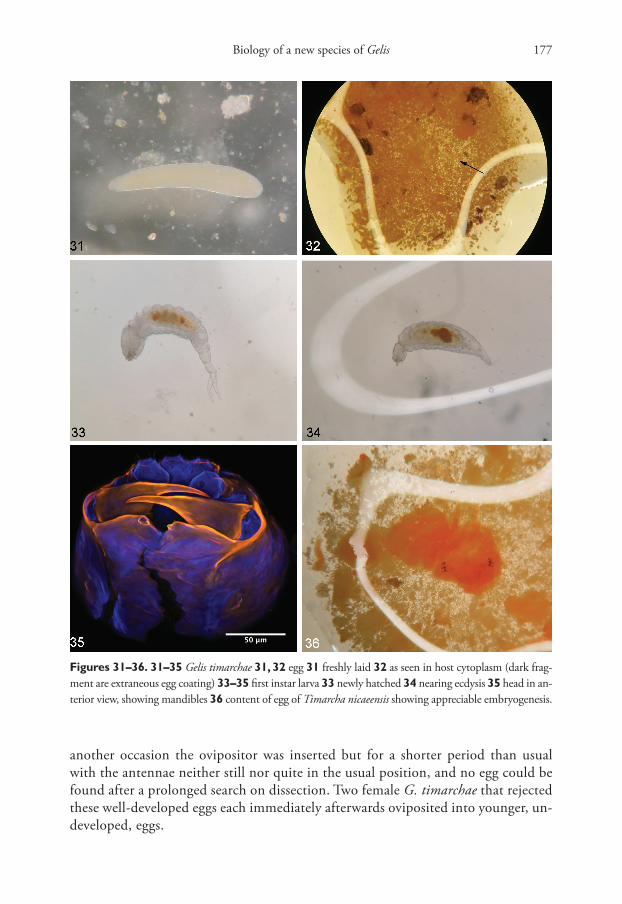
Subsequently, dissection of host eggs immediately after being parasitized during the second set of experiments strongly supported the conclusion of genuine endopara-sitism, as the surprisingly large translucent egg (Fig. 31), measuring about 0.8–0.9 × 0.16 mm, was always apparently entirely within the opaque orange-red cytoplasm, where it was sometimes difficult to find (Fig. 32). There was no evident selection of any particular site on the host egg for insertion of the ovipositor.
First instar larva
Dissections of parasitised host eggs during the second set of experiments (Autumn 2020) revealed that by seven days after oviposition the larva had hatched. The first instar larva, found free in the cytoplasm, has a remarkable form with a bifurcate caudal appendage that is especially prominent when freshly hatched (Fig. 33), though by the time ecdysis approaches this tail is much reduced (Fig. 34). The bifurcate appendage was observed in all cases, from three separate female parents. Newly hatched, the larva is about 1.1–1.3 mm long including its tail, and has the long sharp mandibles typical of many first instar larvae of endoparasitoid Ichneu-monidae (Fig. 35).
Suitable developmental stage of the host
Unparasitized host eggs were also dissected during the second experimental period, and it was found that virtually no embryogenesis took place for at least a month under outdoor (Edinburgh) temperatures, but eggs brought indoors after a cou-ple of weeks did start to develop such that after a further 2 weeks at ca 18–22 °C
Mark R. Shaw et al. / Journal of Hymenoptera Research 82: 161–186 (2021)176
embryogenesis was clearly evident, head with ocelli, and legs, becoming discernible in the surrounding granular fluid (Fig. 36). Eggs in this state that were offered to females were usually simply rejected from external examination, but on one occa-sion oviposition did occur and the parasitoid egg was recovered by dissection. On
Figures 25–30. 25 female Gelis proximus, habitus lateral 26 eggs of Timarcha nicaeensis that had pro-duced T. nicaeensis larva (left) and adult G. timarchae (right) 27, 28 G. timarchae inserting ovipositor 27 before and 28 after red fluid issues at its base 29 parasitized T. nicaeensis egg opened after preservation, showing separation of cytoplasm from chorion 30 smooth interior of T. nicaeensis egg chorion, with pos-sible puncture mark.
Biology of a new species of Gelis 177
another occasion the ovipositor was inserted but for a shorter period than usual with the antennae neither still nor quite in the usual position, and no egg could be found after a prolonged search on dissection. Two female G. timarchae that rejected these well-developed eggs each immediately afterwards oviposited into younger, un-developed, eggs.
Figures 31–36. 31–35 Gelis timarchae 31, 32 egg 31 freshly laid 32 as seen in host cytoplasm (dark frag-ment are extraneous egg coating) 33–35 first instar larva 33 newly hatched 34 nearing ecdysis 35 head in an-terior view, showing mandibles 36 content of egg of Timarcha nicaeensis showing appreciable embryogenesis.
Mark R. Shaw et al. / Journal of Hymenoptera Research 82: 161–186 (2021)178
Unfortunately the G. timarchae females available when there were developing eggs to hand were nearing the end of their lives (which sometimes leads to erratic behaviour in parasitioids, MRS pers. obs.), and there was simply insufficient living material to investigate more thoroughly the level of host larval development that renders the eggs unsuitable. However, we have not found developed (sclerotised) host remains in any of the dissected host eggs containing G. timarchae cocoons (N = ca 15) where such remains would be expected, i.e. between the host egg chorion and the G. timarchae cocoon, which strongly suggests that the period of host suitability is restricted to the time during which the egg content is essentially cytoplasm. In fact, this space between the G. timarchae cocoon and the host egg’s shell was always found to be very clean.
Autumn development of G. timarchae larvae
It is clear from Table 1 that there is no simple phenology in the development of G. timarchae, and several dissections of experimentally parasitized host eggs were un-dertaken to try to elucidate this.
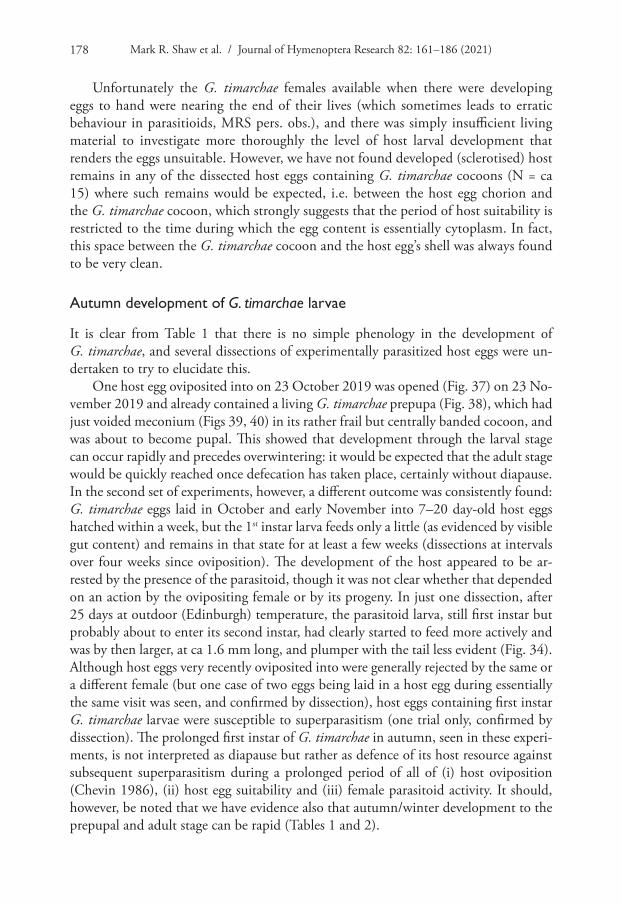
One host egg oviposited into on 23 October 2019 was opened (Fig. 37) on 23 No-vember 2019 and already contained a living G. timarchae prepupa (Fig. 38), which had just voided meconium (Figs 39, 40) in its rather frail but centrally banded cocoon, and was about to become pupal. This showed that development through the larval stage can occur rapidly and precedes overwintering: it would be expected that the adult stage would be quickly reached once defecation has taken place, certainly without diapause. In the second set of experiments, however, a different outcome was consistently found: G. timarchae eggs laid in October and early November into 7–20 day-old host eggs hatched within a week, but the 1st instar larva feeds only a little (as evidenced by visible gut content) and remains in that state for at least a few weeks (dissections at intervals over four weeks since oviposition). The development of the host appeared to be ar-rested by the presence of the parasitoid, though it was not clear whether that depended on an action by the ovipositing female or by its progeny. In just one dissection, after 25 days at outdoor (Edinburgh) temperature, the parasitoid larva, still first instar but probably about to enter its second instar, had clearly started to feed more actively and was by then larger, at ca 1.6 mm long, and plumper with the tail less evident (Fig. 34). Although host eggs very recently oviposited into were generally rejected by the same or a different female (but one case of two eggs being laid in a host egg during essentially the same visit was seen, and confirmed by dissection), host eggs containing first instar G. timarchae larvae were susceptible to superparasitism (one trial only, confirmed by dissection). The prolonged first instar of G. timarchae in autumn, seen in these experi-ments, is not interpreted as diapause but rather as defence of its host resource against subsequent superparasitism during a prolonged period of all of (i) host oviposition (Chevin 1986), (ii) host egg suitability and (iii) female parasitoid activity. It should, however, be noted that we have evidence also that autumn/winter development to the prepupal and adult stage can be rapid (Tables 1 and 2).

Biology of a new species of Gelis 179
Summer diapause
Although there is clear evidence of rapid development to the adult stage following oviposition into host eggs early in the year (Tables 1 and 2), dissections of host eggs collected in late spring 2020 that had not quickly produced adult parasitoids showed that in mid-August many contained living prepupae of G. timarchae in their cocoons, with the gut not yet voided. These were clearly in diapause, and many other such eggs collected in spring or early summer produced adults of G. timarchae in the autumn (Table 1). In all of these host eggs that were opened (N = 4), as well as in various others from which G. timarchae had emerged, the Gelis cocoon within the Timarcha egg was unequivocally single-layered (Figs 37, 39).
Eclosion of male (FilmingVarWild video 3)
We witnessed the eclosion of a male G. timarchae from one of the eggs parasitized in culture and were struck by the length of time it took (several hours) to unfurl and successfully clean its wings. Clearly they were saturated, to an extent abnormal in the emergence of most ichneumonids, which might have been because the moisture content of the environment in which it developed was high – with no easy way for
Figures 37–40. Gelis timarchae 37, 38 prepupa 37 in opened cocoon 38 facial view 39, 40 cocoon of with freshly voided meconial pellets 39 from side, also showing central band of more densely spun silk 40 from posterior end.
Mark R. Shaw et al. / Journal of Hymenoptera Research 82: 161–186 (2021)180
moisture to be lost – and it brought to mind the unusual emergence of Trichogramma gicai Pintureau & Stefanescu from eggs of the papilionid butterfly Papilio machaon Linnaeus filmed by BKvLS and PK, in which there is no attempt to expand wings until some time after eclosion (FilmingVarWild video 4).
The Gelis proximus rearing
The common and widespread species Gelis proximus has a very broad host range com-prising various small cocoons and cocoon-like structures in low vegetation (Schwarz and Shaw 1999), and evidently does not specialise on Timarcha eggs. When the host egg from which it had emerged was carefully opened it was found to contain a cocoon with meconium (clearly that of the parasitoid that emerged) within another, similarly constructed, single-envelope cocoon that we believe had been made by G. timarchae and, to judge from the lack of a clear band, to have been parasitized in the process of construction. Thus we conclude that G. proximus was simply behaving as a pseudo-hyperparasitoid, as it frequently does (Schwarz and Shaw 1999; see also Salt 1952 as G. corruptor), and had adventitiously parasitized a cocoon of G. timarchae rather than acting as a primary parasitoid of the Timarcha egg.
Discussion
Parasitism of various kinds is well-documented for Timarcha spp. (Cox 1994; Jolivet and Poinar 2007), but there is no mention of egg parasitism, let alone any Gelis species reared from eggs. [As an aside, some corrections to taxa recorded by Jolivet and Poinar (2007) are needed: the species widely recorded as Perilitus falciger (Ruthe) as a parasitoid of Timarcha is probably a misidentification of P. foveolatus Reinhard, of which P. sicheli Giard is a synonym (Haeselbarth 1998)]. In the Natural History Museum, London (NHMUK) we have seen 2 ♀, 1 ♂ of a different species of Gelis, G. forticornis (Förster) (determined by MS in 2017), labelled as reared from Timarcha, but this species has a relatively robust ovipositor, unsuited for oviposition into an egg. Despite lack of detail in the labelling and the absence of host remains, a simple interpretation is that they had been adventitious pseudohyperparasitoids, perhaps of gregarious Perilitus (Braconidae: Euphorinae) cocoons, in the fashion normal for many Gelis species (Schwarz and Shaw 1999). However, Schwarz (1998), and Schöller (1999) based on the same material, re-cord G. forticornis as a parasitoid of the chrysomelid Cryptocephalus moraei (Linnaeus) and, although no host stage was given, the records taken together may suggest a possible specialisation on that family, whether as a primary or secondary parasitoid.
First record of endoparasitism of insect eggs by an ichneumonid
We have not found a published account of any ichneumonid species of any subfamily developing to the adult stage as an endoparasitoid fully within an insect egg, and it
Biology of a new species of Gelis 181
is remarkable behaviour for a Gelis species in particular, as indeed is the use of a host not in the least associated with silk. Egg placement by G. timarchae in the host egg is evidently, from dissections as well as from the depth and vertical direction of insertion of the ovipositor, within the cytoplasm, rather than in the space between the chorion and the inner membrane.
Oviposition and development
Oviposition sequences for Gelis species appear not to have been described in much detail but, as the majority of species are either effectively predators of successive spider eggs in an egg sac or external parasitoids within cases or cocoons made by holometabol-ous insects (Schwarz and Shaw 1999), it is likely that the oviposition process in most Gelis species would be much less specialised than that recorded here. Certainly G. timar-chae is one of few species possessing a very fine sharp ovipositor, and it seems possible that other species with this feature (e.g. G. brevis and G. rotundiventris) might also turn out to be internal parasitoids of (perhaps fresh) insect eggs once their biology is known.
Primary parasitism of Timarcha species is practised by specialist parasitoids, and this is probably at least to some extent connected with the toxicity, due to anthraquinones, present in the red haemolymph fluid familiarly seen in the “reflex bleeding” of the adult beetles when disturbed (Jolivet and Verma 2002). The same colour of fluid is seen aris-ing at the base of the ovipositor of G. timarchae, evidently via a voluntary opening at the base of the ovipositor to the exterior, at an early stage of the oviposition sequence into a fresh Timarcha egg (Fig. 28). The whole sequence involves only a single insertion of the ovipositor and we interpret the appearance of fluid as a release of hydrostatic pres-sure present in the host egg, that would prevent the passage of the parasitoid egg down the egg canal until the pressure is released. This mechanism presumably allows closer control of the amount of fluid released than would arise if the ovipositor was simply in-serted and then withdrawn in a pre-oviposition step, and the unusually slender oviposi-tor of G. timarchae is a clear specialisation for such controlled egg-piercing behaviour. The release of fluid pressure seen during every oviposition into very young eggs strongly supports the conclusion that the egg is placed in the cytoplasm, rather than in the space between the chorion and membrane surrounding it. In rather older eggs (but still undeveloped and containing only cytoplasm) there is less tendency for this fluid to be released, presumably because the pressure within the egg is by then lower. Interestingly, the freshly voided meconial pellets (Figs 39, 40) are of the same bright reddish colour.
Egg and egg-limitation
Interestingly, in view of the clear specialisations seen in the first instar larva, the egg seems to be unspecialised (e.g. lacking any marked size-reduction). Thus the large egg of G. timarchae (0.8–0.9 mm long) is similar in shape and size to that of the relative-ly unspecialised ectoparasitoid G. agilis (“approaching 1 mm”, Harvey 2008), which has a comparable though sometimes slightly larger adult body length of 1.9–4.4 mm
Mark R. Shaw et al. / Journal of Hymenoptera Research 82: 161–186 (2021)182
(Schwarz 1998). In the present case the female Gelis adult is 3.2–3.7 mm long and, although G. timarchae was clearly a major cause of mortality in the population of Timarcha at the study site, it is hardly surprising to see strong evidence of egg-limita-tion. Although no doubt temperature-dependent this would probably amount to no more than an average of around four ovipositions per female per day (cf. Muesebeck and Dohanian 1927; Harvey et al. 2019). Thus there were a few instances in which a functional-age and well-fed female G. timarchae failed to oviposit into a 2-day old host egg under experimental circumstances that otherwise seemed suitable and, because we also found small egg batches in the wild from which both Timarcha larvae and G. timarchae emerged, this clearly happens in the field as well. Egg-limited oviposition behaviour is not unusual for a wide range of synovigenic species laying relatively large anhydropic eggs (Jervis and Kidd 1986; MRS pers. obs.), and can also be deduced from field data in several of the relatively unspecialised species of Gelis when they encounter aggregated resources such as cocoon batches of gregarious microgastrine braconids (Schwarz and Shaw 1999; see also Harvey 2008; Harvey et al. 2019).
Apparent lack of host feeding
We observed about 8% failure to produce anything, host larva or parasitoid, in field-collected intact Timarcha eggs, which might indicate that some eggs had been predat-ed, possibly through host-feeding by G. timarchae, before collection. As far as is known Gelis species are broadly synovigenic (that is, the females develop eggs successively dur-ing the adult lifespan). At least in the case of species parasitizing holometabolous in-sects, they generally host-feed (Muesebeck and Dohanian 1927; Harvey 2008), taking protein/lipid meals to aid egg maturation, typically (though probably not exclusively) from a proportion of the normal hosts that are killed but not then parasitized, by imbibing fluid issuing from puncture wounds made by the ovipositor following a stab and withdraw process. As it is obvious that the developing larva of G. timarchae must have a sufficient means of coping with the anthraquinones that are presumably present in the host eggs (Jolivet and Verma 2002), it seems likely that the adult females would be similarly endowed and so be able to host-feed. Despite their continuous access to dilute honey, and the small number of their ovipositions, it seems remarkable that in our experiments no such host-feeding by G. timarchae was observed. Our observations included females that lived for up to 72 days in captivity, with access only to dilute honey until being periodically offered host eggs under full observation, into which they were able to oviposit until almost the last day of their lives. It would be interesting to know whether, and if so how, they would source proteins and lipids in the wild.
First instar larva
The caudate form of the first instar larva of G. timarchae is surprising for a normally ectoparasitoid group and, together with its elongate sharp mandibles, strongly suggests that the larva in this case is specifically adapted for an endoparasitoid life, able to move
Biology of a new species of Gelis 183
effectively in its liquid medium to attack any other parasitoid that may subsequently arrive. The length of time that it (sometimes) spends in that instar is a further indica-tion of adaptation for fighting to hold ownership; probably important in view of the apparent willingness of G. timarchae to superparasitize host eggs, and the long life of the adult females. The bifurcate nature of the caudal appendage was a further surprise and we do not know of similar cases in any other ichneumonid (notwithstanding the clearly different paired structures seen in Agriotypus, illustrated by Clausen (1931)). Unfortunately we were unable to find descriptions or illustrations of first instar larvae of ectoparasitoid Gelis species in the literature apart from that of Gelis tenellus (Say), which lacks a caudal appendage of any kind (Clancy 1946). It also has relatively small mandibles, possibly reflecting no need to defend its by then paralysed and hence unat-tractive host from later-arriving competitors.
Phenology
It is clear that there are two main periods in which adults of G. timarchae are present in the field; from very early in spring into early summer, and again in the autumn into early winter. This broadly corresponds to the times that fresh host eggs are available; at our study site this seemed to be approximately from the end of October through to early December, then again from late January to at least May – roughly but not exactly as depicted by Chevin (1986). Further, it is evident that the autumn-laid (and pre-sumably also early spring-laid) host eggs develop only slowly, at least in cold weather, in addition to being laid over a long time in each of the two oviposition periods. In response, G. timarchae is predominantly bivoltine. In the spring/summer generation it seems to develop rapidly to the prepupal stage which then diapauses within the host egg leading to adult emergence in autumn. In the subsequent generation resulting from autumn ovipositions the parasitoid at first remains as a first instar larva in order to defend its position, then probably develops slowly (once oviposition activity by female parasitoids has subsided), perhaps without diapause (but see below), to emerge early in spring when fresh host eggs are again available. No tendency for adult females to overwinter was seen.
However, that rather simple bivoltine pattern of oviposition in spring leading to adults in autumn, and ovipositions by them in the late part of the year producing the spring adults, is evidently only a part of the reality because, in both of the main ovi-position periods, some individuals develop rapidly to become adults (then presumably ovipositing) during the same period of host availability (Tables 1 and 2). Thus up to four generations could arise, albeit partial ones. An opposite trend also seen from our data is delayed development, with a normal emergence period being skipped leading to essentially univoltine behaviour (see long development times of progeny of female 657 recorded in Table 2 in particular: in these cases diapause clearly started during winter or spring, before the commonly seen summer diapause period). Facultative prolongation of this nature is rather unusual in temperate parasitoids and, when seen on a regular basis, it is usually ascribed to an uncertain occurrence of hosts in a harsh
Mark R. Shaw et al. / Journal of Hymenoptera Research 82: 161–186 (2021)184
or unpredictable environment (e.g. Shaw and Wahl 1989; Askew and Shaw 2005). The unusual and protracted oviposition by the host, arguably itself a bet-hedging strategy, may be the stimulus in the present case.
Our experiments and rearings, undertaken under rather ill-controlled environ-mental conditions, show what can happen regarding phenology, but more work would need to be carried out to elucidate any environmental controls or perhaps genetic influences underlying our observations.
Acknowledgements
We are grateful to István Mikó for preparing Fig. 35 at the University of New Hamp-shire Instrumentation Center, to Geraldine Groussier for obtaining a CO1 sequence of G. timarchae at INRAE Institut Sophia Agrobiotech, to Karin Traxler for assem-bling the plates, to Richard Lyskowski for determining specimens of Timarcha, and to Gavin Broad for drawing our attention to the specimens of Gelis forticornis in NHMUK. Gavin Broad, Seraina Klopfstein and Ika Österblad made helpful sugges-tions in review.
References
Askew RR, Shaw MR (2005) Observations on the biology of Baryscapus (Hymenoptera: Eu-lophidae: Tetrastichinae) with description of a new koinobiont hyperparasitoid with de-layed development. Acta Societas Zoologicae Bohemicae 69: 11–14.
Chevin H (1985) Contribution à la biologie des Timarcha (Col. Chrysomelidae). Cahiers Lia-sion OPIE 19(2) 57: 7–14.
Chevin H (1986) Contribution à la biologie des Timarcha (Col. Chrysomelidae): Timarcha nicaeensis Villa. Cahiers Liasion OPIE 20(1) 60: 17–21.
Chevin H (1994) Food selection and life-cycle of the Old World Timarcha Latreille, 1829 (Col. Chrysomelinae). In: Jolivet PH, Cox ML, Petitpierre E (Eds) Novel aspects of the biology of Chrysomelidae. Kluwer Academic Publishers, Dordrecht, 533–539. https://doi.org/10.1007/978-94-011-1781-4_40
Clancy DW (1946) The insect parasitoids of the Chrysopidae (Neuroptera). University of Cali-fornia Publications in Entomology 7: 403–496.
Clausen CP (1931) Biological observations on Agriotypus (Hymenoptera). Proceedings of the Entomological Society of Washington 33: 29–37.
Cox ML (1994) The Hymenoptera and Diptera parasitoids of Chrysomelidae. In: Jolivet PH, Cox ML, Petitpierre E (Eds) Novel aspects of the biology of Chrysomelidae. Klu-wer Academic Publishers, Dordrecht, 419–467. https://doi.org/10.1007/978-94-011-1781-4_35
Fauna Europaea (2020) Fauna Europaea. www.fauna-eu.org [consulted 12.iv.2020]
Biology of a new species of Gelis 185
FilmingVarWild video 1 (2020) B. Kan-van Limburg Stirum & P. Kan, Gelis timarchae (Phygadeuontinae) parasitizes a Timarcha nicaeensis egg. https://www.youtube.com/watch?v=iz3PyEBH0zc&ab_channel=FilmingVarWild
FilmingVarWild video 2 (2020) B. Kan-van Limburg Stirum & P. Kan, Gelis timarchae (Phygadeuontinae) oviposits in two Timarcha nicaeensis eggs. https://www.youtube.com/watch?v=8AVSPFkukBk&ab_channel=FilmingVarWild
FilmingVarWild video 3 (2020) B. Kan-van Limburg Stirum & P. Kan, Gelis timarchae (Phyga-deuontinae) male emerges from parasitized Timarcha nicaeensis egg. https://www.youtube.com/watch?v=Gra7TqXLXuk&ab_channel=FilmingVarWild
FilmingVarWild video 4 (2015) B. Kan-van Limburg Stirum & P. Kan, Trichogrammati-dae: Trichogramma gicai parasitizes eggs of Papilio machaon. https://www.youtube.com/watch?v=GdBKDTYR57o&ab_channel=FilmingVarWild
Haeselbarth E (1998) Zur Braconiden-Gattung Perilitus Nees, 1818 1. Beitrag: Die Perilitus falciger-Gruppe (Hymenoptera, Braconidae). Entomofauna 19: 197–208.
Harvey JA (2008) Comparing and contrasting development and reproductive strategies in the pupal hyperparasitoids Lysibia nana and Gelis agilis (Hymenoptera: Ichneumonidae). Evolutionary Ecology 22: 153–166. https://doi.org/10.1007/s10682-007-9164-x
Harvey JA, de Haan L, Verdeny-Vilata O, Visser B, Gols R (2019) Reproduction and offspring sex ratios differ markedly among closely related hyperparasitoids living in the same microhabi-tats. Journal of Insect Behaviour 32: 243–251. https://doi.org/10.1007/s10905-019-09730-z
Horstmann K (1986) Die westpaläarktischen Arten der Gattung Gelis Thunberg, 1827, mit macropteren oder brachypteren Weibchen (Hymenoptera, Ichneumonidae). Entomofauna 7: 329–424.
Jervis MA, Kidd NAC (1986) Host-feeding strategies in hymenopteran parasitoids. Biological Reviews 61: 395–434. https://doi.org/10.1111/j.1469-185X.1986.tb00660.x
Jolivet P, Poinar G (2007) Parasites, commensals and phoretics of Timarcha (Coleoptera: Chrysomelidae). Genus 18: 589–596.
Jolivet P, Verma KK (2002) Biology of leaf beetles. Intercept Publishers, Andover, 332 pp.Muesebeck CFW, Dohanian SM (1927) A study in hyperparasitism, with particular reference
to the parasites of Apanteles melanoscelus (Ratzeburg). United States Department of Agri-culture Bulletin 1487: 1–36. https://doi.org/10.5962/bhl.title.108942
Salt G (1952) Trimorphism in the ichneumonid parasite Gelis corruptor. Quarterly Journal of Microscopical Science 93: 453–474.
Schindelin J, Arganda-Carreras I, Frise E, Kaynig V, Longair M, Pietzsch T, Preibisch S, Rueden C, Saalfeld S, Schmid B, Tinevez J-Y, White DJ, Hartenstein V, Eliceiri K, Tomancak P, Cardona A (2012) FIJI: an open-source platform for biological-image analysis. Nature Methods 9(7): 676–682. https://doi.org/10.1038/nmeth.2019
Schöller M (1999) Field studies of Cryptocephalinae biology. Advances in Chrysomelidae Biology 1: 421–436.
Schwarz M (1995) Revision der westpaläarktischen Arten der Gattungen Gelis Thunberg mit apteren Weibchen und Thaumatogelis Schmiedeknecht (Hymenoptera, Ichneumonidae). Teil 1. Linzer biologische Beiträge 27: 5–105.
Mark R. Shaw et al. / Journal of Hymenoptera Research 82: 161–186 (2021)186
Schwarz M (1998) Revision der westpaläarktischen Arten der Gattungen Gelis Thunberg mit apteren Weibchen und Thaumatogelis Schmiedeknecht (Hymenoptera, Ichneumonidae). Teil 2. Linzer biologische Beiträge 30: 629–704.
Schwarz M (2002) Revision der westpaläarktischen Arten der Gattungen Gelis Thunberg mit apteren Weibchen und Thaumatogelis Schwarz (Hymenoptera, Ichneumonidae). Teil 3. Linzer biologische Beiträge 34: 1293–1392.
Schwarz M (2016) Die Schlupfwespengattung Gelis (Hymenoptera, Ichneumonidae, Cryp-tinae) mit macropteren Weibchen in der Westpaläarktis. Linzer biologische Beiträge 48: 1677–1752.
Schwarz M, Shaw MR (1999) Western Palaearctic Cryptinae (Hymenoptera: Ichneumonidae) in the National Museums of Scotland, with nomenclatural changes, taxonomic notes, rear-ing records and special reference to the British check list. Part 2. Genus Gelis Thunberg (Phygadeuontini: Gelina). Entomologist’s Gazette 50: 117–142.
Selman BJ (1994) Eggs and oviposition in chrysomelid beetles. In: Jolivet PH, Cox ML, Petit-pierre E (Eds) Novel aspects of the biology of Chrysomelidae. Kluwer Academic Publish-ers, Dordrecht, 69–74. https://doi.org/10.1007/978-94-011-1781-4_2
Shaw MR, Wahl DB (1989) The biology, egg and larvae of Acaenitus dubitator (Panzer) (Hyme-noptera, Ichneumonidae: Acaenitinae). Systematic Entomology 14: 117–125. https://doi.org/10.1111/j.1365-3113.1989.tb00269.x
UK Beetles (2020) UK Beetles. www.ukbeetles.co.uk [consulted 27.xii.2020]