Embed Size (px)
Citation preview

BIODIVERSITAS ISSN: 1412-033X
Volume 22, Number 2, February 2021 E-ISSN: 2085-4722
Pages: 874-880 DOI: 10.13057/biodiv/d220242
Short Communication:
Histopathology of red chilli fruit (Capsicum annuum) infected with
Colletotrichum acutatum of Java, Indonesia isolates
JUNI SAFITRI MULJOWATI1,♥, LOEKAS SOESANTO2, LAURENTIUS HARTANTO NUGROHO3 1Faculty of Biology, Universitas Jenderal Soedirman. Jl. Dr. Suparno No. 62, Purwokerto Utara, Banyumas 53122, Central Java, Indonesia.
Tel.: +62-281-638794, Fax.: +62-281-631700, email: [email protected] 2Faculty of Agricultural, Universitas Jenderal Soedirman. Jl. Dr. Suparno No. 63, Purwokerto Utara, Banyumas 53122, Central Java, Indonesia
3Faculty of Biology, Universitas Gadjah Mada. Jl. Teknika Selatan, Sekip Utara, Sleman 55281, Yogyakarta, Indonesia
Manuscript received: 29 November 2020. Revision accepted: 19 January 2021.
Abstract. Muljowati JS, Soesanto L, Nugroho LH. 2021. Short Communication: Histopathology of red chilli fruit (Capsicum annuum)
infected with Colletotrichum acutatum of Java, Indonesia isolates. Biodiversitas 22: 874-880. Colletotrichum acutatum isolates from
Malang, Temanggung, Kulonprogo, Brebes, Garut, and Pandeglang in Indonesia varied in their ability to produce pigments. In the
present study, the histopathological status of red chilli was investigated during the early phase of infection by C. acutatum Java isolate.
The results included a description of the histopathological features of red chillies (Capsicum annuum) in the early phase of infection by
C. acutatum isolates and the relationship between the origin of the isolates and the time of onset of infection. The red chilli fruits were
inoculated with fungal conidia suspension and then the histopathology of chilli fruits was observed at 0 hours (uninfected control), 8
hours, 16 hours, 24 hours, and 32 hours after inoculation. The results showed that C. acutatum isolates from Kulonprogo caused host
damage within 8 hours, while the other isolates (from Malang, Brebes, Garut and Pandeglang) began to produce similar symptoms at 16
hours or 24 hours. This study revealed for the first time that pathogenic activity begins at the onset of infection, and the resulting
anatomical damage to red chillies begins at different times.
Keywords: Anatomical structure, Colletotrichum acutatum, Java, pigment, red chilli
INTRODUCTION
Colletotrichum is a widespread genus of pathogenic
fungi that causes anthracnose in host plants (Gautam
2014a,b). In Indonesia, the main causes of anthracnose are
C. capsici, C. gloeosporioides, C. nymphaea, and C.
acutatum (Saxena et al. 2016). Hakim et al. (2014) found
that C. acutatum commonly causes anthracnose, causing
crop losses in the pre-to post-harvest period. Several
environmental factors such as air temperature, humidity,
and light penetration also play important role in the
development of anthracnose disease. Anthracnose on chilli
fruits (Capsicum annuum L.) is more severe in humid areas
than on dry land (Than et al. 2008).
In Indonesia, red chilli production is concentrated on
Java Island, although red chilli cultivation has also begun
outside of Java. Red chilli production centers in Java
(Malang, Temanggung, Kulonprogo, Brebes, Garut and
Pandeglang) have different environmental characteristics.
Differences in environmental characteristics as well as the
methods of cultivation and pest control, may affect the
histopathological effects and virulence of C. acutatum
isolates (Sharma and Kulshrestha, 2015; Ansari et al.
2018).
Plant cell walls are dynamic structures that allow
interactions between plants and pathogens (Underwood
2012; Miedes et al. 2014). For instance, fungal necrotrophs
continuously destroy the cell wall of host plant by
excreting many degradation enzymes. Host plants have
developed defense systems to prevent the development of
pathogenic diseases. However, pathogens may still be able
to use metabolic products to support their growth and
infection (Bellincampi et al. 2014).
In the early phase of infection, Colletotrichum species
excretes cutinase to weaken host plant’s defenses (Pandey
et al. 2012), as well as polygalacturonase and pectate lyase,
which function to degrade the pectin polymer and
hydrolyze the cuticle. Anand et al. (2008) found that
virulent Colletotrichum strains excrete more pectinase than
avirulent strains. In addition to producing enzymes, C.
acutatum also produces and secretes pigments. Based on
this pigment production, C. acutatum classified into
chromogenic (i.e. pigment-producing) C. acutatum and
non-chromogenic C. acutatum (González et al. 2006)
24 hours after exposure to C. acutatum, infected red
chilli fruits may not show any symptoms of plant diseases.
During this asymptomatic phase of infection, the pathogen
invades the plant cell without causing lethal damage and is
considered hemibiotrophic. In the next phase, the pathogen
penetrates the cell wall and continues to grow; in this phase
it is classified as a sub-cuticular intracellular fungus,
indicating its ability to grow in the cell wall before entering
the lumen (Wharton and Schilder 2008).
The C. acutatum isolates used in this study were taken
from Malang, Temanggung, Kulonprogo, Brebes, Garut,
and Pandeglang, which are centers for the production of red

MULJOWATI et al. – Histopathology of Capsicum annuum infected with Colletotrichum acutatum
875
chilli in Indonesia. In this study, we investigated the ability
of C. acutatum isolates to produce anthracnose symptoms
in the initial phase of infection. Since histopathological
status is related to anatomical changes due to C. acutatum
infection, we explored the histopathological status of red
chilli during the early phase of infection by C. acutatum.
Moreover, we assessed which isolate most rapidly caused
lesions on red chilli fruits. The results of this study shed
light on the histopathological status of red chilli fruits in
early phase of infection by C. acutatum isolates, as well as
the pathogenic activities of the fungi.
MATERIALS AND METHODS
Fungus and plant material
Colletotrichum acutatum isolates were obtained from
several regions: Malang, Temanggung, Kulonprogo,
Brebes, Garut, and Pandeglang (Figure 1). Each isolate was
grown in Potato Dextrose Agar medium and incubated at
room temperature for 7 days. A 5 mm diameter piece of C.
acutatum colony was taken from each isolate, and a conidia
suspension was prepared in sterile distilled water at a
concentration of 1 × 10⁵ conidia/mL (Pandey et al. 2012).
The TM 999 red chilli cultivar was used as the test plant
material, as it is the most common cultivar grown by red
chilli farmers in Java. Cultivation of red chillies in the
production center of Brebes was carried out in a non-
shading monoculture. Application of initial fertilizer uses a
dose of 100Kg/Ha Za, 135Kg/Ha TSP, and 100Kg/Ha KCl;
the second was carried out after the plants are 1 month old
at a dose of 100Kg/Ha Za, 65Kg/Ha TSP, and 135Kg/Ha
KCl; and the third was done after the plants are 5 months
old with a dose of 100Kg/Ha Za and 135Kg/Ha KCl. Red
chilli fruits from 60-day-old plants were taken from
cultivation area in Brebes production center, then carried to
the laboratory for the following treatments.
Pathogenicity tests
Using a sterile needle, a 0.5 mm-hole was made 2 cm
below the red chilli fruits’ stalks (Silva et al. 2014). Each
hole was inoculated with 10 µL Java isolates of C.
acutatum conidia suspension using a micropipette, except
for control one. The inoculated red chillies were placed in
an open container, and the inoculated area was covered
using a wet cotton mat. Cotton moisture was maintained by
spraying sterile distilled water on the cotton when it starts
to dry. The chillies were, then, incubated at room
temperature around 27°C and room humidity around 80%
for 0 hour (non-inoculated control), 8 hours, 16 hours, 24
hours, and 32 hours after inoculation (h.a.i), respectively
with 5 replications. Symptoms were recorded at various
times of intervals. The treated chillies, especially the parts
of the pathogen inoculated area were then used for the
preparation of embedded specimens.
Preparation of embedded specimens to examine the
effects of pathogen inoculation
Specimens were prepared for histopathological analysis
as previously described by Ajmal et al. (2016). The red
chilli fruit tissue specimens used to observe the effect of
pathogen inoculation on histological damage were obtained
from the above-mentioned experiments, using the
embedded specimens that were prepared as follows:
Fixation. Red chilli fruits were cut into 1.5-cm pieces,
including the inoculation site. Specimens were then fixed
in formaldehyde–alcohol–acetic acid for 24 hours.
Staining, dehydrating, infiltrating, and embedding. The
specimen was cleaned in 70% alcohol for 24 hours,
incubated with safranin (1% safranin in 70% alcohol) for 7
hours, and dehydrated in increasing concentrations of
alcohol (70%, 80%, 96%) for 10 minutes at each
concentration. Alcoholization was carried out using
ethanol: xylol (3: 1, 1: 1, 1: 3) solutions, followed by
incubation in pure xylol for 30 seconds. Infiltration was
carried out in xylol: paraffin solution (1: 9 ratio by volume)
for 10 hours, and pure paraffin for 1 hour in an oven.
Figure 1. Location of Java island indicating the sampling sites of Colletotrichum acutatum Simmonds, i.e, 1. Malang (7o44′,55,11″-
8o26′,35,45″S, 112o17′,10,90″-112o57′,00,00″E), 2. Temanggung (7o14′-7o32′35″S, 110o23′-110o46′30″ E), 3. Kulonprogo (7o38′42″-7o
59′3″S, 110o1′37″-110o16′26″ E), 4. Brebes (6o44′-7o21′ S, 108o41′-109o11′ E), 5. Garut (6º57′34″S-7º44′57″S, 107º24′3″-108º24′34″
E),6. Pandeglang (6o21′-7O10′ S, 104o48′-106o11′ E)
6
5
4
1
2
3
B IODIVERSITAS 22 (2): 874-880, February 2021
876
Specimens were then left to solidify for 24 hours.
Mounting. Sectioning (thickness: 10 µm) was performed by
slicing the paraffin block containing the plant organ using a
microtome apparatus. The paraffin slices were then placed
on microscope slides smeared with glycerin: albumin (1:
1), dropped by distilled water, and left in a thermostat for
24 hours. The specimens were mounted using Entellan
(Merck). Furthermore, the resulting specimens were
observed under light microscopy to observe the effect of
pathogenic activity on the histology of red chillies.
Observations. Observations of the histopathological status
of anthracnose symptoms were performed according to the
treatment time. The objects were microtome sliced with a
thickness of 10 µm and observed using a light microscope
at magnifications of 40× and 100×.
Detection on formation and analysis of pigment
compound groups
Colletotrichum acutatum isolates from Malang,
Temanggung, Kulonprogo, Brebes, Garut, and Pandeglang
were cultured and incubated for seven days on Potato
Dextrose Broth. The culture filtrate was harvested through
Whatman filter paper no. 41. Afterward, the filtrate was
analyzed for compound groups according to (Geetha et al.
2016). The data obtained were whether C. acutatum
isolates from red pigments or not, and the group of
compounds formed.
Data analysis
The observational data were analyzed descriptively.
RESULTS AND DISCUSSION
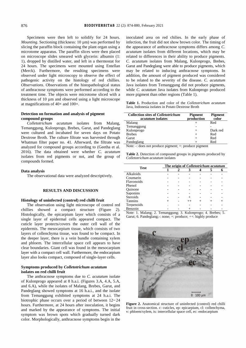
Histology of uninfected (control) red chilli fruit
The observation using light microscope of control red
chillies showed a compact structure (Figure 2).
Histologically, the epicarpium layer which consists of a
single layer of epidermal cells appeared compact. The
cuticle layer protects/covers the outer cell wall of the
epidermis. The mesocarpium tissue, which consists of two
layers of collenchyma tissue, was found to be compact. In
the deeper layer, there is a vein bundle containing xylem
and phloem. The intercellular space cell appears to have
clear boundaries. Giant cell was found in the mesocarpium
layer with a compact cell wall. Furthermore, the endocarpium
layer also looks compact, composed of single-layer cells.
Symptoms produced by Colletotrichum acutatum
isolates on red chilli fruit
The anthracnose symptoms due to C. acutatum isolate
of Kulonprogo appeared at 8 h.a.i. (Figures 3.A, 4.A, 5.A,
and 6.A), while the isolates of Malang, Brebes, Garut, and
Pandeglang showed symptoms at 16 h.a.i., and the isolate
from Temanggung exhibited symptoms at 24 h.a.i. The
biotrophic phase occurs over a period of between 12 ̶ 24
hours. Furthermore, at 24 hours after inoculation, it begins
and marked by the appearance of symptoms. The initial
symptom was brown spots which gradually turned dark
color. Morphologically, anthracnose symptoms begin in the
inoculated area on red chilies. In the early phase of
infection, the fruit did not show brown color. The timing of
the appearance of anthracnose symptoms differs among C.
acutatum isolates from different locations, which may be
related to differences in their ability to produce pigments.
C. acutatum isolates from Malang, Kulonprogo, Brebes,
Garut and Pandeglang were able to produce pigments, which
may be related to inducing anthracnose symptoms. In
addition, the amount of pigment produced was considered
to be related to the severity of the disease. C. acutatum
Java isolates from Temanggung did not produce pigments,
while C. acutatum Java isolates from Kulonprogo produced
more pigment than other regions (Table 1).
Table 1. Production and color of the Colletotrichum acutatum Java, Indonesia isolates in Potato Dextrose Broth
Collection sites of Colletotrichum acutatum isolates
Pigment production
Pigment color
Malang + Red Temanggung - - Kulonprogo + Dark red Brebes + Red Garut + Red Pandeglang + Red Note: -: does not produce pigment; +: produce pigment
Table 2. Detection of compound groups in pigments produced by Colletotrichum acutatum isolates
Test The origin of Colletotrichum acutatum
1 2 3 4 5 6 Alkaloids - - - - - - Coumarin - - - - - - Flavonoids - - - - - - Phenol - - - - - - Quinone - - - - - - Saponins - - - - - - Steroids - - + - - - Tannins + - ++ + + + Terpenoids - - - - - - Benzoin - - + - - - Note: 1. Malang; 2. Temanggung; 3. Kulonprogo; 4. Brebes; 5. Garut; 6. Pandeglang;-: none; +: produce; ++: highly produce
Figure 2. Anatomical structure of uninfected (control) red chilli fruit in cross-section. c: cuticles, ep: epicarpium, cl: collenchyma, v: phloem/xylem, is: intercellular space cell, ec: endocarpium
MULJOWATI et al. – Histopathology of Capsicum annuum infected with Colletotrichum acutatum
877
A B A B
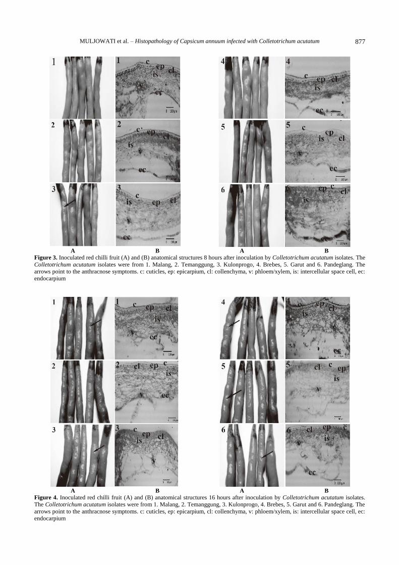
Figure 3. Inoculated red chilli fruit (A) and (B) anatomical structures 8 hours after inoculation by Colletotrichum acutatum isolates. The
Colletotrichum acutatum isolates were from 1. Malang, 2. Temanggung, 3. Kulonprogo, 4. Brebes, 5. Garut and 6. Pandeglang. The
arrows point to the anthracnose symptoms. c: cuticles, ep: epicarpium, cl: collenchyma, v: phloem/xylem, is: intercellular space cell, ec:
endocarpium
A B A B
Figure 4. Inoculated red chilli fruit (A) and (B) anatomical structures 16 hours after inoculation by Colletotrichum acutatum isolates.
The Colletotrichum acutatum isolates were from 1. Malang, 2. Temanggung, 3. Kulonprogo, 4. Brebes, 5. Garut and 6. Pandeglang. The
arrows point to the anthracnose symptoms. c: cuticles, ep: epicarpium, cl: collenchyma, v: phloem/xylem, is: intercellular space cell, ec:
endocarpium

B IODIVERSITAS 22 (2): 874-880, February 2021
878
A B A B
Figure 5. Inoculated red chilli fruit (A) and (B) anatomical structures 24 hours after inoculation by Colletotrichum acutatum isolates.
The Colletotrichum acutatum isolates were from 1. Malang, 2. Temanggung, 3. Kulonprogo, 4. Brebes, 5. Garut and 6. Pandeglang. The
arrows point to the anthracnose symptoms. c: cuticles, ep: epicarpium, cl: collenchyma, v: phloem/xylem, is: intercellular space cell, ec:
endocarpium.
A B A B
Figure 6. Inoculated red chilli fruit (A) and (B) anatomical structures 32 hours after inoculation by Colletotrichum acutatum isolates.
The Colletotrichum acutatum isolates were from 1. Malang, 2. Temanggung, 3. Kulonprogo, 4. Brebes, 5. Garut and 6. Pandeglang. The
arrows point to the anthracnose symptoms. c: cuticles, ep: epicarpium, cl: collenchyma, v: phloem/xylem, is: intercellular space cell, ec:
endocarpium
MULJOWATI et al. – Histopathology of Capsicum annuum infected with Colletotrichum acutatum
879
Histopathology of red chilli fruit infected with
Colletotrichum acutatum isolates
Based on observations using light microscope, the
anatomical structure of red chillies at the incubation period
of 8 h.a.i. (Figure 3.B), found relatively same when
compared to control fruits (Figure 2). But the fruits
inoculated with C. acutatum from Kulonprogo showed
more tenuous than other treatments. At the incubation
period of 16 h.a.i. (Figure 4.B), the cells that make up the
red chilli fruit tissue inoculated with C. acutatum from
Malang, Brebes, Garut, and Pandeglang showed more
tenuous than the control. In the incubation period of 24
h.a.i. (Figure 5.B red chilli fruit tissue also appeared more
tenuous than the control. Meanwhile, inoculation of C.
acutatum from Malang, Brebes, Garut, and Pandeglang
resulted in more tenuous tissue than those inoculated by C.
acutatum from Temanggung. At the incubation period of
32 h.a.i (Figure 6.B), the anatomical structure of the red
chilli fruits looks more tenuous and even appears that the
tissue structure has separated.
Histopathological observations (Figures 3.B, 4..B, 5.B
and 6.B) revealed tissue damage in red chilli fruits
inoculated with C. acutatum Java isolate from Kulonprogo
at 8 h.a.i. This showed that pathogenic activity had begun
in the initial phase of infection, even though melanization
associated with anthracnose symptoms was not observed.
Red chilli tissue damage caused by C. acutatum isolates
from Malang, Brebes, Garut, and Pandeglang began to
appear at 16 h.a.i., while damage by C. acutatum isolates of
Temanggung began at 24 h.a.i.
The compound groups in pigments produced by
Colletotrichum acutatum isolates
Detection of pigments compound groups produced by
C. acutatum Java isolates indicated that the isolates from
Malang, Kulonprogo, Brebes, Garut, and Pandeglang
produced tannins, while the other isolates did not produce
(Table 2). Tannins produced by Kulonprogo isolates had a
stronger pigment color (dark red) than those produced by
the isolates from other regions (Table 1).
Discussion
Histology of uninfected red chilli fruit showed a
compact structure. The epicarpium, mesocarpium, and
endocarpium consist of layer of cells appeared compact.
This result is very similar to that previously reported by
Weryszko-Chmielewska and Zenia (2012).
In the early phase of infection, plant pathogens produce
the pectinase enzyme which plays a role in pathogenesis
(Yakoby et al. 2000). Pectinase degrades the pectin
compounds that make up the middle lamella, resulting in
the separation of cells making up the parenchymal tissue
and increasing the space between cells. C. acutatum Java
isolates can produce pectinase enzyme (Muljowati et al.
2020), which degrades plant cell walls.
The anthracnose symptoms due to C. acutatum Java
isolates showed at different times. C. acutatum isolates
from Kulonprogo caused host damage within 8 hours,
while the other isolates (from Malang, Brebes, Garut and
Pandeglang) began to produce similar symptoms at 16
hours or 24 hours. This result is not in line with the
observation uncovered by Arroyo et al. (2009), that C.
acutatum conidia germination occurred starting at 4 hours
after inoculation. Furthermore, at 24 hours after
inoculation, it begins, which is marked by the appearance
of symptoms. The initial symptoms are brown spots, and
over time the spot darkens. The timing of the appearance of
brown color at the anthracnose symptoms different among
C. acutatum isolates from different locations. According to
Elsenman and Casadevall (2012), melanin is a pigment
with a granular structure, and is a product of the
polymerization of phenol compounds. The function of
melanin is to protect fungi from environmental influences.
It also plays a role in pathogenesis and the invasion of host
tissue.
Besides producing the pectinase enzyme, C. acutatum
isolates from Malang, Kulonprogo, Brebes, Garut and
Pandeglang were able to produce red pigment in culture
medium, while isolates from Temanggung did not produce
pigments. Pigments produced by C. acutatum isolates are
tannins compound group (Geetha et al. 2016). Higher
tannin concentrations result in stronger pigment color (dark
red). Tannin acyl-hydrolase commonly referred to as
tannase, hydrolyses the ester bonds of hydrolyzable
tannins, gallotannins, ellagitannins to produce gallic acid,
glucose and galloil ester (Govindarajan et al. 2016).
Belmares et al. (2004) stated that plants and microbes can
produce tannases. Tannase plays a role in the ability of
pathogens to cause symptoms of plant diseases (Sharma,
2019). Therefore, the pectinase and tannase enzymes in the
red pigment produced by the Java isolate C. acutatum also
play a role in causing symptoms of anthracnose disease in
red chilies. In all incubation periods, anthracnose
symptoms and tissue damage occurred more rapidly in red
chillies inoculated with C. acutatum isolate from
Kulonprogo, while inoculations of C. acutatum isolate
from Temanggung resulted in the slowest anthracnose
symptoms and tissue damage
Anthracnose symptoms and tissue damage in red
chillies inoculated with C. acutatum have been seen in the
early stages of infection. This indicates that pathogenic
activity has started at initial phases.
It remains unclear why the pigment concentration
produced by C. acutatum Java isolates was not the same.
Therefore, it has been suggested that the factors affect the
ability of C. acutatum isolates to produc pigments should
be investigated.
ACKNOWLEDGEMENTS
The authors would like to express their gratitude to the
Research and Community Service Directorate: Directorate
General of Research and Development, Ministry of
Research, Technology, and Higher Education, which
allowed the authors to conduct this research under Doctoral
Dissertation Scheme Number 10984/UN23.14/PN/2017.
The authors would also like to thank the Head of

B IODIVERSITAS 22 (2): 874-880, February 2021
880
Mycology, Plant Structure, and Development Laboratories
of the Faculty of Biology at Jenderal Soedirman
University, Purwokerto, Indonesia, for their technical
support in this research.
REFERENCES
Ajmal M, Abida A, Anum A, Shaista A, Brian GN. 2016. Stem histopathology of sesame seedlings infected with Alternaria
alternata. Microsc Res 4: 11-19.
Anand T, Ramanujam B, Thiruvengadam R, Gandhi K, Manikam R, Govindasamy S. 2008. Production of cell wall degrading enzymes
and toxins by Colletotrichum capsici and Alternaria alternata causing fruit rot of chillies. J Plant Prot Res 48: 437-451.
Ansari A, Khanzada MA, Rajput MA, Sultan Maitlo1, Rajput AQ, Ujjan
A. 2018. Effect of different abiotic factors on the growth and sporulation of Colletotrichum gloeosporioides causing anthracnose of
mango. Plant Protection 02 (01): 23-30.
Arroyo FT, Javier M, Gregorio G, Berta DS, Carmen B, Maria P, Cesar B, Fernando R. 2005. Ultrastructure of the early stages of Colletotrichum
acutatum infection of strawberry tissue. Can J Bot 83: 491-500.
Bellincampi D, Felice C, Vincenzo L. 2014. Plant cell wall dynamics and wall-related susceptibility in planta-pathogen interactions. Front Plant
Sci 5: 1-9.
Belmares R, Contreras-Esquivel JC, Rodriguez-Hererra R, Coronel AR, Aquilar CN. 2004. Microbial production of tannase: an enzyme with
potential use in food industry. Lebensm-wiss u- Technol 37: 857-864.
Elsenman HC, Arturo C. 2012. Synthesis and assembly of fungal melanin. Appl Microbiol Biotechnol 93: 931-40.
Gautam AK. 2014a. Colletotrichum gloeosporioides: biology,
pathogenicity and management in India. Plant Physiol Pathol 2: 2. DOI: 10.4172/2329-955X.1000125
Gautam AK. 2014b. The Genera Colletotrichum: An incitant of numerous
new plant diseases in India. J New Biol Rep 3: 9-21. Geetha S, Kokkaiah I, Sinthia G, Palanichamy M. 2016. Preliminary
phytochemical screening of different solvent extracts and stem of
Crataeva religiosa Hook Frost. Intl J Curr Microbiol Appl Sci 5: 116-122.
González E, Turner BS, James CC. 2006. Clarification of the etiology of
Glomerella leaf spot and bitter rot of apple caused by Colletotrichum spp. based on morphology and genetic, molecular, and pathogenicity
tests. Phytopathology 96 (9): 982-992.
Govindarajan RK, Reevathi S, Rameshkumar N, Khrisnan M, Kayalvizhi
N. 2016. Microbial tannase: current perspectives and biotechnological
advances. Biocatalys Agric Biotechnol 6: 168-175. Hakim A, Muhamad S, Widodo. 2014. Ketahanan penyakit antraknosa
terhadap cabai lokal dan cabai introduksi. Bul Agrohorti 2 (1): 31-36.
[Indonesian] Liu GY, Victor N. 2009. Color me bad: microbial pigments as virulence
factors. Trends Microbiol 17: 406-413.
Miedes E, Vanholme R, Boerjan W, Molina A. 2014. The role of the secondary cell wall in plant resistance to pathogens. Front Plant Sci 5
(e1001123): 358. DOI: 10.3389/fpls.2014.00358.
Muljowati JS, Soesanto L, Nugroho LH. 2020. Production of pectinase enzymes by Colletotrichum acutatum Simmonds causing anthracnose
in red chilli ( Capsicum annuum, L). IOP Conf Ser Earth Environ Sci
593 (1): 012036. DOI: 10.1088/1755-1315/593/1/012036. Pandey A, Pandey BK, Muthukumar M, Yadava LP, Chauhan UK. 2012.
Histopathological study of infection process of Colletotrichum
gloeosporioides Prnz and Sacc. on Mangifera indica L. Plant Pathol J 11: 18-24.
Saxena A, Richa R, Vijai KG, Harikesh BS. 2016. Chilli anthracnose: the
epidemiology and management. Front Microbiol 7: 1-18. Sharma KP. 2019. Tannin degradation by phytopathogen’s tannase: A
plant’s defense perspective. Biocatalys Agric Biotechnol 21: 101342.
DOI: 10.1016/j.bcab.2019.101342. Sharma M, Kulshrestha S. 2015. Colletotrichum gloeosporioides: An
anthracnose causing pathogen of fruits and vegetables. Biosci
Biotechnol Res Asia 12 (2): 1233-1246. DOI: 10.13005/bbra/1776. Silva SAM, Rosana R, Leandro SAGs, Cláudia PS, Cíntia SB, Margarida
GFC, Artur MM. 2014. Resistance in Capsicum spp. to anthracnose
affected by different stages of fruit development during pre-and post-harvest. Trop Plant Pathol 39: 335-341.
Than PP, Jeewon R, Hyde KD, Pongsupasamit S, Mongkolporn O, Taylor
PWJ. 2008. Characterization and pathogenicity of Colletotrichum species associated with anthracnose on chilli (Capsicum spp.) in
Thailand. Plant Pathol 57: 562-572.
Underwood W. 2012. The plant cell wall: A dynamic barrier against
pathogen invasion. Front Plant Sci 3 (85): 85. DOI:
10.3389/fpls.2012.00085. Weryszko-Chmielewska E, Zenia M. 2012. Anatomical traits of sweet
pepper (Capsicum annuum L.) fruit. Acta Agrobot 64: 181-188.
Wharton PS, Schilder AC. 2008. Novel infection strategies of Colletotrichum acutatum on ripe blueberry fruit. Plant Pathol 57: 122-
134.
Yakoby N, Stanley F, Amos D, Noel TK, Dov P. 2000. Expression of pectate lyase from Colletotrichum gloeosporioides in C. magna
promotes pathogenicity. Mol Plant-Microb Interact 13: 887-891.