Embed Size (px)
Citation preview
GENOMICS 1,287-291 (1987)
SHORT COMMUNICATION
Relationship of the Genes for Chediak-Higashi Syndrome (Beige) and the T-Cell Receptor y Chain in Mouse and Man
RANDALL F. tiOLCOMBE,*St WILLIAM STRAuss,t FRANCES L. OWEN,* LAURENCE A. BOXER,~ ROBERT W. WARREN” Mary ELLEN CONLEY,# JAMES FERRARA,’ RANDI Y. LEAVITT,**
ANTHONY S. FAUCI, * * BENJAMIN A. TAYLOR,tt AND J. G. SEIDMANt
*Division of Hematology, Department of Medicine, Brigham & Women’s Hospital, Boston, Massachusetts 02175; tDepartment of Genetics, Harvard Medical School, Boston, Massachusetts 02115; *Department of Pathology, Tufts University School of
Medicine, Boston, Massachusetts 02 111; §Division of Pediatric Hematology/Oncology, Mott Hospital, University of Michigan, Ann Arbor, Michigan 48109; “Division of Rheumatology and Immunology, Department of Pediatrics, University of North Carolina, Chapel Hill, North Carolina 27599; #Department of Pediatrics, Children’s Hospital of Philadelphia, Philadelphia,
Pennsylvania 19 704; lIDivision of Hematology/Oncology, Children’s Hospital Medical Center, Boston, Massachusetts 02 115; ‘*N/AID, National Institutes of Health, Bethesda, Maryland 20892; and t t The Jackson Laboratory, Bar Harbor, Maine 04609
Received September 8, 1987; revised November 24, 1987
The genetic linkage of Chediak-Higashi syndrome and its murine analog, beige (bg), to the T-cell recep- tor (TCR-7) y chain gene is further deilned. Previous studies using recombinant inbred strains of mice dem- onstrated that the murine bg gene is genetically linked to a murine TCR-7 gene. We report that in the mouse the frequency of recombination between these two markers is 0.025. Further, we tested the hypoth- esis that these two genes are linked in the human ge- nome by analyzing restriction fragment length poly- morphisms (RFLPs) in five families with children af- flicted with Chediak-Higashi syndrome. In three families, RFLPs in TCR-y genes were inherited dis- cordantly from Chediak-Higashi syndrome, demon- strating nonlinkage. We postulate that there is an evolutionary chromosomal breakpoint between the bg gene and the TCR-y gene. 0 1987 Academic Press, Inc.
Chediak-Higashi syndrome (CHS) is a rare auto- somal recessive disorder characterized by oculocuta- neous albinism, a predisposition to pyogenic infec- tions, and large abnormal cytoplasmic masses in all granule-containing cells (Blume and Wolff, 1972). Death may occur at an early age due to infection or a lymphoma-like accelerated phase which may be of T- cell origin (Argyle et aZ., 1982) or rather a lymphohis- tiocytic proliferation related to EBV infection (Rubin et al., 1985). Other cellular processes that have been shown to be abnormal include absent or decreased natural killer (NK) activity (Haliotis et aZ., 1980; Klein et al., 1980; Merino et al., 1983; Roder and Duwe, 1979), altered responsiveness to EBV infection
(Merino et al., 1986), decreased antibody-dependent cell-mediated cytotoxicity (ADCC) (Klein et aL, 1980), and altered neutrophil digestive capabilities (Quie and Cates, 1977). Beige (bg) mice have many of these fea- tures, and several groups have proposed that the mu- rine bg mutation is analogous to the mutation in pa- tients with CHS (Brandt et aL, 1975; Lutzner et al., 1966; Windhorst and Padgett, 1973). Here we further define the map distance between the murine bg gene and the murine T-cell receptor (TCR) y chain gene and demonstrate that the human CHS gene is not closely linked to the human y chain gene.
We have previously shown that a murine TCR-7 chain gene is closely linked to the bg gene in the crin- kled (CT), bg, and extra-toes (Xt) cluster by mapping this locus using recombinant inbred lines and taking advantage of a restriction fragment length polymor- phism (RFLP) that exists at this locus (Owen et aZ., 1986). The TCR-7 chain is an immunoglobulin-like polypeptide that comprises one chain of the T-cell y-6 receptor. Murine y chains are encoded by three closely linked immunoglobulin-like genes located on murine chromosome 13 (Kranz et al., 1985). Because there is diminished NK activity in beige mice (Roder and Duwe, 1979), and because it has been shown that T-cells with a r-6 T-cell receptor are capable of non- MHC restricted (or NK-like) killing (Ang et al., 1987; Borst et al., 1987; Brenner et al., 1987), the possibility existed that the gene for the TCR-y chain was in- volved in the pathogenesis of CHS/beige. In this study we present data from breeding studies in mice demon- strating that the bg mutation can be genetically sepa-
287 oS88-7543/87 $3.00 Copyright Q 1987 by Academic Preee, Inc.
All rights of reproduction in any form reserved.
288 SHORT COMMUNICATION
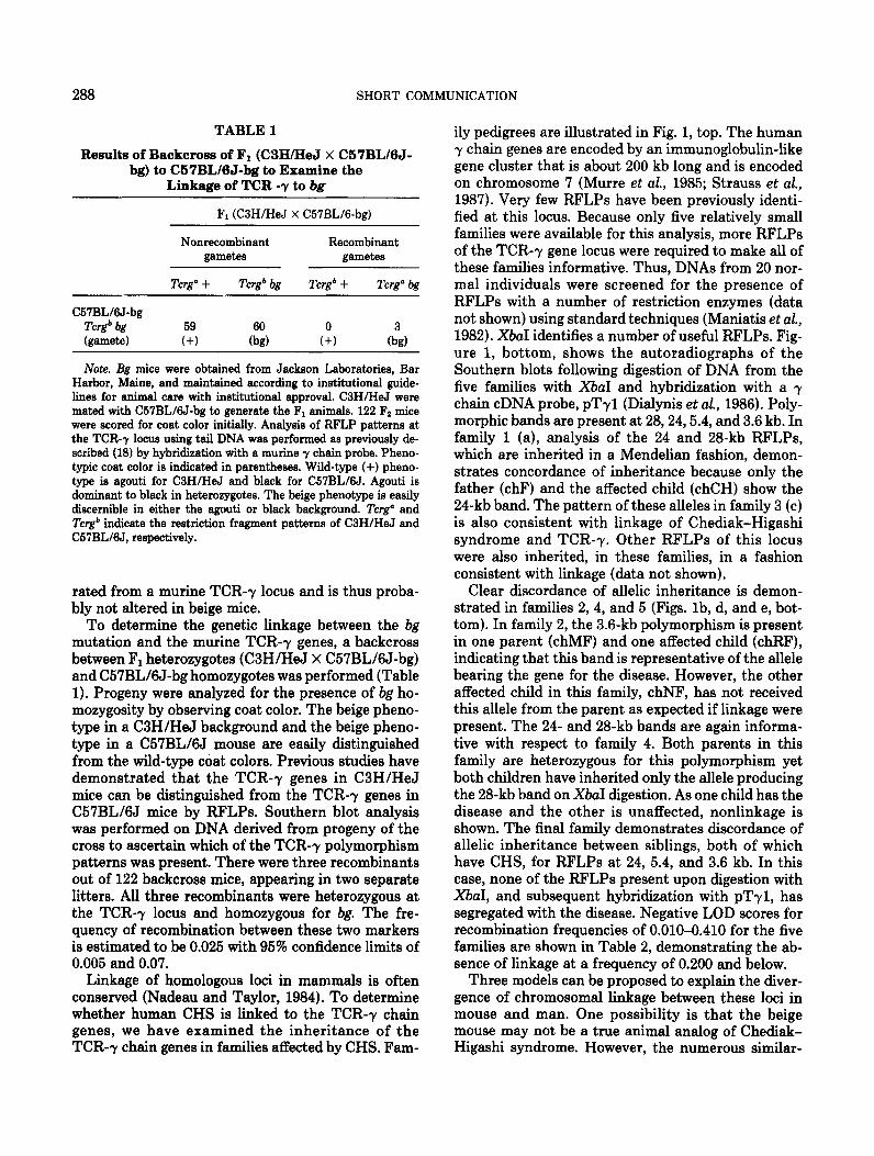
TABLE 1
Results of Backcross of F1 (C3HIHeJ X C57BL/6J- bg) to C57BL/BJ-bg to Examine the
Linkage of TCR -y to bg
F1 (C3H/HeJ X C57BL/6-bg)
C57BL&J-bg
Nonrecombinant gametes
Tcrg” + Tcrg* bg
Recombinant gametes
Tcrgb + Tcrg” bg
Tcrg* bg
kamete) :+3 ii, A &I
Note. Bg mice were obtained from Jackson Laboratories, Bar Harbor, Maine, and maintained according to institutional guide- lines for animal care with institutional approval. C3H/HeJ were mated with C57BL/6J-bg to generate the F1 animals. 122 Fz mice were scored for coat color initially. Analysis of RFLP patterns at the TCR-7 locus using tail DNA was performed as previously de- scribed (18) by hybridization with a murine y chain probe. Pheno- typic coat color is indicated in parentheses. Wild-type (+) pheno- type is agouti for C3H/HeJ and black for C57BL/6J. Agouti is dominant to black in heterozygotes. The beige phenotype is easily discernible in either the agouti or black background. Tcrg” and Tcrg* indicate the restriction fragment patterns of C3H/HeJ and C57BL/6J, respectively.
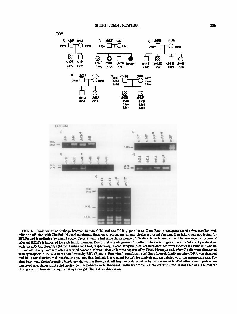
ily pedigrees are illustrated in Fig. 1, top. The human y chain genes are encoded by an immunoglobulin-like gene cluster that is about 200 kb long and is encoded on chromosome 7 (Murre et aZ., 1985; Strauss et al., 1987). Very few RFLPs have been previously identi- fied at this locus. Because only five relatively small families were available for this analysis, more RFLPs of the TCR-7 gene locus were required to make all of these families informative. Thus, DNAs from 20 nor- mal individuals were screened for the presence of RFLPs with a number of restriction enzymes (data not shown) using standard techniques (Maniatis et aZ., 1982). XbaI identifies a number of useful RFLPs. Fig- ure 1, bottom, shows the autoradiographs of the Southern blots following digestion of DNA from the five families with XbaI and hybridization with a y chain cDNA probe, pTy1 (Dialynis et al., 1986). Poly- morphic bands are present at 28,24,5.4, and 3.6 kb. In family 1 (a), analysis of the 24 and 28-kb RFLPs, which are inherited in a Mendelian fashion, demon- strates concordance of inheritance because only the father (chF) and the affected child (chCH) show the 24-kb band. The pattern of these alleles in family 3 (c) is also consistent with linkage of Chediak-Higashi syndrome and TCR-7. Other RFLPs of this locus were also inherited, in these families, in a fashion consistent with linkage (data not shown).
rated from a murine TCR-7 locus and is thus proba- bly not altered in beige mice.
To determine the genetic linkage between the bg mutation and the murine TCR-7 genes, a backcross between F1 heterozygotes (C3H/HeJ X C57BL/6J-bg) and C57BL&J-bg homozygotes was performed (Table 1). Progeny were analyzed for the presence of bg ho- mozygosity by observing coat color. The beige pheno- type in a C3H/HeJ background and the beige pheno- type in a C57BL&J mouse are easily distinguished from the wild-type coat colors. Previous studies have demonstrated that the TCR-7 genes in C3H/HeJ mice can be distinguished from the TCR-7 genes in C57BLKI mice by RFLPs. Southern blot analysis was performed on DNA derived from progeny of the cross to ascertain which of the TCR-7 polymorphism patterns was present. There were three recombinants out of 122 backcross mice, appearing in two separate litters. All three recombinants were heterozygous at the TCR-7 locus and homozygous for bg. The fre- quency of recombination between these two markers is estimated to be 0.025 with 95% confidence limits of 0.005 and 0.07.
Clear discordance of allelic inheritance is demon- strated in families 2,4, and 5 (Figs. lb, d, and e, bot- tom). In family 2, the 3.6-kb polymorphism is present in one parent (chMF) and one affected child (chRF), indicating that this band is representative of the allele bearing the gene for the disease. However, the other affected child in this family, chNF, has not received this allele from the parent as expected if linkage were present. The 24- and 28-kb bands are again informa- tive with respect to family 4. Both parents in this family are heterozygous for this polymorphism yet both children have inherited only the allele producing the 28-kb band on XbaI digestion. As one child has the disease and the other is unaffected, nonlinkage is shown. The final family demonstrates discordance of allelic inheritance between siblings, both of which have CHS, for RFLPs at 24, 5.4, and 3.6 kb. In this case, none of the RFLPs present upon digestion with XbaI, and subsequent hybridization with pTy1, has segregated with the disease. Negative LOD scores for recombination frequencies of 0.010-0.410 for the five families are shown in Table 2, demonstrating the ab- sence of linkage at a frequency of 0.200 and below.
Linkage of homologous loci in mammals is often Three models can be proposed to explain the diver- conserved (Nadeau and Taylor, 1984). To determine gence of chromosomal linkage between these loci in whether human CHS is linked to the TCR-7 chain mouse and man. One possibility is that the beige genes, we have examined the inheritance of the mouse may not be a true animal analog of Chediak- TCR-7 chain genes in families affected by CHS. Fam- Higashi syndrome. However, the numerous similar-
SHORT COMMUNICATION 289
TOP
a) chF chM
20R4
rx
zwa b) chKF &MF
X6(-) 3.6(t)
c) chRE chJE
28R4T 24R4
chCH chB 28R4 26R0
chNF &RF chZF infant chNE chME chBE chHE 3.6~) 3.6(+) 3.8(+) 24R4 24R4 28R4 28R4
4 chGJ ch DJ
28~4 TX@4 ‘kf *-iTT z
t&l &A dlER chLR
28RB 20R4 5.4(-) 5.4(+) 3.6(-) 3.6(+)
FIG. 1. Evidence of nonlinkage between human CHS and the TCR-7 gene locus. Top: Family pedigrees for the five families with offspring acted with Chediak-Higaehi syndrome. Squares repreeent males, and circles represent females. One infant wae not tested for RFLPe and is indicated by a solid circle. Cross-hatching indicates the presence of Chediak-Higaehi syndrome. The preeence or absence of relevant RFLPe is indicated for each family member. Bottom: Autoradiogmme of Southern blots after digestion with X&I and hybridization with the cDNA probe pTrl(8) for families l-5 (a-e, respectively). Blood samples (5-20 cc) were obtained from index caees with CHS and all immediate family members after informed conzent. Mononuclear cells were separated by FicoWI-Iypaque and, after T-cells were eliminated with cyclosporin A, B-cellz were transformed by EBV (Epstein-Barr virus), establishing cell lines for each family member. DNA wae obtained and 10 pg wae digeeted with restriction enzymes. Bars indicate the relevant RFLPs for analysis and are labeled with the appropriate size. For simplicity, only the informative bande are shown in a through d. All fragments detected by hybridization with pTr1 after &I digestion are dieplayed in e. Superecript solid circles identify patients with Chediak-Higashi syndrome. X DNA cut with HindI was used as a eize marker during electrophoresis through a 1% agaroze gel. See text for discussion.
290 SHORT COMMUNICATION
TABLE 2
Degree of Nonlinkage between the Genes for Human Chediak-Higashi Syndrome and the TCR-r Chain
6 LOD score
0.010 -21.79 0.060 -8.47 0.110 -4.64 0.160 -2.68 0.210 -1.54 0.260 -0.85 0.310 -0.43 0.360 -0.20 0.410 -0.07
Note. A computer program entitled Linkage 3.5, written by Jurg Ott, was used for calculation of LOD scores. Because of peculiari- ties within the program, “affection status” was converted to a bi- nary phenotype with internally consistent estimation of heterozy- gosity of unaffected siblings. 0 represents recombination frequency.
ities between the conditions both in morphology and function of affected cells and in physical characteris- tics make this explanation less likely. A second possi- bility may be that the human CHS is a multigenic disease. This might explain the somewhat variable clinical course seen among affected individuals. In- deed, though most affected children die during the first decade of life, some individuals have been known to live into the fourth decade without manifesting a lymphoma-like syndrome or fatal pyogenic infections. Again, by analogy to the murine model, this seems unlikely. Several alleles of bg have been described previously, each with identical phenotypic manifesta- tions (Brandt and Swank, 1976). The third and most likely model is that there has been a chromosomal breakpoint during evolution between the TCR-7 locus and the beige locus, resulting in the genes being sepa- rated by larger distances in the human genome and possibly even on different chromosomes.
Because the linkage between CHS and TCR-7 is not present in man, a probe useful for prenatal diag- nosis is not currently available. However, we imagine that segments of DNA linked to the murine bg locus may be linked to the human CHS defect and may offer an approach to identifying a DNA probe that will be a useful diagnostic tool for this disease.
ACKNOWLEDGMENTS
R.F.H. is supported by National Heart Lung and Blood Institute Training Grant HL-07446. This work was supported by NIH Grants AI19148 and AI18436 to J.G.S., AI20065 to L.A.B., and GM18684 to B.A.T. F.L.O. is supported by Grant IM394 from the American Cancer Society. M.E.C. has received a grant from the Immune Deficiency Foundation. Thanks to Dr. Margaret Johnson who assisted in locating patients, Dr. Thomas Quertermous for his
advice, and Eric Hoffman and Dr. Samuel Latt for their statistical guidance.
1.
2.
3.
4.
5.
6.
7.
8.
9.
10.
11.
12.
13.
14.
15.
16.
17.
REFERENCES
ANG, S. L., SEIDMAN, J. G., PETERMAN, G. M., et al. (1987) Functional gamma chain-associated T cell receptors on cere- brospinal fluid-derived natural killer-like T cell clones. J. Enp. Med. 166(5): 1453-1458. ARGYLE, J. C., KJELDSBERG, C. R., STY, J., SHIGEOKA, A. O., AND HILL, H. R. (1982). T-cell lymphoma and the Che- diak-Higashi syndrome. Blood 60: 672-676. BLUME, R. S., AND WOLFF, S. M. (1972). The Chediak-Higa- shi syndrome: Studies in four patients and a review of the literature. Medicine (Baltimore) 51: 247-280. BORST, J., VAN DE GRIEND, R. J., VAN OOSTVEEN, J. W., et al. (1987). A T-cell receptor gamma/CD3 complex found on cloned functional lymphocytes. Nature (‘London) 325(6106): 683-688. BRANDT, E. J., ELLIOTT, R. W., AND SWANK, R. T. (1975). Defective lysosomal enzyme secretion in kidneys of Chediak- Higashi (Beige) mice. J. Cell Biol. 67: 774-788. BRANDT, E. J., AND SWANK, R. T. (1976). The Chediak-Higa- shi (Beige) mutation in two mouse strains: Allelism and simi- larity in lysosomal dysfunction. Amer. J. Pa&l. 82: 573-588. BRENNER, M. B., MCLEAN, J., Scm, H., et al. (1987). Two forms of the T-cell receptor gamma protein found on periph- eral blood cytotoxic T lymphocytes. Nature (London,J 325(6106): 689-694. DIALYNIS, D. P., MURRE, C., QUERTERMOUS, T., et al. (1986). Cloning and sequence analysis of complementary DNA en- coding an aberrantly rearranged human T-cell y chain. Proc. Natl. Acad. Sci. USA 83: 2619-2623. HALIOTIS, T., RODER, J., KLEIN, M., ORTALDO, J., FAUCI, A. S., AND HERBERMAN, R. B. (1980). Chediak-Higashi gene in humans. I. Impairment of natural-killer function. J. Exp. Med. 151: 1039-1048. KLEIN, M., RODER, J., HALIOTIS, T., et al. (1980). Chediak- Higashi gene in humans. II. The selectivity of the defect in natural-killer and antibody-dependent cell-mediated cytotox- icity function. J. Exp. Med. 151: 1049-1058. KRANZ, D. M., SAITO, H., DISTECHE, C. M., et al. (1985). Chro- mosomal location of the murine T-cell receptor alpha-chain gene and the T-cell gamma gene. Science 221: 941-945. LUTZNER, M. A., LOWRIE, C. T., AND JORDAN, H. W. (1966). Giant granules in leukocytes of the beige mouse. J. Hered. 68: 299-300. MANIATIS, T., FRITSCH, E. F., AND SAMBROOK, J. (1982). “Molecular Cloning: A Laboratory Manual,” Cold Spring Harbor Laboratory, Cold Spring Harbor, NY. MERINO, F., AMESTY, C., HENLE, W., L/AYRISSE, Z., BIANCO, N., AND R&UREZ-DUQUE, P. (1986). Chediak-Higashi syn- drome: Immunological responses to Epstein-Barr virus stud- ies in gene heteroxygotes. J. Clin Immurwl. 6: 242-248. MERINO, F., KLEIN, G. O., HENLE, W., R.AMIREZ-DUQUE, P., FORSGREN, M., AND -STY, C. (1983). Low NK activity and elevated anti-EBV antibodies in Chediak-Higashi patients. Clin. Immurwl. Immurwpathol. 27: 326-339. Mu=, C., WALDMAN, R. C., MORTON, C. C., et al. (1985). Human gamma-chain genes are rearranged in leukemic T cells and map to the short arm of chromosome 7. Nature (London) 3 16: 549-552. NADEAU, J. H., AND TAYLOR, B. A. (1984). Lengths of chromo-
SHORT COMMUNICATION 291
somal segments conserved since divergence of man and mouse. Pmt. Natl. Acad. Sci. USA 81: 814-818.
18. OWEN, F. L., TAYLOR, B. A., ZWEIDLER, A., AND SEIDMAN, J. G. (1986). The murine y-chain of the T cell receptor is closely linked to a spermatocyte specific histone gene and the beige coat color locus on chromosome 13. J. Zmmurwl. 13'7: 1044-1646.
19. QIJIE, P. G., AND CATES, K. L. (1977). Clinical conditions associated with defective polymorphonuclear leukocyte che- motaxis. Amer. J. Pathd. 88: 711-125.
20. RODER, J., AND DUWE, A. (1979). The beige mutation in the mouse selectively impairs natural killer cell function. Nature (London) 278: 451-453.
21. RODER, J. C., HALIOTIS, T., KLEIN, M., et al. (1979). A new immunodeficiency disorder in humans involving NK cells. Nature (London) 284: 553-555.
22. RABIN, C. M., BURKE, B. A., MCKENNA, R. W., et al. (1985). The accelerated phase of Chediak-Higashi syndrome: An ex- pression of the virus-associated hemophagocytic syndrome? Cancer 56: 524-530.
23. STRAUSS, W. M., QUERTERMOUS, T., AND SEIDMAN, J. G. (1987). Measuring the human T cell receptor -y-chain locus. Science 237: 1217-1219.
24. WINDHORST, D. B., AND PADGE?T, B. (1973). The Chediak- Higashi syndrome and the homologous trait in animals. J. Invest. Dermatol. 60: 529-537.

![Genome-wide characterization of the relationship between essential and TATA … · 2016. 12. 9. · that essential genes tended to have higher CAIs than non-essential genes [6]. However,](https://img.pdfslide.net/doc/110x75/60d37a5b3dea784a25050fe2/genome-wide-characterization-of-the-relationship-between-essential-and-tata-2016.jpg)


![Short Communication The Relationship between Terminal ... Relationship...ultraviolet light [23], salinity [24], low temperature [3], high temperature [4], and heavy metal stress [25]](https://img.pdfslide.net/doc/110x75/60ecda0f0e887a386157c3b9/short-communication-the-relationship-between-terminal-relationship-ultraviolet.jpg)