Embed Size (px)
DESCRIPTION
Genetics, Biochemistry
Citation preview

Sickle Hemoglobin Confers Toleranceto Plasmodium InfectionAna Ferreira,1 Ivo Marguti,1 Ingo Bechmann,2,3 Viktoria Jeney,1 Angelo Chora,1 Nuno R. Palha,1 Sofia Rebelo,1
Annie Henri,4,5,6 Yves Beuzard,4,5,6 and Miguel P. Soares1,*1Instituto Gulbenkian de Ciencia, 2780-156 Oeiras, Portugal2Institute of Anatomy, University of Leipzig, Liebigstr. 13, 04103 Leipzig, Germany3Dr. Senckenbergische Anatomie, Institute for Clinical Neuroanatomy, JohannWolfgangGoethe-University, 60 -596 Frankfurt/Main, Germany4Institut Universitaire d’Hematologie, Universite Paris 7, France5CEA, Institute of Emerging Diseases and Innovative Therapies, Fontenay-aux-Roses, France6INSERM U962 and University Paris Sud 11, France
*Correspondence: [email protected] 10.1016/j.cell.2011.03.049
SUMMARY
Sickle human hemoglobin (Hb) confers a survivaladvantage to individuals living in endemic areas ofmalaria, the disease caused by Plasmodium infec-tion.Asdemonstratedhereby,miceexpressingsickleHb do not succumb to experimental cerebral malaria(ECM). This protective effect is exerted irrespectivelyof parasite load, revealing that sickle Hb confers hosttolerance toPlasmodium infection. SickleHb inducesthe expression of heme oxygenase-1 (HO-1) in hema-topoietic cells, via a mechanism involving the tran-scription factor NF-E2-related factor 2 (Nrf2). Carbonmonoxide (CO), a byproduct of heme catabolism byHO-1, prevents further accumulation of circulatingfree heme after Plasmodium infection, suppressingthe pathogenesis of ECM. Moreover, sickle Hbinhibits activation and/or expansion of pathogenicCD8+ T cells recognizing antigens expressedbyPlas-modium, an immunoregulatory effect that does notinvolveNrf2 and/orHO-1.Our findingsprovide insightinto molecular mechanisms via which sickle Hbconfers host tolerance to severe forms of malaria.
INTRODUCTION
Several point mutations in the b-chain of human Hb, e.g. HbS
(b6Glu->Val) (Jallow et al., 2009; Williams et al., 2005b), HbC
(b6Glu->Lys)(Modiano et al., 2001) and HbE (b26Glu->Lys)(Huta-
galung et al., 1999), can confer a survival advantage to
Plasmodium (P.) infection (Williams, 2006). When present in the
homozygous form, some of these mutations, e.g. HbS, become
pathologic causing hemolytic anemia, leading to accumulation
of high levels of cell-free Hb and heme in plasma (Reiter et al.,
2002). Individuals carrying the HbSmutation in the heterozygous
form, i.e. the A/S sickle cell trait, also accumulate low (nonpatho-
logic) levels of heme in plasma (Muller-Eberhard et al., 1968) and
are protected against malaria (Allison, 1954; Beet, 1947; Jallow
et al., 2009; Williams, 2006).
398 Cell 145, 398–409, April 29, 2011 ª2011 Elsevier Inc.
Once released from Hb, a phenomenon favored in the case of
HbS (Hebbel et al., 1988), free heme becomes cytotoxic (Balla
et al., 1992; Seixas et al., 2009; Gozzelino et al., 2010). This
deleterious effect is countered by the expression of heme
oxygenase-1 (HO-1, encoded by the Hmox1 gene)(Balla et al.,
1992; Ferreira et al., 2008; Gozzelino et al., 2010), a stress-
responsive enzyme that catabolizes free heme into biliverdin,
iron and carbon monoxide (CO)(Tenhunen et al., 1968). HO-1
expression is induced by free heme, through a mechanism that
involves the ubiquitination-degradation of Kelch-like ECH-asso-
ciated protein 1 (Keap1) (Kensler et al., 2007), a cytoplasmic
repressor of the transcription factor NF-E2-related factor-2
(Nrf2)(Alam et al., 1999). Upon nuclear translocation, Nrf2 binds
to the stress-responsive elements in the Hmox1 promoter
(Ogawa et al., 2001), a regulatory mechanism that plays a central
role in the control of Hmox1 expression in response to heme
(Kensler et al., 2007).
HO-1 is protective against a wide variety of immune mediated
inflammatory diseases (Nath et al., 1992; Soares and Bach,
2009) including experimental cerebral malaria (ECM)(Pamplona
et al., 2007), a lethal neuroinflammatory syndrome that develops
in P. berghei ANKA infected C57BL/6 mice and that mimics
some of the pathologic features of human cerebral malaria
(CM)(Mishra and Newton, 2009). This protective effect is
mediated by CO, which binds cell-free Hb and inhibits its oxida-
tion, thus preventing heme release from oxidized Hb (Hebbel
et al., 1988) and hence the pathogenesis of ECM (Pamplona
et al., 2007). Given that expression of HO-1 (Belcher et al.,
2006; Jison et al., 2004) and production of CO (Sears et al.,
2001) are induced in individuals carrying the HbS mutation we
hypothesized that this might explain why individuals carrying
the A/S sickle cell trait have a survival advantage against malaria
(Allison, 1954; May et al., 2007; Williams et al., 2005b). We
provide evidence demonstrating that this is the case.
RESULTS
Sickle Hb Confers a Survival Advantageagainst Malaria in MiceInoculation of C57BL/6 mice (Hbwt) with P. berghei ANKA
infected red blood cells (RBC) led within 6 to 12 days to the

Figure 1. HbSAD Prevents ECM Onset
(A) Survival of P. berghei ANKA infected Hbwt (n = 91) and HbSAD (n = 76) mice (10 independent experiments with survival advantage p < 0.05). Grey shading:
expected time of ECM.
(B) Representative H&E stained microvessel in the BBB of infected Hbwt and HbSAD mice, at ECM onset in Hbwt mice (n = 3/group). EC: endothelial cell; PVC:
perivascular compartment; GL: glia limitans (dotted line); RBC: red blood cells; iRBC: infected RBC. Magnification: 100x.
(C) Mean brain edema in naıve versus infected Hbwt and HbSADmice ± standard deviation (n=4/group), at the time of ECM onset in Hbwt mice. ns: not significant.
(D) Brain leukocyte sequestration in naıve versus infected Hbwt and HbSAD mice, at the time of ECM onset in Hbwt mice. Dots represent single mice (n = 4 -14/
group). Red lines represent mean values. nd: not determined.
(E) Mean percentage of infected RBC in Hbwt and HbSAD mice ± standard deviation. Same mice as in (A).
(F) Mean number of infected RBC in Hbwt (n=7) and HbSAD (n=9) mice ± standard deviation at the time of ECM onset in Hbwt mice.
See also Figure S1 and Figure S2.
development of clinical signs of ECM (Figure 1A). Incidence of
ECM was significantly reduced in hemizygous C57BL/6 HbSAD
mice (Figure 1A) expressing a b-chain of human Hb carrying theb6Glu->Val (HbS) mutation as well as two additional mutations,b23Ala->Ile (Antilles23I) and b121Asp->Gln (D-Punjab121Q),
known to enhance HbS polymerization in humans and mice
(Trudel et al., 1991). Naıve HbSAD mice present a very mild sickle
cell syndrome, which does not lead to anemia (Table S1A avail-
able online; Trudel et al., 1994), similar to the asymptomatic
human A/S sickle cell trait that affords protection against malaria
(Allison, 1954; Beet, 1947; Jallow et al., 2009). HbSAD mice that
did not develop ECM, succumbed 20-25 days postinfection
from hyperparasitemia-induced anemia (data not shown),
a condition unrelated to ECM (Schofield and Grau, 2005).
When infected with P. berghei ANKA, C57BL/6.Sv/129 HbA/a
mice expressing normal human Hb as well as endogenous
mouse Hb (Wu et al., 2006) developed clinical signs of ECM
and succumbed 6 to 12 days after infection (Figure S1A). Litter-
mate control C57BL/6.Sv/129 Hba/a mice, expressing only the
endogenous mouse Hb developed ECM but with lower inci-
dence, as compared to HbA/a mice (Figure S1A). This suggests
that normal human Hb might promote, rather than prevent, the
pathogenesis of ECM in C57BL/6.Sv/129 mice. While the
reason for this is not clear, it is possible that human Hb alters
mouse RBC physiology in a manner that would promote the
development of ECM. However, this effect becomes negligible
as C57BL/6.Sv/129 mice are backcrossed into the C57BL/6
genetic background (Figure S1D), in which ECM incidence is
higher than 95% (Figure 1A). Given that HbSAD suppresses
the pathogenesis of ECM in C57BL/6 mice and that the delete-
rious effect of normal human Hb becomes negligible under this
genetic background, it is reasonable to infer that the protective
effect of HbSAD is attributable to the mutations in the human
b-globin chain rather than to human Hb per se.
HbSADmice thatdid not succumbwithin6–12dayspostinfection
also did not develop the pathologic hallmarks of ECM, including
blood brain barrier (BBB) disruption (Figures 1B), perivascular
RBC accumulation in brain (Figure 1B) and brain edema
Cell 145, 398–409, April 29, 2011 ª2011 Elsevier Inc. 399
(Figure 1C). These pathologic features were present in Hbwt
(Figures 1B and 1C) andHbA/amice, i.e. brain edema (Figure S1B).
Given that BBBdisruption and brain edema occur inP. berghei
ANKA infected C57BL/6 mice via a CD8+ T cell-dependent
mechanism (Belnoue et al., 2002; Schofield and Grau, 2005),
we asked whether the protective effect of HbSAD against ECM
was associated with inhibition of CD8+ T cell sequestration in
the brain. The number of CD45high leukocytes and CD8+
T cells, including granzyme B-positive (GrB+) CD8+ T cells, was
reduced in P. berghei ANKA infected HbSAD, as compared to
Hbwt mice at ECM onset (Figure 1D).
When infected with P. berghei ANKA, Hbwt mice also devel-
oped severe lung injury and a mild form of liver injury (Figure S2)
with no apparent injury to the kidneys or to the heart (data not
shown). The extent of lung and liver injury was reduced in
P. berghei ANKA infected HbSAD versus Hbwt mice (Figure S2).
Sickle Hb Confers Tolerance to Plasmodium Infectionin MiceProtection of HbSAD mice against ECM was not associated with
reduction of pathogen load, as assessed by the percentage of in-
fected RBC, i.e., parasitemia (Figure 1E) aswell as by the number
of circulating infected RBC (Figure 1F) versus control Hbwt
(Figures 1E and 1F) or HbA/a mice (Figures S1C and S1E). While
the protective effect of the human sickle cell trait against malaria
has been associated with decreased pathogen load (Allison,
1954; May et al., 2007; Williams et al., 2005b), there are several
instances where this does not appear to be the case (Crompton
et al., 2008; Livincstone, 1971; Motulsky et al., 1966), which is in
keeping with the observation that HbSAD confers protection
against ECM without interfering with pathogen load. These
observations suggest that mutations in the b-chain of human
Hb, such as those in HbSAD can afford tolerance to Plasmodium
infection, a host defense strategy that limits disease severity by
preventing tissue damage, without targeting the pathogen. This
contrasts to resistance to infection, the well-recognized host
defense strategy that limits disease severity by decreasing path-
ogen load (Raberg et al., 2007; Schneider and Ayres, 2008).
Parasite sequestration was similar in Hbwt, HbSADHmox1+/+,
and HbSADHmox1+/- mice, as assessed using a transgenic lucif-
erase-P. berghei ANKA strain (Figure 4S). This supports further
the notion that induction of HO-1 by sickle Hb confers host toler-
ance to Plasmodium infection.
Sickle Hb Induces the Expression of HO-1 that ConfersTolerance to Plasmodium InfectionHumans and rodents carrying the HbS mutation express high
levels of HO-1 in the hematopoietic compartment (Belcher
et al., 2006; Jison et al., 2004). Consistent with this, naıve HbSAD
mice express high levels of Hmox1 mRNA in bone marrow and
peripheral blood cells, as compared to naıve Hbwt mice (Fig-
ure 2A). Naıve HbSAD mice also expressed higher levels of
Hmox1 mRNA in the kidneys (Figure S3A), which is consistent
with the chronic development of kidney injury in these mice, re-
vealed clinically upon aging (Sabaa et al., 2008). HbSAD mice
expressed similar levels of Hmox1 mRNA in the liver, heart,
lung and spleen (Figure S3A), as compared to Hbwt mice.
HbA/a mice expressed similar levels of Hmox1 mRNA in the
400 Cell 145, 398–409, April 29, 2011 ª2011 Elsevier Inc.
bone marrow and peripheral blood versus littermate control
Hba/a mice (Suppl. Figure 3B), demonstrating that expression
of a bS related variant but not a normal b-globin chain is required
to induce Hmox1 expression.
Given that HO-1 is protective against severe forms of malaria
in mice (Pamplona et al., 2007; Seixas et al., 2009), we asked
whether its induction in HbSAD mice (Figure 2A) is required to
suppress the development of ECM (Figure 1A). Deletion of one
Hmox1 allele (Hmox1+/-) reduced Hmox1 mRNA expression in
bone marrow and whole blood leukocytes of HbSAD mice (Fig-
ure S3C), without causing overt postnatal lethality (Table S2A).
When challenged by P. berghei ANKA infection, HbSADHmox1+/-
mice succumbed to ECM (Figure 2B), with concomitant develop-
ment of BBB disruption (Figure 2C), brain edema (Figure 2D) and
sequestration of CD45high leukocytes (data not shown), CD8+
T cells and activated GrB+CD8+ T cells in the brain (Figure 2E)
but without noticeable hematological changes (Table S1B).
The protective effect of HbSAD against lung and liver injury,
associated with P. berghei ANKA infection, was lost in
HbSADHmox1+/� mice (Figure S2). This was not associated
with increased parasite load (Figure 2F).
Pharmacologic inhibition of HO activity by zinc protoporphyrin
IX (ZnPPIX) (Figures S5A and S5B), increased ECM incidence in
HbSAD mice versus vehicle-treated controls (Figure S5C). This
effect was not associated with modulation of parasitemia (Fig-
ure S5D), suggesting that heme catabolism by HO-1 confers
tolerance to Plasmodium infection in HbSAD mice.
Induction of HO-1 by Sickle Hb Inhibits the Productionof Chemokines Involved in the Pathogenesis of ECMSeveral chemokines can contribute to the pathogenesis of ECM
and presumably to that of human CM (Campanella et al., 2008;
Schofield andGrau, 2005;Mishra andNewton, 2009). Expression
of mRNA encoding Ccl2 (Mcp-1), Ccl3 (MIP1a), Ccl5 (Rantes),
and Cxcl10 (Ip-10) were decreased in the brain of HbSAD mice
that did not develop ECM versus Hbwt mice that succumbed to
ECM (Figure 3A). This inhibitory effect involved HO-1, since
expression of mRNA encoding these chemokines was increased
in the brain of infected HbSADHmox1+/- versus HbSADHmox1+/+
(Figure 3A). The involvement of CXCL10/IP-10 in the pathogen-
esis of ECM (Campanella et al., 2008) suggests that its inhibition
might contribute functionally the protective effect of HbSAD
against ECM. Expression ofmRNAsencoding other chemokines,
such asCxcl11 (Ip-9) or the chemokine receptorsCcr2 andCxcr3
wasalso inhibitedbyHbSADbut in amanner thatwasnot impaired
in HbSADHmox1+/- versus HbSADHmox1+/+ mice (Figure 3B). This
suggests that the inhibitory effect ofHbSAD over the expression of
these genes, probably does not involve HO-1. Expression of
mRNA encoding the chemokine Ccl19 (MIP-3b) and the chemo-
kine receptor Ccr7 was not modulated by HbSAD and/or did not
involve HO-1 (Figure 3C). This was also the case for several
other genes previously involved or not in the pathogenesis of
ECM (Figure S6 and Figure S7).
Sickle Hb Confers Tolerance to Plasmodium Infectionvia HO-1 Expression in Hematopoietic CellsWe performed syngenic bone marrow transplants from
HbSADHmox1+/+ or HbSADHmox1+/- mice into lethally irradiated
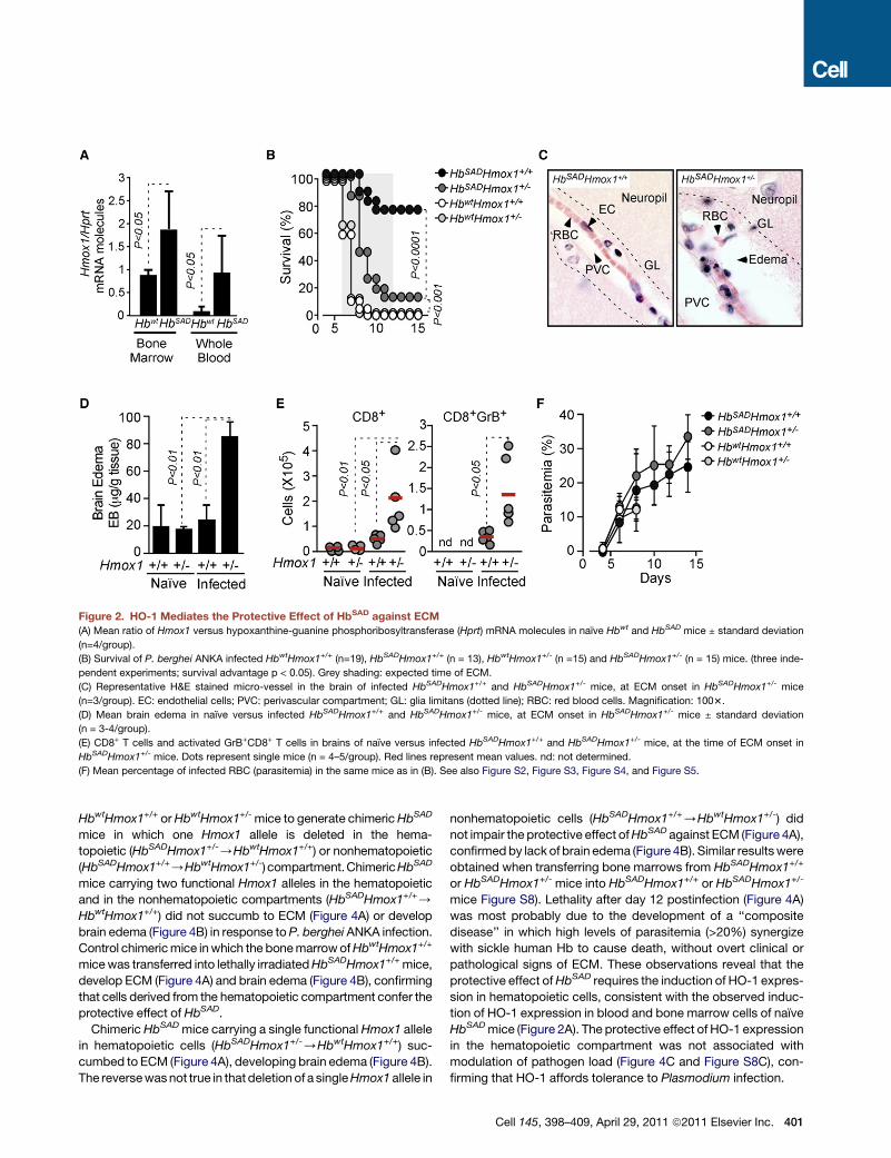
Figure 2. HO-1 Mediates the Protective Effect of HbSAD against ECM
(A) Mean ratio of Hmox1 versus hypoxanthine-guanine phosphoribosyltransferase (Hprt) mRNA molecules in naıve Hbwt and HbSAD mice ± standard deviation
(n=4/group).
(B) Survival of P. berghei ANKA infected HbwtHmox1+/+ (n=19), HbSADHmox1+/+ (n = 13), HbwtHmox1+/- (n =15) and HbSADHmox1+/- (n = 15) mice. (three inde-
pendent experiments; survival advantage p < 0.05). Grey shading: expected time of ECM.
(C) Representative H&E stained micro-vessel in the brain of infected HbSADHmox1+/+ and HbSADHmox1+/- mice, at ECM onset in HbSADHmox1+/- mice
(n=3/group). EC: endothelial cells; PVC: perivascular compartment; GL: glia limitans (dotted line); RBC: red blood cells. Magnification: 1003.
(D) Mean brain edema in naıve versus infected HbSADHmox1+/+ and HbSADHmox1+/- mice, at ECM onset in HbSADHmox1+/- mice ± standard deviation
(n = 3-4/group).
(E) CD8+ T cells and activated GrB+CD8+ T cells in brains of naıve versus infected HbSADHmox1+/+ and HbSADHmox1+/- mice, at the time of ECM onset in
HbSADHmox1+/- mice. Dots represent single mice (n = 4–5/group). Red lines represent mean values. nd: not determined.
(F) Mean percentage of infected RBC (parasitemia) in the same mice as in (B). See also Figure S2, Figure S3, Figure S4, and Figure S5.
HbwtHmox1+/+ orHbwtHmox1+/- mice to generate chimericHbSAD
mice in which one Hmox1 allele is deleted in the hema-
topoietic (HbSADHmox1+/-/HbwtHmox1+/+) or nonhematopoietic
(HbSADHmox1+/+/HbwtHmox1+/-) compartment.ChimericHbSAD
mice carrying two functional Hmox1 alleles in the hematopoietic
and in the nonhematopoietic compartments (HbSADHmox1+/+/
HbwtHmox1+/+) did not succumb to ECM (Figure 4A) or develop
brain edema (Figure 4B) in response toP. berghei ANKA infection.
Control chimericmice inwhich the bonemarrowofHbwtHmox1+/+
micewas transferred into lethally irradiatedHbSADHmox1+/+mice,
develop ECM (Figure 4A) and brain edema (Figure 4B), confirming
that cells derived from the hematopoietic compartment confer the
protective effect of HbSAD.
Chimeric HbSADmice carrying a single functional Hmox1 allele
in hematopoietic cells (HbSADHmox1+/-/HbwtHmox1+/+) suc-
cumbed to ECM (Figure 4A), developing brain edema (Figure 4B).
The reversewasnot true in that deletion of a singleHmox1allele in
nonhematopoietic cells (HbSADHmox1+/+/HbwtHmox1+/-) did
not impair the protective effect ofHbSAD against ECM (Figure 4A),
confirmedby lack of brain edema (Figure 4B). Similar resultswere
obtained when transferring bone marrows from HbSADHmox1+/+
or HbSADHmox1+/- mice into HbSADHmox1+/+ or HbSADHmox1+/-
mice Figure S8). Lethality after day 12 postinfection (Figure 4A)
was most probably due to the development of a ‘‘composite
disease’’ in which high levels of parasitemia (>20%) synergize
with sickle human Hb to cause death, without overt clinical or
pathological signs of ECM. These observations reveal that the
protective effect ofHbSAD requires the induction of HO-1 expres-
sion in hematopoietic cells, consistent with the observed induc-
tion of HO-1 expression in blood and bone marrow cells of naıve
HbSADmice (Figure 2A). The protective effect of HO-1 expression
in the hematopoietic compartment was not associated with
modulation of pathogen load (Figure 4C and Figure S8C), con-
firming that HO-1 affords tolerance to Plasmodium infection.
Cell 145, 398–409, April 29, 2011 ª2011 Elsevier Inc. 401

Figure 3. Induction of HO-1 by HbSAD Inhibits Chemokine Production in the Brain
Quantification of mRNA encoding chemokines and chemokine receptors in the brains of naıve (-) and P. berghei ANKA infected (+) mice carrying one (-) or two (+)
functional Hmox1 alleles and expressing (+) HbSAD or not (-). Results are shown as mean fold induction over naıve HbwtHmox1+/+ mice ± standard deviation
(n = 4-8/group), analyzed at ECM onset in Hbwt or HbSADHmox1+/- control groups.
(A) Genes inhibited by HbSAD under the control of HO-1.
(B) Genes inhibited by HbSAD, presumably not under the control of HO-1.
(C) Genes not regulated by HbSAD. *p < 0.05; **p < 0.01; ns P > 0.05. See also Figure S6 and Figure S7.
Sickle Hb Inhibits the Activation/Expansion of CD8+ TCells Recognizing Antigens Expressed by Plasmodium
The number of splenic CD8+ T cells recognizing specifically
a MHC I-restricted epitope derived from glycoprotein B
(gB498-505) of herpes simplex virus-1 expressed by transgenic
P. berghei ANKA (Lundie et al., 2008) was reduced in HbSAD
versusHbwtmice, as assessed five days after infection (Figure 4A
and Figure S9A). The number of splenic GrB+CD8+ T cells was
also reduced in HbSAD versus Hbwt mice five days after infection
(Figure 4E and Figure S9B). This reveals that HbSAD prevents
overt expansion of pathogenic CD8+ T cells, an effect that should
contribute to the protective effect of HbSAD against ECM (Bel-
noue et al., 2002; Lundie et al., 2008). We then asked whether
this immunoregulatory effect of HbSAD involved the expression
of HO-1. The number of gB498-505-specific CD8+ T cells
and GrB+CD8+ T cells was not different in the spleen of
HbSADHmox1+/- versus HbSADHmox1+/+ mice five days after
infection (Figures 4D and 4E and Figures S9A and S9B). This
suggests that HbSAD controls the activation and/or expansion
of splenic CD8+ T cells, via a mechanism that probably does
not involve HO-1.
Sickle Hb Induces HO-1 Expression via a MechanismInvolving Nrf2Given that Nrf2 plays a central role in the transcriptional regula-
tion of HO-1 expression (Alam et al., 1999) we asked whether
induction of HO-1 expression in whole blood leukocytes of naıve
HbSAD mice (Figure 2A) involved this transcription factor. Dele-
tion of oneNrf2 allele inHbSADmice (HbSADNrf2+/-) was sufficient
to reduce the level of Hmox1 mRNA expression in whole blood
leukocytes, to those of naıve HbwtNrf2+/+ mice (Figure 5A).
This suggests that sickle Hb induces Hmox1 transcription and
expression via a mechanism involving Nrf2. Incidence of ECM
increased significantly in P. berghei ANKA infected HbSADNrf2+/-
402 Cell 145, 398–409, April 29, 2011 ª2011 Elsevier Inc.
versus HbSADNrf2+/+ mice (Figure 5B), confirmed by the devel-
opment of brain edema (Figure 5C). A similar effect was
observed in a limited number of HbSAD mice in which both
Nrf2 alleles were functionally deleted, i.e. HbSADNrf2-/- mice
(n = 5; 20% survival). It should be noted that deletion of both
Nrf2 alleles in HbSAD mice lead to overt postnatal lethality
(Table S2B). Loss of protection against ECM in HbSADNrf2+/-
versus HbSADNrf2+/+ mice was not associated with a regain of
CD8+ T cell activation and/or expansion in the spleen, as as-
sessed five days after infection (Figure S10). This suggests
that the immunoregulatory of HbSAD probably does not involve
Nrf2, which is consistent with the observation that this effect
also does not seem to involve HO-1, a gene regulated by HbSAD
via Nrf2 (Figure 5A).
The protective effect of HbSAD against lung and liver injury
associated to P. berghei ANKA infection was lost inHbSADNrf2+/-
versus HbSADNrf2+/+ mice (Figure S2). This was not associated
with increased parasite load in HbSADNrf2+/- versus
HbSADNrf2+/+ mice (Figure 5D), which is consistent with the
notion that induction of HO-1 expression by Nrf2 confers toler-
ance to Plasmodium infection.
Sickle Hb Confers Tolerance to Plasmodium Infectionvia a Mechanism Involving CO Produced through HemeCatabolism by HO-1Consistent with similar observations in individuals carrying the
HbS mutation in the homozygous (Reiter et al., 2002) or hetero-
zygous (Muller-Eberhard et al., 1968) form, naıve HbSAD mice
had higher concentration of free heme in plasma, as compared
to age-matched control naıve Hbwt mice (Figure 6A). When
pre-exposed in vitro to low levels of free heme cells are protected
against a subsequent heme challenge (Balla et al., 1992). We
asked whether free heme would exert a similar protective effect
in vivo. Administration of free heme to Hbwt mice prior to

Figure 4. The Protective But Not The Immunoregulatory Effect of Sickle Hb against ECM Involves the Expression of HO-1 in HematopoieticCells
(A) Survival of P. berghei ANKA infected chimeric mice resulting from the adoptive transfer of bone marrow from HbSADHmox1+/+ mice into HbwtHmox1+/+
recipients (n = 6); from Hbwt Hmox1+/+ mice into HbSADHmox1+/+ recipients (n = 5); from HbSADHmox1+/+ mice into HbwtHmox1+/- recipients (n = 8) and from
HbSADHmox1+/- into HbwtHmox1+/+ recipients (n = 7). Recipients were lethally irradiated before the adoptive transfer. Grey shading indicates expected time of
ECM. Pooled from four independent experiments.
(B) Mean brain edema ± standard deviation (n = 3/group) in chimeric mice, produced as in (A).
(C) Mean percentage of infected RBC in chimeric mice ± standard deviation, same mice as in (A).
(D) Number of splenic CD8+ T cells specific for gB498-505 in HbwtHmox1+/+, HbSADHmox1+/+ and HbSADHmox1+/� mice not infected (naıve) or 5 days after
P. berghei ANKA infection.
(E) Number of splenic CD8+GrB+ T in the samemice as in (d). Circles in (d) and (e) represent single mice and red linesmean values (n = 10 and n = 16 in noninfected
and infected groups, respectively), pooled from 4 independent experiments with similar results.
See also Figure S8 and Figure S9.
P. berghei ANKA infection suppressed ECM incidence, as
compared to vehicle-treated Hbwt mice (Figure 6B).
The protective effect of heme was dose-dependent, with
higher dosage leading to (1) increased HO-1 expression in
whole blood cells (Figure S1A) and spleen (Figure S1B) and to
a lesser extent in the bone marrow (Figure S11C) and (2)
suppression of ECM (Figure 11D). This protective effect was
not associated with modulation of parasitemia (Figure S11D),
suggesting that low concentration of free heme in the
plasma of naıve HbSAD mice (Figure 6a) can confer tolerance
to Plasmodium infection.
We asked whether accumulation of low levels of free heme in
HbSAD contributes to the immunoregulatory effect exerted by
HbSAD on CD8+ T cells (Figures 4D and 4E). Administration of
free heme to Hbwt mice, prior to infection with transgenic
P. berghei ANKA expressing gB498-505, reduced the number of
splenic gB498-505-specific CD8+ T cells (Figures S12A and
S12B) as well as GrB+CD8+ T cells, as compared to vehicle
treated Hbwt mice five days after infection (Figures S12C and
S12D). This supports further the notion that the protective effect
of HbSAD against ECM is mediated, to a large extent, via the
accumulation of low levels of circulating free heme.
Plasma free heme concentration increased significantly
following P. berghei ANKA infection in Hbwt mice (Figure 6A),
an effect we have previously shown to contribute in a critical
manner to the pathogenesis of ECM (Ferreira et al., 2008;
Pamplona et al., 2007). Albeit less pronounced this increase
was also observed in HbSAD mice (Figure 6A). When challenged
with free heme after infection, HbSAD succumbed to ECM (Fig-
ure 6C), confirmed by the occurrence of brain edema (Figure 6d).
This reveals that free heme has a dual effect in the control of ECM
onset, being protective when present at slightly above normal
concentration before infection (Figure 6B) while highly patho-
genic when present at higher levels after infection (Figure 6C).
Free heme did not interfere with pathogen load (Figures S11E
and S11F), revealing that when present at slightly above normal
concentration before infection free heme promotes tolerance to
malaria, while impairing tolerance to malaria when present at
Cell 145, 398–409, April 29, 2011 ª2011 Elsevier Inc. 403

Figure 5. Sickle Human Hb Prevents the
Onset of ECM via the Induction of HO-1
expression by Nrf2
(A) Mean ratio of Hmox1 versus hypoxanthine-
guanine phosphoribosyltransferase (Hprt) mRNA
molecules in peripheral blood mononuclear cells
of naıve HbwtNrf2+/+, HbwtNrf2+/-, HbSADNrf2+/+
and HbSADNrf2+/- mice ± standard deviation
(n = 6–8/group).
(B) Survival of P. berghei ANKA-infected
HbwtNrf2+/+ (n = 6), HbwtNrf2+/- (n = 13),
HbSADNrf2+/+ (n = 10) and HbSADNrf2+/- (n = 14)
mice, three independent experiments. Grey
shading indicates expected time of ECM.
(C) Brain edemawasmeasured by Evans blue (EB)
accumulation in brains of infected HbwtNrf2+/+,
HbwtNrf2+/-, HbSADNrf2+/+ and HbSADNrf2+/- mice,
at the time of ECM onset. Mean ± standard devi-
ation (n=4-5/group).
(D) Mean percentage of infected RBC (para-
sitemia) ± standard deviation, samemice as in (B).
See also Figure S10.
higher concentrations after infection. Heme administration at the
same dosage and schedule to naıve Hbwt or HbSAD mice did not
result in lethality (data not shown).
When applied via inhalation to wild type mice, CO suppresses
the pathogenesis of ECM via amechanism that relies on the inhi-
bition of heme release fromHb (Pamplona et al., 2007).We asked
whether the protective effect of HbSAD against ECM was medi-
ated via this mechanism. Inhaled CO suppressed the incidence
of ECM in HbSADHmox1+/- mice (Figure 6E), confirmed by the
lack of brain edema (Figure 6F). A similar protective effect was
observed when CO was applied to P. berghei infected HbSAD
mice treated with the enzymatic HO inhibitor ZnPPIX (Figures
S5E and S5F). CO did not modulate parasitemia (Figure 1G
and Figures S11G and S11H). Instead, its protective effect was
associated with reduction of plasma free heme concentration,
below that of naıveHbSADHmox1+/-mice (Figure 6G). Administra-
tion of free heme to infectedHbSADHmox1+/-mice abrogated the
protective effect of CO, restoring ECM incidence (Figure 6H),
confirmed by brain edema (Figure 6I). Heme was not toxic
when administered at the same dosage and schedule to naıve
HbSADHmox1+/- mice receiving CO, i.e. 0% mortality. These
observations demonstrate that sickle Hb suppresses the onset
of ECMvia the induction ofHO-1 and theproduction ofCO,which
inhibits the accumulation of free heme thus affording tolerance to
Plasmodium infection (Figure 7).
DISCUSSION
The protective effect of sickle human Hb against malaria is
thought to rely on the reduction of parasite load (Allison, 1954;
404 Cell 145, 398–409, April 29, 2011 ª2011 Elsevier Inc.
May et al., 2007; Williams et al., 2005b),
implying that sickle human Hb affords re-
sistance (Raberg et al., 2007; Schneider
and Ayres, 2008) to Plasmodium infec-
tion. Consistent with this notion, sickle
human Hb decreases RBC permissive-
ness to Plasmodium invasion and growth (Friedman, 1978;
Pasvol et al., 1978) while increasing phagocytosis of infected
RBC, as assessed ex vivo (Ayi et al., 2004).Whether these effects
account for the protective effect of sickle human Hb in vivo
remained to be established. We used a well-established mouse
model of malaria allowing for genetic manipulation of the host,
to test in vivo the relative contribution of host specific genes to
the protective effect of sickle Hb against Plasmodium infection.
For technical and ethical reasons these studies can only be
performed in rodent models of malaria.
As demonstrated hereby, sickle Hb affords protection
against Plasmodium infection in mice (Figure 1A and Figure 2),
a finding consistent with previous reports using different mouse
and Plasmodium strains (Hood et al., 1996; Shear et al., 1993).
This survival advantage occurs irrespectively of parasite load
(Figures 1E and 1F) and is not associated with modulation of
parasite sequestering in different organs (Figure S4), revealing
that sickle human Hb confers tolerance (Raberg et al., 2007;
Schneider and Ayres, 2008) to Plasmodium infection. This is
consistent with our recent observation that heme catabolism
by HO-1 also suppresses development of multiple organ
dysfunction associated with the pathogenesis of severe sepsis
in mice (Larsen et al., 2010), a lethal outcome of polymicrobial
infection that resembles, in some aspects, severe malaria
(Clark et al., 2004). Since the survival advantage conferred
by HbS against malaria in human populations can occur
without overt decrease of parasite load (Crompton et al.,
2008; Livincstone, 1971; Motulsky et al., 1966), tolerance to
Plasmodium infection might also operate in individuals ex-
pressing sickle Hb.

Figure 6. HbSAD Inhibits Free Heme Accu-
mulation via the Production of CO
(A) Mean plasma free heme concentration in naıve
versus P. berghei ANKA infected Hbwt and HbSAD
mice at ECM onset in Hbwt mice ± standard
deviation (n = 4–15/group).
(B) Survival of infectedHbwtmice receiving vehicle
(n = 25) or heme (35–40 mg/kg, every 48 hr, day 2
preinfection to 4 postinfection) (n = 17). Pooled
from four independent experiments, with similar
results.
(C) Survival of infected HbSAD mice receiving
vehicle (n = 8) or heme (20mg/kg, every 12 hr, day
4–7 postinfection) (n = 8). Pooled from two inde-
pendent experiments, with similar results.
(D) Mean brain edema in HbSAD mice treated as in
(c) at ECM onset in heme-treated HbSAD mice ±
standard deviation (n = 4/group).
(E) Survival of infected HbSADHmox1+/- mice
exposed to air (n = 8) or CO (250 ppm, days 4-7
post infection) (n = 12). Pooled from three inde-
pendent experiments, with similar results.
(F) Mean brain edema in HbSADHmox1+/- mice
treated as in (E), at ECM onset in air-treated
mice ± standard deviation (n =3–4/group).
(G) Mean free heme in plasma of HbSADHmox1+/-
mice treated as in (E) ± standard deviation (n =
4–6/group).
(H) Survival of infected HbSADHmox1+/- mice
exposed to CO (250ppm; days 4–7 postinfection)
and receiving vehicle (n = 8) or heme (20 mg/kg,
every 12 hr, days 4–7 postinfection) (n = 8). Pooled
from two independent experiments, with similar
results.
(I) Mean brain edema in HbSADHmox1+/- mice
treated as in (H), at ECM onset in heme-treated
mice ± standard deviation (n = 3–4/group). Grey
shading in (B, C, E, andH) indicates expected time
of ECM. See also Figure S11 and Figure S12.
We provide evidence for the existence of a specific molecular
mechanism via which sickle humanHb confers tolerance toPlas-
modium infection. When expressed at nonpathological levels in
mice, sickle Hb leads to the accumulation of low concentrations
of free heme in plasma (Figure 6A). The same is true for individ-
uals carrying the sickle cell trait (Muller-Eberhard et al., 1968),
which affords protection against malaria (Allison, 1954; Beet,
1947; Jallow et al., 2009; Williams, 2006). Presumably, this is
due to the higher rate of heme release from sickle versus normal
human Hb (Hebbel et al., 1988). In the absence of overt inflam-
mation, free heme induces HO-1 expression without causing
cytotoxicity (Balla et al., 1992; Gozzelino et al., 2010). Presum-
ably, this explains how sickle human Hb induces the expression
of HO-1 in human (Jison et al., 2004) and mouse (Figure 2A)
peripheral bloodmononuclear cells aswell as in human endothe-
lial cells (Bains et al., 2010; Nath et al., 2001). Expression of HO-1
Cell 145, 398–4
prevents the cytotoxic effects of free
heme (Balla et al., 1992; Gozzelino
et al., 2010), hence limiting the patholog-
ical outcome of sickle cell anemia in mice
(Belcher et al., 2006).
The mechanism via which sickle Hb induces the expression of
HO-1 in vivo involves Nrf2 (Figure 5A), a transcription factor
previously shown to regulate Hmox1 expression (Alam et al.,
1999; Kensler et al., 2007). Induction of HO-1 via Nrf2 affords
protection against malaria in HbSAD mice expressing sickle Hb
(Figure 2B and Figure 5B). This protective effect occurs irrespec-
tively of parasite load (Figure 2F and Figure 5D) or parasite
sequestration in different organs (Figure S4), revealing that sickle
human Hb affords tolerance to Plasmodium infection via the
Nrf2/HO-1 system.
The protective effect of HO-1 against sickle cell anemia
(Belcher et al., 2006) and against malaria is mediated by the
same end-product of heme catabolism, namely CO (Figures 6E
and 6F). This gasotransmitter inhibits Hb oxidation and subse-
quently heme release from Hb (Hebbel et al., 1988) (Figure 6G),
thus preventing free heme from participating in the pathogenesis
09, April 29, 2011 ª2011 Elsevier Inc. 405
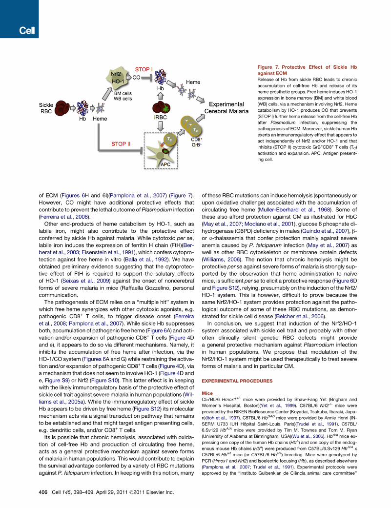
Figure 7. Protective Effect of Sickle Hb
against ECM
Release of Hb from sickle RBC leads to chronic
accumulation of cell-free Hb and release of its
heme prosthetic groups. Free heme induces HO-1
expression in bone marrow (BM) and white blood
(WB) cells, via a mechanism involving Nrf2. Heme
catabolism by HO-1 produces CO that prevents
(STOP I) further heme release from the cell-free Hb
after Plasmodium infection, suppressing the
pathogenesis of ECM.Moreover, sickle humanHb
exerts an immunoregulatory effect that appears to
act independently of Nrf2 and/or HO-1 and that
inhibits (STOP II) cytotoxic GrB+CD8+ T cells (TC)
activation and expansion. APC: Antigen present-
ing cell.
of ECM (Figures 6H and 6I)(Pamplona et al., 2007) (Figure 7).
However, CO might have additional protective effects that
contribute to prevent the lethal outcome ofPlasmodium infection
(Ferreira et al., 2008).
Other end-products of heme catabolism by HO-1, such as
labile iron, might also contribute to the protective effect
conferred by sickle Hb against malaria. While cytotoxic per se,
labile iron induces the expression of ferritin H chain (FtH)(Ber-
berat et al., 2003; Eisenstein et al., 1991), which confers cytopro-
tection against free heme in vitro (Balla et al., 1992). We have
obtained preliminary evidence suggesting that the cytoprotec-
tive effect of FtH is required to support the salutary effects
of HO-1 (Seixas et al., 2009) against the onset of noncerebral
forms of severe malaria in mice (Raffaella Gozzelino, personal
communication.
The pathogenesis of ECM relies on a ‘‘multiple hit’’ system in
which free heme synergizes with other cytotoxic agonists, e.g.
pathogenic CD8+ T cells, to trigger disease onset (Ferreira
et al., 2008; Pamplona et al., 2007). While sickle Hb suppresses
both, accumulation of pathogenic free heme (Figure 6A) and acti-
vation and/or expansion of pathogenic CD8+ T cells (Figure 4D
and e), it appears to do so via different mechanisms. Namely, it
inhibits the accumulation of free heme after infection, via the
HO-1/CO system (Figures 6A and G) while restraining the activa-
tion and/or expansion of pathogenic CD8+ T cells (Figure 4D), via
a mechanism that does not seem to involve HO-1 (Figure 4D and
e, Figure S9) or Nrf2 (Figure S10). This latter effect is in keeping
with the likely immunoregulatory basis of the protective effect of
sickle cell trait against severe malaria in human populations (Wil-
liams et al., 2005a). While the immunoregulatory effect of sickle
Hb appears to be driven by free heme (Figure S12) its molecular
mechanism acts via a signal transduction pathway that remains
to be established and that might target antigen presenting cells,
e.g. dendritic cells, and/or CD8+ T cells.
Its is possible that chronic hemolysis, associated with oxida-
tion of cell-free Hb and production of circulating free heme,
acts as a general protective mechanism against severe forms
ofmalaria in human populations. This would contribute to explain
the survival advantage conferred by a variety of RBC mutations
against P. falciparum infection. In keeping with this notion, many
406 Cell 145, 398–409, April 29, 2011 ª2011 Elsevier Inc.
of these RBCmutations can induce hemolysis (spontaneously or
upon oxidative challenge) associated with the accumulation of
circulating free heme (Muller-Eberhard et al., 1968). Some of
these also afford protection against CM as illustrated for HbC
(May et al., 2007; Modiano et al., 2001), glucose 6 phosphate di-
hydrogenase (G6PD) deficiency in males (Guindo et al., 2007), b-
or a-thalassemia that confer protection mainly against severe
anemia caused by P. falciparum infection (May et al., 2007) as
well as other RBC cytoskeleton or membrane protein defects
(Williams, 2006). The notion that chronic hemolysis might be
protective per se against severe forms of malaria is strongly sup-
ported by the observation that heme administration to naıve
mice, is sufficient per se to elicit a protective response (Figure 6D
and Figure S12), relying, presumably on the induction of the Nrf2/
HO-1 system. This is however, difficult to prove because the
same Nrf2/HO-1 system provides protection against the patho-
logical outcome of some of these RBC mutations, as demon-
strated for sickle cell disease (Belcher et al., 2006).
In conclusion, we suggest that induction of the Nrf2/HO-1
system associated with sickle cell trait and probably with other
often clinically silent genetic RBC defects might provide
a general protective mechanism against Plasmodium infection
in human populations. We propose that modulation of the
Nrf2/HO-1 system might be used therapeutically to treat severe
forms of malaria and in particular CM.
EXPERIMENTAL PROCEDURES
Mice
C57BL/6 Hmox1+/- mice were provided by Shaw-Fang Yet (Brigham and
Women’s Hospital, Boston)(Yet et al., 1999). C57BL/6 Nrf2-/- mice were
provided by the RIKEN BioResource Center (Koyadai, Tsukuba, Ibaraki, Japa-
n)(Itoh et al., 1997). C57BL/6 HbSAD mice were provided by Annie Henri (IN-
SERM U733 IUH Hopital Saint-Louis, Paris)(Trudel et al., 1991). C57BL/
6.Sv129 HbA/A mice were provided by Tim M. Townes and Tom M. Ryan
(University of Alabama at Birmingham, USA)(Wu et al., 2006). HbA/a mice ex-
pressing one copy of the human Hb chains (HbA) and one copy of the endog-
enous mouse Hb chains (Hba) were produced from C57BL/6.Sv129 HbA/A x
C57BL/6 Hbwt mice (or C57BL/6 Hba/a) breeding. Mice were genotyped by
PCR (Hmox1 and Nrf2) and isoelectric focusing (Hb), as described elsewhere
(Pamplona et al., 2007; Trudel et al., 1991). Experimental protocols were
approved by the ‘‘Instituto Gulbenkian de Ciencia animal care committee’’

and by the ‘‘Direccao Geral de Veterenaria (DGV)’’ of the Portuguese Ministry
of Agriculture, Rural Development and Fisheries (License 018831-2010-09-03).
Bone Marrow Chimeras
Bone marrow chimeras were generated in Hmox1+/+, Hmox1+/- mice express-
ing or not the HbSAD allele (8–10 weeks) as described (Seixas et al., 2009).
Parasites, Infection, and Disease assessment
Mice were infected with (GFP)-P. berghei ANKA (Pamplona et al., 2007),
GFP-Luciferase P. berghei ANKA (MR4-866) or a (GFP)-P. berghei transgenic
parasite expressing different MHC II and MHC I restricted epitopes including
the MHC I-restricted epitope derived from glycoprotein B of herpes simplex
virus-1 (gB498-505)(Lundie et al., 2008), provided by William R. Heath (Walter
and Eliza Hall, Melbourne, Victoria, Australia). Parasitemias were determined
by flow cytometry (Pamplona et al., 2007). Infected mice were monitored twice
daily for clinical symptoms of ECM.
Visualization and Quantification of Luciferase Activity
P. berghei ANKA Infected Mice
Luciferase activity was visualized by imaging of dissected tissues using an
electron multiplying-charge-coupled device (EM-CCD) photon-counting
camera (ImagEM, Hamamatsu).
Protoporphyrins
Iron-protoporphyrin IX (FePPIX; heme) and zinc-protoporphyrin IX (ZnPPIX)
were dissolved in 0.2 M NaOH, neutralized (pH 7.4) with 0.2 M HCl and admin-
istered (i.p.), as described (Pamplona et al., 2007).
CO Treatment
Mice were placed in a gastight 60 L capacity chamber and exposed continu-
ously between days 4–7 postinfection to CO at a flow rate of �12 L/min
(final concentration of 250 parts per million; ppm), as described (Pamplona
et al., 2007; Sato et al., 2001). CO concentration was monitored using a CO
analyzer (Interscan Corporation, Chatsworth).
Histology
Brains were harvested, when clinical signs of ECM were noticed in control
mice. Tissue was fixed in buffered 4% (vol/vol) paraformaldehyde and histo-
logical analysis was performed on perfusion-fixed tissues.
BBB Permeability
Mice were injected intravenously (i.v.) with 0.1 ml of 2% Evans Blue (Sigma)
when clinical symptoms of ECM were noticed in control mice (Pamplona
et al., 2007).
Analyzes of Splenic CD8+ T Cell Activation
Intracellular granzyme B staining were performed as described elsewhere
(Lundie et al., 2008). Analyzes of CD8+ T cells recognizing theMHC I-restricted
epitope gB498-505 (SSIEFARL) from glycoprotein B of herpes simplex virus-1,
was performed as described elsewhere (Lundie et al., 2008).
Leukocyte Brain Infiltration
Leukocytes were isolated from the brain of P. berghei ANKA infected mice
when clinical symptoms of ECM were detectable in control groups. Brain
leukocyte infiltration was quantified by flow cytometry (Pamplona et al., 2007).
Quantitative Real-Time Reverse Transcription PCR
Mice were sacrificed at day ECMonset inHbwtmice.Hmox1mRNAwas quan-
tified by Quantitative real-time reverse transcription PCR (qRT-PCR) (Roche
System)(Pamplona et al., 2007). TaqMan� Gene Signature Mouse Immune
Array (Applied Biosystems) was used to quantify all other mRNAs (7900HT
ABI system), according to manufactures recommendations.
Serum Biochemistry
Hematograms were measured by focused flow technology (Hemavet
Multispecies Hematology System, HV950FS, Drew Scientific Inc., Centro
Diagnostivo Veterinario, Lisboa, Portugal). Plasma Hb was determined by
spectroscopy at l=577. Total plasma heme was measured using the 3,30,5,50 tetramethylbenzidine (TMB) peroxidase assay (BD Biosciences), at
l=655 nm.
Statistical Analysis
Nonparametric Mann-Whitney U test was used to assess statistical signifi-
cance between averages in different samples in which n<5. In samples with
nR5 the unpaired Student’s t-test for unequal variances was used. Normal
distributions were confirmed using the Kolmogorov-Smirnov test. Significant
differences in survival were evaluated by the generation of Kaplan-Meier plots
and by performing log-rank analysis for all experiments in which survival was
assessed as an end-point. Statistical analysis for the progeny-expected ratios
was performed using Pearson’s chi-squared tests. *P<0.05 or **P<0.01 were
considered statistically significant.
SUPPLEMENTAL INFORMATION
Supplemental Information includes Extended Experimental Procedures,
twelve figures, and two tables and can be found with this article online at
doi:10.1016/j.cell.2011.03.049.
ACKNOWLEDGMENTS
We thank Ruslan Medzhitov for intellectual support and encouragement by
means of many insightful discussions, Thiago Carvalho (Instituto Gulbenkian
de Cie ncia) and Rui Costa Fundacao Champalimaud as well asMarcelo Bozza
(Universidade Federal do Rio De Janeiro), Robert P. Hebbel and Gregory Ver-
cellotti (University of Minnesota, USA) for critical review of the manuscript,
Nuno Sepulveda for support in statistical analysis, Tim M. Townes and Tom
M. Ryan (University of Alabama at Birmingham) for providing the HbA/A
mice. Sılvia Cardoso and Matteo Villa for mouse breeding and genotyping.
This work was supported by ‘‘Fundacao para a Ciencia e a Tecnologia’’,
Portugal grants PTDC/SAU-MII/71140/2006 and SFRH/BPD/21707/2005
(AF), SFHR/BD/33218/2007 (IM), PTDC/SAU-MII/71140/2006, PTDC/BIA-
BCM/101311/2008, PTDC/SAU-FCF/100762/2008, GEMI Fund Linde Health-
care, European Community and LSH-2005-1.2.5-1 (MPS), FP7-PEOPLE-
2007-2-1-IEF (VJ). I.B. is supported by the DFG, BMBF, Dr. Senckenberg-Stif-
tung, Kassel-Stiftung andMesser-Stiftung, Germany. Annie Henri is supported
by INSERMandYvesBeuzard by Paris VII University, Commissariat a l’Energie
Atomique and Agence Nationale de la Recherche Scientifique, France.
Author contribution: A.F. contributed to study design, performed and/or
contributed critically to all experiments, analyzed data and was assisted to
do so by NRP. A.C. performed experiments and analysis of leukocyte infiltra-
tion and generation of bonemarrow chimeric animals with A.F. I.M.: performed
experiments and interpreted data revealing the immunoregulatory effect of
sickle hemoglobin with A.F. I.B. provided expert analysis, advice and teaching
on immunopathology. V.J. determined free heme concentrations in plasma
and quantified HO activity. S.R. generated and maintained all mouse colonies
used. A.H. provided the HbSAD mice. YB provided mentorship and advise on
sickle cell mouse model. M.P.S. formulated the original hypothesis, drove
most of the study design, analyzed and provided mentorship. The manuscript
was written by M.P.S. with assistance from A.F. and Y.B.
Received: June 21, 2010
Revised: January 3, 2011
Accepted: March 28, 2011
Published: April 28, 2011
REFERENCES
Alam, J., Stewart, D., Touchard, C., Boinapally, S., Choi, A.M., and Cook, J.L.
(1999). Nrf2, a Cap’n’Collar transcription factor, regulates induction of the
heme oxygenase-1 gene. J. Biol. Chem. 274, 26071–26078.
Allison, A.C. (1954). Protection afforded by sickle-cell trait against subtertian
malareal infection. BMJ 1, 290–294.
Cell 145, 398–409, April 29, 2011 ª2011 Elsevier Inc. 407
Ayi, K., Turrini, F., Piga, A., and Arese, P. (2004). Enhanced phagocytosis of
ring-parasitized mutant erythrocytes: a common mechanism that may explain
protection against falciparum malaria in sickle trait and beta-thalassemia trait.
Blood 104, 3364–3371.
Bains, S.K., Foresti, R., Howard, J., Atwal, S., Green, C.J., and Motterlini, R.
(2010). Human sickle cell blood modulates endothelial heme oxygenase
activity: effects on vascular adhesion and reactivity. Arterioscler. Thromb.
Vasc. Biol. 30, 305–312.
Balla, G., Jacob, H.S., Balla, J., Rosenberg, M., Nath, K., Apple, F., Eaton,
J.W., and Vercellotti, G.M. (1992). Ferritin: a cytoprotective antioxidant strate-
gem of endothelium. J. Biol. Chem. 267, 18148–18153.
Beet, E.A. (1947). Sickle cell disease in Northern Rhodesia. East Afr. Med. J.
24, 212–222.
Belcher, J.D., Mahaseth, H., Welch, T.E., Otterbein, L.E., Hebbel, R.P., and
Vercellotti, G.M. (2006). Heme oxygenase-1 is a modulator of inflammation
and vaso-occlusion in transgenic sickle mice. J. Clin. Invest. 116, 808–816.
Belnoue, E., Kayibanda, M., Vigario, A.M., Deschemin, J.C., van Rooijen, N.,
Viguier, M., Snounou, G., and Renia, L. (2002). On the pathogenic role of
brain-sequestered alphabeta CD8+ T cells in experimental cerebral malaria.
J. Immunol. 169, 6369–6375.
Berberat, P.O., Katori, M., Kaczmarek, E., Anselmo, D., Lassman, C., Ke, B.,
Shen, X., Busuttil, R.W., Yamashita, K., Csizmadia, E., et al. (2003). Heavy
chain ferritin acts as an antiapoptotic gene that protects livers from ischemia
reperfusion injury. FASEB J. 17, 1724–1726.
Campanella, G.S., Tager, A.M., El Khoury, J.K., Thomas, S.Y., Abrazinski, T.A.,
Manice, L.A., Colvin, R.A., and Luster, A.D. (2008). Chemokine receptor
CXCR3 and its ligands CXCL9 and CXCL10 are required for the development
of murine cerebral malaria. Proc. Natl. Acad. Sci. USA 105, 4814–4819.
Clark, I.A., Alleva, L.M., Mills, A.C., and Cowden, W.B. (2004). Pathogenesis of
malaria and clinically similar conditions. Clin. Microbiol. Rev. 17, 509–539.
Crompton, P.D., Traore, B., Kayentao, K., Doumbo, S., Ongoiba, A., Diakite,
S.A., Krause, M.A., Doumtabe, D., Kone, Y., Weiss, G., et al. (2008). Sickle
cell trait is associated with a delayed onset of malaria: implications for time-
to-event analysis in clinical studies of malaria. J. Infect. Dis. 198, 1265–1275.
Eisenstein, R.S., Garcia, M.D., Pettingell, W., and Munro, H.N. (1991). Regula-
tion of ferritin and heme oxygenase synthesis in rat fibroblasts by different
forms of iron. Proc. Natl. Acad. Sci. USA 88, 688–692.
Ferreira, A., Balla, J., Jeney, V., Balla, G., and Soares, M.P. (2008). A central
role for free heme in the pathogenesis of severe malaria: the missing link? J.
Mol. Med. 86, 1097–1111.
Friedman, M.J. (1978). Erythrocytic mechanism of sickle cell resistance to ma-
laria. Proc. Natl. Acad. Sci. USA 75, 1994–1997.
Gozzelino, R., Jeney, V., and Soares, M.P. (2010). Mechanisms of cell protec-
tion by heme oxygenase-1. Annu. Rev. Pharmacol. Toxicol. 50, 323–354.
Guindo, A., Fairhurst, R.M., Doumbo, O.K., Wellems, T.E., and Diallo, D.A.
(2007). X-linked G6PD deficiency protects hemizygous males but not hetero-
zygous females against severe malaria. PLoS Med. 4, e66.
Hebbel, R.P., Morgan, W.T., Eaton, J.W., and Hedlund, B.E. (1988). Acceler-
ated autoxidation and heme loss due to instability of sickle hemoglobin.
Proc. Natl. Acad. Sci. USA 85, 237–241.
Hood, A.T., Fabry, M.E., Costantini, F., Nagel, R.L., and Shear, H.L. (1996).
Protection from lethal malaria in transgenic mice expressing sickle hemo-
globin. Blood 87, 1600–1603.
Hutagalung, R., Wilairatana, P., Looareesuwan, S., Brittenham, G.M., Aikawa,
M., and Gordeuk, V.R. (1999). Influence of hemoglobin E trait on the severity of
Falciparum malaria. J. Infect. Dis. 179, 283–286.
Itoh, K., Chiba, T., Takahashi, S., Ishii, T., Igarashi, K., Katoh, Y., Oyake, T.,
Hayashi, N., Satoh, K., Hatayama, I., et al. (1997). An Nrf2/small Maf hetero-
dimer mediates the induction of phase II detoxifying enzyme genes through
antioxidant response elements. Biochem. Biophys. Res. Commun. 236,
313–322.
408 Cell 145, 398–409, April 29, 2011 ª2011 Elsevier Inc.
Jallow, M., Teo, Y.Y., Small, K.S., Rockett, K.A., Deloukas, P., Clark, T.G., Ki-
vinen, K., Bojang, K.A., Conway, D.J., Pinder, M., et al. (2009). Genome-wide
and fine-resolution association analysis of malaria in West Africa (Nat Genet).
Jison, M.L., Munson, P.J., Barb, J.J., Suffredini, A.F., Talwar, S., Logun, C.,
Raghavachari, N., Beigel, J.H., Shelhamer, J.H., Danner, R.L., et al. (2004).
Blood mononuclear cell gene expression profiles characterize the oxidant,
hemolytic, and inflammatory stress of sickle cell disease. Blood 104, 270–280.
Kensler, T.W., Wakabayashi, N., and Biswal, S. (2007). Cell survival responses
to environmental stresses via the Keap1-Nrf2-ARE pathway. Annu. Rev. Phar-
macol. Toxicol. 47, 89–116.
Larsen, R., Gozzelino, R., Jeney, V., Tokaji, L., Bozza, F.A., Japiassu, A.M., Bo-
naparte, D., Cavalcante, M.M., Chora, A., Ferreira, A., et al. (2010). A central
role for free heme in the pathogenesis of severe sepsis. Sci Transl Med 2,
51ra71.
Livincstone, F.B. (1971). Malaria and human polymorphisms. Annu. Rev.
Genet. 5, 33–64.
Lundie, R.J., de Koning-Ward, T.F., Davey, G.M., Nie, C.Q., Hansen, D.S., Lau,
L.S., Mintern, J.D., Belz, G.T., Schofield, L., Carbone, F.R., et al. (2008). Blood-
stage Plasmodium infection induces CD8+ T lymphocytes to parasite-ex-
pressed antigens, largely regulated by CD8alpha+ dendritic cells. Proc. Natl.
Acad. Sci. USA 105, 14509–14514.
May, J., Evans, J.A., Timmann, C., Ehmen, C., Busch, W., Thye, T., Agbe-
nyega, T., and Horstmann, R.D. (2007). Hemoglobin variants and disease
manifestations in severe falciparum malaria. JAMA 297, 2220–2226.
Mishra, S.K., and Newton, C.R. (2009). Diagnosis and management of the
neurological complications of falciparum malaria. Nat Rev Neurol 5, 189–198.
Modiano, D., Luoni, G., Sirima, B.S., Simpore, J., Verra, F., Konate, A., Ras-
trelli, E., Olivieri, A., Calissano, C., Paganotti, G.M., et al. (2001). Haemoglobin
C protects against clinical Plasmodium falciparum malaria. Nature 414,
305–308.
Motulsky, A.G., Vandepitte, J., and Fraser, G.R. (1966). Population genetic
studies in the Congo. I. Glucose-6-phosphate dehydrogenase deficiency,
hemoglobin S, and malaria. Am. J. Hum. Genet. 18, 514–537.
Muller-Eberhard, U., Javid, J., Liem, H.H., Hanstein, A., and Hanna, M. (1968).
Plasma concentrations of hemopexin, haptoglobin and heme in patients with
various hemolytic diseases. Blood 32, 811–815.
Nath, K.A., Balla, G., Vercellotti, G.M., Balla, J., Jacob, H.S., Levitt, M.D., and
Rosenberg, M.E. (1992). Induction of heme oxygenase is a rapid, protective
response in rhabdomyolysis in the rat. J. Clin. Invest. 90, 267–270.
Nath, K.A., Grande, J.P., Haggard, J.J., Croatt, A.J., Katusic, Z.S., Solovey, A.,
and Hebbel, R.P. (2001). Oxidative stress and induction of heme oxygenase-1
in the kidney in sickle cell disease. Am. J. Pathol. 158, 893–903.
Ogawa, K., Sun, J., Taketani, S., Nakajima, O., Nishitani, C., Sassa, S., Haya-
shi, N., Yamamoto, M., Shibahara, S., Fujita, H., et al. (2001). Heme mediates
derepression of Maf recognition element through direct binding to transcrip-
tion repressor Bach1. EMBO J. 20, 2835–2843.
Pamplona, A., Ferreira, A., Balla, J., Jeney, V., Balla, G., Epiphanio, S., Chora,
A., Rodrigues, C.D., Gregoire, I.P., Cunha-Rodrigues, M., et al. (2007). Heme
oxygenase-1 and carbon monoxide suppress the pathogenesis of experi-
mental cerebral malaria. Nat. Med. 13, 703–710.
Pasvol, G., Weatherall, D.J., and Wilson, R.J. (1978). Cellular mechanism for
the protective effect of haemoglobin S against P. falciparum malaria. Nature
274, 701–703.
Raberg, L., Sim, D., and Read, A.F. (2007). Disentangling genetic variation for
resistance and tolerance to infectious diseases in animals. Science 318,
812–814.
Reiter, C.D., Wang, X., Tanus-Santos, J.E., Hogg, N., Cannon, R.O., 3rd,
Schechter, A.N., and Gladwin, M.T. (2002). Cell-free hemoglobin limits nitric
oxide bioavailability in sickle-cell disease. Nat. Med. 8, 1383–1389.
Sabaa, N., de Franceschi, L., Bonnin, P., Castier, Y., Malpeli, G., Debbabi, H.,
Galaup, A., Maier-Redelsperger, M., Vandermeersch, S., Scarpa, A., et al.
(2008). Endothelin receptor antagonism prevents hypoxia-induced mortality

and morbidity in a mouse model of sickle-cell disease. J. Clin. Invest. 118,
1924–1933.
Sato, K., Balla, J., Otterbein, L., Snith, N.R., Brouard, S., Lin, Y., Czismadia, E.,
Sevigny, J., Robson, S.C., Vercellotti, G., et al. (2001). Carbon monoxide
generated by heme oxygenase-1 suppresses the rejection of mouse to rat
cardiac transplants. J. Immunol. 166, 4185–4194.
Schneider, D.S., and Ayres, J.S. (2008). Two ways to survive infection: what
resistance and tolerance can teach us about treating infectious diseases.
Nat. Rev. Immunol. 8, 889–895.
Schofield, L., and Grau, G.E. (2005). Immunological processes in malaria path-
ogenesis. Nat. Rev. Immunol. 5, 722–735.
Sears, D.A., Udden, M.M., and Thomas, L.J. (2001). Carboxyhemoglobin
levels in patients with sickle-cell anemia: relationship to hemolytic and vasooc-
clusive severity. Am. J. Med. Sci. 322, 345–348.
Seixas, E., Gozzelino, R., Chora, A., Ferreira, A., Silva, G., Larsen, R., Rebelo,
S., Penido, C., Smith, N.R., Coutinho, A., et al. (2009). Heme oxygenase-1
affords protection against noncerebral forms of severe malaria. Proc. Natl.
Acad. Sci. USA 106, 15837–15842.
Shear, H.L., Roth, E.F., Jr., Fabry, M.E., Costantini, F.D., Pachnis, A., Hood, A.,
and Nagel, R.L. (1993). Transgenic mice expressing human sickle hemoglobin
are partially resistant to rodent malaria. Blood 81, 222–226.
Soares, M.P., and Bach, F.H. (2009). Heme oxygenase-1: from biology to ther-
apeutic potential. Trends Mol. Med. 15, 50–58.
Tenhunen, R., Marver, H.S., and Schmid, R. (1968). The enzymatic conversion
of heme to bilirubin by microsomal heme oxygenase. Proc. Natl. Acad. Sci.
USA 61, 748–755.
Trudel, M., De Paepe, M.E., Chretien, N., Saadane, N., Jacmain, J., Sorette,
M., Hoang, T., and Beuzard, Y. (1994). Sickle cell disease of transgenic SAD
mice. Blood 84, 3189–3197.
Trudel, M., Saadane, N., Garel, M.C., Bardakdjian-Michau, J., Blouquit, Y.,
Guerquin-Kern, J.L., Rouyer-Fessard, P., Vidaud, D., Pachnis, A., Romeo,
P.H., et al. (1991). Towards a transgenic mouse model of sickle cell disease:
hemoglobin SAD. EMBO J. 10, 3157–3165.
Williams, T.N. (2006). Human red blood cell polymorphisms and malaria. Curr.
Opin. Microbiol. 9, 388–394.
Williams, T.N., Mwangi, T.W., Roberts, D.J., Alexander, N.D., Weatherall, D.J.,
Wambua, S., Kortok, M., Snow, R.W., andMarsh, K. (2005a). An immune basis
for malaria protection by the sickle cell trait. PLoS Med. 2, e128.
Williams, T.N., Mwangi, T.W.,Wambua, S., Alexander, N.D., Kortok, M., Snow,
R.W., and Marsh, K. (2005b). Sickle cell trait and the risk of Plasmodium falci-
parum malaria and other childhood diseases. J. Infect. Dis. 192, 178–186.
Wu, L.C., Sun, C.W., Ryan, T.M., Pawlik, K.M., Ren, J., and Townes, T.M.
(2006). Correction of sickle cell disease by homologous recombination in
embryonic stem cells. Blood 108, 1183–1188.
Yet, S.F., Perrella, M.A., Layne, M.D., Hsieh, C.M., Maemura, K., Kobzik, L.,
Wiesel, P., Christou, H., Kourembanas, S., and Lee, M.E. (1999). Hypoxia
induces severe right ventricular dilatation and infarction in heme oxygenase-
1 null mice. Journal of Clinical Investigation 103, R23–R29.
Cell 145, 398–409, April 29, 2011 ª2011 Elsevier Inc. 409