Embed Size (px)
DESCRIPTION
Hunger and sickness, war and religion Multidisciplinary Perspectives
Citation preview
Sickness, Hunger, War, and Religion Multidisciplinary Perspectives
Edited by
MICHAELA HARBECKKRISTIN VON HEYKINGHEINER SCHWARZBERG
Perspectives
2012 / 3

RCC Perspectives
Sickness, Hunger, War, and Religion
Multidisciplinary Perspectives
Edited by
Michaela HarbeckKristin von HeykingHeiner Schwarzberg
2012 / 3
2 RCC Perspectives
Volume Editors
Michaela Harbeck studied biology, forensic medicine, and biochemistry at the University
of Kiel. She received a doctorate in physical anthropology from LMU Munich, where she
then worked as an assistant professor in the Department of Anthropology and Human
Genetics. Since 2010 Harbeck has worked as a curator for the Bavarian State Collection
of Anthropology and Palaeoanatomy in Munich. She has published numerous articles on
applying the methodology of modern natural sciences to research in osteology. Currently,
she is using molecular biology and archaeometric analyses of skeletal material to investi-
gate the social structures, the burden of disease, and living conditions of past populations
of the early Middle Ages in particular.
Kristin von Heyking majored in anthropology and human genetics and minored in bio-
logy at LMU Munich, where she went on to work as a biological and technical assistant
for the Working Group on Anthropology and Environmental History in the Department of
Anthropology and Human Genetics. She also participated in the international project “A
Global History of Health” and was a research associate at the Rachel Carson Center for
Environment and Society. In her soon to be completed PhD in physical anthropology, von
Heyking applies a broad range of scientiic methods to analyze the health of individuals
from an urban, late medieval poorhouse. Currently working as a freelance anthropologist,
she has published her irst results from this research as well as further anthropological
analyses of medieval lifestyles.
Heiner Schwarzberg studied prehistoric archaeology, prehistoric anthropology, and his-
tory of art at the Universities of Halle (Saale), Jena, and Berlin. After working at the German
Archaeological Institute in Berlin and the Martin-Luther-University Halle, he took on an
assistant professorship at LMU Munich in 2008. He has specialized in the archaeology of
Neolithic early farming communities in Europe and Asia Minor. In a Turkish-German joint
project funded by the German Research Foundation, Schwarzberg is cooperating with
Istanbul University, the Prussian Cultural Heritage Foundation and the German Archaeo-
logical Institute to excavate the sixth- and seventh-millennium BCE settlement mound
of Aşağı Pınar in Eastern Thrace—a key site for Neolithization processes of southeastern
Europe. Schwarzberg has published several books and papers on the Early Neolithic and
the prehistoric cults and religion of the earliest European farmers.
3Sickness, Hunger, War, and Religion
Contents
Introduction
Michaela Harbeck, Kristin von Heyking, and Heiner Schwarzberg
Life in Ancient Egypt: Akhentanen, the Armarna Period, and Tutankhamun
Barry Kemp and Albert Zink
Mesolithic-Neolithic Transformations: The Populations of the
Danube Gorges
Dušan Borić, Marija Radović, and Soija Stefanović
Syphilis in South America: A Closer Look at Pre-Contact Bolivia
Heiko Prümers, Martin Trautmann, Iris Trautmann, Sandra Lösch, and
Carsten Pusch
History of the Plague
The Plague—An Introduction
Ingrid Wiechmann
The Origin and Early Spread of Yersinia pestis and of Epidemic Plague:
Paleobiological and Historical Viewpoints
Ole Benedictow
The Archaeology of the Second Plague Pandemic: An Overview of French
Funerary Contexts
Raffaella Bianucci and Sacha Kacki
Hypothyroidism in Switzerland
Christina Papageorgopoulou, Kaspar Staub, and Frank Rühli
The Ancient Battleield at Kalkriese
Birgit Großkopf, Achim Rost, and Susanne Wilbers-Rost
05
09
25
41
63
75
91

5Sickness, Hunger, War, and Religion
Michaela Harbeck, Kristin von Heyking, and Heiner Schwarzberg
Introduction
“A peste, fame et bello libera nos, Domine! From pestilence, famine and war, O Lord,
deliver us!” This prayer from the Litany of Saints, with its origins in the Middle Ages,
contains four fundamental elements that have shaped human existence over centuries:
disease, hunger, war, and religion.
But while these big issues are central to many academic disciplines, there are surpris-
ingly few attempts to examine them across disciplines. This volume brings together
the discussions from an innovative and lively workshop held at the Museum Mensch
und Natur (Museum of Man and Nature) in Munich in March 2011.1 The workshop,
which was the brainchild of Christof Mauch, Director of the Rachel Carson Center for
Environment and Society, and anthropologist Kristin von Heyking, brought together
scholars from the disciplines of prehistoric, early historic, and medieval archaeology,
from history, and from physical anthropology, in order to present and synthesize their
approaches to a number of key historical questions.
The event, which was jointly organized by the RCC and the Society for Anthropology’s
Arbeitsgemeinschaft Prähistorische Anthropologie und Paläoanatomie (Working
Group for Prehistoric Anthropology and Paleoanatomy, APPA), deviated from the
standard procedure of inviting individual contributions, instead asking for groups of
researchers representing different disciplines to send in their own suggestions for a
panel, in which a representative of each discipline would present their perspectives on
a common theme. The response to this call was indicative of a widespread recognition
of and excitement about the interdisciplinary approach, with a high number of excel-
lent suggestions ultimately whittled down by the selection committee2 to six panels.
1 We would like to take the opportunity to reiterate our gratitude to Michael Apel and Gilla Simon from the Mu-seum für Mensch und Natur for their energetic support. Our thanks also go to the Bavarian State Collection for Anthropology and Paleoanatomy for their logistical assistance. A particular thank you is due to the volunteers (Janina Deppe, Andrea Grigat, Ramona Schleuder, Anja Staskiewicz, Caroline Lang, Tanja Plötz, Negi Moghad-dam, and Annika Wisknowsky) who were responsible for the smooth running of the workshop.
2 The committee was made up of the following people: Gisela Grupe, Michaela Harbeck (Working Group for Anthropology and Environmental History, LMU Munich and/or Bavarian State Collection for Anthropology and Paleoanatomy), Uwe Lübken, Christof Mauch (Rachel Carson Center for Environment and Society), Carola Metzner-Nebelsick, Heiner Schwarzberg, Wolf-Rüdiger Teegen (LMU Munich, Institute for Prehis-toric and Protohistoric Archaeology and for Archaeology of the Roman Provinces). Thanks are due to the members of the committee for performing their dificult task so well.
6 RCC Perspectives
Thus, we have here topics ranging across the historical spectrum, from the transition
period of Mesolithic and Neolithic societies, via Ancient Egypt and the Roman Empire,
to historical manifestations of diseases still circulating today.
The disciplines involved center around very different source materials, which approach
and explain the past: while, to put it simply, historians are concerned with written
sources, archaeologists examine the material remains of past cultures. The subgroup
(pre)historic anthropology, which is a part of the discipline of physical anthropology,
has a very speciic interest in skeletal remains of anatomically modern human beings.
The fact that each of these disciplines focuses on different sources to draw its con-
clusions means that each can only tell us about certain aspects of historical reality.
Bringing them together presents us with a richer, more faceted picture of the past.
The workshop featured thirty-eight speakers from a range of European countries,
and, again indicative of the level of interest, was attended by a large number of in-
terested workshop participants who did not deliver a paper, hailing not just from
Munich, but from academic institutions across several countries. In total, the work-
shop was attended by around one hundred people. Given this enthusiastic attend-
ance, we are delighted that we were able to reassemble almost all of the original
panels in this issue of the RCC Perspectives. Presenters have provided a synthesis
of the panel; of the six panels, only the one devoted to the history of the Plague is
incomplete. However, the individual abstracts that appear here are complemented
by an introduction by our colleague here in Munich, Ingrid Wiechmann. In total,
the contributions presented here have involved nineteen different authors located
across a range of countries.
Life in Ancient Egypt during the Eighteenth Dynasty (sixteenth to fourteenth centu-
ries BC) is the subject of the contributions by Albert Zink and his colleagues, and by
Barry Kemp. Kemp describes the historical situation during the reign of the Pharaoh
Akhenaten, who brought about changes in religious custom that had a huge effect on
the lives of the population. Albert Zink and colleagues have identiied the mummies
of the ruling family centered around Akhentanen und Tutankhamun, and in doing so
have cleared up the debate about kinship which date back some century and a half of
research. They were moreover able to show which illnesses plagued members of the
ruling family, in particular Tutankhamun.
7Sickness, Hunger, War, and Religion
Moving back further in time to the Mesolithic populations of the Balkans, Dušan Borić,
Marija Radović, and Soija Stefanović are able to show that the combination of ar-
chaeological and anthropological data can give us a much richer picture of nutrition,
illness, migration, and cultural changes in prehistoric societies. They examine the
progression from the culture of the last hunter-gatherer societies to subsistence farm-
ing communities between the eighth and sixth millennia BC using burial sites at the
Iron Gate, a distinctive gorge of approximately 130 kilometers in length, situated on
the Danube, where it marks the border between modern-day Serbia and Romania.
Among other things, they are able to connect changes in diet with possibly religiously
motivated changes in burial practices.
The question of the living conditions of the pre-hispanic population of Bolivia is the
central question for Heiko Prümers, Martin Trautmann, Sandra Lösch, and Iris Traut-
mann, in particular the remarkably high incidence of syphilis. The successful molecular
genetic characterization of the precursor of the modern bacterial source of syphilis,
Treponema pallidum, conirms the incidence of this infectious disease in pre-conquest
South America and contributes to a much greater understanding of the origins and
history of syphilis.
The history of another devastating disease that wrought havoc on the medieval world,
in this case the Plague, was the subject of discussion by a further panel. Out of the
original panel contributions, which were part of a rather more broadly deined set of
questions, we present here the papers by Ole Benedictow, Raffaella Bianucci, and
Sacha Kacki, complemented by Ingrid Wiechmann’s introduction to the topic. Bene-
dictow discusses the seemingly irreconcilable indings from molecular biology and
historical sources regarding the origins and transmission of the Plague. Bianucci and
Kacki identify differing burial practices during Plague epidemics for urban and rural
dwellers respectively, and explain these differences.
Christina Papageorgopoulou, Kaspar Staub, and Frank Rühli concern themselves with
the epidemiology of hypothyroidism, a condition caused by iodine deiciency; speciic-
ally, in its occurrence in Switzerland during the medieval and early modern periods.
They explain the medical basis of the condition, and go on to look at its endemic occur-
rence and its effects in Switzerland since the nineteenth century; for the irst time, they
8 RCC Perspectives
can document the incidence of hypothyroidism in the medieval population by looking
at osteological data.
Finally, Birgit Großkopf, Susanne Wilbers-Rost, and Achim Rost describe and inter-
pret the remains of an ancient battleield near Kalkriese, close to Osnabrück in the
German federal state of Lower Saxony. The authors are able to link this battleield to
the historical accounts of the “Battle of Teutoberg Forest” in the year 9 AD, in which
three Roman legions suffered a devastating defeat at the hands of Germanic troops.
The authors discuss how to account for the disparities between the historical sources
describing the course of the battle and the treatment of the dead after the end of the
battle, and their own archaeological and anthropological indings.
Taken together, these contributions show the huge potential provided by an interdis-
ciplinary examination of the interrelatedness of sickness, hunger, war, religion, and
human populations in attempts to reconstruct historical processes and events. We very
much hope that this approach will be adopted more frequently and consistently in the
future, so that the fascinating and illuminating research within the various disciplines
can contribute to a more detailed and complete picture of our common past.
Life in Ancient Egypt
Akhentanen, the Amarna Period, and Tutankhamun
Barry Kemp and Albert Zink
Authors
Barry Kemp
The McDonald Institute for Archaeological Research
University of Cambridge
Downing Street
Cambridge CB2 3ER
Great Britain
Albert Zink
EURAC Research
Institute for Mummies and the Iceman
Viale Druso 1
39100 Bolzano
Italy
10 RCC Perspectives
11Sickness, Hunger, War, and Religion
Barry Kemp
An Unintended Social Experiment: Pharaoh Akhenaten at Amarna
The royal mummies of the family that included Tutankhamun span a period of some
ifty years that centers on the seventeen-year reign of Pharaoh Akhenaten (ca. 1351–
1334 BC). The people who lived at the time experienced the effects of a religious
revolution. Instigated by Akhenaten, it led to the rapid creation of a new capital city:
the modern archaeological site of Tell el-Amarna. Simultaneously, the country seems
to have been affected by an epidemic that spread into neighbouring countries, at least
as far as the Hittite kingdom in Anatolia.
Akhenaten lived at a time of great prosperity and international exchanges, in which
trade, diplomacy, and limited warfare were constants. It was Egypt’s principal impe-
rial age, celebrated at home with temple building on a large scale (much of Karnak
and Luxor temples belong to this period, as do the “Colossi of Memnon,” created for
Akhenaten’s father, Amenhotep III).
We know next to nothing about the inluences that led Akhenaten to behave as he did.
The sources give the impression of a man driven by a vision that dictated a course of
action that was bound to lead to conlict. That vision was for the zealous puriication
of the one source of power that lay beyond human reach, yet was visible: namely, the
sun and its light.
His puriication took two forms. One was to ignore conventional theology and, in the
case of the principal deviation from a purist’s view of the sun—the human-shaped
god Amun, who had partially absorbed the solar cult—to go on the attack and attempt
to erase his existence. This Akhenaten pursued thoroughly (though his attack on the
existence of other gods and goddesses was minor and inconsistent).
The other was to identify a place where the sun could be worshipped, uncontaminated
by prior human or divine associations. As chosen by Akhenaten, this was a semicir-
cle of desert on the east bank of the Nile, bounded by cliffs cut by wadi mouths that
created a horizon line above which the sun rose clearly and dramatically. He called
the place Akhetaten, “The Horizon of the Aten.” It is the modern archaeological site
12 RCC Perspectives
of Tell el-Amarna. We know something of what was in his mind from a irst-person
narrative recorded in a series of boundary texts carved into the faces of the cliffs. It
includes his list of important constructions, comprising temples to the Aten, palaces
and other places for the royal family, and tombs for them all and for those most closely
associated with him (his “priests”).
Nothing is said about a city. He must have known, nonetheless, that many thousands
of people would come to live there: junior oficials, soldiers, people involved in man-
ufacture, and even more whose place in life was to serve others. Although they were
necessary, their needs fell outside his vision. They were left to build their own city,
which they did rapidly and in a way that remains a remarkable witness to the power of
self-organization. Perhaps as many as twenty or thirty thousand people found them-
selves making a home within a long, narrow city that had the river on one side, and
empty desert on the other. They became the subjects (and in many cases the victims)
of an experiment in community creation, which was an unintended by-product of
Akhenaten’s determination to convert his vision into a large-scale reality.
To judge from the language that he used, he acted out of piety, a wish to re-establish the
spiritual basis of Egypt on purer lines. It is likely that he instructed those around him
to live more righteous lives, but it was not in his mind to create a popular movement
of “Atenists” who would stand apart from unbelievers. This kind of division, which has
been a striking characteristic of religion since Hellenistic times, still lay in the future.
This is the background to the following study made by Albert Zink and his colleagues,
which looks at the identities of the mummies preserved on the site. The individuals
who make up the group of mummies include Akhenaten himself (this identiication
is a major result of the research), his father (Amenhotep III) and grandparents (Yuya
and Thuya), and his successor king-but-one, Tutankhamun (almost certainly a son), in
whose reign Akhenaten’s experiment was brought to an end.
Since 2006, archaeological excavation at Amarna has begun to yield a parallel human
population: the people of the city itself, who willingly or unwillingly bore the brunt of
Akhenaten’s grand scheme. The site of the excavation is one of several cemeteries of
Amarna’s people. We have called it the South Tombs Cemetery, because it lies behind
one of the two groups of rock-cut tombs made for Akhenaten’s oficials.
13Sickness, Hunger, War, and Religion
The cemetery had been robbed in ancient times and most of the burials disturbed.
But careful recovery and recording is enabling a team of anthropologists from the
University of Arkansas (led by Professor Jerry Rose) to reassemble skeletons, partially
or wholly. As a result, we have a total of around two hundred individuals to work with,
with a target of four hundred to aim for. A proile of their health is emerging that
has several marked features. Inadequacy of diet in childhood showed itself in several
ways, including retardation of body growth. Injuries were common, often spinal and
produced by having to bear loads that were too heavy. An abnormal peak of deaths
amongst people in their late teens and twenties is consistent with written sources from
the eastern Mediterranean that speak of an epidemic.
The picture stands in marked contrast to the images of abundance which accompanied
the cult of the Aten. The temples were designed to supply food on a large scale, but the
system seems to have been inadequate. Perhaps it needed a longer time to settle down to
being the city’s main provider of food. The epidemic must have hindered its development.
Anthropology offers a new angle from which to view Akhenaten’s reign, a source that
is independent of the evidence we have previously relied on. It would be of great ben-
eit if representatives of our citizen population could be examined in the same way that
the royal mummies have been. Then we might better know to what extent kings and
commoners shared some of life’s experiences.
Albert Zink*1
King Tutankhamun and the Royal Family of the Eighteenth Dynasty of
Ancient Egypt
The genealogy of Tutankhamun is one of the greatest remaining unsolved mysteries
in Egyptology (Carter and Mace 1927). For decades, experts all over the world have
studied and debated the pharaoh’s true lineage. However, due to the lack of concrete
archaeological and Egyptological evidence, no conclusion was reached until now.
* In collaboration with Zahi Hawass (Egyptian Supreme Council of Antiquities), Yehia Z. Gad (Egyptian National Research Center), Somaia Ismail (Central Hospital Bolzano), Paul Gostner (Kasr Al Ainy Faculty of Medicine), Ashraf Selim, and Carsten M. Pusch (University of Tübingen)
14 RCC Perspectives
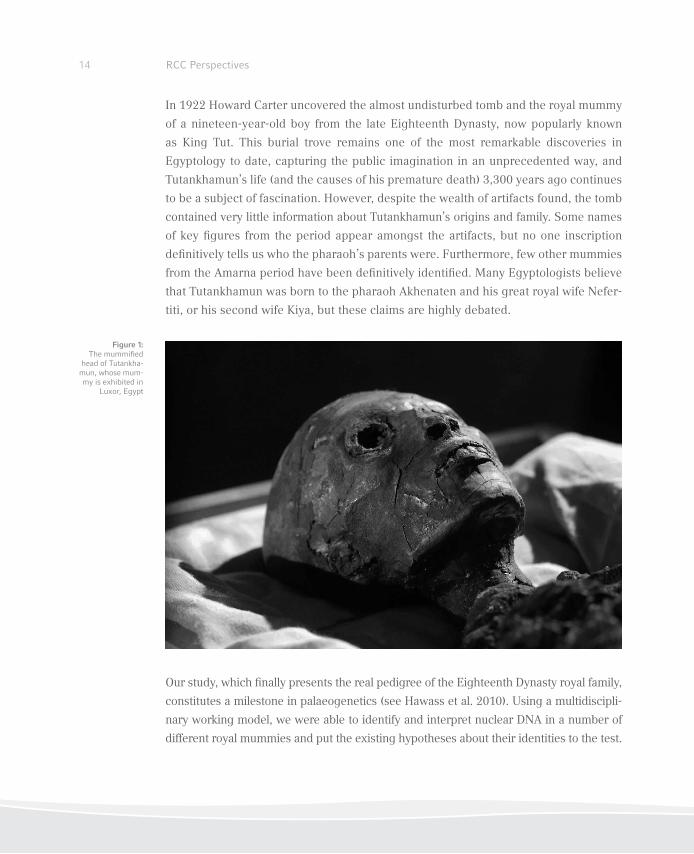
In 1922 Howard Carter uncovered the almost undisturbed tomb and the royal mummy
of a nineteen-year-old boy from the late Eighteenth Dynasty, now popularly known
as King Tut. This burial trove remains one of the most remarkable discoveries in
Egyptology to date, capturing the public imagination in an unprecedented way, and
Tutankhamun’s life (and the causes of his premature death) 3,300 years ago continues
to be a subject of fascination. However, despite the wealth of artifacts found, the tomb
contained very little information about Tutankhamun’s origins and family. Some names
of key igures from the period appear amongst the artifacts, but no one inscription
deinitively tells us who the pharaoh’s parents were. Furthermore, few other mummies
from the Amarna period have been deinitively identiied. Many Egyptologists believe
that Tutankhamun was born to the pharaoh Akhenaten and his great royal wife Nefer-
titi, or his second wife Kiya, but these claims are highly debated.
Our study, which inally presents the real pedigree of the Eighteenth Dynasty royal family,
constitutes a milestone in palaeogenetics (see Hawass et al. 2010). Using a multidiscipli-
nary working model, we were able to identify and interpret nuclear DNA in a number of
different royal mummies and put the existing hypotheses about their identities to the test.
Figure 1:The mummiied
head of Tutankha-mun, whose mum-my is exhibited in
Luxor, Egypt
15Sickness, Hunger, War, and Religion
This was facilitated by the preservation of nucleic acids in the corpses, which we speculate
was a result of the particular (but as yet poorly understood) embalming techniques of the
ancient priests. As these techniques reached their peak with the Eighteenth Dynasty, we
were provided with DNA of extraordinary quantity and quality.
Previous Identiications
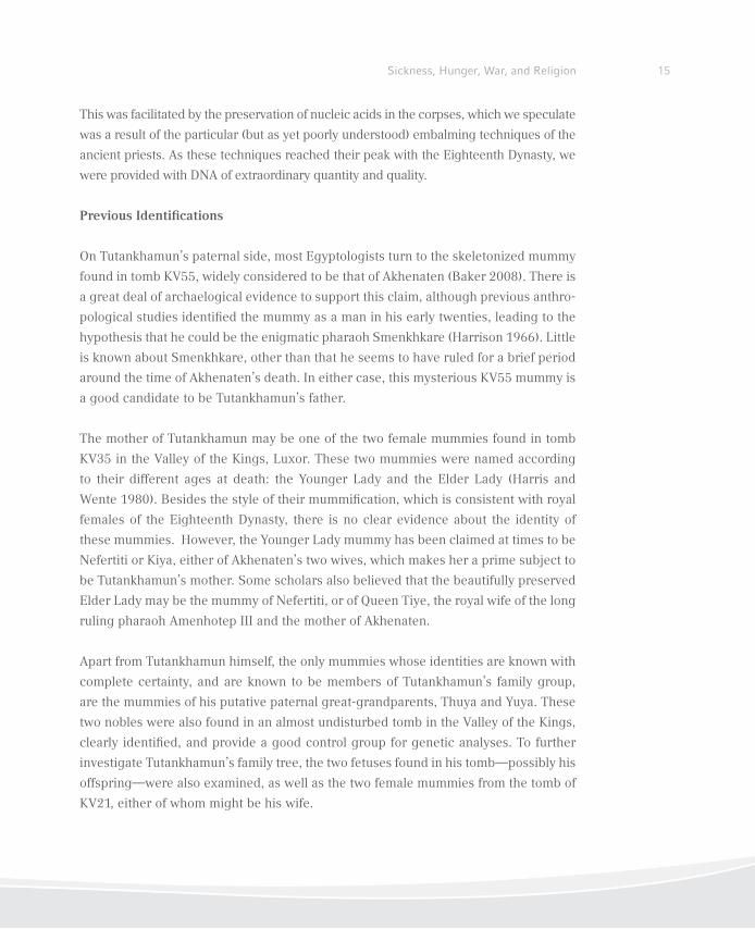
On Tutankhamun’s paternal side, most Egyptologists turn to the skeletonized mummy
found in tomb KV55, widely considered to be that of Akhenaten (Baker 2008). There is
a great deal of archaelogical evidence to support this claim, although previous anthro-
pological studies identiied the mummy as a man in his early twenties, leading to the
hypothesis that he could be the enigmatic pharaoh Smenkhkare (Harrison 1966). Little
is known about Smenkhkare, other than that he seems to have ruled for a brief period
around the time of Akhenaten’s death. In either case, this mysterious KV55 mummy is
a good candidate to be Tutankhamun’s father.
The mother of Tutankhamun may be one of the two female mummies found in tomb
KV35 in the Valley of the Kings, Luxor. These two mummies were named according
to their different ages at death: the Younger Lady and the Elder Lady (Harris and
Wente 1980). Besides the style of their mummiication, which is consistent with royal
females of the Eighteenth Dynasty, there is no clear evidence about the identity of
these mummies. However, the Younger Lady mummy has been claimed at times to be
Nefertiti or Kiya, either of Akhenaten’s two wives, which makes her a prime subject to
be Tutankhamun’s mother. Some scholars also believed that the beautifully preserved
Elder Lady may be the mummy of Nefertiti, or of Queen Tiye, the royal wife of the long
ruling pharaoh Amenhotep III and the mother of Akhenaten.
Apart from Tutankhamun himself, the only mummies whose identities are known with
complete certainty, and are known to be members of Tutankhamun’s family group,
are the mummies of his putative paternal great-grandparents, Thuya and Yuya. These
two nobles were also found in an almost undisturbed tomb in the Valley of the Kings,
clearly identiied, and provide a good control group for genetic analyses. To further
investigate Tutankhamun’s family tree, the two fetuses found in his tomb—possibly his
offspring—were also examined, as well as the two female mummies from the tomb of
KV21, either of whom might be his wife.
16 RCC Perspectives
Material and Methods
Eleven mummies of the Eighteenth Dynasty (ca. 1550–1295 BCE) of the New Kingdom
(ca. 1570–1070 BCE) underwent a detailed anthropological and radiological study in
order to determine the preservation status, the individual’s age and sex, and also to
reveal any evidence of disease or the cause of death. For the radiological analysis, the
mummies were scanned using a multidetector CT unit “emotion 6” by Siemens Med-
ical Systems. This information was further used to identify the exact locations to take
tissue samples for later DNA extraction in the laboratories.
Subsequently, the mummies were sampled by tak-
ing small bone punch biopsies from at least four
different areas of the corpse. The samples were
stored in sterile tubes and transferred to dedicat-
ed ancient DNA laboratories for further processing.
The ancient DNA extractions of the bone samples
and all further analytical steps, such as polyme-
rase chain reaction (PCR) ampliication, cloning,
and sequencing, were all performed following strict
and widely accepted guidelines (Richards, Sykes,
and Hedges 1995). Along with these precautions,
detailed contamination monitoring protocols for
the PCR experiments were included in the research
(mock and negative controls, separate working
areas, etc.). Moreover, all involved lab members were tested for their Y-chromosomal and
autosomal markers. For authentication of the results, all analytical steps were repeated at
least ive times; in addition, a subset of the data was independently replicated in a newly
equipped lab exclusively dedicated to ancient DNA work.
The analyses of the ancient Egyptian mummy samples included laborious optimization
strategies by applying several different DNA extraction and puriication protocols. In-
hibition PCR experiments were performed in a third lab, located at the National Re-
search Center, Cairo, to titrate for the proper amount of ampliiable ancient DNA. After
the successful extraction of DNA, we performed an intensive genetic testing of nuclear
DNA loci including sixteen Y-chromosomal (AMPFLSTR Yiler PCR Ampliication kit,
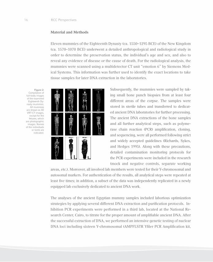
Figure 2:Compilation of
full-body images of the examined Eighteenth Dy-
nasty mummies. All mummies
are shown as 3D VRT-CT images,
except for the fetuses, which
are photographic images. Names
of the individuals or tomb are
indicated.
17Sickness, Hunger, War, and Religion
Applied Biosystems) as well as eight autosomal microsatellite markers (AMPFLSTR
Miniler PCR Ampliication kit, Applied Biosystems).
Results and Discussion
Kinship Analyses
The optimization protocols for extraction and puriication of DNA, PCR ampliication,
sequencing and fragment length analyses yielded results for all mummies under in-
vestigation. The genetic data clearly identiied the mummies of KV35 Elder Lady, and
KV55. Our results, in conjunction with archaeological data, provide substantial evi-
dence that the mummy found in KV55 is indeed Akhenaten, and that the KV35 Elder
Lady is Tutankhamun’s paternal grandmother, Queen Tiye. Moreover, the KV55 mum-
my and the KV35 Younger Lady mummy can safely be identiied as the father and
mother of Tutankhamun. This is demonstrated by the following results:
1. The established Y-chromosomal proiles show identical patterns in Amenhotep III,
KV55, and Tutankhamun. This provides evidence that these individuals share the same
paternal lineage. Control mummies examined along with Tutankhamun’s putative fami-
ly members yielded different Y-speciic alleles.
2. Fine analysis of the genetic relationship between the mummies was achieved by a
genetic ingerprint typing exploring autosomal alleles. We obtained complete inger-
print proiles of all individuals except for one of the KV62 fetuses and both mummies
from KV21, who yielded partial data sets. By evaluating the segregation of alleles
through the generations, we reconstructed the most plausible royal pedigree: a ive-
generation family tree.
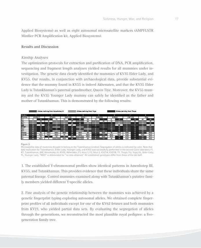
Figure 3:Microsatellite data of mummies thought to belong to the Tutankhamun kindred. Segregation of alleles is indicated by color. Note that data replication for Tutankhamun, Elder Lady, Younger Lady, and KV55 was successfully performed in the second Cairo laboratory (*). KT, Tutankhamun; AM, Amenhotep III; KV55, Akhenaten; F1, fetus 1; F2, fetus 2; KV21A; KV21B; TY, Thuya; YU, Yuya; EL, Elder Lady; YL, Younger Lady. “NDO” is abbreviated for “no data obtained.” All established genotypes differ from those of the lab staff.
RCC Perspectives18
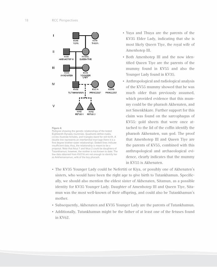
• Yuya and Thuya are the parents of the
KV35 Elder Lady, indicating that she is
most likely Queen Tiye, the royal wife of
Amenhotep III.
• Both Amenhotep III and the now iden-
tiied Queen Tiye are the parents of the
mummy found in KV55 and also the
Younger Lady found in KV35.
• Anthropological and radiological analysis
of the KV55 mummy showed that he was
much older than previously assumed,
which provided evidence that this mum-
my could be the pharaoh Akhenaten, and
not Smenkhkare. Further support for this
claim was found on the sarcophagus of
KV55: gold sheets that were once at-
tached to the lid of the cofin identify the
pharaoh Akhenaten, sun god. The proof
that Amenhotep III and Queen Tiye are
the parents of KV55, combined with this
anthropological and archaeological evi-
dence, clearly indicates that the mummy
in KV55 is Akhenaten.
• The KV35 Younger Lady could be Nefertiti or Kiya, or possibly one of Akhenaten’s
sisters, who would have been the right age to give birth to Tutankhamun. Speciic-
ally, we should also mention the eldest sister of Akhenaten, Sitamun, as a possible
identity for KV35 Younger Lady. Daughter of Amenhotep III and Queen Tiye, Sita-
mun was the most well-known of their offspring, and could also be Tutankhamun’s
mother.
• Subsequently, Akhenaten and KV35 Younger Lady are the parents of Tutankhamun.
• Additionally, Tutankhamun might be the father of at least one of the fetuses found
in KV62.
Figure 4:Pedigree showing the genetic relationships of the tested Eighteenth Dynasty mummies. Quadrants deine males, circles illustrate females, and triangles stand for still-birth. A double line represents an interfamilial marriage (here it is a irst degree brother-sister relationship). Dotted lines indicate insuficient data; thus, the relationship is meant to be a proposal. Note that fetus 1 and fetus 2 could be daughters of Tutankhamun; however, the mother is not known to date. The few data obtained from KV21A are not enough to identify her as Ankhensenamun, wife of the boy pharaoh.
19Sickness, Hunger, War, and Religion
Gynecomastia and Syndromes
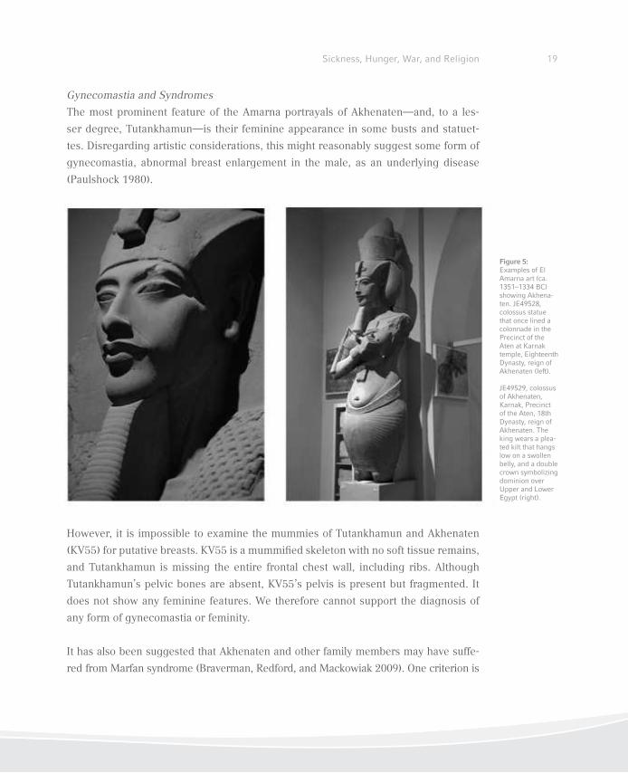
The most prominent feature of the Amarna portrayals of Akhenaten—and, to a les-
ser degree, Tutankhamun—is their feminine appearance in some busts and statuet-
tes. Disregarding artistic considerations, this might reasonably suggest some form of
gynecomastia, abnormal breast enlargement in the male, as an underlying disease
(Paulshock 1980).
However, it is impossible to examine the mummies of Tutankhamun and Akhenaten
(KV55) for putative breasts. KV55 is a mummiied skeleton with no soft tissue remains,
and Tutankhamun is missing the entire frontal chest wall, including ribs. Although
Tutankhamun’s pelvic bones are absent, KV55’s pelvis is present but fragmented. It
does not show any feminine features. We therefore cannot support the diagnosis of
any form of gynecomastia or feminity.
It has also been suggested that Akhenaten and other family members may have suffe-
red from Marfan syndrome (Braverman, Redford, and Mackowiak 2009). One criterion is
Figure 5: Examples of El Amarna art (ca. 1351–1334 BC) showing Akhena-ten. JE49528, colossus statue that once lined a colonnade in the Precinct of the Aten at Karnak temple, Eighteenth Dynasty, reign of Akhenaten (left).
JE49529, colossus of Akhenaten, Karnak, Precinct of the Aten, 18th Dynasty, reign of Akhenaten. The king wears a plea-ted kilt that hangs low on a swollen belly, and a double crown symbolizing dominion over Upper and Lower Egypt (right).
the presence of dolichocephaly, that is an abnormally long head (Pyeritz and McKusick
1979). We tested for this by establishing the cephalic indices for 15 mummies. Many
scholars believe that dolichocephaly is present in individual members of the Eighteenth
Dynasty. Dolichocephaly is frequently seen in busts and statuettes of the Amarna period
(Nefertiti, Akhenaten and Tutankhamun are prominent examples). Technically, dolicho-
cephaly is deined as a skull with a cephalic index (CI) of 75 or less. Apart from Yuya
(CI = 70.3), no mummies from the Tutankhamun lineage satisfy the criterion. However,
Akhenaten’s CI is 81.0 and Tutankhamun’s 83.9, which deines their skulls as brachyce-
phalic, or abnormally wide.
The diagnosis of Marfan syndrome is based on a combination of the major and minor
clinical features (De Paepe et al. 1996). The presence of either two major features or
one minor feature, or of one major feature and four minor features, supports a dia-
gnosis of Marfan syndrome. Following this classiication, we could not ind evidence
to add weight to a Marfan diagnosis.
Radiological Findings
Previous X-ray analyses have revealed much about the life of the pharaoh; however,
they have also left plenty of questions unanswered over the years. Our study was
designed either to conirm or refute the conclusions of previous examinations, and it
focused on details that earlier studies might have overlooked. We speciically looked
for life-threatening elements that might have directly caused the king’s death, or been
linked to his cause of death. While our inspection of the rest of his body did not result
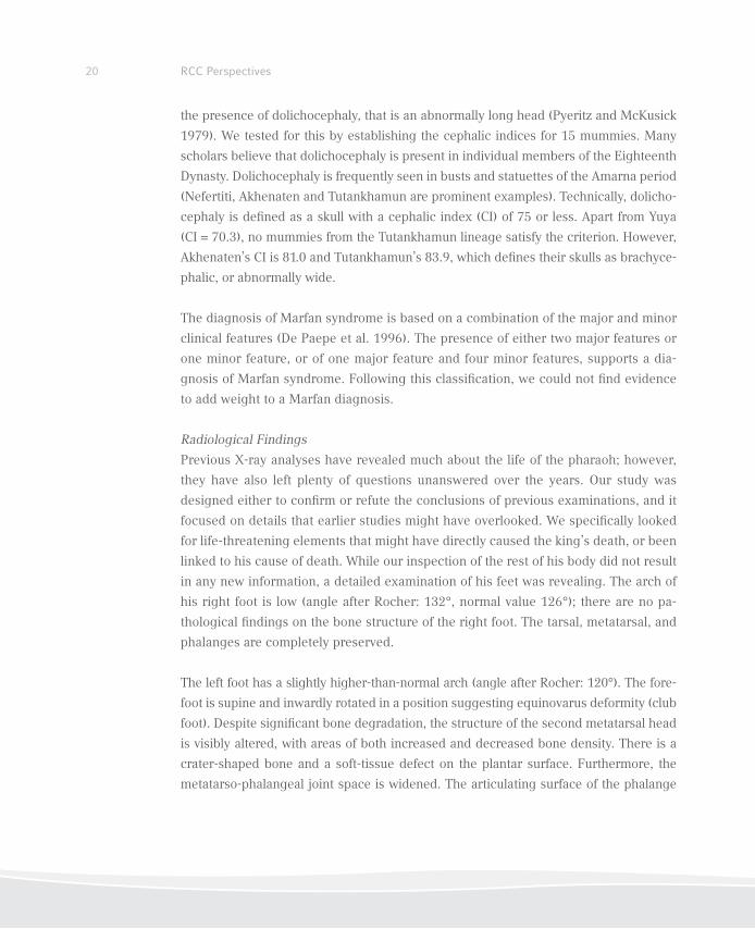
in any new information, a detailed examination of his feet was revealing. The arch of
his right foot is low (angle after Rocher: 132°, normal value 126°); there are no pa-
thological indings on the bone structure of the right foot. The tarsal, metatarsal, and
phalanges are completely preserved.
The left foot has a slightly higher-than-normal arch (angle after Rocher: 120°). The fore-
foot is supine and inwardly rotated in a position suggesting equinovarus deformity (club
foot). Despite signiicant bone degradation, the structure of the second metatarsal head
is visibly altered, with areas of both increased and decreased bone density. There is a
crater-shaped bone and a soft-tissue defect on the plantar surface. Furthermore, the
metatarso-phalangeal joint space is widened. The articulating surface of the phalange
RCC Perspectives20
21Sickness, Hunger, War, and Religion
is normal. The third metatarsal head is only slightly deformed, but its structure shows
signs of apparent bone necrosis. The remaining metatarsal heads of the left foot appear
normal; the second and third toes are in abduction. The second toe is shortened, because
it lacks a second phalanx; the irst phalanx joins directly with the ungual phalanx. These
indings show that Tutankhamun suffered from a juvenile aseptic bone necrosis of the
second and third metatarsal bones of his left foot (Köhler´s di-
sease II, Freiberg-Köhler syndrome). The widened joint space
and the secondary changes to the second and third metatarsal
heads indicate that the disease was still lourishing. Bone and
soft tissue loss at the second metatarsophalangeal joint could
further indicate an acute inlammatory condition resulting
from ulcerative osteoarthritis and osteomyelitis.
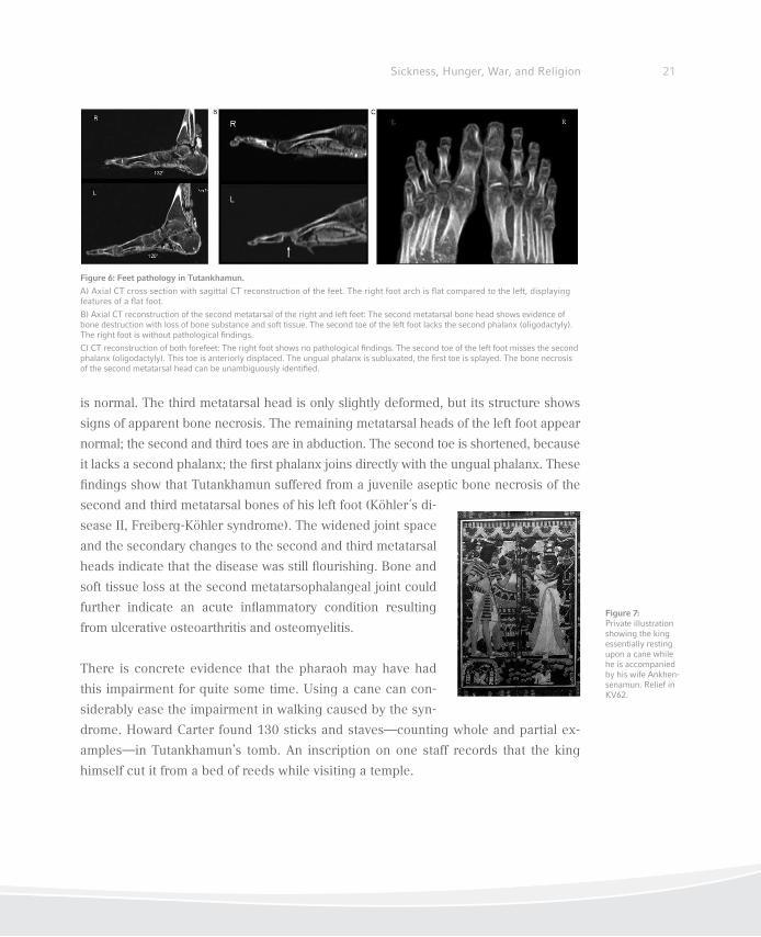
There is concrete evidence that the pharaoh may have had
this impairment for quite some time. Using a cane can con-
siderably ease the impairment in walking caused by the syn-
drome. Howard Carter found 130 sticks and staves—counting whole and partial ex-
amples—in Tutankhamun’s tomb. An inscription on one staff records that the king
himself cut it from a bed of reeds while visiting a temple.
Figure 6: Feet pathology in Tutankhamun.
A) Axial CT cross section with sagittal CT reconstruction of the feet. The right foot arch is lat compared to the left, displaying features of a lat foot.
B) Axial CT reconstruction of the second metatarsal of the right and left feet: The second metatarsal bone head shows evidence of bone destruction with loss of bone substance and soft tissue. The second toe of the left foot lacks the second phalanx (oligodactyly). The right foot is without pathological indings.
C) CT reconstruction of both forefeet: The right foot shows no pathological indings. The second toe of the left foot misses the second phalanx (oligodactyly). This toe is anteriorly displaced. The ungual phalanx is subluxated, the irst toe is splayed. The bone necrosis of the second metatarsal head can be unambiguously identiied.
Figure 7: Private illustration showing the king essentially resting upon a cane while he is accompanied by his wife Ankhen-senamun. Relief in KV62.
22 RCC Perspectives
Infectious Diseases
As the various macroscopic inspections, X-rays, and CT examinations conducted in
the past did not yield any conclusive data, we considered various life-threatening dis-
eases as potential causes of death. In order to test for the malaria-causing parasite
Plasmodium falciparum, DNA PCR primers were designed that speciically amplify
small Stevor, Ama1, and Msp1 gene fragments, thereby yielding amplicons in the range
of circa 100–250 bp. PCR products and cloned DNA fragments were sequenced. We
identiied Plasmodium falciparum DNA in the mummies of Tutankhamun, Yuya, and
Thuya. Since we applied primers that are highly speciic for the P. falciparum genome,
we can safely conclude that our positively typed mummies suffered from malaria tro-
pica, the most severe form of malaria.
Cause of Death
Tutankhamun suffered from multiple physical disorders, and it is possible that some of
them may have cumulated in an inlammatory, immunosuppressive syndrome, which
would seriously undermine his health. We can imagine a young, frail king, who walked
with a cane due to Köhler’s Disease II (osteonecrotic and sometimes painful) together
with oligodactyly in the right foot and club foot in the left. A sudden leg fracture,
perhaps from a fall, would be life-threatening when combined with a malaria tropica
infection.
Conclusions
This multidisciplinary study (incorporating genetic, archaeological, anthropological,
and Egyptological research) is the irst concretely to clarify the lineage of Tutankha-
mun. As most of the archaeological and Egyptological data are still subject to debate,
we established thorough genetic ingerprints of King Tutankhamun and his putative
family members. By conducting a detailed ancient DNA study, we identiied the mum-
mies’ origins and shed light on the pharaoh’s family bonds.
23Sickness, Hunger, War, and Religion
References
Baker, Darrell. 2008. Encyclopedia of the Pharaohs, Volume I: Predynastic to the Twentieth Dy-
nasty. Cairo: American University in Cairo Press.
Braverman, Irvin M., Donald B. Redford, and Philip A. Mackowiak. 2009. “Akhenaten and the
Strange Physiques of Egypt’s 18th Dynasty.” Annals of Internal Medicine 150 (8): 556–60.
Carter, Howard and Arthur C. Mace. 1927. The Tomb of Tut-Ankh-amun. Vol. 2. London: Cassell
and Co.
De Paepe, Anne, Richard B. Devereux, Harry C. Dietz, Raoul C. Hennekam, and Reed E. Pyeritz.
1996. “Revised Diagnostic Criteria for the Marfan Syndrome.” American Journal of Medical
Genetics 62 (4): 417–26.
Harris, James E. and Eduard F. Wente. 1980. An X-Ray Atlas of the Royal Mummies. Chicago:
University of Chicago Press.
Harrison, Ronald G. 1966. “An Anatomical Examination of the Pharaonic Remains Purported to
Be Akhenaten.” Journal of Egyptian Archaeology 52: 95–119.
Hawass, Zahi, Yehia Z. Gad, Somaia Ismail, Rabab Khairat, Dina Fathallah, Naglaa Hasan, Amal
Ahmed, Hisham Elleithy, Markus Ball, Fawzi Gaballah, Sally Wasef, Mohamed Fateen, Hany
Amer, Paul Gostner, Ashraf Selim, Albert Zink, and Carsten M. Pusch. 2010. “Ancestry, Pa-
thology and Cause of Death in King Tutankhamun’s Family.” Journal of the American Medical
Association 303: 638–47.
Paulshock, Bernadine Z. 1980. “Tutankhamun and His Brothers: Familial Gynecomastia in the
Eighteenth Dynasty.” Journal of the American Medical Association 244 (2): 160–64.
Pyeritz, Reed E. and Victor A. McKusick. 1979. “The Marfan Syndrome: Diagnosis and Manage-
ment.” New England Journal of Medicine 300 (14): 772–77.
Richards, Martin B., Bryan C. Sykes, and Robert E. M. Hedges. 1995. “Authenticating DNA Ex-
tracted from Ancient Skeletal Remains.” Journal of Archaeological Science 22 (2): 291–99.

Mesolithic-Neolithic Transformations
The Populations of the Danube Gorges
Dušan Borić, Marija Radović, and Soija Stefanović

Authors
Dušan BorićCardiff University
School of History, Archaeology, and Religion
Colum Drive
Cardiff CF10 3EU
Great Britain
Marija Radović Belgrade University
Department of Archaeology
Laboratory for Bioarchaeology
ćika Ljubina 18–20
11000 Belgrade
Serbia
Soija StefanovićBelgrade University
Department of Archaeology
Laboratory for Bioarchaeology
ćika Ljubina 18–20
11000 Belgrade
Serbia
26 RCC Perspectives
27Sickness, Hunger, War, and Religion
Dušan Borić, Marija Radović, and Soija Stefanović
Introduction
In the Danube Gorges that lie between Serbia and Romania, several archeological
sites critical for the understanding of the transitions between the Mesolithic and Neo-
lithic in southeastern Europe have been discovered. In particular, several preserved
burial sites, containing around 500 individual skeletal remains, offer a unique oppor-
tunity to examine the life- and deathways of these communities. Through an analysis
of skeletal remains and patterns of interment, this paper discusses questions of local
versus non-local identities, as well as changes in diet throughout the Neolithization.
One site in particular, Lepenski Vir, is the basis for research into the paleopathology of
local populations. This study concludes that skeletal health parameters suggest a re-
latively good health status of this population over time, although treponemal infection
(a group of diseases including syphilis, bejel, pinta, and yaws) affected large numbers
of individuals at the Danube Gorges, and occur as a major pathological condition in
all periods. Dental evidence also suggests relatively good health of the community, in
contrast to other populations of the forager-farmer transition. Results of dental-based
study indicate that changes in the biology of this population led to an increase of gen-
eral health, though these changes were not the same for females and males.
Dušan Borić
Isotopic and Symbolic Identities: Mesolithic-Neolithic Transformations
Among the Inhabitants of the Danube Gorges
In the past decade or so, several research teams carried out isotopic analyses on hu-
man burial remains and animal bones from the Danube Gorges Mesolithic and Neo-
lithic sites.1 This suite of new data has contributed signiicantly to our current un-
derstanding of dietary preferences and historical changes in this micro-region at the
time of Mesolithic-Neolithic transformations (ca. 6300–5900 BCE).2 This new research
1 The following isotopes have been analyzed: δ13C, δ15N (Bonsall et al. 1997; Cook et al. 2009; Borić et al. 2004; Grupe et al. 2003) and δ34S (Nehlich et al. 2010) for dietary patterns and trophic levels; δ18O for ecological provenance (Grupe and Borić 2010); and 87Sr/86Sr for patterns of individual and population mobility (Borić and Price forthcoming).
2 This article uses the dating systems BCE and CE (Before the Common Era and Common Era), which are an alternative to BC and AD.
28 RCC Perspectives
suggests that the pattern of ish-dominated diet—characteristic of the Late Mesolithic
in the Danube Gorges—remained largely unaltered during Mesolithic-Neolithic trans-
formations. However, dietary signatures of several individuals from the central site of
Lepenski Vir indicate diets less reliant on ish, representing the trend that continued
with the appearance of the irst crouched (Neolithic) burials at the same site. Moreo-
ver, the same individuals contained non-local strontium signals, suggesting that they
might have been (Neolithic?) migrants that came from the surrounding regions or
from further aield (Borić and Price forthcoming). Based on these indings, this paper
discusses the possible impact of Neolithic migrants on local populations and identities.
The earliest human remains in the Danube Gorges area date to the Early Mesolithic
(ca. 9500–7300 BCE).3 At Padina, for instance, one whole area at Sector III of this site
was used for burial within several layers of a linear stone construction during the Early
Mesolithic (Borić and Miracle 2004; Borić 2011). While some burials date to the last
half of the tenth millennium BCE, others are from the turn of the ninth and eighth mil-
lennia BCE. Some are characterized by speciic body positions, such as seated position
with crossed legs—the “lotus” body posture.
Isotopic analysis of these human burial remains suggests a shift in dietary patterns in
the region. For instance, isotopic analyses of nitrogen (δ15N) of burials from Padina,
Lepenski Vir, and Vlasac show higher trophic levels (>13%) and indicate heavy pro-
tein intake, most likely due to the consumption of ish, a staple food in the Late Meso-
lithic of the Danube Gorges. However, this pattern of ish-dominated diet for the Early
Mesolithic burials from Padina has been recently challenged by new isotope results
(sulfur 34S) that indicate no signiicant intake of ish at Padina, Vlasac, or Lepenski Vir
during this period (Nehlich et al. 2010).4 While these indings contradict our initial
understanding of dietary preferences in the Early Mesolithic on the basis of carbon
and nitrogen isotopic values, a more robust sample of individuals is needed to conirm
these initial sulfur isotope results. Such work is currently in progress.
3 Only one burial—from the site of Climente II— might be from the Epipalaeolithic period (ca. 13,000–9500 BCE) but it has not been AMS-dated at present.
4 Early Mesolithic humans from Padina (Burials 12, 16, and 19a), Vlasac (Burials 16 and 17) and Lepenski Vir (Burials 67 and 68) have low 34S values despite δ15N values that are for the same individuals higher than 13% (Nehlich et al. 2010).
29Sickness, Hunger, War, and Religion
On the other hand, isotopic indings from a large number of later burials, dated to the
Late Mesolithic (ca. 7300–6200 BCE), suggest the importance of ish in the diet of
communities inhabiting the region. Burial practices may also relect the signiicance
of ish in local diets. The dominant burial position during this period is supine, with
many individuals placed parallel to the Danube, often with their heads pointing in the
downstream direction. This position may be related to the importance of anadromous
sturgeon ish that migrate upstream to breed and might have held totemic, as well as
dietary, signiicance (Borić 2005; Radovanović 1997).
This same burial position remains dominant in the period of Mesolithic-Neolithic
transformations (ca. 6200–5900 BCE) during which the Danube Gorges foragers came
into close contact with highly mobile Early Neolithic groups. Mesolithic supine burials
along the Danube Gorges contain ample evidence of this contact, from the mixing of
the local type of body decoration made from Cyprinidae (carp) pharyngeal teeth to
new types of ornaments, such as Spondylus beads or red and white discoid limestone
beads (Borić 2007). During this same period, the proliferation of carved sandstone
boulders with hybrid human-ish depictions suggests an elaboration on local Mesoli-
thic beliefs that stressed the importance of certain species of ish (Borić 2005).
These new cultural contacts altered dietary habits. Isotopic data reveal the seeds of
change. In particular, a number of individuals found at the site of Lepenski Vir have re-
duced trophic levels, possibly indicating a reduction of ish in their diet (Bonsall et al.
1997; Cook et al. 2009). Strontium isotopic results suggest that several of these same
individuals were migrants into this region, even though their burial position remained
supine. Another migrant, buried in a crouched position, was discovered at the Neolithic
site of Ajmana, situated some hundred kilometers downstream from Lepenski Vir. Aj-
mana and similar sites might have been contemporaneous with the continuation of local
forager settlements in the upstream area of the region. In the period following circa 5900
BCE and lasting until the mid-sixth millennium BCE, new waves of migrants entered the
region, their traces found at Lepenski Vir in particular (Borić and Price forthcoming).
This pattern is also corroborated by lower δ15N values, suggesting that ish became less
important to the diet of these migrants. That these apparent changes in diet correspond to
the spread of crouched rather than supine burials suggests that the community’s symbolic
identities were being constructed along new and foreign ideological, cultural, or religious
values when compared to the Mesolithic.
30 RCC Perspectives
Soija Stefanović
Pathological Conditions on Skeletal Remains from the Site of Lepenski Vir
While research on the Danube Gorges populations suggests a relatively good health stat-
us over time, treponemal infections were widespread and constituted a major patholog-
ical condition in all periods. Although various studies indicate increases in infections
as a consequence of Neolithic transitions, there are still no detailed studies explaining
which infections might have affected human health at the start of the Neolithic period.
Evidence for one kind of infection, caused by the bacterium Treponema pallidum, has
been found at the Danube Gorges sites from the Mesolithic period, with a slight increase
in the Neolithic period.
In this study, health changes in Neolithic transition were analyzed on 60 adult skeletons
from the site of Lepenski Vir situated in the Danube Gorges. The skeletons date to the Ear-
ly Mesolithic, Mesolithic-Neolithic transformation, and the Early/Middle Neolithic periods.
(No Late Mesolithic burials or layers have been conirmed at Lepenski Vir.) Three kinds
of pathological conditions have been identiied: 1) trauma (observed on skulls and major
bones of limbs); 2) cribra orbitalia and porotic hyperostosis (skull lesions, generally caused
by anemia and iron deiciency, both characteristics of the transition from hunther-gatherer
to agricultural populations); and 3) infection and periosteal reaction (observed on skulls
and postcranial bones).
Evidence of trauma comes only from two individuals: one Early Mesolithic male with
a trauma on the left tibia, and one Early/Middle Neolithic female with a trauma on the
skull. Low frequencies of traumas indicate low rates of violence in all periods.
Evidence of skull lesions associated with anemia is more widespread. Cribra orbitalia is
not present among the adults from Lepenski Vir during the Early Mesolithic and Early/
Middle Neolithic, while seven individuals (four females and three males) have traces of
cribra in the Mesolithic-Neolithic transformations. Cribra orbitalia in the form of gross
lesions with excessive expansion only appears in three individuals, each of whom proba-
bly experienced severe health problems as a result. Based on available evidence, then, it
appears that individuals from Lepenski Vir did not experience the same health problems
during the Neolithic transition as seen in many other transitional populations.
31Sickness, Hunger, War, and Religion
In contrast to cribra, porotic hyperostosis was much more frequent among individuals
from Lepenski Vir, appearing in all periods (three individuals from the Early Mesolithic,
ten from the Transformation phase, and ive from the Early/Middle Neolithic). However,
only modest lesions and scattered ine-pitting on parietals and occipital bones were
present, without cases of gross lesions with excessive cranial expansion and areas of
exposed diploe. The causes of cribra and porotic hyperostosis are complex: they may
have been caused by iron deiciency, various diseases, and/or parasites (Goodman and
Martin 2002). Also, porotic hyperostosis might have been caused by inlammatory pro-
cesses after scalping (Schultz 1993; Schultz 2001). Walker et al. (2009) suggest that po-
rotic hyperostosis and many traces of cribra might have been caused by megaloblastic
anemia (a form of anemia that results from inhibition of DNA synthesis in red blood cell
production) but with different etiologies—cribra is caused by vitamin C deiciency while
porotic hyperostosis is the outcome of the depletion of vitamin B12 reserves. With little
evidence of cribra, inhabitants of Lepenski Vir appear to have been well supplied with
vitamin C, with some exceptions during the Transformation phase. On the other hand, a
high prevalence of porotic hyperostosis suggests that vitamin B12 reserves were deplet-
ed. However, since few cases of cribra and only modest forms of porotic hyperostosis
have been found, such deiciencies probably had only a limited affect on the overall
health of the prehistoric inhabitants of Lepenski Vir.
In addition to trauma and anemia, infection is also an indicator of the state of overall
health in the Danube Gorges populations. Traces of infection caused by bacteria Tre-
ponema pallidum—found on cranial and postcranial bones, of 121 individuals—present
important evidence of treponemal disease (Stefanović 2012). Traces of infection were
studied on all bones and bone fragments and a numerical value was assigned to each
type of pathological change. In the cases of postcranial skeletons, either an osteoblastic
or osteoclastic reaction was determined, as well as whether the reaction was active or
healed (or both). On skulls, lesions and other changes were recorded—necrotic and os-
teolytic damages, stellate scars, necrotic destructions and healing processes, and new
bone formation.
Lesions were found on 24 individuals (seven in the Early Mesolithic, eight in the Trans-
formation phase and nine in the Early/Middle Neolithic). On postcranial bones, the lesions
are predominantly osteoblastic and affect the diaphyses of the many various bones. The
pattern of many affected bones of an individual with bilateral symmetric lesions sug-
32 RCC Perspectives
gests system infection. No individual has been found with periostal reaction affecting
only the upper limbs. Osteoclastic lesions are much less present and also attack mostly
lower limbs, especially the tibia. All the individuals with an osteoclastic reaction also
have an osteoblastic reaction on many of their bones. Presence of bilateral symmetrical
osteoclastic lesions suggests that treponemal infection contributed to the development
of lesions, and we may assume the same etiology for most other osteoblastic reactions.
Although the sample size from Lepenski Vir is too small to compare the presence of di-
sease in Mesolithic and Neolithic periods, the fact that there were many more individu-
als with an advanced stage of the disease at Lepenski Vir than at the (mostly) Mesolithic
site Vlasac suggests that intensity of the infection increased during the Neolithic period.
It is possible that population growth in the Transformational period, combined with an
inlux of migrants, may have increased pathogens in the Danube Gorges population.
Whether prehistoric people had attempted healing treatments for various infections is
hard to ascertain, but some cut marks on skulls from Lepenski Vir indicate such a pos-
sibility. On some skulls, traces of cutting by thin and sharp tools have been detected, as
well as possible instances of scalping. If these cuts were not post-mortem, they might
indicate efforts to heal through the removal of soft tissue on the infected patients. If such
interventions took place, it is possible that inlammation after cutting might have caused
porotic hyperostosis in some cases, especially since porotic hyperostosis on many individ-
uals has the form of scattered ine-pitting, and was unconnected with dietary problems.
The results of this study suggest a relatively good health status of inhabitants of Lepen-
ski Vir over time, with the exception of infections, which occur as a major pathologi-
cal condition in all periods represented in the sample of analyzed individuals. Changes
in health status observed in other populations spanning the forager-farmer transition
around the world—such as a decline in overall quality of nutrition—were not detected
at Lepenski Vir. While higher rates of cribra orbitalia and porotic hyperostosis are found
in the Transformation period, the small number of cases and their moderate appearance
indicate stability in health over time. At the very least, they suggest no dramatic changes
in the quality of life.

33Sickness, Hunger, War, and Religion
Marija Radović
Dental Pathologies and Tooth Wear at the Site of Lepenski Vir
The shift towards Neolithic lifestyles affected human biology signiicantly. In many
instances, the transition to agriculture resulted in the increase of “stress,” deined as a
series of interrelated pathological conditions coupled with changes in dietary patterns
and living conditions. Here, I examine differences in dietary patterns and subsistence
on the basis of dental pathologies and tooth wear from the Danube Gorges during the
period of Mesolithic-Neolithic transformations. I chose the site of Lepenski Vir as a
case study for this research, and studied the dental status on 29 adult individuals, 14
males, and 15 females.5 I examined 195 teeth for enamel hypoplasia (enamel defects),
the rate of dental wear, and dental calculus, or plaque—all indicators of oral and gen-
eral health in relation to food composition. I studied the defects in order to trace
physiological stress and growth disturbance in childhood
Changes in lifestyle adopted from the “Neolithic package”6 may explain the evident de-
crease of systematic childhood stress from the Mesolithic to the Neolithic (Table 1). The
distribution frequency of linear enamel hypoplasia (LEH) by sex of the individuals shows
5 Of the 29 specimens, six males date from the Mesolithic; ive males and 11 females date from the Transforma-tion phase; and three males and four females date from the Neolithic.
6 The term “Neolithic package” means a general shift to a sedentary way of life, cultivation of plants, domestica-tion of animals, and/or labor specialization.
Table 1: Prevalence of linear enamel hypoplasia, LEH, through chro-nological periods.
RCC Perspectives34
that females were less affected by growth disturbance or less exposed to physiological
stress, though the sample is somewhat biased due to the absence of (Early) Mesolithic
females at Lepenski Vir. Another possible explanation for differences in the rate of LEH
between males and females may relate to females’ premature mortality. The etiology
of LEH does not indicate a single crisis episode. LEH defects generally irst appear
between two and ive years of age. These results indicate weaning stress as a possible
explanation.
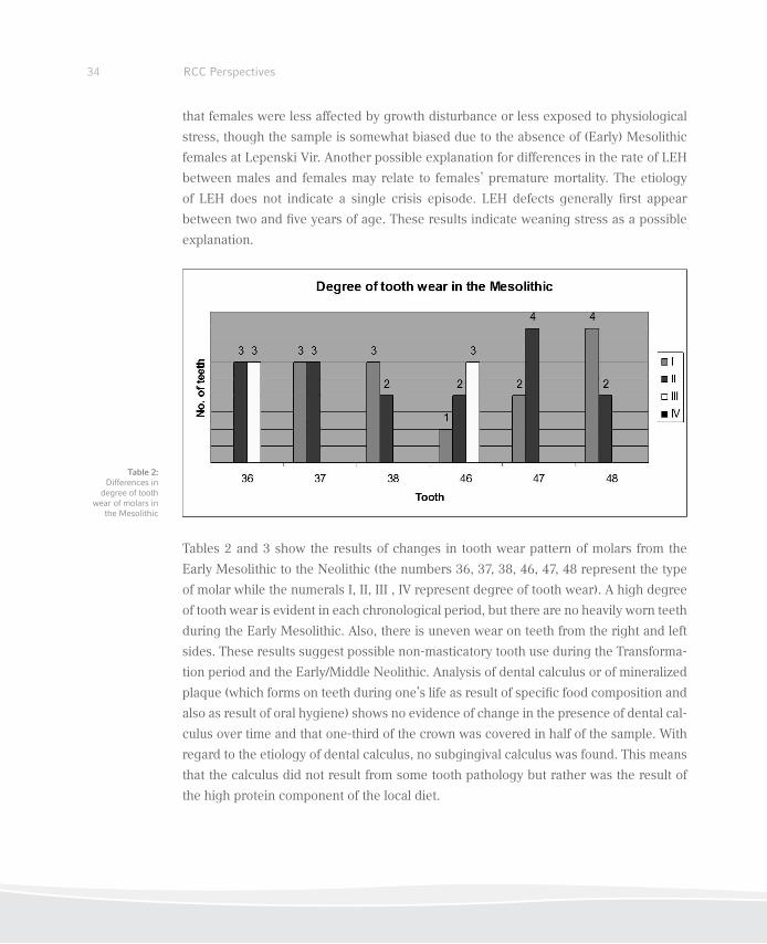
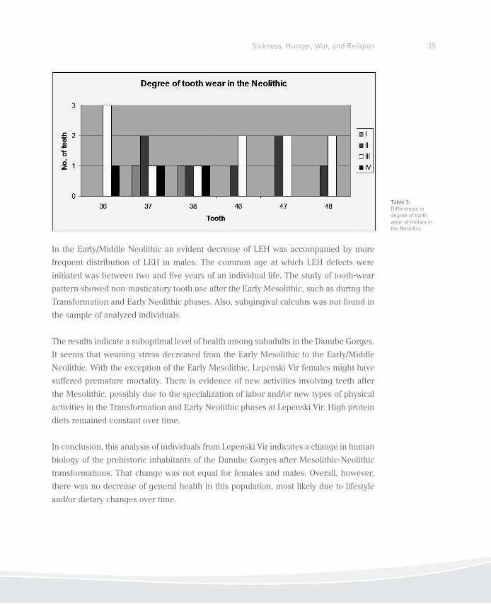
Tables 2 and 3 show the results of changes in tooth wear pattern of molars from the
Early Mesolithic to the Neolithic (the numbers 36, 37, 38, 46, 47, 48 represent the type
of molar while the numerals I, II, III , IV represent degree of tooth wear). A high degree
of tooth wear is evident in each chronological period, but there are no heavily worn teeth
during the Early Mesolithic. Also, there is uneven wear on teeth from the right and left
sides. These results suggest possible non-masticatory tooth use during the Transforma-
tion period and the Early/Middle Neolithic. Analysis of dental calculus or of mineralized
plaque (which forms on teeth during one’s life as result of speciic food composition and
also as result of oral hygiene) shows no evidence of change in the presence of dental cal-
culus over time and that one-third of the crown was covered in half of the sample. With
regard to the etiology of dental calculus, no subgingival calculus was found. This means
that the calculus did not result from some tooth pathology but rather was the result of
the high protein component of the local diet.
Table 2: Differences in
degree of tooth wear of molars in
the Mesolithic
35Sickness, Hunger, War, and Religion
In the Early/Middle Neolithic an evident decrease of LEH was accompanied by more
frequent distribution of LEH in males. The common age at which LEH defects were
initiated was between two and ive years of an individual life. The study of tooth-wear
pattern showed non-masticatory tooth use after the Early Mesolithic, such as during the
Transformation and Early Neolithic phases. Also, subgingival calculus was not found in
the sample of analyzed individuals.
The results indicate a suboptimal level of health among subadults in the Danube Gorges.
It seems that weaning stress decreased from the Early Mesolithic to the Early/Middle
Neolithic. With the exception of the Early Mesolithic, Lepenski Vir females might have
suffered premature mortality. There is evidence of new activities involving teeth after
the Mesolithic, possibly due to the specialization of labor and/or new types of physical
activities in the Transformation and Early Neolithic phases at Lepenski Vir. High protein
diets remained constant over time.
In conclusion, this analysis of individuals from Lepenski Vir indicates a change in human
biology of the prehistoric inhabitants of the Danube Gorges after Mesolithic-Neolithic
transformations. That change was not equal for females and males. Overall, however,
there was no decrease of general health in this population, most likely due to lifestyle
and/or dietary changes over time.
Table 3: Differences in degree of tooth wear of molars in the Neolithic
Conclusions: Integration of Archaeological and Osteological Data from the
Danube Gorges
Spanning the period from the Early Mesolithic through the Early/Middle Neolithic, the
abundance of human remains found in the Danube Gorges offers a unique opportunity
to examine life- and deathways of these communities through an analysis of skeletal
remains and burial practices. Our three contributions have looked at both aspects of
the data, trying to integrate archaeological indings with an array of new data obtained
by studying skeletal remains. These include studies of isotopes for dietary patterns and
mobility, pathological conditions (such as the incidence of traumas, cribra orbitalia and
porotic hyperostosis, traces of infections and periosteal reaction), and a dental examina-
tion of linear enamel hypoplasia (LEH) and tooth wear.
While we used isotopic data to analyze several key Mesolithic-Neolithic sites in the
Danube Gorges, our discussion of pathological conditions and teeth was restricted to
Lepenski Vir, considered the most representative site of the later Mesolithic–Neolithic
sequence (comprising the Mesolithic-Neolithic transformation, ca. 6200–5900 BCE, and
Early/Middle Neolithic, ca. 5900–5500 BCE). However, we should add that at present
Late Mesolithic (ca. 7300–6200 BCE) occupation has not been found at this site, either
by direct dating of human remains or settlement deposits. On the other hand, there is a
prominent presence of Late Mesolithic groups at other sites along the Danube Gorges,
evidenced by a large number of burials, among other archaeological features. The lack
of Late Mesolithic burials at Lepenski Vir to some extent limits our discussion of chang-
ing patterns of health status. For instance, while human remains from Lepenski Vir show
a low level of violent injuries in all three bracketed periods, we have clear evidence of a
number of violent injuries by bone projectile points found at the site of Schela Cladovei
and Vlasac, all dated to the Late Mesolithic (Roksandić 2004).
No signs of major pathological conditions appear in populations at Lepenski Vir. How-
ever, there are clear skeletal traces of various infections, possibly indicating treponemal
infections. In addition, evidence of apparent cuts on some skeletal elements, in particu-
lar skull bones, may point to attempts to heal such infections by removing soft tissue.
This procedure, in turn, might have caused a higher level of porotic hyperostosis, which
peaks in the Transformation period. It remains unclear and open to speculation whether
RCC Perspectives36
37Sickness, Hunger, War, and Religion
such infections during this period might have related to contacts with possible (Neo-
lithic?) migrants, suggested by the presence of strontium at Lepenski Vir.
In particular, the examination of teeth shows little variation throughout the periods
with regard to the changes in the quality of life. While enamel defects (LEH) appeared
to affect males more, this pattern might be due to females’ higher mortality, the like-
ly consequence of frequent deaths while giving birth. It is interesting to note that the
decrease of enamel defects in the Early/Middle Neolithic may be the consequence
of improvements in dietary practices during this period. Isotopic data suggest that
there was a general reduction in the reliance on ish in this phase. The level of sub-
adult health is suboptimal in all periods, but is biased by the small number of burials.
The level of wear on teeth is high in all periods, but there are no heavily worn teeth
in the Early Mesolithic, while in the Transformation period, and the Early/Middle
Neolithic teeth were used in a non-masticatory way for specialized everyday activities,
which caused very speciic wear patterns on certain teeth.
In sum, with some variations over time, the sample of human burials from the Mesoli-
thic-Neolithic Danube Gorges of the Balkans suggests relative stability and good overall
health conditions despite other major changes that the introduction of a food-producing
economy and the arrival of foreign groups might have triggered. Such stability might
have been the consequence of a rich and diverse environment along the Danube, which
allowed the intake of high levels of protein, primarily coming from ish. The overall in-
take of ish was reduced in the Neolithic but, judging by the decrease in the incidence
of LEH in this period, the introduction of food-producing practices was a risk-buffering
step that might have reduced dietary shortages and episodes of famine. Still, one should
not rule out the possibility that occasionally dietary stress and any subsequent health-
related issues might also have been the consequence of cultural practices with social,
ideological, and religious reasons ruling people’s attitudes to foods and health.
38 RCC Perspectives
References
Bonsall, Clive, Rosemarie Lennon, Kathleen McSweeney, Catriona Stewart, Douglass Harkness,
Vasile Boroneanţ, László Bartosiewicz, Robert Payton, and John Chapman. 1997. “Mesolithic and
Early Neolithic in the Iron Gates: A Palaeodietary Perspective.“ Journal of European Archaeology
5 (1): 50–92.
Borić, Dušan. 2005. “Body Metamorphosis and Animality: Volatile Bodies and Boulder Artworks
from Lepenski Vir.” Cambridge Archaeological Journal 15 (1): 35–69.
———. 2007. “Mesolithic-Neolithic Interactions in the Danube Gorges.” In Mesolithic-Neolithic
Interactions in the Danube Basin, edited by Janusz K. Kozłowski and Marek Nowak, 31–45.
Oxford: Archaeopress.
———. 2011. “Adaptations and Transformations of the Danube Gorges Foragers (c.13,000–5500
cal. BC): An Overview.” In Beginnings—New Research in the Appearance of the Neolithic bet-
ween Northwest Anatolia and the Carpathian Basin, edited by Raiko Krauß, 157–203. Rahden/
Westf: Verlag Marie Leidorf.
Borić, Dušan and Preston Miracle. 2004. “Mesolithic and Neolithic (Dis)continuities in the Danu-
be Gorges: New AMS Dates from Padina and Hajdučka Vodenica (Serbia).” Oxford Journal of
Archaeology 23 (4): 341–71.
Borić, Dušan, Gisela Grupe, Joris Peters and Živko Mikić. 2004. “Is the Mesolithic-Neolithic Sub-
sistence Dichotomy Real? New Stable Isotope Evidence from the Danube Gorges.” European
Journal of Archaeology 7 (3): 221–48.
Borić, Dušan and T. Douglass Price. Unpublished. “Mobility in the Mesolithic-Neolithic Danube
Gorges: Strontium Isotope Analyses.”
Cook, Gordon, Clive Bonsall, Catriona Pickard, Kathleen McSweeney, László Bartosiewicz, and
Adina Boroneanţ. 2009. “The Mesolithic-Neolithic Transition in the Iron Gates, Southeast Eu-
rope: Calibration and Dietary Issues.” In Chronology and Evolution within the Mesolithic of
North-west Europe, edited by Philippe Crombé, Mark van Strydonck, Joris Sergant, Matthieu
Boudin, and Machteld Bats, 497–515. Newcastle upon Tyne: Cambridge Scholar Publishing.
Eshed, Vered, Avi Gopher, Ron Pinhasi, and Israel Hershkovitz. 2010. “Paleopathology and the
Origin of Agriculture in the Levant.” American Journal of Physical Anthropology 143: 121–133.
Goodman, Alan H. and Debra L. Martin. 2002. “Reconstructing Health Proiles from Skeletal Re-
mains.” In The Backbone of History: Health and Nutrition in the Western Hemisphere, edited
by Richard H. Steckel and Jerome C. Rose, 11–60. Cambridge: Cambridge University Press.
39Sickness, Hunger, War, and Religion
Grupe, Gisela, Henriette Manhart, Živko Mikić, and Joris Peters. 2003. “Vertebrate Food Webs and
Subsistence Strategies of Meso- and Neolithic Populations of Central Europe.” In Documenta Ar-
chaeobiologiae 1. Yearbook of the State Collection of Anthropology and Palaeoanatomy, München,
Germany, edited by Gisela Grupe and Joris Peters, 193–213. Rahden/Westf.: Marie Leidorf.
Grupe, Gisela and Dušan Borić. 2010. “Human Subsistence Strategies in the Danube Gorges
throughout the Mesolithic and in the Course of the Mesolithic-Neolithic Transformations.”
Paper presented at the MESO 2010 (September 13–17), Santander, Spain.
Nehlich, Olaf, Dušan Borić, Soija Stefanović, and Michael P. Richards. 2010. “Sulphur Isotope
Evidence for Freshwater Fish Consumption: A case study from the Danube Gorges, SE Europe.”
Journal of Archaeological Science 37: 1131–39.
Radovanović, Ivana. 1997. “The Lepenski Vir Culture: A Contribution to Interpretation of its Ideo-
logical Aspects.” In Antidoron Dragoslavo Srejović completis LXV annis ab amicis collegis
discipulis oblatum, edited by Miroslav Lazić, 85–93. Belgrade: Centre for Archaeological Re-
search, University of Belgrade.
Roksandić, Mirjana. 2004. “Contextualizing the Evidence of Violent Death in the Mesolithic: Burials
Associated with Victims of Violence in the Iron Gates Gorge.” In Evidence and Meaning of Violent
Interactions in Mesolithic Europe, edited by Mirjana Roksandić, 53–74. Oxford: Archaeopress.
Schultz, Michael. 1993. “Initial Stages of Systemic Bone Disease.” In Histology of Ancient Hu-
man Bone: Methods and Diagnosis, edited by Gisela Grupe and A. Neill Garland, 185–203.
Berlin: Springer.
———. 2001. “Paleohistopathology of Bone: A New Approach to the Study of Ancient Diseases.”
Yearbook of Physical Anthropology 44: 106–47.
Steckel, Richard H. and Jerome C. Rose. 2002. “A Health Index from Skeletal Remains.” In The
Backbone of History: Health and Nutrition in the Western Hemisphere, edited by Richard H.
Steckel and Jerome C. Rose, 61–93. Cambridge: Cambridge University Press.
Stefanović, Soija. 2012. Siilis kontroverza: Treponematozne infekcije u evropskoj prasitoriji,
Belgrade: University of Belgrade.
Walker, Phillipp L., Rhonda Bathurst, Rebecca Richman, Thor Gjerdrum, and Valerie A. Andrush-
ko. 2009. “The Causes of Porotic Hyperostosis and Cribra Orbitalia: A Reappraisal of the Iron-
Deiciency-Anemia Hypothesis.” American Journal of Physical Anthropology 139: 109–25.

Syphilis in South America
A Closer Look at Pre-Contact Bolivia
Heiko Prümers, Martin Trautmann, Iris Trautmann,
Sandra Lösch, and Carsten Pusch
Authors
Sandra Lösch
Muncipal Hospital Munich Bogenhausen
Englschalkinger Str. 77
81925 München
Germany
Heiko Prümers
Deutsches Archäologisches Institut
Commission for Archaeology of Non-European Cultures
Dürenstr. 35–37
53173 Bonn
Germany
Carsten Pusch
Eberhard Karls Universität
Institute for Human Genetics
Department of Molecular Genetics
Wilhelmstr. 27
72074 Tübingen
Germany
Iris Trautmann
A und O - Anthropologie und Osteoarchäologie
Praxis für Bioarchäologie
Stolzeneckstr. 7
81245 Munich
Germany
Martin Trautmann
A und O - Anthropologie und Osteoarchäologie
Praxis für Bioarchäologie
Stolzeneckstr. 7
81245 Munich
Germany
RCC Perspectives42
43Sickness, Hunger, War, and Religion
Martin Trautmann and Iris Trautmann
Introduction1
The origin of syphilis, the apparently new venereal epidemic that swept Europe in the
sixteenth century, is a longstanding question in the history of medicine.
Treponemal diseases are among the most widespread infections found in humans, and
may have affected hominines since the Pleistocene (Rothschild 2005). Venereal syphi-
lis, caused by Treponema pallidum ssp. pallidum, was especially dreaded because of
its insidious contagion and its painful, disiguring, and potentially lethal course. This
disease was irst recognized and described in Europe in the early sixteenth century AD
(Fracastoro 1530). At the time, it was already thought to be a plague introduced from the
New World. And, indeed, while skeletons from several pre-contact sites in the Americas
show symptoms of one of the treponemal diseases, there are no unambiguous cases
from Europe before 1500 AD (Roberts 1994).
Here we will present some indings from our interdisciplinary study of the Loma Salva-
tierra archaeological site that contribute to this discussion by further clarifying a likely
origin and route of transmission of syphilis from the Old World to the New.
Heiko Prümers
Archaeology
From 2003 to 2006, the German Archaeological Institute, in collaboration with the Bolivi-
an “Unidad Nacional de Arqueología,” undertook excavations at the pre-Hispanic site of
Loma Salvatierra (ig. 1) in the northern lowlands of Bolivia (Prümers 2004, 2008, 2009).
The region, called Llanos de Moxos, is one of the largest seasonally inundated savan-
nahs in the world. It forms part of the hydrological system of the Amazon basin, but
constitutes a geographic subregion on its own.
Acknowledgments We want to thank our colleague Dr. Zuzana Obertová, who was involved in the anthropological examination of the irst sample of skeletons and found the irst tell-tale saber-shin tibia, with which everything started.
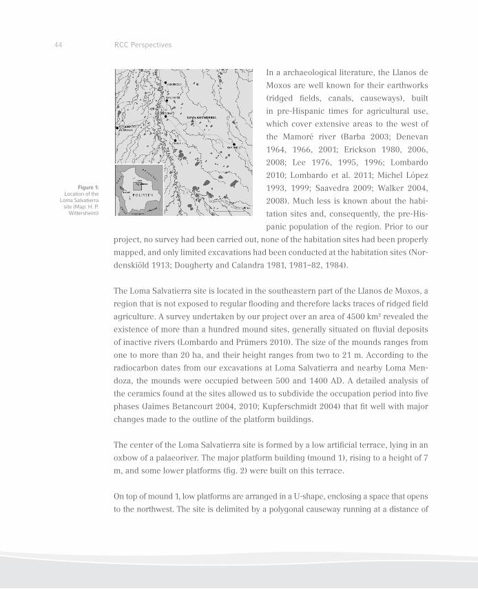
44 RCC Perspectives
In a archaeological literature, the Llanos de
Moxos are well known for their earthworks
(ridged ields, canals, causeways), built
in pre-Hispanic times for agricultural use,
which cover extensive areas to the west of
the Mamoré river (Barba 2003; Denevan
1964, 1966, 2001; Erickson 1980, 2006,
2008; Lee 1976, 1995, 1996; Lombardo
2010; Lombardo et al. 2011; Michel López
1993, 1999; Saavedra 2009; Walker 2004,
2008). Much less is known about the habi-
tation sites and, consequently, the pre-His-
panic population of the region. Prior to our
project, no survey had been carried out, none of the habitation sites had been properly
mapped, and only limited excavations had been conducted at the habitation sites (Nor-
denskiöld 1913; Dougherty and Calandra 1981, 1981–82, 1984).
The Loma Salvatierra site is located in the southeastern part of the Llanos de Moxos, a
region that is not exposed to regular looding and therefore lacks traces of ridged ield
agriculture. A survey undertaken by our project over an area of 4500 km2 revealed the
existence of more than a hundred mound sites, generally situated on luvial deposits
of inactive rivers (Lombardo and Prümers 2010). The size of the mounds ranges from
one to more than 20 ha, and their height ranges from two to 21 m. According to the
radiocarbon dates from our excavations at Loma Salvatierra and nearby Loma Men-
doza, the mounds were occupied between 500 and 1400 AD. A detailed analysis of
the ceramics found at the sites allowed us to subdivide the occupation period into ive
phases (Jaimes Betancourt 2004, 2010; Kupferschmidt 2004) that it well with major
changes made to the outline of the platform buildings.
The center of the Loma Salvatierra site is formed by a low artiicial terrace, lying in an
oxbow of a palaeoriver. The major platform building (mound 1), rising to a height of 7
m, and some lower platforms (ig. 2) were built on this terrace.
On top of mound 1, low platforms are arranged in a U-shape, enclosing a space that opens
to the northwest. The site is delimited by a polygonal causeway running at a distance of
Figure 1: Location of the
Loma Salvatierra site (Map: H. P.
Wittersheim)
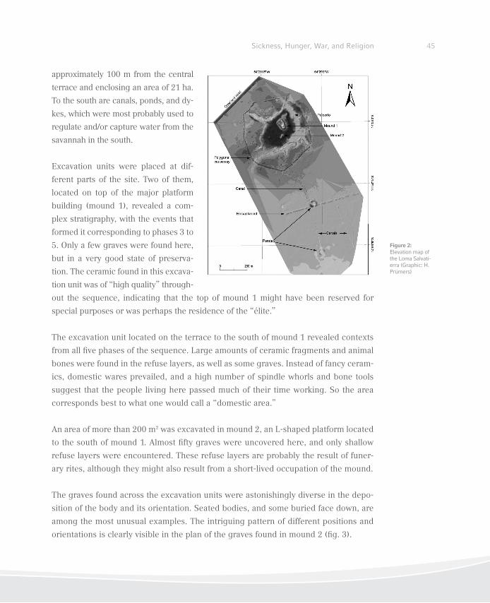
45Sickness, Hunger, War, and Religion
approximately 100 m from the central
terrace and enclosing an area of 21 ha.
To the south are canals, ponds, and dy-
kes, which were most probably used to
regulate and/or capture water from the
savannah in the south.
Excavation units were placed at dif-
ferent parts of the site. Two of them,
located on top of the major platform
building (mound 1), revealed a com-
plex stratigraphy, with the events that
formed it corresponding to phases 3 to
5. Only a few graves were found here,
but in a very good state of preserva-
tion. The ceramic found in this excava-
tion unit was of “high quality” through-
out the sequence, indicating that the top of mound 1 might have been reserved for
special purposes or was perhaps the residence of the “élite.”
The excavation unit located on the terrace to the south of mound 1 revealed contexts
from all ive phases of the sequence. Large amounts of ceramic fragments and animal
bones were found in the refuse layers, as well as some graves. Instead of fancy ceram-
ics, domestic wares prevailed, and a high number of spindle whorls and bone tools
suggest that the people living here passed much of their time working. So the area
corresponds best to what one would call a “domestic area.”
An area of more than 200 m2 was excavated in mound 2, an L-shaped platform located
to the south of mound 1. Almost ifty graves were uncovered here, and only shallow
refuse layers were encountered. These refuse layers are probably the result of funer-
ary rites, although they might also result from a short-lived occupation of the mound.
The graves found across the excavation units were astonishingly diverse in the depo-
sition of the body and its orientation. Seated bodies, and some buried face down, are
among the most unusual examples. The intriguing pattern of different positions and
orientations is clearly visible in the plan of the graves found in mound 2 (ig. 3).
Figure 2: Elevation map of the Loma Salvati-erra (Graphic: H. Prümers)
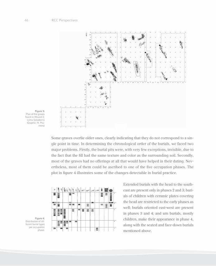
46 RCC Perspectives
Some graves overlie older ones, clearly indicating that they do not correspond to a sin-
gle point in time. In determining the chronological order of the burials, we faced two
major problems. Firstly, the burial pits were, with very few exceptions, invisible, due to
the fact that the ill had the same texture and color as the surrounding soil. Secondly,
most of the graves had no offerings at all that would have helped in their dating. Nev-
ertheless, most of them could be ascribed to one of the ive occupation phases. The
plot in igure 4 illustrates some of the changes detectable in burial practice.
Extended burials with the head to the south-
east are present only in phases 2 and 3; buri-
als of children with ceramic plates covering
the head are restricted to the early phases as
well; burials oriented east-west are present
in phases 3 and 4; and urn burials, mostly
children, make their appearance in phase 4,
along with the seated and face-down burials
mentioned above.
Figure 3: Plan of the graves
found in Mound 2, Loma Salvatierra (Graphic: H. Prü-
mers)
Figure 4: Distribution of dif-ferent burial types
per occupation phase.
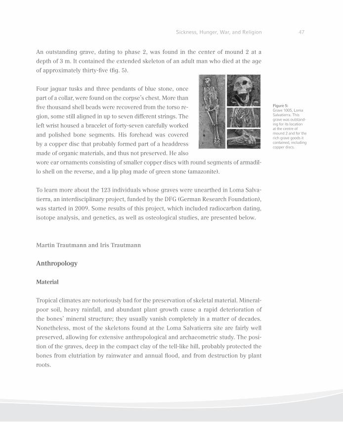
47Sickness, Hunger, War, and Religion
An outstanding grave, dating to phase 2, was found in the center of mound 2 at a
depth of 3 m. It contained the extended skeleton of an adult man who died at the age
of approximately thirty-ive (ig. 5).
Four jaguar tusks and three pendants of blue stone, once
part of a collar, were found on the corpse’s chest. More than
ive thousand shell beads were recovered from the torso re-
gion, some still aligned in up to seven different strings. The
left wrist housed a bracelet of forty-seven carefully worked
and polished bone segments. His forehead was covered
by a copper disc that probably formed part of a headdress
made of organic materials, and thus not preserved. He also
wore ear ornaments consisting of smaller copper discs with round segments of armadil-
lo shell on the reverse, and a lip plug made of green stone (amazonite).
To learn more about the 123 individuals whose graves were unearthed in Loma Salva-
tierra, an interdisciplinary project, funded by the DFG (German Research Foundation),
was started in 2009. Some results of this project, which included radiocarbon dating,
isotope analysis, and genetics, as well as osteological studies, are presented below.
Martin Trautmann and Iris Trautmann
Anthropology
Material
Tropical climates are notoriously bad for the preservation of skeletal material. Mineral-
poor soil, heavy rainfall, and abundant plant growth cause a rapid deterioration of
the bones’ mineral structure; they usually vanish completely in a matter of decades.
Nonetheless, most of the skeletons found at the Loma Salvatierra site are fairly well
preserved, allowing for extensive anthropological and archaeometric study. The posi-
tion of the graves, deep in the compact clay of the tell-like hill, probably protected the
bones from elutriation by rainwater and annual lood, and from destruction by plant
roots.
Figure 5: Grave 1005, Loma Salvatierra. This grave was outstand-ing for its location at the centre of mound 2 and for the rich grave goods it contained, including copper discs.
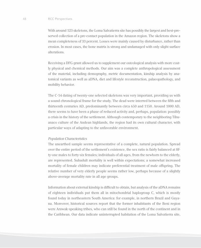
48 RCC Perspectives
With around 123 skeletons, the Loma Salvatierra site has possibly the largest and best-pre-
served collection of a pre-contact population in the Amazon region. The skeletons show a
mean completeness of 33 percent. Losses were mainly caused by disturbance, rather than
erosion. In most cases, the bone matrix is strong and undamaged with only slight surface
alterations.
Receiving a DFG grant allowed us to supplement our osteological analysis with more cost-
ly physical and chemical methods. Our aim was a complete anthropological assessment
of the material, including demography, metric documentation, kinship analysis by ana-
tomical variants as well as aDNA, diet and lifestyle reconstruction, palaeopathology, and
mobility behavior.
The C-14 dating of twenty-one selected skeletons was very important, providing us with
a sound chronological frame for the study. The dead were interred between the ifth and
thirteenth centuries AD, predominantly between circa 650 and 1150. Around 1000 AD,
there seems to have been a phase of reduced activity and, perhaps, population: possibly
a crisis in the history of the settlement. Although contemporary to the neighboring Tihu-
anaco culture of the Andean highlands, the region had its own cultural character, with
particular ways of adapting to the unfavorable environment.
Population Characteristics
The unearthed sample seems representative of a complete, natural population. Spread
over the entire period of the settlement’s existence, the sex ratio is fairly balanced at if-
ty-one males to forty-six females; individuals of all ages, from the newborn to the elderly,
are represented. Subadult mortality is well within expectations; a somewhat increased
mortality of female children may indicate preferential treatment of male offspring. The
relative number of very elderly people seems rather low, perhaps because of a slightly
above-average mortality rate in all age groups.
Information about external kinship is dificult to obtain, but analysis of the aDNA remains
of eighteen individuals put them all in mitochondrial haplogroup C, which is mostly
found today in northeastern South America: for example, in northern Brazil and Guya-
na. Moreover, historical sources report that the former inhabitants of the Beni region
were Arawak-speaking tribes, who can still be found in the north of the continent and in
the Caribbean. Our data indicate uninterrupted habitation of the Loma Salvatierra site,
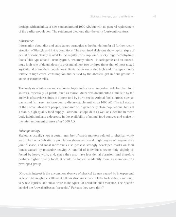
49Sickness, Hunger, War, and Religion
perhaps with an inlux of new settlers around 1000 AD, but with no general replacement
of the earlier population. The settlement died out after the early fourteenth century.
Subsistence
Information about diet and subsistence strategies is the foundation for all further recon-
struction of lifestyle and living conditions. The examined skeletons show typical signs of
dental disease closely related to the regular consumption of sticky, high-carbohydrate
foods. This type of food—usually grain, or starchy tubers—is cariogenic, and an exceed-
ingly high rate of dental decay is present: almost two or three times that of most mixed
agricultural premodern populations. Dental abrasion is also high and of a type charac-
teristic of high cereal consumption and caused by the abrasive grit in lour ground in
stone or ceramic mills.
The analysis of nitrogen and carbon isotopes indicates an important role for plant food
sources, especially C4 plants, such as maize. Maize was documented at the site by the
analysis of starch residues in pottery and by burnt seeds. Animal food sources, such as
game and ish, seem to have been a dietary staple until circa 1000 AD. The tall stature
of the Loma Salvatierra people, compared with genetically close populations, hints at
a stable, high-quality food supply. Later on, isotope data as well as a decline in mean
body height indicate a decrease in the availability of animal food sources and maize in
the later settlement phases after 1000 AD.
Palaeopathology
Skeletons usually show a certain number of stress markers related to physical work-
load. The Loma Salvatierra population shows an overall high degree of degenerative
joint disease, and most individuals also possess strongly developed marks on their
bones caused by muscular activity. A handful of individuals seems only slightly af-
fected by heavy work, and, since they also have less dental abrasion (and therefore
perhaps higher quality food), it would be logical to identify them as members of a
privileged group.
Of special interest is the uncommon absence of physical trauma caused by interpersonal
violence. Although the settlement hill has structures that could be fortiications, we found
very few injuries, and those were more typical of accidents than violence. The Spanish
labeled the Arawak tribes as “peaceful.” Perhaps they were right?
50 RCC Perspectives
There is no clear evidence of malnutrition: although stress markers suggestive of arrested
development phases during childhood—such as cribra orbitalia and enamel hypoplasia—
are frequent, the generally tall and robust stature of the subjects indicates a suficient
diet. Therefore, we assume that the stress markers were caused by periods of illness. The
Loma Salvatierra population was clearly exposed to a multitude of infectious diseases. For
example, the skulls of ive children show signs of heavy periostitis (inlammation of the
connective tissue surrounding the bones) with plaques of woven bone on the interior side
of the skull: probably a symptom of infectious meningitis.
Up to twenty-ive individuals show patches of reaction to inlammation on their long
bones, especially the tibiae. Although there are many causes for periosteal woven bone
growth, the symptoms we found here are peculiar to treponemal infection.
There are multiple active and healed foci of periostitis with woven bone growth and vas-
cularisation, extensive excrescences, and, in some cases, bloated diaphyses with rough
thickening of the compacta and spongy illing of the medulla in tibiae, ibulae, ulnae,
femora, and humeri.
We also observed a sabre-shin deformation with thickened anterior compacta in the
tibiae of at least two individuals, and short and slightly barrel-shaped upper and lower
irst deciduous incisors, with a central notch (Hutchinson’s sign), in one case.
Figure 6-9: Skeletal symptoms
of treponemal infection
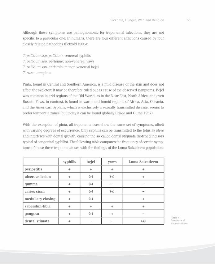
51Sickness, Hunger, War, and Religion
Although these symptoms are pathognomonic for treponemal infections, they are not
speciic to a particular one. In humans, there are four different aflictions caused by four
closely related pathogens (Petzold 2005):
T. pallidum ssp. pallidum: venereal syphilis
T. pallidum ssp. pertenue: non-venereal yaws
T. pallidum ssp. endemicum: non-venereal bejel
T. carateum: pinta
Pinta, found in Central and Southern America, is a mild disease of the skin and does not
affect the skeleton; it may be therefore ruled out as cause of the observed symptoms. Bejel
was common in arid regions of the Old World, as in the Near East, North Africa, and even
Bosnia. Yaws, in contrast, is found in warm and humid regions of Africa, Asia, Oceania,
and the Americas. Syphilis, which is exclusively a sexually transmitted disease, seems to
prefer temperate zones; but today it can be found globally (Idsøe and Guthe 1967).
With the exception of pinta, all treponematoses show the same set of symptoms, albeit
with varying degrees of occurrence. Only syphilis can be transmitted to the fetus in utero
and interferes with dental growth, causing the so-called dental stigmata (notched incisors
typical of congenital syphilis). The following table compares the frequency of certain symp-
toms of these three treponematoses with the indings of the Loma Salvatierra population:
Table 1: Symptoms of treponematoses
syphilis bejel yaws Loma Salvatierra
periostitis + + + +
ulcerous lesion + (+) (+) +
gumma + (+) – –
caries sicca + (+) (+) –
medullary closing + (+) +
sabershin-tibia + + + +
gangosa + (+) + –
dental stimata + – – (+)
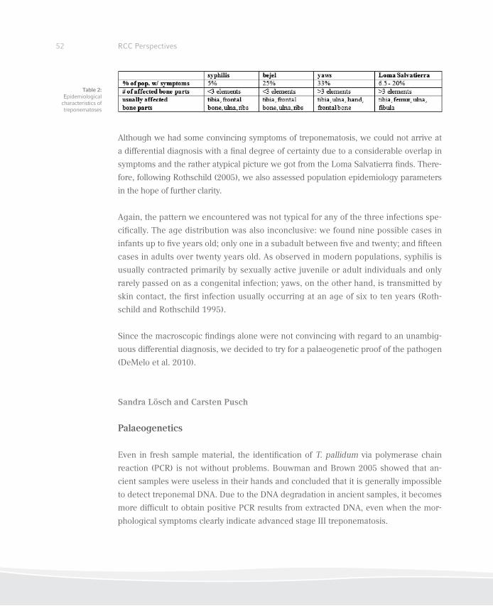
52 RCC Perspectives
Although we had some convincing symptoms of treponematosis, we could not arrive at
a differential diagnosis with a inal degree of certainty due to a considerable overlap in
symptoms and the rather atypical picture we got from the Loma Salvatierra inds. There-
fore, following Rothschild (2005), we also assessed population epidemiology parameters
in the hope of further clarity.
Again, the pattern we encountered was not typical for any of the three infections spe-
ciically. The age distribution was also inconclusive: we found nine possible cases in
infants up to ive years old; only one in a subadult between ive and twenty; and ifteen
cases in adults over twenty years old. As observed in modern populations, syphilis is
usually contracted primarily by sexually active juvenile or adult individuals and only
rarely passed on as a congenital infection; yaws, on the other hand, is transmitted by
skin contact, the irst infection usually occurring at an age of six to ten years (Roth-
schild and Rothschild 1995).
Since the macroscopic indings alone were not convincing with regard to an unambig-
uous differential diagnosis, we decided to try for a palaeogenetic proof of the pathogen
(DeMelo et al. 2010).
Sandra Lösch and Carsten Pusch
Palaeogenetics
Even in fresh sample material, the identiication of T. pallidum via polymerase chain
reaction (PCR) is not without problems. Bouwman and Brown 2005 showed that an-
cient samples were useless in their hands and concluded that it is generally impossible
to detect treponemal DNA. Due to the DNA degradation in ancient samples, it becomes
more dificult to obtain positive PCR results from extracted DNA, even when the mor-
phological symptoms clearly indicate advanced stage III treponematosis.
Table 2: Epidemiological
characteristics of treponematoses
53Sickness, Hunger, War, and Religion
However, we were able to recover and speciically amplify the polymerase A gene
of T. pallidum from skeleton samples of the Loma Salvatierra site independently in
separate aDNA laboratories in Tübingen (C. Pusch, M. Ball, N. Gaultier) and Munich
(A. Nerlich, S. Lösch).
Sampling and Preparation
Our investigation used selected samples of thirty individuals from the burial site for
testing.
Negative control samples were also obtained from individuals of this population who
showed no symptoms of a treponemal infection. All samples were obtained after exca-
vation, during the subsequent thorough examination of the skeletal remains.
Undamaged teeth—mostly molars—were extracted from the jaw and used as sample
material. All necessary precautions to avoid contamination were taken (Roberts and
Ingham 2008). Sampling was performed under strictly sterile conditions in labora-
tories dedicated to work with ancient DNA; all involved were wearing clean clothes,
sterile gloves, and masks in an environment uncontaminated by modern samples from
patients affected by syphilis. Samples were prepared and sent directly from sample
suppliers to cooperating laboratories.
To eliminate contamination, all bone surfaces were irst cleaned with a 0.5% sodium
hypochlorite solution, and then the outer surface was removed mechanically by sterile
instruments. After that, the bones were irradiated with UV-light with a wavelength of
255 nm. Material was then taken from the dentine inner part next to the pulpa cavity.
This was then used to produce a homogeneous bone powder using a mixer mill.
Other precautions against contamination were also taken, including a strict separation
of work areas between sample handling, DNA extraction, PCR set-up, and post-PCR
procedures using different laboratories. Fresh sterile ilter tips were routinely used,
and surfaces and equipment in contact with sample tubes were cleaned before and af-
ter each assay. The results were replicated independently in the Munich laboratories.
Here, a different methodological approach was used.
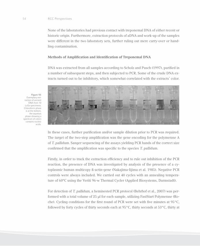
54 RCC Perspectives
None of the laboratories had previous contact with treponemal DNA of either recent or
historic origin. Furthermore, extraction protocols of aDNA and work-up of the samples
were different in the two laboratory sets, further ruling out mere carry-over or hand-
ling contamination.
Methods of Ampliication and Identiication of Treponemal DNA
DNA was extracted from all samples according to Scholz and Pusch (1997), puriied in
a number of subsequent steps, and then subjected to PCR. Some of the crude DNA ex-
tracts turned out to be inhibitory, which somewhat correlated with the extracts’ color.
In these cases, further puriication and/or sample dilution prior to PCR was required.
The target of the two-step ampliication was the gene encoding for the polymerase A
of T. pallidum. Sanger sequencing of the assays yielding PCR bands of the correct size
conirmed that the ampliication was speciic to the species T. pallidum.
Firstly, in order to track the extraction eficiency and to rule out inhibition of the PCR
reaction, the presence of DNA was investigated by analysis of the presence of a cy-
toplasmic human multicopy ß-actin-gene (Nakajima-Iijima et al. 1985). Negative PCR
controls were always included. We carried out 40 cycles with an annealing tempera-
ture of 60°C using the Veriti 96-w Thermal Cycler (Applied Biosystems, Darmstadt).
For detection of T. pallidum, a heminested PCR protocol (Behrhof et al., 2007) was per-
formed with a total volume of 25 µl for each sample, utilizing FastStart Polymerase (Ro-
che). Cycling conditions for the irst round of PCR were set with ive minutes at 95ºC,
followed by forty cycles of thirty seconds each at 95ºC, thirty seconds at 53ºC, thirty at
Figure 10: Exemplary ext-
raction of ancient DNA from 10
LoSa specimens. Chloroform phase
is at the bottom, the aqueous
phase showing a spectrum of colors
contains nucleic acids.
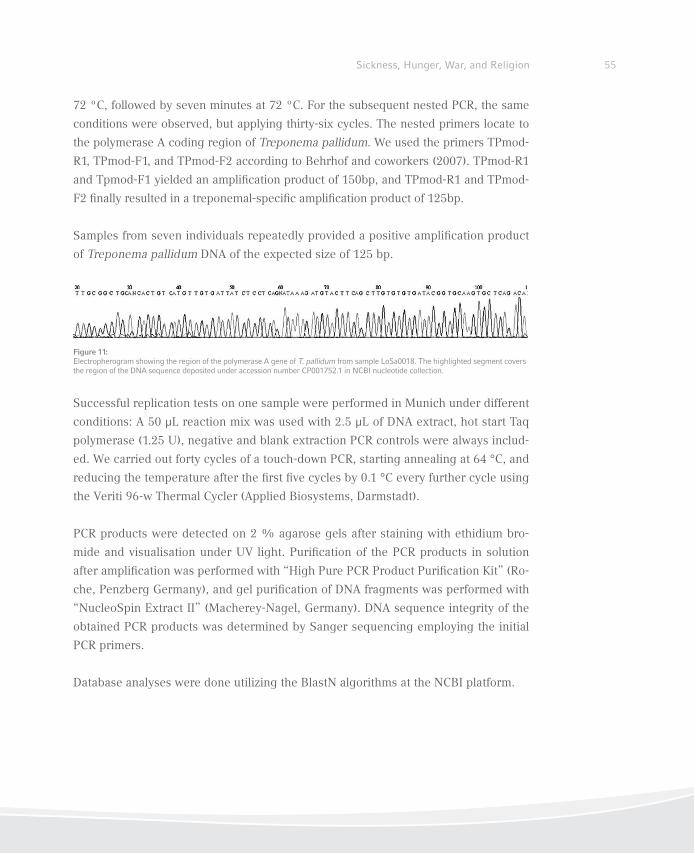
55Sickness, Hunger, War, and Religion
72 ºC, followed by seven minutes at 72 ºC. For the subsequent nested PCR, the same
conditions were observed, but applying thirty-six cycles. The nested primers locate to
the polymerase A coding region of Treponema pallidum. We used the primers TPmod-
R1, TPmod-F1, and TPmod-F2 according to Behrhof and coworkers (2007). TPmod-R1
and Tpmod-F1 yielded an ampliication product of 150bp, and TPmod-R1 and TPmod-
F2 inally resulted in a treponemal-speciic ampliication product of 125bp.
Samples from seven individuals repeatedly provided a positive ampliication product
of Treponema pallidum DNA of the expected size of 125 bp.
Successful replication tests on one sample were performed in Munich under different
conditions: A 50 μL reaction mix was used with 2.5 μL of DNA extract, hot start Taq
polymerase (1.25 U), negative and blank extraction PCR controls were always includ-
ed. We carried out forty cycles of a touch-down PCR, starting annealing at 64 °C, and
reducing the temperature after the irst ive cycles by 0.1 °C every further cycle using
the Veriti 96-w Thermal Cycler (Applied Biosystems, Darmstadt).
PCR products were detected on 2 % agarose gels after staining with ethidium bro-
mide and visualisation under UV light. Puriication of the PCR products in solution
after ampliication was performed with “High Pure PCR Product Puriication Kit” (Ro-
che, Penzberg Germany), and gel puriication of DNA fragments was performed with
“NucleoSpin Extract II” (Macherey-Nagel, Germany). DNA sequence integrity of the
obtained PCR products was determined by Sanger sequencing employing the initial
PCR primers.
Database analyses were done utilizing the BlastN algorithms at the NCBI platform.
Figure 11: Electropherogram showing the region of the polymerase A gene of T. pallidum from sample LoSa0018. The highlighted segment covers the region of the DNA sequence deposited under accession number CP001752.1 in NCBI nucleotide collection.
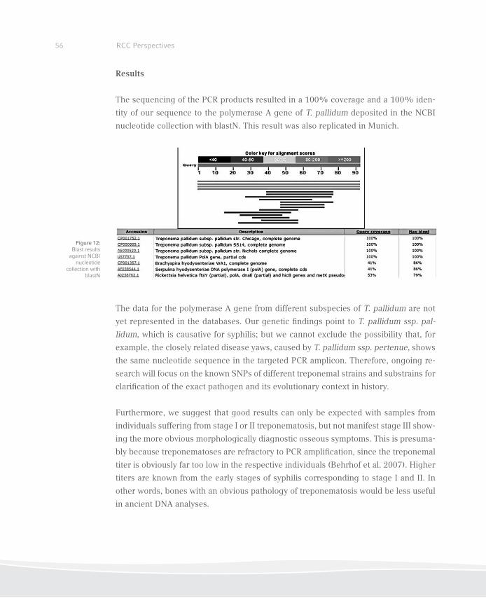
56 RCC Perspectives
Results
The sequencing of the PCR products resulted in a 100% coverage and a 100% iden-
tity of our sequence to the polymerase A gene of T. pallidum deposited in the NCBI
nucleotide collection with blastN. This result was also replicated in Munich.
The data for the polymerase A gene from different subspecies of T. pallidum are not
yet represented in the databases. Our genetic indings point to T. pallidum ssp. pal-
lidum, which is causative for syphilis; but we cannot exclude the possibility that, for
example, the closely related disease yaws, caused by T. pallidum ssp. pertenue, shows
the same nucleotide sequence in the targeted PCR amplicon. Therefore, ongoing re-
search will focus on the known SNPs of different treponemal strains and substrains for
clariication of the exact pathogen and its evolutionary context in history.
Furthermore, we suggest that good results can only be expected with samples from
individuals suffering from stage I or II treponematosis, but not manifest stage III show-
ing the more obvious morphologically diagnostic osseous symptoms. This is presuma-
bly because treponematoses are refractory to PCR ampliication, since the treponemal
titer is obviously far too low in the respective individuals (Behrhof et al. 2007). Higher
titers are known from the early stages of syphilis corresponding to stage I and II. In
other words, bones with an obvious pathology of treponematosis would be less useful
in ancient DNA analyses.
Figure 12: Blast results
against NCBI nucleotide
collection with blastN
57Sickness, Hunger, War, and Religion
Conclusions
The excavations at the pre-contact settlement site of Loma Salvatierra in lowland Bolivia
turned out to be an archaeological source of the highest importance. It became evident
that large, independent, specialized, and complex societies existed in the Amazonian
basin, contemporary to the Andean civilizations, showing a high degree of adaptation to
a challenging environment.
Even in the irst millennium AD, these populations were plagued by an insidious di-
sease: venereal syphilis, which caused a global epidemic starting in the Age of Ex-
ploration, about 1500 AD. Our results demonstrate conclusively that the pathogen
Treponema pallidum ssp. pallidum was already present in populations that settled
northeastern South America, and presumably also the Caribbean.
In accordance with Harper et al. (2008), who reconstructed a phylogenetic tree of Tre-
ponema, we suggest that an American branch of yaws may have developed into an
early strain of syphilis. The close vicinity of warm, humid conditions to cool and arid
ones in the Amazonian-Andean transition zone would have been the perfect stimulus
for the pathogen’s adaptive changes. After 1300 AD, when the Llanos de Moxos were
deserted, the disease could have spread—presumably through migrating Arawak-
speaking tribes—towards the Caribbean.
This model would strongly support the hypothesis that Columbus’s crew contracted
the disease from contact with native peoples and brought it to Europe, from whence
it spread worldwide.
58 RCC Perspectives
References
Barba, Josep. 2003. “Campos elevados.” In Moxos: Una Limnocultura. Cultura y Medio Natural en
la Amazonía Boliviana, 89–92. Barcelona: Centre d´Estudis Amazònics.
Behrhof, Wera, Erik Springer, Wolfgang Bräuninger, James C. Kirkpatrick, and Achim Weber. 2007.
“PCR Testing for Treponema pallidum in Parafin-embedded Skin Biopsy Specimens: Test De-
sign and Impact on the Diagnosis of Syphilis.” Journal of Clinical Pathology 61: 390–95.
Bouwman, Abigail S. and Terence A. Brown. 2005. “The Limits of Biomolecular Palaeopathology:
Ancient DNA Cannot Be Used to Study Veneral Syphilis.” Journal of Archaeological Science 32
(5): 703–13.
De Melo, Fernando L., Joana C. M. de Mello, Ana M. Fraga, Kelly Nunes, and Sabine Eggers. 2010.
“Syphilis at the Crossroad of Phylogenetics and Paleopathology.” PLoS Neglected Tropical Di-
seases 4 (1): 1–11.
Denevan, William M. 1964. “Pre-Spanish Earthworks in the Llanos de Mojos of Northeastern Boli-
via.” Revista Geográica 34 (60): 17–25.
———. 1966. The Aboriginal Cultural Geography of the Llanos de Mojos of Bolivia. Berkeley: Uni-
versity of California Press.
———. 2001. Cultivated Landscapes of Native Amazonia and the Andes. Oxford Geographical and
Environmental Studies. New York: Oxford University Press.
Dougherty, Bernard and Horacio A. Calandra. 1981. “Nota preliminar sobre investigaciones ar-
queológicas en Llanos de Moxos, Departamento del Beni, República de Bolivia.” Revista del
Museo de La Plata, VIII, sección antropología 53: 87-106. La Plata.
———. 1981–82. “Excavaciones arqueológicas en la Loma Alta de Casarabe, Llanos de Moxos,
Departamento del Beni, Bolivia.” Relaciones de la Sociedad Argentina de Antropología, n.s., 14
(2): 9–48.
———. 1984. “Prehispanic Human Settlement in the Llanos de Moxos, Bolivia.” Quaternary of
South America and Antarctic Peninsula 2: 163–99.
Erickson, Clark L. 1980. “Sistemas Agrícolas Prehispánicas en los Llanos de Mojos.” América In-
dígena 40 (4): 731–55.
59Sickness, Hunger, War, and Religion
———. 2006. “The Domesticated Landscapes of the Bolivian Amazon.” In Time and Complexity
in Historical Ecology: Studies in the Neotropical Lowlands, edited by William Balée and Clark L.
Erickson, 235–78. New York: Columbia University Press.
———. 2008. “Amazonia: The Historical Ecology of a Domesticated Landscape.” In Handbook of
South American Archaeology, edited by Helaine Silverman and William Isbell, 157–83. New
York: Springer.
Fracastoro, Girolamo. 1530. Syphilidis, sive Morbi Gallici
Harper, Kristin N., Paolo S. Ocampo, Bret M. Steiner, Robert W. George, Michael S. Silverman,
Shelly Bolotin, Allan Pillay, Nigel J. Saunders, and George J. Armelagos. 2008. “On the Origin of
the Treponematoses: A Phylogenetic Approach.” PloS Neglected Tropical Diseases 2 (1): e148.
Idsøe, Olav and Thorstein Guthe. 1967. “The Rise and Fall of the Treponematoses. I. Ecological
Aspects and International Trends in Venereal Syphilis.” British Journal of Venereal Diseases 43
(4): 227–43.
Jaimes Betancourt, Carla. 2004. Secuencia Cerámica del Corte 1 de la Loma Mendoza. Tésis de
Licenciatura. Universidad Mayor de San Andrés, La Paz.
———. 2010. La cerámica de la Lima Salvatierra. PhD diss., Universität Bonn. http://hss.ulb.uni-
bonn.de/2010/2354/2354.htm.
Kupferschmidt, Denise. 2004. Analyse der frühen Keramik des präkolumbischen Siedlungsplatzes
Loma Mendoza, Bolivien. Magisterarbeit, Rheinische Friedrich-Wilhelms-Universität, Bonn.
Lee, Kenneth. 1976. “7000 años de historia del hombre de Mojos: Agricultura de pampas estériles
(Informe preliminar).” Panorama Universitario 1: 23–26.
———. 1995. Complejo hidraulico de las llanuras de Baures (Area a ser protegida). Unplublished
Manuscript, June 1995.
———. 1996. “Apuntes sobre las obras hidrauclicas prehispánicas de las llanuras de Moxos.” Pai-
titi 11 (1): 24–26.
Lombardo, Umberto. 2010. “Raised Fields of Northwestern Bolivia: A GIS based Analysis.” Zeit-
schrift für Archäologie Außereuropäischer Kulturen 3: 127–49.
60 RCC Perspectives
Lombardo, Umberto, Elisa Canal-Beeby, Seraina Fehr, and Heinz Veit. 2011. “Raised Fields in the
Bolivian Amazonia: A prehistoric Green Revolution or a Flood Risk Mitigation Strategy?” Journal
of Archaeological Science 38 (3): 502–12.
Lombardo, Umberto and Heiko Prümers. 2010. “Pre-Columbian Human Occupation Patterns in the
Eastern Plains of the Llanos de Moxos, Bolivian Amazonia.” Journal of Archaeological Science
37 (8): 1875–85.
Michel López, Marcos. 1993. Prospección arqueológica de San Ignacio de Moxos. Prov. Moxos,
Departamento de Beni. Bolivia. Tésis de Licenciatura, Universidad Mayor de San Andrés, La Paz.
———. 1999. “Desarrollo temprano de la agricultura de campos elevados en los Llanos de Moxos,
Depto. de Beni, Bolivia.” In El Formativo Sudamericano, una Revaluación: Ponencias presenta-
das en el Simposio de Arqueología Sudamericana, Cuenca - Ecuador, 13-17 de enero de 1992,
edited by Paulina Ledergerber-Crespo, 271–81. Quito: Abya-yala.
Nakajima-Iijima, Sadayo, Hiroshi Hamada, Premkumar Reddy, and Takeo Kakunaga. 1985. “Mole-
cular Structure of the Human Cytoplasmic Beta-actin Gene: Interspecies Homology of Sequences
in the Introns.” Proceedings of the National Academy of Sciences of the United States of America
82: 6133–37.
Nordenskiöld von, Erland. 1913. “Urnengräber und Mounds im bolivianischen Flachland.” Baess-
ler-Archiv 3 (6): 205–55.
Petzoldt, D. 2005. “Syphilis.” In Dermatologie und Venerologie II, edited by Otto Braun-Falco, Gerd
Plewig, Helmut Heinrich Wolff, Walter Burgdor, and Michael Landthaler, 227–45. Heidelberg:
Springer Medizin Verlag.
Prümers, Heiko. 2004. “Hügel umgeben von ‘schönen Monstern’: Ausgrabungen in der Loma Men-
doza (Bolivien).” In Expeditionen in Vergessene Welten: 25 Jahre archäologische Forschungen in
Amerika, Afrika und Asien. (AVA-Forschungen, Bd. 10), 47–78. Cologne: Linden Soft.
———. 2008. “Der Wall führt zum See. Die Ausgrabungen 2005–2006 in der Loma Salvatierra
(Bolivien).” Zeitschrift für Archäologie Außereuropäischer Kulturen 2: 371–79.
Prümers, Heiko. 2009. “‘Charlatanocracia’ en Moxos?” In: Procesos y expresiones de poder, identi-
dad y orden tempranos en Sudamérica. Vol.2 , edited by Peter Kaulicke and Tom D. Dillehay.
Boletín de Arqueología PUCP 11 (2007): 103–16.
61Sickness, Hunger, War, and Religion
Roberts, Charlotte. 1994. “Treponematosis in Gloucester, England: A Theoretical and Practical Ap-
proach to the Pre-Columbian Theory.” In: The Origin of Syphilis in Europe: Before or fter 1493?,
edited by Olivier Dutour, György Pali, Jacques Berato, and Jean-Pierre Brun, 101–08. Paris:
Editions Errance.
Roberts, Charlotte and Sarah Ingham. 2008. “Using Ancient DNA Analysis in Palaeopathology: A
Critical Analysis of Published Papers, with Recommendations for Future Work.” International
Journal of Osteoarchaeology 18: 600–13.
Rothschild, Bruce. 2005. “History of Syphilis.” Clinical Infectious Diseases 40: 1454–63.
Rothschild, Bruce and Christine Rothschild. 1995. “Treponemal Disease Revisited: Skeletal Discri-
minators for Yaws, Bejel, and Venereal Syphilis.” Clinical Infectious Diseases 20: 1402–08.
Saavedra Arteaga, Oscar. 2009. Culturas Hidráulicas de la Amazonía Boliviana. La Paz: Oxfam.
Scholz, Michael and Carsten Pusch. 1997. “An eficient Isolation Method for High-quality DNA from
Ancient Bones.” Trends in Genetics 13: 249.
Walker, John Hamilton. 2004. Agricultural Change in the Bolivian Amazon—Cambio Agrícola en la
Amazonía Boliviana. Memoirs in Latin American Archaeology, No. 13. Trinidad, Beni: University
of Pittsburgh Latin American Archaeology Publications & Fundación Kenneth Lee.
———. 2008. “The Llanos de Mojos.” In Handbook of South American Archaeology, edited by
Helaine Silverman and William Isbell, 927–39. New York: Springer.


History of the Plague
Ole Jørgen Benedictow, Raffaella Bianucci,
Sacha Kacki, and Ingrid Wiechmann

64 RCC Perspectives
Authors
I. The Plague—An Introduction
Ingrid Wiechmann
Ludwig Maximilian University of Munich,
Department Biology I, Biodiversity research/Anthropology
Grosshaderner Str. 2
82152 Planegg-Martinsried
Germany
II. The Origin and Early Spread of Yersinia pestis and of Epidemic Plague:
Paleobiological and Historical Viewpoints
Ole Jørgen Benedictow
University of Oslo, Prof. Emeritus
III. The Archaeology of the Second Plague Pandemic: An Overview of French
Funerary Contexts
Raffaella Bianucci
University of Torino
Department of Anatomy, Pharmacology, and Legal Medicine
Laboratory of Criminalistic Sciences
Via Verdi 8
10124 Torino
Italy
Sacha Kacki
Inrap, Villeneuve-d‘Ascq Archaeological Center
Université Bordeaux 1
PACEA, UMR 5199
France
Anthropologie des Populations Passées et Présentes
351 cours de la libération
33405 Talence Cedex
France
65Sickness, Hunger, War, and Religion
Ingrid Wiechmann
The Plague—An Introduction
The infectious disease known as the Plague is caused by the bacterium Yersinia pes-
tis, a gram-negative, facultative anaerobic, rod-shaped bacterium belonging to the
Enterobacteriaceae family. It is a zoonosis found chiely in feral rodents, spread in the
main by infected leas. Plague epidemics in human populations can, however, occur
when the Yersinia pestis pathogen invades rat populations that live in close proximity
to humans. Once the rats have been largely killed off by the disease, the infected leas
move to human hosts.
In human infections, we differentiate between four possible forms of the plague: bu-
bonic plague, septicemic plague, pneumonic plague, and abortive plague. Bubonic
and pneumonic plague are the two most common variants. Bubonic plague is usually
triggered by a bite from an infected lea. Following an incubation period of two to sev-
en days, the irst symptoms appear: severe malaise, fever, headaches and joint pain,
and dizziness. Lymph nodes and lymphatic vessels in the area of the lea bite swell to
form sores with a diameter of up to ten centimeters. These sores change color in the
course of the sickness, often appearing blue or black due to internal bleeding in the
lymph nodes. Without treatment, bubonic plague often leads to septicemic plague, in
which the pathogens (Yersinia pestis) enter the bloodstream and thus attack the whole
body. Septicemic plague causes large-scale bleeding in the skin and internal organs,
and is almost always fatal without treatment. If pathogens spread to the lungs, pneu-
monic plague can develop (secondary pneumonic plague). In this form, pathogens can
also be transmitted from person to person by droplet infection (primary pneumonic
plague). The incubation period until the irst symptoms appear can be from only a few
hours up to a full day. Without treatment, pneumonic plague leads inevitably to death
in between two and ive days. By comparison, the abortive plague is rather less dras-
tic; its symptoms are usually a mild fever and some swelling in the lymph nodes, and
it provides long-term immunity to the plague bacterium.
The Plague, and thus its originator Yersinia pestis, have been linked to many epidem-
ics in the history of humankind. The only completely proven incidence of the Plague,
however, is the most recent worldwide outbreak, which occurred at the end of the

66 RCC Perspectives
nineteenth century (the Third Pandemic); the Yersinia pestis pathogen was irst iden-
tiied during this pandemic (Yersin 1894).
Whether the Plague of Justinian in late antiquity (the First Pandemic) was caused by the
same bacterium remains to be seen: molecular geneticists have so far not been able to
ind conclusive proof. The outbreaks known as the Antonine Plague (165–180 AD) and
the Plague of Athens (430–426 BC) are generally assumed to have been expressions of
other infectious diseases rather than the plague in its strictly medical sense. However,
in recent years, the ields of molecular genetics and immunology have been able to pro-
vide ever more convincing evidence that Yersinia pestis was the pathogen that caused
the Black Death (the Second Pandemic) in the Middle Ages (Raoult et al. 2000; Garrelt
and Wiechmann 2003; Drancourt et al. 2007; Wiechmann, Harbeck and Grupe 2010;
Haensch et al. 2010; Tran et al. 2011; Kacki et al. 2011; Schuenemann et al. 2011).
References
Drancourt, Michel, Michel Signoli, La Vu Dang, Bruno Bizot, Véronique Roux, Stéfan Tzortzis,
and Didier Raoult. 2007. “Yersinia pestis Orientalis in Remains of Ancient Plague Patients.”
Emerging Infectious Diseases 13 (2): 332–33.
Garrelt, Christina, and Ingrid Wiechmann. 2003. “Detection of Yersinia pestis DNA in Early and Late
Medieval Bavarian Burials.” In Documenta Archaeobiologiae 1. Decyphering Ancient Bones: the
Research Potential of Bioarchaeological Collections, edited by Gisela Grupe and Joris Peters, 247–
54. Rahden/Westfalen: Leidorf.
Haensch, Stephanie, Raffaela Bianucci, Michel Signoli, Minoarisoa Rajerison, Michael Schultz,
Sacha Kacki, Marco Vermunt, Darlene A. Weston, Derek Hurst, Mark Achtman, Elisabeth Car-
niel, and Barbara Bramanti. 2010. “Distinct Clones of Yersinia pestis Caused the Black Death.”
PLoS Pathogens 6 (10): e1001134. doi:10.1371/journal.ppat.1001134.
Kacki Sacha, Lila Rahalison, Minoarisoa Rajerison, Ezio Ferroglio, and Raffaela Bianucci. 2011.
“Black Death in the Rural Cemetery of Saint-Laurent-de-la-Cabrerisse Aude-Languedoc, Southern
France, 14th century: immunological evidence.” Journal of Archaeological Science 38: 581–87.
Raoult, Didier, Gérard Aboudharam, Eric Crubézy, Georges Larrouy, Bertrand Ludes, and Michel
Drancourt. 2000. “Molecular Identiication by ‘Suicide PCR’ of Yersinia pestis as the Agent of
Medieval Black Death.” Proceedings of the National Academy of Sciences of the United States
of America 97 (23): 12800–03.
67Sickness, Hunger, War, and Religion
Schuenemann, Verena J., Kirsten Bos, Sharon DeWitte, Sarah Schmedes, Joslyn Jamieson, Alissa
Mittnik, Stephen Forrest, Brian K. Coombes, James W. Wood, David J. D. Earn, William White,
Johannes Krause, and Hendrik N. Poinar. 2011. “Targeted Enrichment of Ancient Pathogens
Yielding the pPCP1 Plasmid of Yersinia pestis from Victims of the Black Death.” Proceedings of
the National Academy of Sciences of the United States of America 108 (38): E746–52.
Tran, Thi-Nguyen-Ny, Michel Signoli, Luigi Fozzati, Gérard Aboudharam, Didier Raoult, and Mi-
chel Drancourt. 2011. “High Throughput, Multiplexed Pathogen Detection Authenticates Plague
Waves in Medieval Venice, Italy.” PLoS One 6 (3): e16735. doi:10.1371/journal.pone.0016735.
Wiechmann, Ingrid, Michaela Harbeck, and Gisela Grupe. 2010. “Yersinia pestis DNA Sequences
in Late Medieval Skeletal Finds, Bavaria.” Emerging Infectious Diseases 16 (11): 1806–07.
Yersin, Alexandre. 1894. “La peste bubonique à Hong Kong.” Annales de l’Institut Pasteur 8: 662–67.
Ole Jørgen Benedictow
The Origin and Early Spread of Yersinia pestis and of Epidemic Plague:
Palaeobiological and Historical Viewpoints
A recent paper by a team of microbiologists headed by G. Morelli, Y. Song, and C. J.
Mazzoni demonstrates the great opportunities offered by the new science of palaeobio-
logy for advancing knowledge of the evolutionary development of infectious diseases—in
this case, the bubonic plague—and the indispensable importance of interdisciplina-
ry cooperation with historians (Morelli et al. 2010). They conclude convincingly that
“plague originated in East Asia, perhaps in China Proper, more likely in Mongolia or
Eastern Russia (Eastern Siberia) “[over] 2,600 years ago” and “spread through multiple
radiations to Europe, South America, Africa, and Southeast Asia.” However, they also re-
fer to the development of biovars and various strains in claiming that “the geographical
sources and evolutionary branch order of [the biovar of] 2.MED[ievalis] subpopulations,
which arose [over] 545 years ago” and that other data “supports the westward spread
of 2.MED from China through trade articles that were carried along the Silk Road,” the
extensive trade route from China to Western Asia “between 200 [BCE]1 and 1400.” They
also claim that “the estimated age of [the biovar of] 1.ANT[iqua]1 (628–6,914 years ago)
slightly predates the extensive voyages from China led by Zheng He between 1409 and
68 RCC Perspectives
1433. They conclude that, “It seems highly likely that these ships were infested by rats,
which could have transmitted Yersinia pestis from China to Africa.”
A historian of epidemic diseases will note that, irstly, the indicated routes of spread
are not supported by concrete, tenable evidence; in fact, they are at variance with the
evidence, and out of chronological/historical sync with other data and events. Secondly,
the references to bubonic plague in Chinese sources are both late and sparse when com-
pared with the much earlier, quite numerous references in classical Greek, Hellenistic
Greek, and ancient Roman sources. There are also substantially earlier references to
bubonic plague, especially the Biblical account of epidemic events associated with the
war between the Philistines and the Israelites in approximately 1100 BCE.
Morelli et al.’s biological determination of an East Asian origin of Yersinia pestis seems
convincing and undoubtedly represents a major scholarly achievement. However, the
fact that the earliest references by far to the bubonic plague concern regions on the
Mediterranean littoral needs explanation, in order to make these seemingly disparate
pieces of information compatible.
Morelli et al.’s attempts at explaining the historical spread of Yersinia pestis across
the Eurasian continent by transportation along the Silk Roads, or transportation to
Eastern Africa by Zheng He’s maritime expeditions in the period 1409–1433, must be
deemed untenable. There are also serious problems or at least weaknesses associated
with their dating of the time of its origin, and the spatio-temporal pattern of its spread,
using biomolecular clocking techniques.
This calls for a possible alternative explanation(s) of how Yersinia pestis could have
spread out of its original homeland in East Asia, which should be compatible with
the spatio-temporal perspectives provided by historical sources. Such an explanation
could be based on a notion of the natural pace of the spread of plague disease, caused
by Yersinia pestis, in sylvatic (wild) rodent populations.
A similar event can serve as a useful basis for clariication of this question. In June
1900, plague entered a new continent, namely North America, in San Francisco. Seventy-
ive years later, plague had penetrated into 12 states and had crossed the middle of
the United States at 100o longitude, a distance of around 2000 km (1250 miles) from
69Sickness, Hunger, War, and Religion
San Francisco, and had also invaded Canada in the north and Mexico in the south. The
average spread rate had thus been of the order of 25 km (16 miles) a year. McNeill
(1979, 152–55, 272) concludes his discussion by saying that “the geographic spread
of plague infection in North America occurred naturally.” It was basically passed on
between colonies of ground-burrowing rodents.
At a similar pace of spread, plague would have covered the roughly 8000 km from a
central location in East Asia to the Middle East in about 320 years; or about 400 years
at 20 km per year. This estimate can now be juxtaposed with Morelli et al.’s conclu-
sions that “Yersinia pestis evolved in or near China” over 2,600 years ago, but also
that 1.ANT1 developed 628–6,914 years ago; they seem to envisage an age of Yersinia
pestis in East Asia 6,914 years ago, and that the real age would include the time of
its spread to East Africa and the development of the biovar. The natural spread rate
of plague is compatible with the historical data, and within the enormous margins of
uncertainty inherent in biomolecular dating techniques.
In the Middle East, Yersinia pestis would become connected to the black rat and Xeno-
psylla cheopis, “the rat lea par excellence,” as it has often been called. Black rats and
their leas have been central in all plague epidemics studied by modern physicians and
epidemiologists and the speciic deining features of rat-borne plague can be observed
with respect to historical plague epidemics. Maps of the global distribution of plague
foci of various species of wild rodents show that they stretch almost continuously from
Manchuria to the Middle East. After arrival in the Middle East, Yersinia pestis could
easily have been spread by human agency to western Arabia and further to Central
East Africa, triggering the formation of plague foci there.
Clearly, the bubonic plague pathogen originated in East Asia and spread westwards
from there. The epidemic powers of plague (as distinct from the epizootic powers) may
have evolved in the Middle East, where the black rat and Xenopsylla cheopis were pre-
sent, producing the basic requirements for the adaptation and spread of the plague
in human populations. In order to become an epidemic disease spreading in human
habitats the disease would have to be introduced among rodents in human habitats
with leas that would also attack and transmit the disease to human beings. Conditions
in the Middle East were well suited to allow a transformation of the patterns of the
spread of plague. If so, this could have triggered a new phase of plague that spread back
70 RCC Perspectives
eastwards, with profound epidemic implications that may not be easily identiiable with
palaeobiological tools.
Clearly, wherever plague’s ancestral homeland was, the temporal perspectives provide
superabundant time for plague to spread westwards all the way across the Eurasian conti-
nent, cross into Africa, and establish the plague foci of the Middle East, the western coast-
al areas of Iran and Arabia, and Central Eastern Africa. The establishment of these foci
would constitute the basis for the biblical account of pre-classical plague, and the classical
plague epidemics and clinical descriptions of plague in classical medical works.
This transcontinental spread of Yersinia pestis could quite likely have occurred long be-
fore antiquity and have caused spread all the way back to China, in time to establish the
epidemic combination of the black rat and Xenopsylla cheopis and the necessary condi-
tions for plague epidemics much earlier—perhaps even thousands of years earlier—than
the “2,600 years ago” suggested by Morelli et al. It raises essential and interesting ques-
tions about the possible adaptations between the black rat and Xenopsylla cheopis and
the biovars and various strains of Yersinia pestis encountered on such a journey. Would
signiicant adaptation by selection be necessary at all? Or would the existing adaptations
selected by Yersinia pestis in relation to various combinations of rodent leas and their
sylvatic rodent hosts function readily and well with Xenopsylla cheopis and the black rat?
References
Benedictow, Ole J. 2004. The Black Death 1346–1353: The Complete History. Woodbridge: The
Boydell Press.
———. 2010. What Disease was Plague? On the Controversy over the Microbiological Identity of
Plague Epidemics of the Past. Leiden: Brill.
McNeill, William H. 1979. Plagues and Peoples. Harmondsworth: Penguin Books.
Morelli, Giovanni, Yajun Song, Camila Mazzoni, Mark Eppinger, Philippe Roumagnac, David
Wagner, Mirjam Feldkamp, Barica Kusecek, Amy Vogler, Li Yanjun, Yajun Cui, Nicholas Thom-
son, Thibaut Jobart, Raphael Leblois, Peter Lichtner, Lila Rahalison, Jeannine Petersen, Fran-
cois Balloux, Paul Keim, Thierry Wirth, Jacques Ravel, Ruifu Yang, Elisabeth Carniel, and Mark
Achtman. 2010. “Yersinia pestis Genome Sequencing identiies Patterns of Global Phylogene-
tic Diversity.” Nature Genetics 42 (12): 1140–43. doi:10.1038/ng.960.
71Sickness, Hunger, War, and Religion
Raffaella Bianucci and Sacha Kacki
The Archaeology of the Second Plague Pandemic: An Overview of French
Funerary Contexts
Over the last 20 years, several plague mass graves have been unearthed in France,
thus enhancing our knowledge of historical plague pandemics. Moreover, recent ar-
chaeo-anthropological and palaeoimmunological investigations have shown that abrupt
mortality crises caused by plague have been handled differently in urban and rural com-
munities (Kacki et al. 2011). In this paper, we report on the different funerary contexts
and the related practices adopted by some French urban and rural communities during
the Second Pandemic (1348–1722 CE).
Several eighteenth-century urban plague burial sites have been uncovered in France.
Most of them date to the “Great Plague,” which occurred in Provence and Languedoc
during 1720–1722. It has been estimated that the Marseilles “Great Plague,” which
started on 20 June 1720, affected 242 localities and resulted in 119,811 deaths out
of a population of 394,369 individuals (Signoli et al. 2002). In urban centers, such as
Marseilles (“The Observance Pit” and “The Major Pit”) and Martigues (“Le Délos” and
“Le Couvent des Capucins”), plague epidemics caused the death of dozens or even
hundreds of people daily. This led to the digging of large pits or trenches, where the
deceased were buried. We can pinpoint a number of differences in the funerary treat-
ments of these eighteenth-century victims, mainly linked to the patients’ admission
through the plague inirmaries. To sum up, two different kinds of plague victims can
be identiied within the mass burials:
1. Those skeletons found together with personal belongings correspond to the plague
victims who died outside the inirmaries and who were found dead several hours, or
even days, later. They were buried fully dressed, along with their belongings, because
gravediggers wanted to minimize contact with the infected bodies.
2. Skeletons found without any personal belongings correspond to the victims who
died in the plague inirmaries and were buried according to the sanitary restrictions
adopted during the plague epidemics, i.e., undressed and wrapped in shrouds. The
transit through the plague inirmaries resulted in a slower rhythm of inhumations,
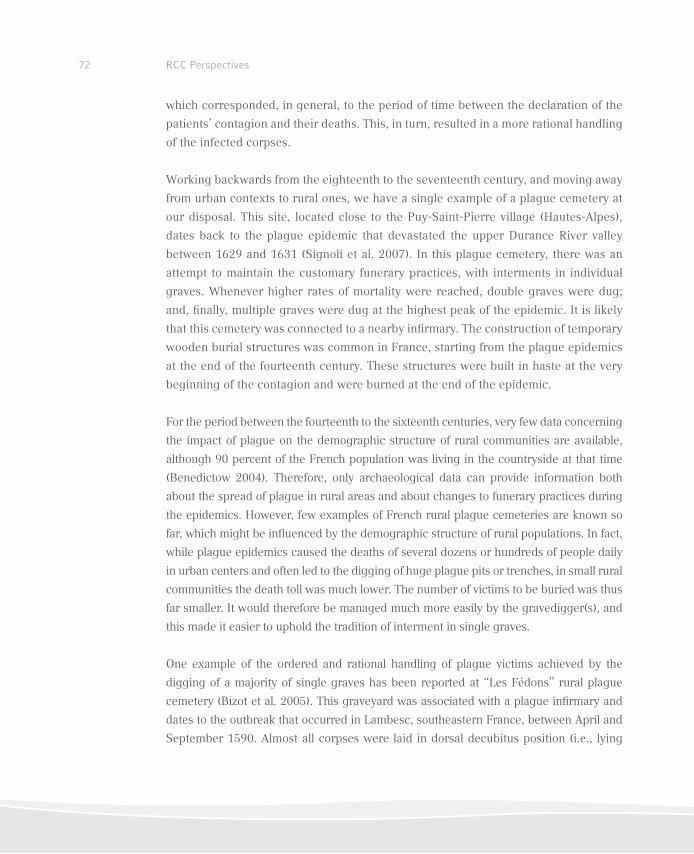
72 RCC Perspectives
which corresponded, in general, to the period of time between the declaration of the
patients’ contagion and their deaths. This, in turn, resulted in a more rational handling
of the infected corpses.
Working backwards from the eighteenth to the seventeenth century, and moving away
from urban contexts to rural ones, we have a single example of a plague cemetery at
our disposal. This site, located close to the Puy-Saint-Pierre village (Hautes-Alpes),
dates back to the plague epidemic that devastated the upper Durance River valley
between 1629 and 1631 (Signoli et al. 2007). In this plague cemetery, there was an
attempt to maintain the customary funerary practices, with interments in individual
graves. Whenever higher rates of mortality were reached, double graves were dug;
and, inally, multiple graves were dug at the highest peak of the epidemic. It is likely
that this cemetery was connected to a nearby inirmary. The construction of temporary
wooden burial structures was common in France, starting from the plague epidemics
at the end of the fourteenth century. These structures were built in haste at the very
beginning of the contagion and were burned at the end of the epidemic.
For the period between the fourteenth to the sixteenth centuries, very few data concerning
the impact of plague on the demographic structure of rural communities are available,
although 90 percent of the French population was living in the countryside at that time
(Benedictow 2004). Therefore, only archaeological data can provide information both
about the spread of plague in rural areas and about changes to funerary practices during
the epidemics. However, few examples of French rural plague cemeteries are known so
far, which might be inluenced by the demographic structure of rural populations. In fact,
while plague epidemics caused the deaths of several dozens or hundreds of people daily
in urban centers and often led to the digging of huge plague pits or trenches, in small rural
communities the death toll was much lower. The number of victims to be buried was thus
far smaller. It would therefore be managed much more easily by the gravedigger(s), and
this made it easier to uphold the tradition of interment in single graves.
One example of the ordered and rational handling of plague victims achieved by the
digging of a majority of single graves has been reported at “Les Fédons” rural plague
cemetery (Bizot et al. 2005). This graveyard was associated with a plague inirmary and
dates to the outbreak that occurred in Lambesc, southeastern France, between April and
September 1590. Almost all corpses were laid in dorsal decubitus position (i.e., lying
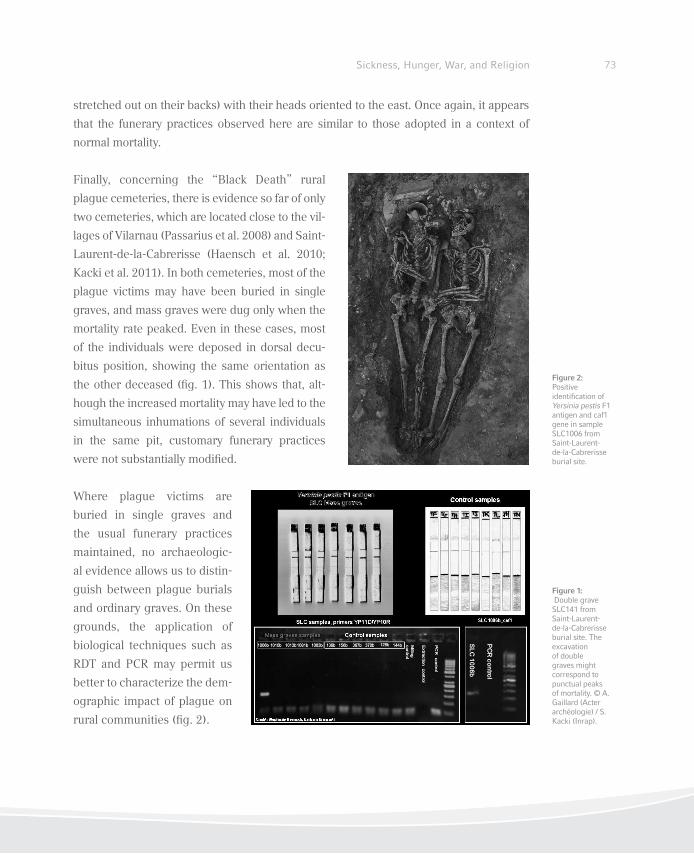
73Sickness, Hunger, War, and Religion
stretched out on their backs) with their heads oriented to the east. Once again, it appears
that the funerary practices observed here are similar to those adopted in a context of
normal mortality.
Finally, concerning the “Black Death” rural
plague cemeteries, there is evidence so far of only
two cemeteries, which are located close to the vil-
lages of Vilarnau (Passarius et al. 2008) and Saint-
Laurent-de-la-Cabrerisse (Haensch et al. 2010;
Kacki et al. 2011). In both cemeteries, most of the
plague victims may have been buried in single
graves, and mass graves were dug only when the
mortality rate peaked. Even in these cases, most
of the individuals were deposed in dorsal decu-
bitus position, showing the same orientation as
the other deceased (ig. 1). This shows that, alt-
hough the increased mortality may have led to the
simultaneous inhumations of several individuals
in the same pit, customary funerary practices
were not substantially modiied.
Where plague victims are
buried in single graves and
the usual funerary practices
maintained, no archaeologic-
al evidence allows us to distin-
guish between plague burials
and ordinary graves. On these
grounds, the application of
biological techniques such as
RDT and PCR may permit us
better to characterize the dem-
ographic impact of plague on
rural communities (ig. 2).
Figure 1: Double grave SLC141 from Saint-Laurent-de-la-Cabrerisse burial site. The excavation of double graves might correspond to punctual peaks of mortality. © A. Gaillard (Acter archéologie) / S. Kacki (Inrap).
Figure 2: Positive identiication of Yersinia pestis F1 antigen and caf1 gene in sample SLC1006 from Saint-Laurent-de-la-Cabrerisse burial site.

74 RCC Perspectives
References
Benedictow, Ole J. 2004. The Black Death 1346–1353: The Complete History. Woodbridge: Boydell
Press.
Bianucci, Raffaella, Lila Rahalison, Emma Rabino Massa, Alberto Peluso, Ezio Ferroglio, and
Michel Signoli. 2008. “Technical Note: A Rapid Diagnostic Test Detects Plague in Ancient
Human Remains: An Example of Interaction between Archaeological and Biological Approa-
ches (South-eastern France, 16th, 17th and 18th Centuries).” American Journal of Physical
Anthropology 136 (3): 361–67.
Bizot, Bruno, Dominique Castex, Patrick Reynaud, and Michel Signoli. 2005. La saison d’une
peste (Avril-Septembre 1590): Le cimetière des Fédons à Lambesc. Paris: CNRS Editions.
Haensch, Stephanie, Raffaella Bianucci, Michel Signoli, Minoarisoa Rajerison, Michael Schultz,
Sacha Kacki, Marco Vermunt, Darlene A. Weston, Derek Hurst, Mark Achtmann, Elisabeth Car-
niel, and Barbara Bramani. 2010. “Distinct Clones of Yersinia pestis Caused the Black Death.”
PLoS Pathogens 6 (10): 1001134. doi:10.1371/journal.ppat.101134.
Kacki, Sacha, Lila Rahalison, Minoarisoa Rajerison, Ezio Ferroglio, and Raffaella Bianucci. 2011.
“Black Death in the Rural Cemetery of Saint-Laurent-de-la-Cabrerisse, Aude-Languedoc, Sou-
thern France, 14th Century: Immunological Evidence.” Journal of Archaeological Science 38:
581–87.
Passarrius, Olivier, Richard Donat, and Aymat Catafau. 2008. Vilarnau, un village du Moyen Âge
en Roussillon. Éditions Trabucaire, Collection Archéologie Départementale.
Signoli, Michel, Isabelle Séguy, Jean-Noël Biraben, and Olivier Dutour. 2002. “Paleodemography
and Historical Demography in the Context of an Epidemic: Plague in Provence in the Eigh-
teenth Century.” Population - E2002 57 (6): 829–54.
Signoli, Michel, Stephan Tzortzis, Bruno Bizot, Yann Ardagna, Catherine Rigeade, and Isabel-
le Séguy. 2007. “Découverte d’un cimetière de pestiférés du XVIIe siècle (Puy-Saint-Pierre,
Hautes-Alpes, France).” In Peste: entre épidémies et sociétés, edited by Michel Signoli, Do-
minique Chevé, Adalian Pascal, Giles Boëtsch, and Olivier Dutour, 131–35. Florence: Firenze
University Press.
Hypothyroidism in Switzerland
Christina Papageorgopoulou, Kaspar Staub,
and Frank Rühli
76 RCC Perspectives
Authors
Christina Papageorgopoulou
Demokritus University of Thrace
Department of History and Ethnology
Laboratory of Anthropology
P. Tsaldari 1
69100-Komotini
Greece
Kaspar Staub
University of Zurich
Institute of Anatomy
Centre for Evolutionary Medicine
Winterthurerstr. 190
CH-8057 Zurich
Switzerland
Frank Rühli
University of Zurich
Institute of Anatomy
Centre for Evolutionary Medicine
Winterthurerstr. 190
CH-8057 Zurich
Switzerland
77Sickness, Hunger, War, and Religion
Introduction
Hypothyroidism is a condition of mild to severe impairment of physical and mental de-
velopment due to an untreated deiciency of thyroid hormones, and can be endemic,
genetic, or sporadic. Endemic hypothyroidism arises from a diet deicient in iodine.
The most common iodine deiciency disorders are goiter (enlarged thyroid gland) and
hypothyroidism at all ages, as well as endemic cretinism,1 mental impairment, delayed
physical development in growing children (Iglesias and Diez 2008; Zimmermann,
Jooste, and Pandav 2008; Zimmermann 2009, 2010).
Populations living in iodine deprived areas such as the Himalayas, the Andes, and the
European Alps, are most at risk. In the Alpine region of Switzerland endemic hypothy-
roidism was very common until the beginning of the twentieth century. The aim of this
study is to present the theme from three different, but complementary perspectives. The
medical perspective lays the groundwork regarding the pathophysiology, the clinical
picture, and the differential diagnosis of the condition. The historical perspective pre-
sents contemporary scientiic studies on conscription and published data on goiter and
cretinism as endemic manifestations of hypothyroidism (since 1900) and the archaeo-
anthropological perspective reports one of the irst documentations of the condition in
an archaeological population from Switzerland (11th–15th century AD). Together, they
offer insights into a signiicant health problem and contribute signiicantly to the bio-
logical history of the Alpine regions.
The interdisciplinary approach of this contribution brings together different ields with
different methods and sources of information. This is necessary, since medical, historical,
and anthropological sources for the period before 1900 are extremely limited and, in some
cases, completely absent. By comparing medical background, skeletal manifestations
All three authors would like thank the Rachel Carson Center for Environment and Society and the Work Group for Palaeoanthropology and Prehistoric Anthropology (Society for Anthropology, Germany) for the invitation to the Sickness, Hunger, War, and Religion Workshop in Munich in March 2011. Christina Papageorgopoulou would like to thank the archaeological Service of Canton Graubünden for inancing the study of the skeletal material, and Thomas Böni for useful discussion. The present manuscript was written during a postdoctoral fellowship granted from the Alexander v. Humboldt Foundation. Frank Rühli and Kaspar Staub are supported by the Mäxi Foundation and wish to thank Prof. em. Hans Bürgi for helpful comments. All authors would like to thank Karl Link, University of Zurich for proofreading the manuscript.
1 The use of the term cretinism and cretin—referring to severely stunted physical and mental development due to lack of thyroid hormones—is made for the sake of medical, historical, and social facets that the disease shaped during the beginning of the twentieth century and by no means entails a negative connotation.
78 RCC Perspectives
in the Swiss population past and present, and historic and modern survey data, we gained
new insights into a highly complicated, but also signiicant, health and social problem,
namely hypothyroidism. The overlap between the three disciplines both in terms of actual
data—e.g., variation in stature between hypothyroid and non-hypothyroid individuals—
and also in terms of theoretical background underlines the need for interdisciplinary
studies such as this. To our knowledge, this is the irst time that hypothyroidism in
Switzerland has been the subject of a multidisciplinary study, and we hope that this
paper will offer insights and motivation for the study of the condition in the neighboring
Alpine regions of Germany, Italy, and Austria.
Frank Rühli
Hypothyroidism: A Medical (Bone) Perspective
The aim of this irst, medically oriented section is not only to list the clinical classii-
cation of various types or etiologies of hypothyroidism, but also to present speciically
selected cases of hypothyroidism as found in the so called “Galler collection,” a bone
pathology reference series from the University Hospital, Zurich, mostly circa 1940–
1970 (for some general information on this, see Rühli, Hotz, and Böni 2003).
Within the thyrotrophin cycle, hypothyroidism is deined as a lack of lack of triiodo-
thyronine (T3) and thyroxine (T4), and can severely affect tissue development and
remodeling. The main skeletal manifestation of hypothyroidism is delayed epiphy-
seal fusion (affecting the axial skeleton and long bones), resulting in short hands,
limb asymmetries, changes in skull form such as sella turcica, or osteoporosis. Other
manifestations (psychological, soft tissue) are: fatigue, decreased heart rate, weight
gain, problems with memory and concentration, depression, goiter (enlarged thyroid
gland), muscle pain, dry swollen skin, and irregular menstrual periods (in women).
Thyrotrophin-releasing hormone (TRH) is released from the hypothalamus via specif-
ic vessels to the hypophysis (pituitary gland), and, in turn, thyrotrophin or thyroid-
stimulating hormone (TSH) from the hypophysis regulates the thyroid function. Iodine
is needed (ca. 200 µg/day) in order to correctly synthesize the hormones T3 and T4
produced by the thyroid. T3 and T4 increase the general metabolism, sensitivity to
79Sickness, Hunger, War, and Religion
catecholamines (organic compounds that act upon the heart among other organs);
or calcium, and phosphate metabolism. Inborn variants of hypothyroidism (thyroid
hypofunction) are aplasia (absence of an organ), dysplasia (alteration of cells or of a
structure), and hormone resistance (receptor defects), and acquired variants are ia-
trogenic (i.e., resulting from medical treatment, such as through iodine exposition or
following an operation). Primary variants are a lack of iodine or autoimmune thyroidi-
tis (inlammation of thyroid gland; “Morbus Hashimoto”); a secondary variant is a lack
of TSH (hypopituitarism); tertiary variants are a lack of TRH or disruption of the portal
system (Pickardt syndrome, or interruption of the vein system connecting hypothala-
mus and pituitary, e.g. due to tumors). In Switzerland, 757 cases of hypothyroidism
in newborns have been reported since 1977 (ca. 1:3500). There is regular screening
of infants at day four or ive after birth. Severity grades are: latent/compensated (only
TSH is elevated); subclinical (T3 and T4 lowered, but no symptoms); and manifest
(symptoms).
An exemplary case of “cretinism” from the Galler pathological bone-reference series
from the nineteenth and twentieth centuries in Switzerland (Rühli, Hotz, and Böni
2003) is presented here, together with clinical case reports, as well as autopsy ind-
ings. One 79-year-old male individual in the Galler series, who was 160 cm tall, had
particularly strong signs of hypothyroidism. The autopsy indings reported, among
other indications, a cretinoid habitus as well as a struma diffusa on the x-rays.
In general, skeletal manifestations of hypothyroidism (thyroid hypofunction) in the
Galler cases are: kyphoscoliosis, large cranium, proportional dwarism, cretinoid ha-
bitus, renal osteopathy (ibro-osteoclasia), hyperostosis frontalis interna (thickening of
the inner table of the frontal bone), coxarthritis, osteoporosis (mild/severe), and club
foot. Causes of death were: subdural hematoma (cerebral hemorrhage) from a fall,
pneumonia, heart failure, and uterus corpus carcinoma.
80 RCC Perspectives
Kaspar Staub
Historical Perspectives on Hypothyroidism in Switzerland: Endemic Iodi-
ne Deiciency, Goiter, and Cretinism since the Late Nineteenth Century
Switzerland was one of the few countries to become completely iodine suficient be-
fore 1990 (together with some of the Scandinavian countries, Australia, USA, and
Canada), and was a world pioneer, introducing its iodized salt program in 1922 (irst
in Canton Appenzell AR, then subsequently all over Switzerland, operating uninter-
rupted since then). The consensus among researchers is that iodine prophylaxis was
the decisive factor in eradicating substantial risks of iodine deiciency and preventing
endemic goiter and cretinism during the irst half of the twentieth century (Bürgi, Su-
persaxo, and Selz 1990; Zimmermann 2008). This section will summarize this process
since its inception in the late nineteenth century.
Prior to the introduction of the iodized salt program, Switzerland was severely iodine
deicient. Goiter and “cretinism” were widespread in alpine areas and the foothills of the
Alps. Surveys have been made since the beginning of the nineteenth century. For ex-
ample, a census ordered by Napoleon reported four thousand cretins among the 70,000
inhabitants of Canton Valais in 1800. Many travelers to Switzerland reported the devas-
tating endemic of goiter and cretinism; for example, Mark Twain, who stated in 1884:
“I am satisied. I have seen the principal features of Swiss scenery—Mount Blanc and
the goiter—and now for home.” Goiters appeared also in numerous illustrations and
chronicles (Bürgi, Supersaxo, and Selz 1990; Zimmermann 2008; Merke 1971).
Since the 1880s, scientiic studies and surveys based on data from schoolchildren and
military conscripts showed the degree and regional spread of goiter and cretinism. In
1889, Theodor Kocher showed in his goiter survey of around 80,000 schoolchildren in
the Canton of Bern that 20 to 100 percent of the seven to sixteen-year-old children had
goiters. In 1883, Heinrich Bircher had already demonstrated the regional prevalence
of military service exclusion due to large goiters in Swiss conscript data for the period
1875–1880, mapped down to village level. The enormous variation from one village
to another was explained by differences in soil geology and water supply (the pre-
sence of some kind of goitrogenic agent). These early researchers failed to consider
iodine deiciency as the cause, even though the relationship between iodine deicient
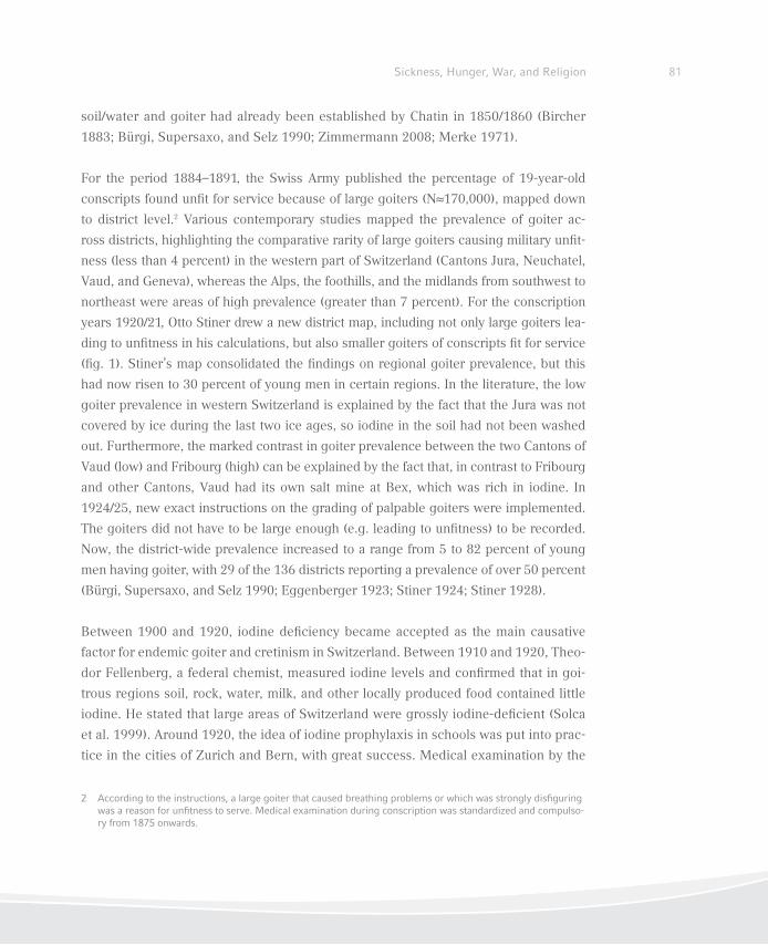
81Sickness, Hunger, War, and Religion
soil/water and goiter had already been established by Chatin in 1850/1860 (Bircher
1883; Bürgi, Supersaxo, and Selz 1990; Zimmermann 2008; Merke 1971).
For the period 1884–1891, the Swiss Army published the percentage of 19-year-old
conscripts found unit for service because of large goiters (N≈170,000), mapped down
to district level.2 Various contemporary studies mapped the prevalence of goiter ac-
ross districts, highlighting the comparative rarity of large goiters causing military unit-
ness (less than 4 percent) in the western part of Switzerland (Cantons Jura, Neuchatel,
Vaud, and Geneva), whereas the Alps, the foothills, and the midlands from southwest to
northeast were areas of high prevalence (greater than 7 percent). For the conscription
years 1920/21, Otto Stiner drew a new district map, including not only large goiters lea-
ding to unitness in his calculations, but also smaller goiters of conscripts it for service
(ig. 1). Stiner’s map consolidated the indings on regional goiter prevalence, but this
had now risen to 30 percent of young men in certain regions. In the literature, the low
goiter prevalence in western Switzerland is explained by the fact that the Jura was not
covered by ice during the last two ice ages, so iodine in the soil had not been washed
out. Furthermore, the marked contrast in goiter prevalence between the two Cantons of
Vaud (low) and Fribourg (high) can be explained by the fact that, in contrast to Fribourg
and other Cantons, Vaud had its own salt mine at Bex, which was rich in iodine. In
1924/25, new exact instructions on the grading of palpable goiters were implemented.
The goiters did not have to be large enough (e.g. leading to unitness) to be recorded.
Now, the district-wide prevalence increased to a range from 5 to 82 percent of young
men having goiter, with 29 of the 136 districts reporting a prevalence of over 50 percent
(Bürgi, Supersaxo, and Selz 1990; Eggenberger 1923; Stiner 1924; Stiner 1928).
Between 1900 and 1920, iodine deiciency became accepted as the main causative
factor for endemic goiter and cretinism in Switzerland. Between 1910 and 1920, Theo-
dor Fellenberg, a federal chemist, measured iodine levels and conirmed that in goi-
trous regions soil, rock, water, milk, and other locally produced food contained little
iodine. He stated that large areas of Switzerland were grossly iodine-deicient (Solca
et al. 1999). Around 1920, the idea of iodine prophylaxis in schools was put into prac-
tice in the cities of Zurich and Bern, with great success. Medical examination by the
2 According to the instructions, a large goiter that caused breathing problems or which was strongly disiguring was a reason for unitness to serve. Medical examination during conscription was standardized and compulso-ry from 1875 onwards.
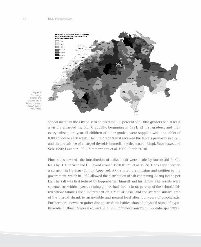
82 RCC Perspectives
school medic in the City of Bern showed that 60 percent of all ifth graders had at least
a visibly enlarged thyroid. Gradually, beginning in 1921, all irst graders, and then
every subsequent year all children of other grades, were supplied with one tablet of
0.003 g iodine each week. The ifth graders irst received the tablets primarily in 1926,
and the prevalence of enlarged thyroids immediately decreased (Bürgi, Supersaxo, and
Selz 1990; Lauener 1936; Zimmermann et al. 2008; Staub 2010).
Final steps towards the introduction of iodized salt were made by successful in situ
tests by H. Hunziker and O. Bayard around 1920 (Bürgi et al. 1979). Hans Eggenberger,
a surgeon in Herisau (Canton Appenzell AR), started a campaign and petition to the
government, which in 1922 allowed the distribution of salt containing 7.5 mg iodine per
kg. The salt was irst iodized by Eggenberger himself and his family. The results were
spectacular: within a year, existing goiters had shrunk in 66 percent of the schoolchild-
ren whose families used iodized salt on a regular basis, and the average surface area
of the thyroid shrank to an invisible and normal level after four years of prophylaxis.
Furthermore, newborn goiter disappeared; no babies showed physical signs of hypo-
thyroidism (Bürgi, Supersaxo, and Selz 1990; Zimmermann 2008; Eggenberger 1922).
Figure 1: Percentage of small and
large goiters in Swiss conscripts 1920/21 (Stiner
1924, 1928).
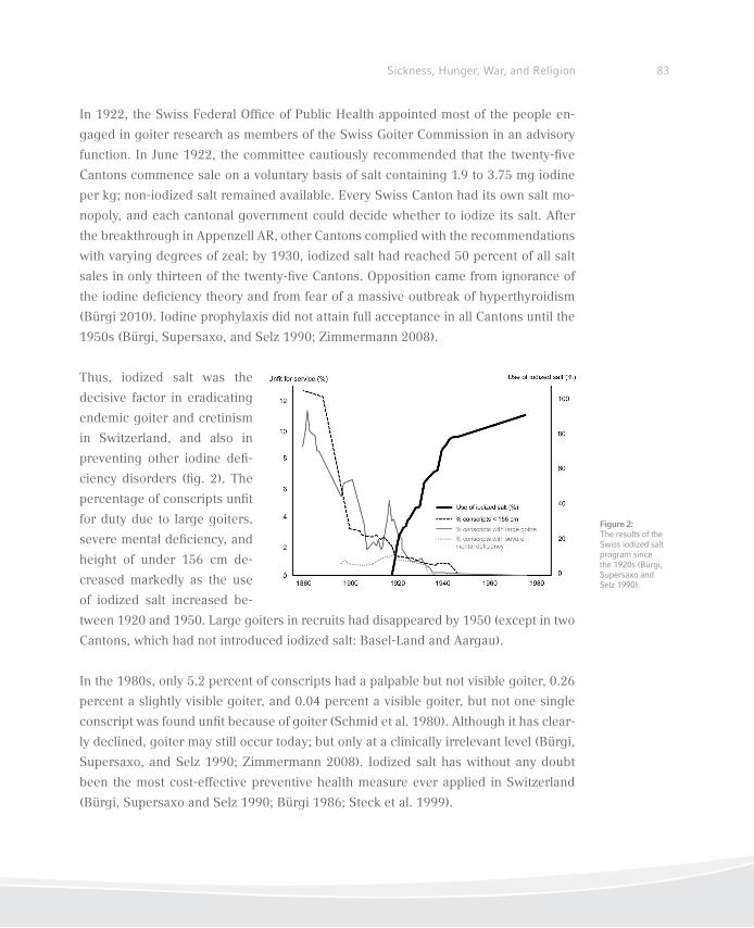
83Sickness, Hunger, War, and Religion
In 1922, the Swiss Federal Ofice of Public Health appointed most of the people en-
gaged in goiter research as members of the Swiss Goiter Commission in an advisory
function. In June 1922, the committee cautiously recommended that the twenty-ive
Cantons commence sale on a voluntary basis of salt containing 1.9 to 3.75 mg iodine
per kg; non-iodized salt remained available. Every Swiss Canton had its own salt mo-
nopoly, and each cantonal government could decide whether to iodize its salt. After
the breakthrough in Appenzell AR, other Cantons complied with the recommendations
with varying degrees of zeal; by 1930, iodized salt had reached 50 percent of all salt
sales in only thirteen of the twenty-ive Cantons. Opposition came from ignorance of
the iodine deiciency theory and from fear of a massive outbreak of hyperthyroidism
(Bürgi 2010). Iodine prophylaxis did not attain full acceptance in all Cantons until the
1950s (Bürgi, Supersaxo, and Selz 1990; Zimmermann 2008).
Thus, iodized salt was the
decisive factor in eradicating
endemic goiter and cretinism
in Switzerland, and also in
preventing other iodine dei-
ciency disorders (ig. 2). The
percentage of conscripts unit
for duty due to large goiters,
severe mental deiciency, and
height of under 156 cm de-
creased markedly as the use
of iodized salt increased be-
tween 1920 and 1950. Large goiters in recruits had disappeared by 1950 (except in two
Cantons, which had not introduced iodized salt: Basel-Land and Aargau).
In the 1980s, only 5.2 percent of conscripts had a palpable but not visible goiter, 0.26
percent a slightly visible goiter, and 0.04 percent a visible goiter, but not one single
conscript was found unit because of goiter (Schmid et al. 1980). Although it has clear-
ly declined, goiter may still occur today; but only at a clinically irrelevant level (Bürgi,
Supersaxo, and Selz 1990; Zimmermann 2008). Iodized salt has without any doubt
been the most cost-effective preventive health measure ever applied in Switzerland
(Bürgi, Supersaxo and Selz 1990; Bürgi 1986; Steck et al. 1999).
Figure 2: The results of the Swiss iodized salt program since the 1920s (Bürgi, Supersaxo and Selz 1990).
84 RCC Perspectives
Christina Papageorgopoulou
Hypothyroidism in Alpine Medieval Switzerland
Hypothyroidism has practically never been described in a historic or prehistoric popula-
tion, except in clinically documented case studies (Ortner and Hotz 2005). This section
reports one of the irst documentations of hypothyroidism due to iodine deiciency in ar-
chaeological populations, in this case the presence of endemic hypothyroidism in a me-
dieval Swiss Alpine community, thus contributing signiicantly to the biological history
of the Alpine regions. Furthermore, the large sample number adequately describes the
variability of the skeletal manifestations and the epidemiological pattern of the disease.
The skeletal sample consists of 404 adult and subadult individuals, originating from
the archaeological site of Tomils/Sogn Murezi and dating from the eleventh to ifteenth
centuries AD. Tomils is located in the Alpine region of the canton Grisons, Switzer-
land, a thousand meters above sea level. The skeletal material was examined for all
standard anthropological variables (e.g., age and sex determination, cranial and post-
cranial metrics, pathological conditions; Papageorgopoulou 2008). Where necessary,
the bones were x-rayed at the University Clinic Balgrist (Zurich, Switzerland).
For the diagnosis of hypothyroidism, a list of observed pathological features character-
istic for the condition were examined and statistically tested based on related literature
(e.g., Borg, Fitzer, and Young 1975; Guggenbühl 1853; Evans 1952; Reilly and Smyth
1937; Scholz 1906) and clinical studies from the region of Switzerland (e.g., De Quer-
vain and Wegelin 1936; König 1968; Weygand 1904; Wieland 1940). We also performed
macroscopic and radiological comparisons of diagnosed cases of hypothyroidism from
Switzerland in the nineteenth and twentieth centuries (Galler pathological reference se-
ries, University of Zurich). The epidemiological pattern of the disease was analyzed, and
further pathological (dental condition, degenerative joint disease, trauma) and archaeo-
logical parameters (interment within the cemetery) were tested.
A high frequency of delayed epiphyseal fusion, in both the axial skeleton and the long
bones; hip and shoulder epiphyseal dysplasias; limb asymmetries and/or abnormal length
of the long bones; and epiphyseal growth plate problems (osteochondritis dissecans) were
present in the skeletal material (table 1). Additionally, cranial characteristics such as short
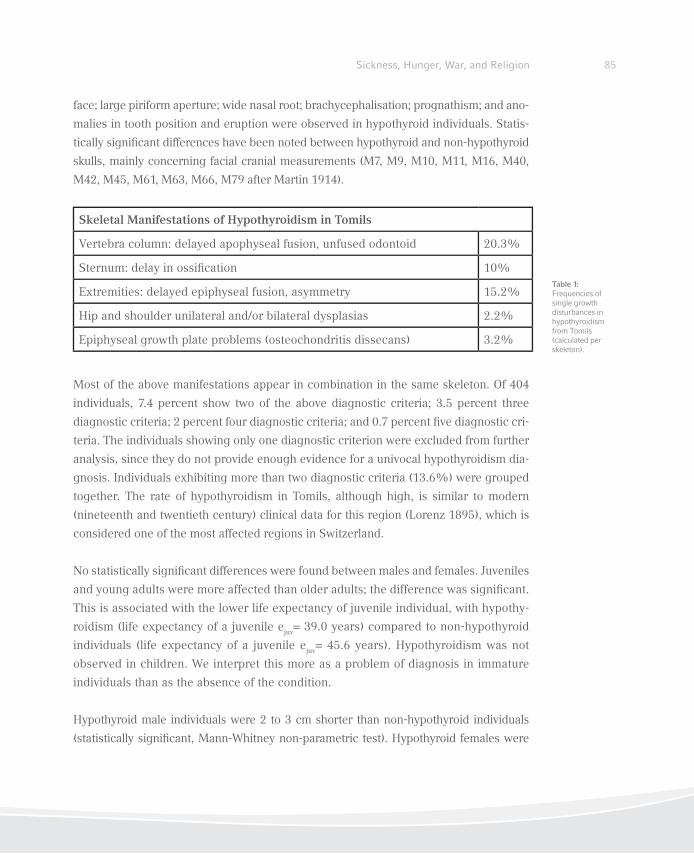
85Sickness, Hunger, War, and Religion
face; large piriform aperture; wide nasal root; brachycephalisation; prognathism; and ano-
malies in tooth position and eruption were observed in hypothyroid individuals. Statis-
tically signiicant differences have been noted between hypothyroid and non-hypothyroid
skulls, mainly concerning facial cranial measurements (M7, M9, M10, M11, M16, M40,
M42, M45, M61, M63, M66, M79 after Martin 1914).
Most of the above manifestations appear in combination in the same skeleton. Of 404
individuals, 7.4 percent show two of the above diagnostic criteria; 3.5 percent three
diagnostic criteria; 2 percent four diagnostic criteria; and 0.7 percent ive diagnostic cri-
teria. The individuals showing only one diagnostic criterion were excluded from further
analysis, since they do not provide enough evidence for a univocal hypothyroidism dia-
gnosis. Individuals exhibiting more than two diagnostic criteria (13.6%) were grouped
together. The rate of hypothyroidism in Tomils, although high, is similar to modern
(nineteenth and twentieth century) clinical data for this region (Lorenz 1895), which is
considered one of the most affected regions in Switzerland.
No statistically signiicant differences were found between males and females. Juveniles
and young adults were more affected than older adults; the difference was signiicant.
This is associated with the lower life expectancy of juvenile individual, with hypothy-
roidism (life expectancy of a juvenile ejuv= 39.0 years) compared to non-hypothyroid
individuals (life expectancy of a juvenile ejuv
= 45.6 years). Hypothyroidism was not
observed in children. We interpret this more as a problem of diagnosis in immature
individuals than as the absence of the condition.
Hypothyroid male individuals were 2 to 3 cm shorter than non-hypothyroid individuals
(statistically signiicant, Mann-Whitney non-parametric test). Hypothyroid females were
Skeletal Manifestations of Hypothyroidism in Tomils
Vertebra column: delayed apophyseal fusion, unfused odontoid 20.3%
Sternum: delay in ossiication 10%
Extremities: delayed epiphyseal fusion, asymmetry 15.2%
Hip and shoulder unilateral and/or bilateral dysplasias 2.2%
Epiphyseal growth plate problems (osteochondritis dissecans) 3.2%
Table 1: Frequencies of single growth disturbances in hypothyroidism from Tomils (calculated per skeleton).
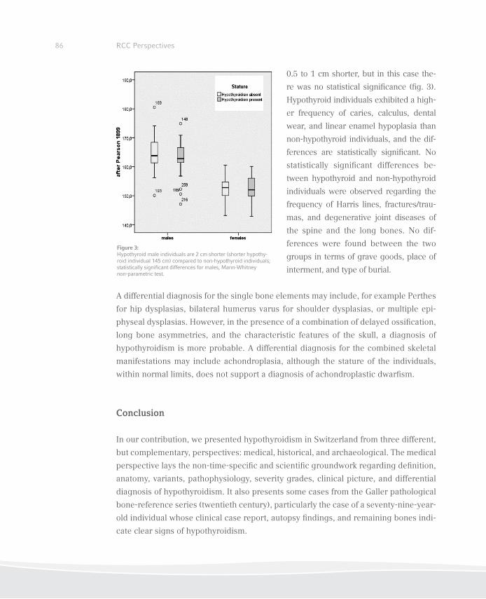
86 RCC Perspectives
0.5 to 1 cm shorter, but in this case the-
re was no statistical signiicance (ig. 3).
Hypothyroid individuals exhibited a high-
er frequency of caries, calculus, dental
wear, and linear enamel hypoplasia than
non-hypothyroid individuals, and the dif-
ferences are statistically signiicant. No
statistically signiicant differences be-
tween hypothyroid and non-hypothyroid
individuals were observed regarding the
frequency of Harris lines, fractures/trau-
mas, and degenerative joint diseases of
the spine and the long bones. No dif-
ferences were found between the two
groups in terms of grave goods, place of
interment, and type of burial.
A differential diagnosis for the single bone elements may include, for example Perthes
for hip dysplasias, bilateral humerus varus for shoulder dysplasias, or multiple epi-
physeal dysplasias. However, in the presence of a combination of delayed ossiication,
long bone asymmetries, and the characteristic features of the skull, a diagnosis of
hypothyroidism is more probable. A differential diagnosis for the combined skeletal
manifestations may include achondroplasia, although the stature of the individuals,
within normal limits, does not support a diagnosis of achondroplastic dwarism.
Conclusion
In our contribution, we presented hypothyroidism in Switzerland from three different,
but complementary, perspectives: medical, historical, and archaeological. The medical
perspective lays the non-time-speciic and scientiic groundwork regarding deinition,
anatomy, variants, pathophysiology, severity grades, clinical picture, and differential
diagnosis of hypothyroidism. It also presents some cases from the Galler pathological
bone-reference series (twentieth century), particularly the case of a seventy-nine-year-
old individual whose clinical case report, autopsy indings, and remaining bones indi-
cate clear signs of hypothyroidism.
Figure 3: Hypothyroid male individuals are 2 cm shorter (shorter hypothy-roid individual 145 cm) compared to non-hypothyroid individuals; statistically signiicant differences for males, Mann-Whitney non-parametric test.

87Sickness, Hunger, War, and Religion
The historical perspective takes other sources of information about the past into con-
sideration: contemporary scientiic studies on conscription, as well as published data on
goiter and cretinism as endemic manifestations of hypothyroidism due to iodine dei-
ciency since the late nineteenth century. In certain regions of Switzerland, 0.5 percent of
inhabitants suffered from hypothyroidism (cretinism), circa 100 percent of schoolchild-
ren had large goiters, and up to 30 percent of young men were unit for military service
before the implementation of iodine in salt (Bürgi, Supersaxo, and Selz 1990).
The anthropological perspective reports one of the irst documentations of hypothyroi-
dism due to iodine deiciency in archaeological populations, contributing signiicantly to
the biological history of the Alpine regions. The large size of the skeletal sample (404 adult
and subadult individuals) originating from the archaeological site of Tomils/Sogn Murezi
and dating to the eleventh to ifteenth centuries AD, adequately describes the presence
and variability of the skeletal manifestations and the epidemiological pattern of endemic
hypothyroidism in a medieval Swiss Alpine population. This can be used as the basis for
diagnosis of skeletal material originating from high-risk regions, which, until now, was ab-
sent in the palaeopathological literature. From an archaeo-anthropological point of view,
the medical and the historical perspectives contribute signiicantly to the identiication
and positive diagnosis of the condition, since they both provide the necessary evidence.
Both the presentation of the Galler hypothyroid cases and the historical documentation
reinforce the anthropological and the archaeological data.
Furthermore, the historical perspective bridges medieval times with the present, when
the implementation of the pioneering iodized salt program eradicated endemic goiter
and cretinism in Switzerland. Iodized salt has with any doubt been the most cost-effec-
tive preventive health measure ever applied in Switzerland. Looking back, the prolonged
administration of iodine or of thyroid hormone has been found highly effective in reduc-
ing the size of endemic iodine deiciency, and thus hypothyroidism. Switzerland was
one of the few countries to be completely iodine suficient before 1990 (together with
some of the Scandinavian countries, Australia, USA, and Canada), and iodine intake in
schoolchildren can be assumed to have been constant since 1980. We demonstrate that
this was not the case in medieval times or in the nineteenth century: iodine deiciency,
goiter, and hypothyroidism were major problems for the local populations.
88 RCC Perspectives
References
Bircher, Heinrich. 1883. Der endemische Kropf. Basel: Benno Schwabe.
Borg, Seth A., Peter M. Fitzer, and Lionel W. Young. 1975. “Roentgenologic Aspects of Adult Cre-
tinism: Two Case Reports and Review of the Literature.” American Journal of Roentgenology
123: 820–28.
Bürgi, Hans. 1986. “Goiter Prevention with Iodized Table Salt in Switzerland: A Brief Review.”
Schweizerische Rundschau für Medizin Praxis 75: 125–27.
———. 2010. “Iodine Excess.” Best Practice & Research Clinical Endocrinology & Metabolism
24: 107–15.
Bürgi, Hans, Max P. Konig, Hugo Studer, and Hans J. Wespi. 1979. “Heinrich Hunziker, M.D. on
his 100th Birthday.” Schweizerische Medizinische Wochenschrift 109 (47): 1905–06.
Bürgi, Hans, Zeno Supersaxo, and Beat Selz. 1990. “Iodine Deiciency Diseases in Switzerland
One Hundred Years after Theodor Kocher’s Survey: A Historical Review with Some New Goiter
Prevalence Data.” Acta Endocrinologica (Copenhagen) 123: 577–90.
De Quervain, Fritz and Karl Wegelin. 1936. Der endemische Kretinismus: Pathologie und Klinik
in Einzeldarstellungen VII. Berlin: Verlag von Julius Springer.
Djemli, Anissa, Guy Van Vliet, and Edgard E. Delvin. 2006. “Congenital Hypothyroidism: From
Paracelsus to Molecular Diagnosis.” Clinical Biochemistry 39: 511–18.
Eggenberger, Hans. 1922. “Die Verhütung des Kropfes und des Kropfrecidives.” Schweizerische
Medizinische Wochenschrift 53: 245–49.
Evans, Philip. 1952. “Deformity of Vertebral Bodies in Cretinism.” Journal of Pediatrics 41: 706–12.
Guggenbühl, Johann. 1853. Die Heilung und Verhütung des Cretinismus und ihre neusten Fort-
schritte. Bern: C. Rätzer
Iglesias, Pedro and Juan Díez. 2008. “Hypothyrodism in Male Patients: A Descriptive, Observati-
onal, and Cross-sectional Study in a Series of 260 Men.” The American Journal of the Medical
Sciences 336: 315–20.
König, Max P. 1968. Die kongenitale Hypothyreose und der endemische Kretinismus. Berlin:
Springer.
89Sickness, Hunger, War, and Religion
Lauener, Paul. 1936. “Fünfzehn Jahre Kropfbekämpfung in den Schulen der Stadt Bern.” Schwei-
zerische Medizinische Wochenschrift 66: 207–09.
Lorenz, Paul. 1895. “Die Ergebnisse der sanitarischen Untersuchungen der Recruten des Kan-
tons Graubünden (Schweiz) in den Jahren 1875 bis 1879.” Jahresbericht der Naturforschen-
den Gesellschaft Graubünden 38: Supplement 2.
Martin, Rudolph. 1914. Lehrbuch der Anthropologie in systematischer Darstellung: Mit beson-
derer Berücksichtigung der anthropologischen Methoden für Studierenden, Ärzte und For-
schungsreisende. Jena: Fischer Verlag.
Merke, Franz. 1971. Geschichte und Ikonograie des endemischen Kropfs und Kretinismus. Bern
and Stuttgart: Verlag Hans Huber.
Ortner, Donald J. and Gerhard Hotz. “Skeletal Manifestations of Hypothyroidism from Switzer-
land.” American Journal of Physical Anthropology 127: 1–6.
Papageorgopoulou, Christina. 2008. “The Medieval Population of Tomils: An Archaeo-anthro-
pological Approach.” PhD diss., University of Basel. Forthcoming in British Archaeological
Reports (BAR) International Series.
Patrick, Lyn. 2008. “Iodine: Deiciency and Therapeutic Considerations.” Alternative Medicine
Review 13: 116–27.
Reilly, William A and Francis S. Smyth. 1937. “Cretinoid Epiphyseal Dysgenesis.” The Journal of
Pediatrics 11: 786–96.
Rühli, Frank, Gerhard Hotz, and Thomas Böni. 2003. “Brief Communication: The Galler Collec-
tion: A Little-known Historic Swiss Bone Pathology Reference Series.” American Journal of
Physical Anthropology 121: 15–18.
Schmid, Markus, Christoph Schulthess, Hans Burgi, and Hugo Studer. 1980. “Iodine Deiciency
is Still Endemic in Switzerland.” Schweizerische Medizinische Wochenschrift 110: 1290–95.
Scholz, Wilhelm. 1906. Klinische und anatomische Untersuchungen über den Cretinismus. Berlin.
Solca, Barbara, Jaeggi-Groisman, Valerio Saglini, and Hans Gerber. 1999. “Iodine Supply in Dif-
ferent Geographical Areas of Switzerland: Comparison Between Rural and Urban Populations
in the Berne and the Ticino Regions.” European Journal of Clinical Nutrition 53: 754–55.
90 RCC Perspectives
Staub, Kaspar. 2010. “Der biologische Lebensstandard in der Schweiz seit 1800: Historisch-
anthropometrische Untersuchung der Körperhöhe (und des Körpergewichts) in der Schweiz
seit 1800, differenziert nach Geschlecht, sozioökonomischem und regionalem Hintergrund.”
PhD diss., University of Bern
Steck, Andreas, Barbara Steck, Max P. Konig, and Hugo Studer. 1972. “Effects of an Improved
Iodine Prophylaxis on Endemic Goiter and Iodine Metabolism.” Schweizerische Medizinische
Wochenschrift 102 (24): 829–37.
Stiner, Otto. 1924. “Die Verbreitung des endemischen Kropfes in der Schweiz nach neueren Ergeb-
nissen der Rekrutenuntersuchungen.” Schweizerische Medizinische Wochenschrift 54: 391–397.
———. 1928. “Der Kropf bei den Stellungsplichtigen der Jahre 1924 und 1925.” Schweizerische
Medizinische Wochenschrift 58: 401–07.
Weygandt, Wilhelm. 1904. Der heutige Stand der Lehre vom Cretinismus. Vol. 4 of Sammlung
zwangloser Abhandlungen aus dem Gebiete der Nerven- und Geisteskrankheiten, no. 6/7. Hal-
le: C. Marhold.
Wieland Burckhardt, Emil. 1940. “Die Athyreosis und Hypothyreosis im Kindesalter: der endemische
Kretinismus.” Zwanglose Abhandlungen aus dem Gebiete der Inneren Sekretion 7. Leipzig: Barth.
Zimmermann, Michael B. 2008. “Research on Iodine Deiciency and Goiter in the 19th and Early
20th Centuries.” Journal of Nutrition 138 (11): 2060–63.
———. 2009. “Iodine Deiciency”. Endocrine Reviews 30: 376–08.
———. 2010. “Symposium on ‘Geographical and Geological Inluences on Nutrition’: Iodine De-
iciency in Industrialised Countries.” Proceedings of the Nutrition Society 69: 133–43.
Zimmermann, Michael B., Peter L. Jooste and Chandrakant S. Pandav. 2008. “Iodine-Deiciency
Disorders.” Lancet 372/9645: 1251–62.
The Ancient Battleield at Kalkriese
Susanne Wilbers-Rost, Birgit Großkopf,
and Achim Rost
92 RCC Perspectives
Authors
Susanne Wilbers-Rost
Varusschlacht im Osnabrücker Land GmbH
Museum und Park Kalkriese
Department of Archaeology
Venner Str. 69
49565 Bramsche-Kalkriese
Germany
Birgit Großkopf
Georg-August-Universität Göttingen
Johann-Friedrich-Blumenbach Institute for Zoology and Anthropology
Historical Anthropology and Human Genetics
Bürgerstr. 50
37073 Göttingen
Germany
Achim Rost
Osnabrück University
Fachbereich Kultur- und Geowissenschaft
Alte Geschichte/Archäologie der Römischen Provinzen
Schlossstr. 8
49074 Osnabrück
Germany
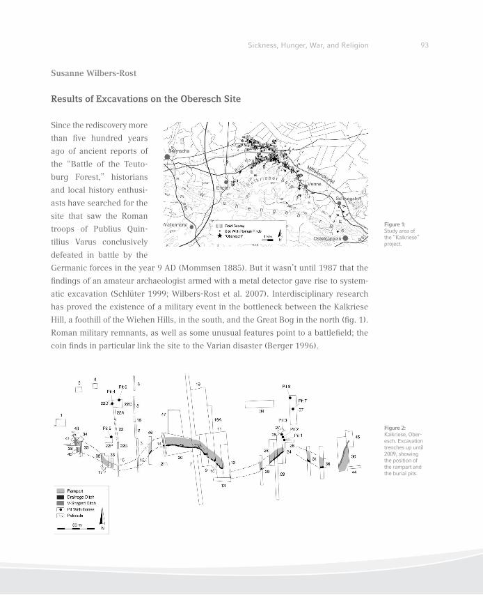
93Sickness, Hunger, War, and Religion
Susanne Wilbers-Rost
Results of Excavations on the Oberesch Site
Since the rediscovery more
than ive hundred years
ago of ancient reports of
the “Battle of the Teuto-
burg Forest,” historians
and local history enthusi-
asts have searched for the
site that saw the Roman
troops of Publius Quin-
tilius Varus conclusively
defeated in battle by the
Germanic forces in the year 9 AD (Mommsen 1885). But it wasn’t until 1987 that the
indings of an amateur archaeologist armed with a metal detector gave rise to system-
atic excavation (Schlüter 1999; Wilbers-Rost et al. 2007). Interdisciplinary research
has proved the existence of a military event in the bottleneck between the Kalkriese
Hill, a foothill of the Wiehen Hills, in the south, and the Great Bog in the north (ig. 1).
Roman military remnants, as well as some unusual features point to a battleield; the
coin inds in particular link the site to the Varian disaster (Berger 1996).
Figure 1: Study area of the “Kalkriese” project.
Figure 2: Kalkriese, Ober-esch. Excavation trenches up until 2009, showing the position of the rampart and the burial pits.
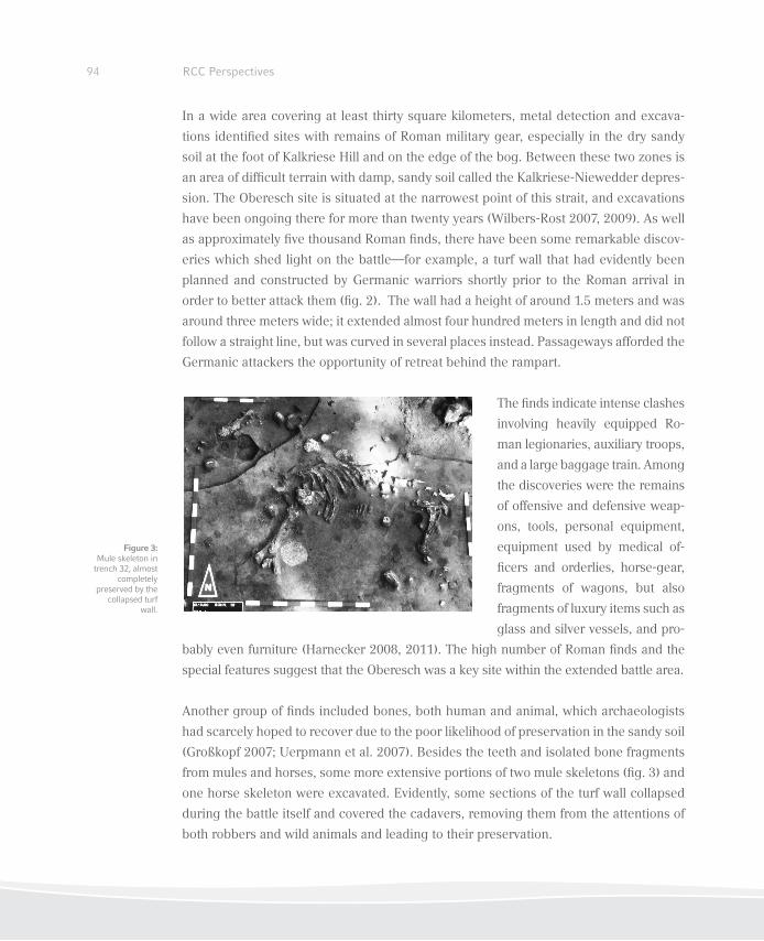
94 RCC Perspectives
In a wide area covering at least thirty square kilometers, metal detection and excava-
tions identiied sites with remains of Roman military gear, especially in the dry sandy
soil at the foot of Kalkriese Hill and on the edge of the bog. Between these two zones is
an area of dificult terrain with damp, sandy soil called the Kalkriese-Niewedder depres-
sion. The Oberesch site is situated at the narrowest point of this strait, and excavations
have been ongoing there for more than twenty years (Wilbers-Rost 2007, 2009). As well
as approximately ive thousand Roman inds, there have been some remarkable discov-
eries which shed light on the battle—for example, a turf wall that had evidently been
planned and constructed by Germanic warriors shortly prior to the Roman arrival in
order to better attack them (ig. 2). The wall had a height of around 1.5 meters and was
around three meters wide; it extended almost four hundred meters in length and did not
follow a straight line, but was curved in several places instead. Passageways afforded the
Germanic attackers the opportunity of retreat behind the rampart.
The inds indicate intense clashes
involving heavily equipped Ro-
man legionaries, auxiliary troops,
and a large baggage train. Among
the discoveries were the remains
of offensive and defensive weap-
ons, tools, personal equipment,
equipment used by medical of-
icers and orderlies, horse-gear,
fragments of wagons, but also
fragments of luxury items such as
glass and silver vessels, and pro-
bably even furniture (Harnecker 2008, 2011). The high number of Roman inds and the
special features suggest that the Oberesch was a key site within the extended battle area.
Another group of inds included bones, both human and animal, which archaeologists
had scarcely hoped to recover due to the poor likelihood of preservation in the sandy soil
(Großkopf 2007; Uerpmann et al. 2007). Besides the teeth and isolated bone fragments
from mules and horses, some more extensive portions of two mule skeletons (ig. 3) and
one horse skeleton were excavated. Evidently, some sections of the turf wall collapsed
during the battle itself and covered the cadavers, removing them from the attentions of
both robbers and wild animals and leading to their preservation.
Figure 3: Mule skeleton in trench 32, almost
completely preserved by the
collapsed turf wall.
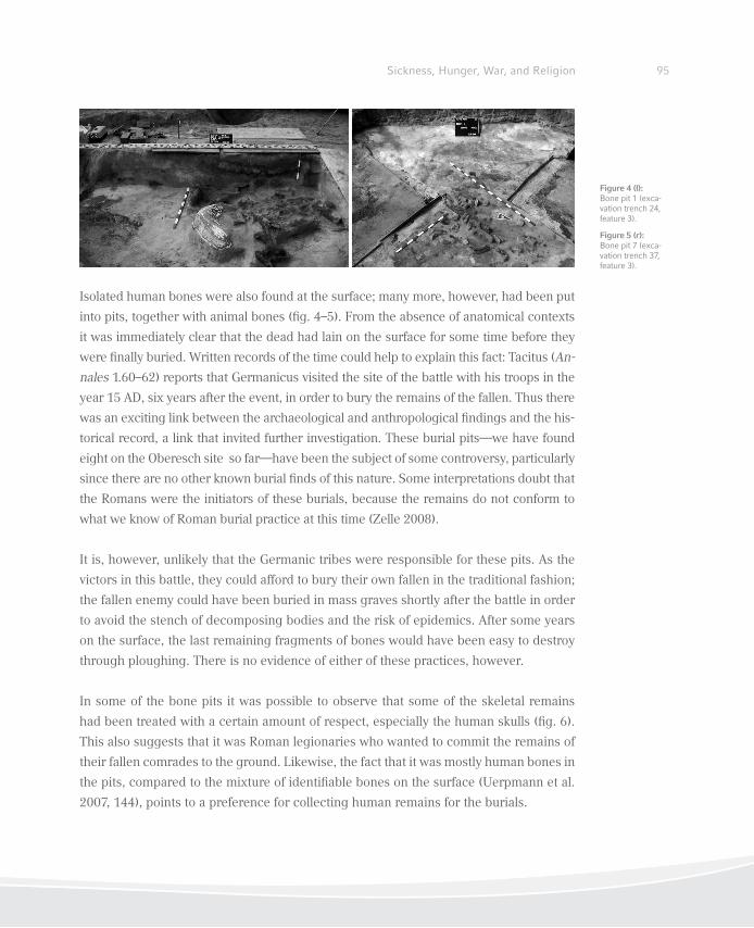
95Sickness, Hunger, War, and Religion
Isolated human bones were also found at the surface; many more, however, had been put
into pits, together with animal bones (ig. 4–5). From the absence of anatomical contexts
it was immediately clear that the dead had lain on the surface for some time before they
were inally buried. Written records of the time could help to explain this fact: Tacitus (An-
nales 1.60–62) reports that Germanicus visited the site of the battle with his troops in the
year 15 AD, six years after the event, in order to bury the remains of the fallen. Thus there
was an exciting link between the archaeological and anthropological indings and the his-
torical record, a link that invited further investigation. These burial pits—we have found
eight on the Oberesch site so far—have been the subject of some controversy, particularly
since there are no other known burial inds of this nature. Some interpretations doubt that
the Romans were the initiators of these burials, because the remains do not conform to
what we know of Roman burial practice at this time (Zelle 2008).
It is, however, unlikely that the Germanic tribes were responsible for these pits. As the
victors in this battle, they could afford to bury their own fallen in the traditional fashion;
the fallen enemy could have been buried in mass graves shortly after the battle in order
to avoid the stench of decomposing bodies and the risk of epidemics. After some years
on the surface, the last remaining fragments of bones would have been easy to destroy
through ploughing. There is no evidence of either of these practices, however.
In some of the bone pits it was possible to observe that some of the skeletal remains
had been treated with a certain amount of respect, especially the human skulls (ig. 6).
This also suggests that it was Roman legionaries who wanted to commit the remains of
their fallen comrades to the ground. Likewise, the fact that it was mostly human bones in
the pits, compared to the mixture of identiiable bones on the surface (Uerpmann et al.
2007, 144), points to a preference for collecting human remains for the burials.
Figure 4 (l): Bone pit 1 (exca-vation trench 24, feature 3).
Figure 5 (r): Bone pit 7 (exca-vation trench 37, feature 3).
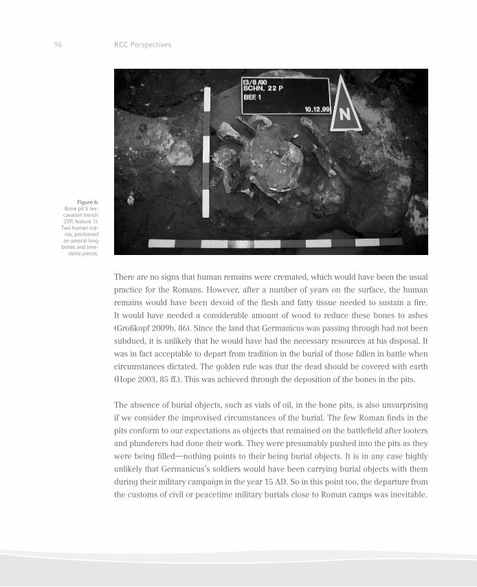
96 RCC Perspectives
There are no signs that human remains were cremated, which would have been the usual
practice for the Romans. However, after a number of years on the surface, the human
remains would have been devoid of the lesh and fatty tissue needed to sustain a ire.
It would have needed a considerable amount of wood to reduce these bones to ashes
(Großkopf 2009b, 86). Since the land that Germanicus was passing through had not been
subdued, it is unlikely that he would have had the necessary resources at his disposal. It
was in fact acceptable to depart from tradition in the burial of those fallen in battle when
circumstances dictated. The golden rule was that the dead should be covered with earth
(Hope 2003, 85 ff.). This was achieved through the deposition of the bones in the pits.
The absence of burial objects, such as vials of oil, in the bone pits, is also unsurprising
if we consider the improvised circumstances of the burial. The few Roman inds in the
pits conform to our expectations as objects that remained on the battleield after looters
and plunderers had done their work. They were presumably pushed into the pits as they
were being illed—nothing points to their being burial objects. It is in any case highly
unlikely that Germanicus’s soldiers would have been carrying burial objects with them
during their military campaign in the year 15 AD. So in this point too, the departure from
the customs of civil or peacetime military burials close to Roman camps was inevitable.
Figure 6: Bone pit 5 (ex-cavation trench 22P, feature 1).
Two human cra-nia, positioned on several long
bones and lime-stone pieces.

97Sickness, Hunger, War, and Religion
The lack of any sign of the grave mound mentioned by Tacitus (Annales 1.62.1), al-
legedly constructed by Germanicus, has also been the subject of discussion. Apart
from the question whether such a mound, which was supposedly lattened relatively
quickly by the Germanic tribes (Tacitus, Annales 2.7.3), could even be archaeologically
proven, we need to bear in mind that Tacitus’s assertion might have been a topos em-
ployed to demonstrate the extent of Germanicus’s devotion to the fallen rather than a
literal description of his actions.
From the archaeological perspective, there is thus no fundamental discrepancy
between the bone pits on the Oberesch site and the written sources that describe
Germanicus’s burial act (Rost/Wilbers-Rost, forthcoming). Six years after the defeat
itself, Germanicus was able to fulil the duty under unusual conditions: the burial of
the fallen. He covered the skeletons with earth and thus paid them his last respects. In
the context of discussions about the interpretation of the burial pits, the osteological
analysis and evaluation of the bone fragments are of great signiicance.
Birgit Großkopf
The Human Remains from the Oberesch Site
Most of the human bone fragments from the
Oberesch site were discovered in pits located in
close proximity to the wall (ig. 7). Only isolated
bone fragments comingled with animal bones
were present in these pits. Articulated, complete
skeletons were not documented. The bones are
fragmentary and exhibit signs of advanced de-
composition, particularly, suricial erosion.
The eight bone pits that have been excavated
so far are markedly different from each other.
The surface areas of the pits vary in size from
40–60 cm to 4 m square. The three neighbour-
ing bone pits (1–3) contained various amounts
Figure 7: Bone pits 1, 2 and 3 (excavati-on trenches 24 and 25), showing different levels of bone content.
98 RCC Perspectives
of ill material. While pit 1 contained numerous bone inds, pits 2 and 3 contained only a
small number of fragments (ig. 7). The degree of bone preservation also differed signii-
cantly. In some bone pits there were pieces of limestone that were found in the soil and
occasionally on the surface. Bone preservation was best when limestone was present. The
bones in pit 5 belonged to two individuals and were suficiently well-preserved to permit
an estimate of stature (Großkopf 2007). Although the bones appeared stable and intact
macroscopically, the internal bone structure displayed signs of signiicant decomposition.
This poor level of preservation prevented DNA analysis. Even the best-preserved bones
and tooth roots proved unusable to provide us with reproducible data.
Anthropological conclusions are therefore based only on evidence collected from isolat-
ed bones or bone fragments. The sexing of skeletons relies in part on speciic features
of the pelvic anatomy. Functional differences of the innominate bone are manifested
as sex-speciic morphological characteristics that in turn provide the most important
criteria for an accurate sex differentiation. Due to the proportionally thin compact bone
structure, preservation of pelvic bones at Oberesch was rarely suficient to allow for sex-
ing. However, skull bones also exhibit numerous markers of individual sex, and these
are readily discernible, even from fragments. Long bones give us information primarily
with regard to robustness and muscle development, however, they can provide addi-
tional information on the sex of an individual. The approximate age of death can be
estimated morphologically based on the degree of skull suture ossiication and diploe
structure. Occlusal wear of the dentition is another criterion for approximating age at
death. Results from the morphological examination were checked against those provid-
ed by the tooth cementum annulation method (Großkopf 1990). This histological tech-
nique for age estimations focuses on the analysis of tooth root cementum.
In addition to determining the age and the sex of those buried at the Oberesch site, a
further step was to identify the minimum number of individuals. One of the irst tasks
was to group bone inds together in order to determine whether bones were fragmented
prior to being buried in the pits. Considering the extent of fragmentation, this was an
ambitious undertaking. Often, the only helpful characteristics were provided by unusual
fragments or fracture edges. The advanced state of bone surface decomposition and
erosion of broken edges (ig. 8) complicated this task further. Based on lower jawbone
fragments, speciically a segment from near the left irst molar, a minimum number of
17 individuals from the burial site was calculated. The number of other jaw and skull
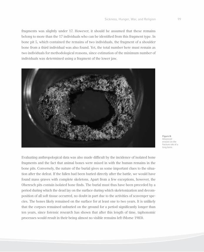
99Sickness, Hunger, War, and Religion
fragments was slightly under 17. However, it should be assumed that these remains
belong to more than the 17 individuals who can be identiied from this fragment type. In
bone pit 5, which contained the remains of two individuals, the fragment of a shoulder
bone from a third individual was also found. Yet, the total number here must remain as
two individuals for methodological reasons, since estimation of the minimum number of
individuals was determined using a fragment of the lower jaw.
Evaluating anthropological data was also made dificult by the incidence of isolated bone
fragments and the fact that animal bones were mixed in with the human remains in the
bone pits. Conversely, the nature of the burial gives us some important clues to the situa-
tion after the defeat. If the fallen had been buried directly after the battle, we would have
found mass graves with complete skeletons. Apart from a few exceptions, however, the
Oberesch pits contain isolated bone inds. The burial must thus have been preceded by a
period during which the dead lay on the surface during which skeletonization and decom-
position of all soft tissue occurred, no doubt in part due to the activities of scavenger spe-
cies. The bones likely remained on the surface for at least one to two years. It is unlikely
that the corpses remained unburied on the ground for a period signiicantly longer than
ten years, since forensic research has shown that after this length of time, taphonomic
processes would result in their being almost no visible remains left (Morse 1983).
Figure 8: Advanced erosion on the fracture site of a long bone.
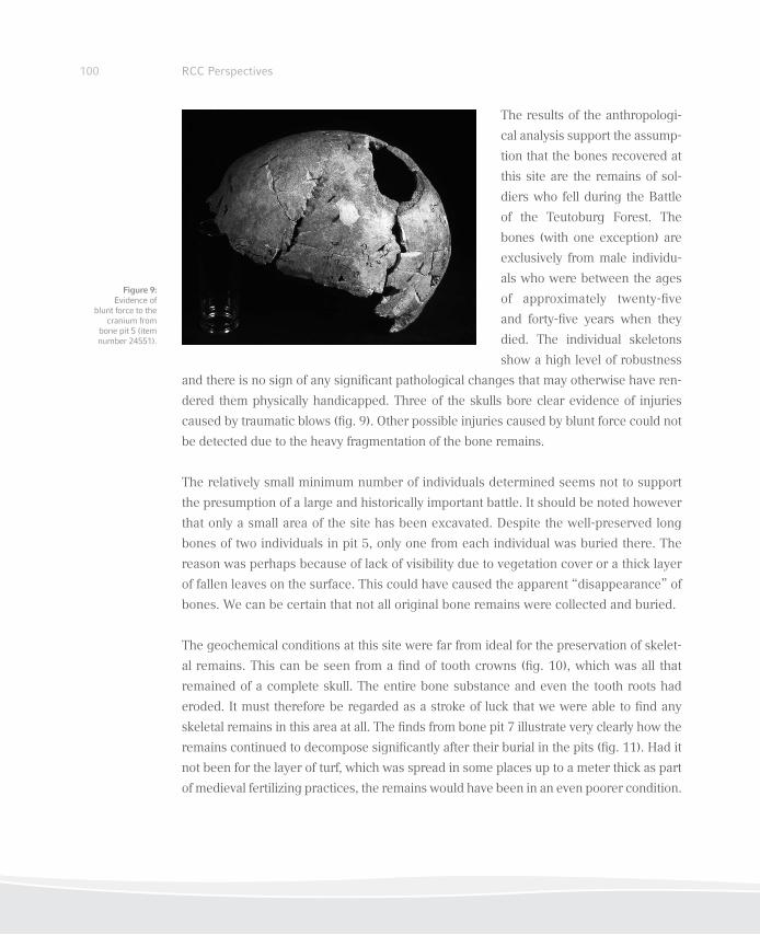
100 RCC Perspectives
The results of the anthropologi-
cal analysis support the assump-
tion that the bones recovered at
this site are the remains of sol-
diers who fell during the Battle
of the Teutoburg Forest. The
bones (with one exception) are
exclusively from male individu-
als who were between the ages
of approximately twenty-ive
and forty-ive years when they
died. The individual skeletons
show a high level of robustness
and there is no sign of any signiicant pathological changes that may otherwise have ren-
dered them physically handicapped. Three of the skulls bore clear evidence of injuries
caused by traumatic blows (ig. 9). Other possible injuries caused by blunt force could not
be detected due to the heavy fragmentation of the bone remains.
The relatively small minimum number of individuals determined seems not to support
the presumption of a large and historically important battle. It should be noted however
that only a small area of the site has been excavated. Despite the well-preserved long
bones of two individuals in pit 5, only one from each individual was buried there. The
reason was perhaps because of lack of visibility due to vegetation cover or a thick layer
of fallen leaves on the surface. This could have caused the apparent “disappearance” of
bones. We can be certain that not all original bone remains were collected and buried.
The geochemical conditions at this site were far from ideal for the preservation of skelet-
al remains. This can be seen from a ind of tooth crowns (ig. 10), which was all that
remained of a complete skull. The entire bone substance and even the tooth roots had
eroded. It must therefore be regarded as a stroke of luck that we were able to ind any
skeletal remains in this area at all. The inds from bone pit 7 illustrate very clearly how the
remains continued to decompose signiicantly after their burial in the pits (ig. 11). Had it
not been for the layer of turf, which was spread in some places up to a meter thick as part
of medieval fertilizing practices, the remains would have been in an even poorer condition.
Figure 9: Evidence of
blunt force to the cranium from
bone pit 5 (item number 24551).
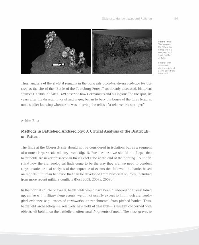
101Sickness, Hunger, War, and Religion
Thus, analysis of the skeletal remains in the bone pits provides strong evidence for this
area as the site of the “Battle of the Teutoburg Forest.” As already discussed, historical
sources (Tacitus, Annales 1.62) describe how Germanicus and his legions “on the spot, six
years after the disaster, in grief and anger, began to bury the bones of the three legions,
not a soldier knowing whether he was interring the relics of a relative or a stranger.”
Achim Rost
Methods in Battleield Archaeology: A Critical Analysis of the Distributi-on Pattern
The inds at the Oberesch site should not be considered in isolation, but as a segment
of a much larger-scale military event (ig. 1). Furthermore, we should not forget that
battleields are never preserved in their exact state at the end of the ighting. To under-
stand how the archaeological inds come to be the way they are, we need to conduct
a systematic, critical analysis of the sequence of events that followed the battle, based
on models of human behavior that can be developed from historical sources, including
from more recent military conlicts (Rost 2008, 2009a, 2009b).
In the normal course of events, battleields would have been plundered or at least tidied
up; unlike with military siege events, we do not usually expect to ind much archaeolo-
gical evidence (e.g., traces of earthworks, entrenchments) from pitched battles. Thus,
battleield archaeology—a relatively new ield of research—is usually concerned with
objects left behind on the battleield, often small fragments of metal. The mass graves to
Figure 10 (l): Tooth crowns, the only remai-ning parts of a complete skull (item number 21209).
Figure 11 (r): Advanced decomposition of a long bone from bone pit 7.
102 RCC Perspectives
bury fallen soldiers, for example those found in Wittstock (the site of a 1636 battle dur-
ing the Thirty Years War; Grothe/Jungklaus 2009), number amongst the most striking
legacies of pitched battles. Modern battleield sites, with their enormous dispersal of
spent irearm munitions, perhaps best represent our sense of what a battleield should
have looked like. But the evidence of military battles in ancient times tend to be rather
thin on the ground, in particular when comparably “disposable” munitions, such as lead
sling bullets, were not used. It has gradually become clear that the battleield needs to
be viewed as a category of its own in terms of archaeological research: conditions for
the preservation of archaeological remains are very different from those for inds from
settlements, graveyards, and cult sites. There is a distinct need to develop a new metho-
dological framework for an appropriate interpretation of battleield sites, including the
sites of ancient battles.
It is remarkable to note that there have been around ive thousand Roman inds on
the Oberesch site, compared with only around ive hundred military inds in the entire
remaining area of the Kalkriese study, even though the excavated areas are of a similar
size (Harnecker and Tolksdorf-Lienemann 2004).
Nevertheless, the analysis of excavations in Kalkriese has proved that the rate of recov-
ery of military items is not linked to the intensity of the battle itself. A large part of the
inds from Kalkriese are shown to be the result of a mixture of processes following the
battle, including salvage and plunder, rather than direct relics of the ighting on the site.
Ancient accounts of the military conlicts between Germanic tribes and Romans in the
irst decades after the birth of Christ have given us some points of reference regarding
the behavior patterns of both parties. We know, for example, that the Germanic com-
batants were motivated to ight by the promise of plunder later (Cassius Dio, Historia
Romana 56.21.4); elsewhere, we learn that the Roman army was trained to rescue their
wounded even under dangerous conditions, and to bring their baggage train into safety
(Tacitus, Annales 1.64.4).
The effects of these actions in terms of archaeological inds can be clearly seen from
the sites at Kalkriese. The battle events on Kalkriese Hill look to have been a series of
guerilla attacks by the Germanic tribesmen on the extended lank of the Roman army,
which was moving from east to west in a long formation. The attacks came along a long,
narrow stretch of land—at least 10 km in length—between the hill and the bog. It would
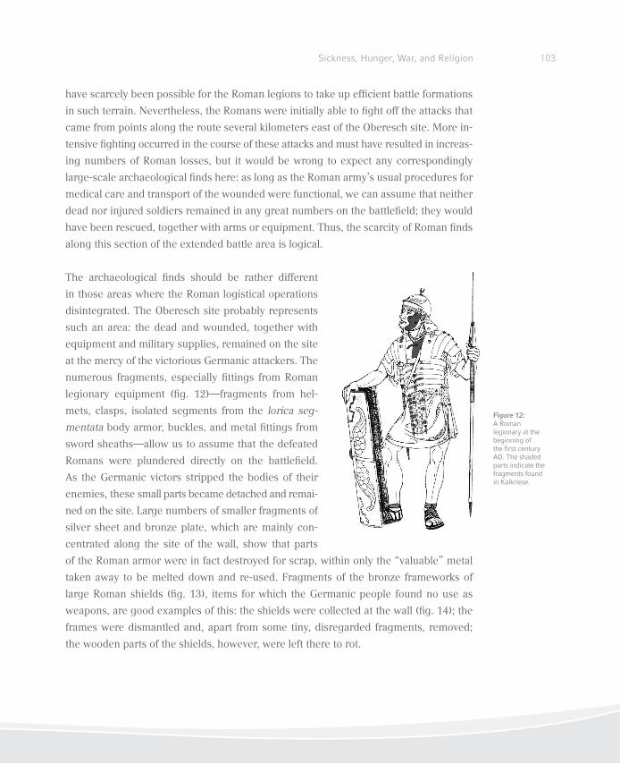
103Sickness, Hunger, War, and Religion
have scarcely been possible for the Roman legions to take up eficient battle formations
in such terrain. Nevertheless, the Romans were initially able to ight off the attacks that
came from points along the route several kilometers east of the Oberesch site. More in-
tensive ighting occurred in the course of these attacks and must have resulted in increas-
ing numbers of Roman losses, but it would be wrong to expect any correspondingly
large-scale archaeological inds here: as long as the Roman army’s usual procedures for
medical care and transport of the wounded were functional, we can assume that neither
dead nor injured soldiers remained in any great numbers on the battleield; they would
have been rescued, together with arms or equipment. Thus, the scarcity of Roman inds
along this section of the extended battle area is logical.
The archaeological inds should be rather different
in those areas where the Roman logistical operations
disintegrated. The Oberesch site probably represents
such an area: the dead and wounded, together with
equipment and military supplies, remained on the site
at the mercy of the victorious Germanic attackers. The
numerous fragments, especially ittings from Roman
legionary equipment (ig. 12)—fragments from hel-
mets, clasps, isolated segments from the lorica seg-
mentata body armor, buckles, and metal ittings from
sword sheaths—allow us to assume that the defeated
Romans were plundered directly on the battleield.
As the Germanic victors stripped the bodies of their
enemies, these small parts became detached and remai-
ned on the site. Large numbers of smaller fragments of
silver sheet and bronze plate, which are mainly con-
centrated along the site of the wall, show that parts
of the Roman armor were in fact destroyed for scrap, within only the “valuable” metal
taken away to be melted down and re-used. Fragments of the bronze frameworks of
large Roman shields (ig. 13), items for which the Germanic people found no use as
weapons, are good examples of this: the shields were collected at the wall (ig. 14); the
frames were dismantled and, apart from some tiny, disregarded fragments, removed;
the wooden parts of the shields, however, were left there to rot.
Figure 12: A Roman legionary at the beginning of the irst century AD. The shaded parts indicate the fragments found in Kalkriese.
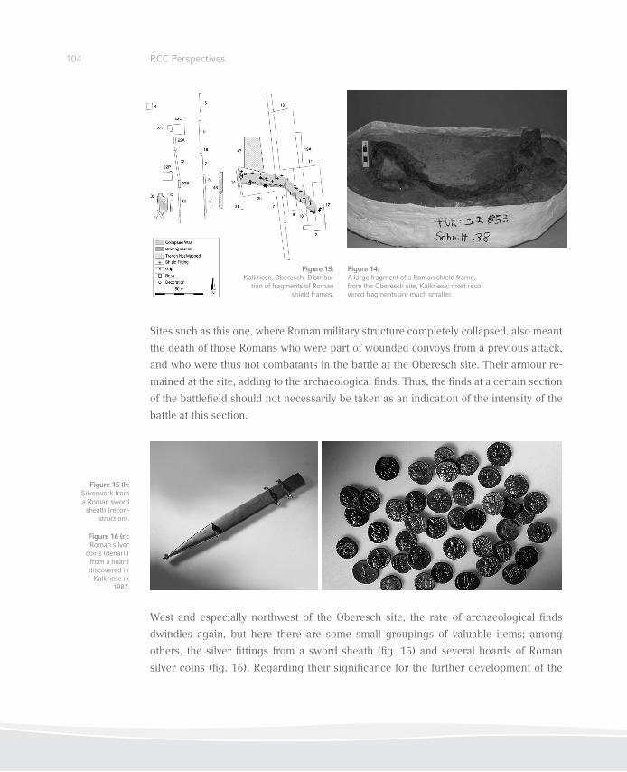
104 RCC Perspectives
Sites such as this one, where Roman military structure completely collapsed, also meant
the death of those Romans who were part of wounded convoys from a previous attack,
and who were thus not combatants in the battle at the Oberesch site. Their armour re-
mained at the site, adding to the archaeological inds. Thus, the inds at a certain section
of the battleield should not necessarily be taken as an indication of the intensity of the
battle at this section.
West and especially northwest of the Oberesch site, the rate of archaeological inds
dwindles again, but here there are some small groupings of valuable items; among
others, the silver ittings from a sword sheath (ig. 15) and several hoards of Roman
silver coins (ig. 16). Regarding their signiicance for the further development of the
Figure 14: A large fragment of a Roman shield frame, from the Oberesch site, Kalkriese; most reco-vered fragments are much smaller.
Figure 15 (l): Silverwork from a Roman sword sheath (recon-
struction).
Figure 13: Kalkriese, Oberesch. Distribu-
tion of fragments of Roman shield frames.
Figure 16 (r): Roman silver
coins (denarii) from a hoard discovered in
Kalkriese in 1987.
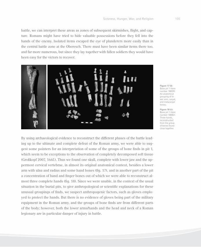
105Sickness, Hunger, War, and Religion
battle, we can interpret these areas as zones of subsequent skirmishes, light, and cap-
ture. Romans might have tried to hide valuable possessions before they fell into the
hands of the enemy. Isolated items escaped the eye of plunderers more easily than in
the central battle zone at the Oberesch. There must have been similar items there too,
and far more numerous, but since they lay together with fallen soldiers they would have
been easy for the victors to recover.
By using archaeological evidence to reconstruct the different phases of the battle lead-
ing up to the ultimate and complete defeat of the Roman army, we were able to sug-
gest some pointers for an interpretation of some of the groups of bone inds in pit 1,
which seem to be exceptions to the observation of completely decomposed soft tissue
(Großkopf 2007, 166f.). Thus we found one skull, complete with lower jaw and the up-
permost cervical vertebrae, in almost its original anatomical context, besides a lower
arm with ulna and radius and some hand bones (ig. 17), and in another part of the pit
a concentration of hand and inger bones out of which we were able to reconstruct al-
most three complete hands (ig. 18). Since we were unable, in the context of the usual
situation in the burial pits, to give anthropological or scientiic explanations for these
unusual groupings of inds, we suspect anthropogenic factors, such as gloves emplo-
yed to protect the hands. But there is no evidence of gloves being part of the military
equipment in the Roman army, and the groups of bone inds are from different parts
of the body; however, both the lower arms/hands and the head and neck of a Roman
legionary are in particular danger of injury in battle.
Figure 17 (l): Bone pit 1 (item number 18059). An anatomical grouping of lo-wer arm, carpal, and metacarpal bones.
Figure 18 (r): Bone pit 1 (item number 18082). Three hands, reconstructed from the group of bones found close together.
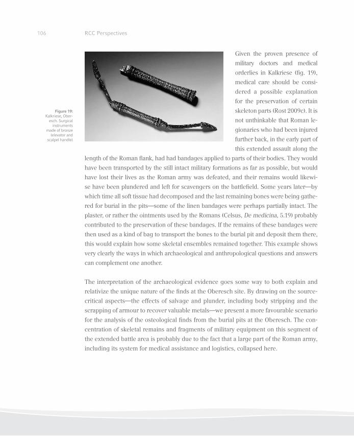
106 RCC Perspectives
Given the proven presence of
military doctors and medical
orderlies in Kalkriese (ig. 19),
medical care should be consi-
dered a possible explanation
for the preservation of certain
skeleton parts (Rost 2009c). It is
not unthinkable that Roman le-
gionaries who had been injured
further back, in the early part of
this extended assault along the
length of the Roman lank, had had bandages applied to parts of their bodies. They would
have been transported by the still intact military formations as far as possible, but would
have lost their lives as the Roman army was defeated, and their remains would likewi-
se have been plundered and left for scavengers on the battleield. Some years later—by
which time all soft tissue had decomposed and the last remaining bones were being gathe-
red for burial in the pits—some of the linen bandages were perhaps partially intact. The
plaster, or rather the ointments used by the Romans (Celsus, De medicina, 5.19) probably
contributed to the preservation of these bandages. If the remains of these bandages were
then used as a kind of bag to transport the bones to the burial pit and deposit them there,
this would explain how some skeletal ensembles remained together. This example shows
very clearly the ways in which archaeological and anthropological questions and answers
can complement one another.
The interpretation of the archaeological evidence goes some way to both explain and
relativize the unique nature of the inds at the Oberesch site. By drawing on the source-
critical aspects—the effects of salvage and plunder, including body stripping and the
scrapping of armour to recover valuable metals—we present a more favourable scenario
for the analysis of the osteological inds from the burial pits at the Oberesch. The con-
centration of skeletal remains and fragments of military equipment on this segment of
the extended battle area is probably due to the fact that a large part of the Roman army,
including its system for medical assistance and logistics, collapsed here.
Figure 19: Kalkriese, Ober-
esch. Surgical instruments
made of bronze (elevator and
scalpel handle)
107Sickness, Hunger, War, and Religion
Conclusion
Battleield archaeology is a relatively new subdiscipline in the ield of archaeology,
which has been able to develop in the last couple of decades due to the systematic use
of metal detectors (Rost 2009a). For this reason, there is currently little experience in the
evaluation of the signiicance of archaeological items excavated from the sites of armed
conlict. Often, written accounts of “world-changing conlicts” provoke us by citing the
huge number of battle dead and the loss of equipment, leading us to harbor expecta-
tions of a correspondingly large amount of inds in our excavations.
The analysis of the inds in Kalkriese has already shown the extent to which post-battle
processes affected the archaeological preservation of military remains, not just contri-
buting to their reduction, but also manipulating and inluencing them in many other
ways. In this context, the osteological analysis of the bones from the eight bone pits
is highly signiicant. The reburial of battle dead is not unusual; however, we cannot
usually draw reliable conclusions about the chronology of the processes following the
battle using archaeological evidence alone. The fact that we were able to prove here
that the bodies of those fallen in battle lay on the surface for a number of years before
their burial represents an unusual preservation history, and, thanks to the putative link
to historical written sources, initiates an exciting discussion.
The archaeological, source-critical considerations regarding the course of the attacks
on the Roman army train give a plausible reason for the astounding concentration of
the bone inds in one segment of the battle area; the lack of grave gifts in the bone
pits could also be explained by reference to cultural and historical factors. On the
other hand, scientiic knowledge goes some way to explaining the fact that the skelet-
al remains were not cremated. Moreover, the delimitation of the time period during
which skeletal remains were on the surface of the battleield prior to being buried was
only possible by means of osteological analysis. These include both anthropological
and archaeozoological studies (Uerpmann et al. 2007). Thus X-ray diffraction meas-
urements on mule teeth showed that skeletal remains from the pits had had longer
exposure to sunlight than the mules, which were covered soon after their death by the
collapse of the turf wall.
108 RCC Perspectives
By using the opportunity to apply different methodological approaches to the same
questions, and to compare and discuss the results of the interdisciplinary analysis from
different angles, we have been gradually able to foster an understanding of the some-
times unexpected patterns of inds excavated at the Kalkriese battleield. The methods
and theses that we have developed at Kalkriese in turn provide a basis for the examina-
tion of other historical, and not necessarily ancient, battleields.
References
Berger, Frank. 1996. Kalkriese 1. Die römischen Fundmünzen. Römisch-Germanische Forschun-
gen 55. Mainz: Philipp v. Zabern.
Carnap-Bornheim, Claus von. 1999. “Archäologisch-historische Überlegungen zum Fundplatz
Kalkrieser-Niewedder Senke in den Jahren zwischen 9 n. Chr. und 15 n. Chr.” In Rom, Ger-
manien und die Ausgrabungen in Kalkriese, edited by Wolfgang Schlüter and Rainer Wiegels,
495–508. Osnabrück: Rasch.
Celsus, Aulus Caelius. 1960–1979. De Medicina. 3 Vols, Translated by W. G. Spencer. Cambridge,
MA: Harvard University Press.
Großkopf, Birgit. 1990. “Individualaltersbestimmung mit Hilfe von Zuwachsringen im Zement
bodengelagerter Zähne.” Zeitschrift für Rechtsmedizin 103: 351–59.
———. 2007. “Die menschlichen Überreste vom Oberesch in Kalkriese.” In Kalkriese 3—Inter-
disziplinäre Untersuchungen auf dem Oberesch in Kalkriese: Archäologische Befunde und na-
turwissenschaftliche Begleituntersuchungen. Römisch-Germanische Forschungen 65, edited
by Susanne Wilbers-Rost, Hans-Peter Uerpmann, Margarethe Uerpmann, Birgit Grosskopf,
and Eva Tolksdorf-Lienemann, 157–78. Mainz: Philipp v. Zabern.
———- 2009a. “Knochenarbeit: Anthropologische Analysen geben Aufschluss über Knochen-
gruben aus Kalkriese.” In Varusschlacht im Osnabrücker Land: Museum und Park Kalkriese,
edited by Varusschlacht im Osnabrücker Land GmbH – Museum und Park Kalkriese, 152–57.
Mainz: Philipp v. Zabern.
———. 2009b. “Kalkriese—Schlachtfeld ohne Massengräber?” In Schlachtfeldarchäologie: Batt-
leieldarchaeology. Tagungen des Landesmuseums für Vorgeschichte Halle 2, edited by Harald
Meller, 82–87. Halle: Landesmuseum für Vorgeschichte.
109Sickness, Hunger, War, and Religion
Grothe, Anja and Bettina Jungklaus. 2008. “In Reih’ und Glied—Archäologische und anthropolo-
gische Aspekte der Söldnerbestattungen von 1636 am Rande des Wittstocker Schlachtfeldes.”
In Schlachtfeldarchäologie: Battleieldarchaeology. Tagungen des Landesmuseums für Vorge-
schichte Halle 2, edited by Harald Meller, 163–71. Halle: Landesmuseum für Vorgeschichte.
Harnecker, Joachim. 2008. Kalkriese 4. Katalog der römischen Funde vom Oberesch: Die Schnit-
te 1 bis 22. Römisch-Germanische Forschungen 66. Mainz: Philipp v. Zabern.
———. 2011. Kalkriese 5—Die römischen Funde vom Oberesch: Die Schnitte 23 bis 39. Rö-
misch-Germanische Forschungen 69. Mainz: Philipp v. Zabern.
Harnecker, Joachim and Eva Tolksdorf-Lienemann. 2004. Kalkriese 2—Sondierungen in der Kalk-
rieser-Niewedder Senke: Archäologie und Bodenkunde. Römisch-Germanische Forschungen
62. Mainz: Philipp v. Zabern.
Hope, Valerie. 2003. “Trophies and Tombstones: Commemorating the Roman Soldier” World
Archaeology 35 (1): 79–97.
Mommsen, Theodor, 1885. Die Örtlichkeit der Varusschlacht. Sitzungsberichte der Preußischen
Akademie der Wissenschaften 1885, 63ff. Berlin: Preußische Akademie der Wissenschaften.
Reprint. Mommsen, Theodor. 1906. Gesammelte Schriften 4(1): 200ff.
Morse, Dan. 1983. “The Time of Death.” In Handbook of Forensic Archaeology and Anthropo-
logy, edited by Dan Morse, Jack Duncan, and James Stoutamire, 124–44. Tallahassee: Bills
Bookstore.
Rost, Achim. 2008. “Quellenkritische Überlegungen zur archäologischen Untersuchung von
Schlachtfeldern am Beispiel von Kalkriese.” In Alésia et la bataille du Teutoburg: Un parallèle
critique des sources, edited by Michel Reddé, Siegmar v. Schnurbein, and Francia Supplemen-
tum 66, 303–13. Ostildern: Thorbecke.
———. 2009a. “Alesia, Kalkriese, Little Big Horn: Das neue Forschungsgebiet der Schlacht-
feldarchäologie.” In Varusschlacht im Osnabrücker Land: Museum und Park Kalkriese, edited
by Varusschlacht im Osnabrücker Land GmbH – Museum und Park Kalkriese, 100–17. Mainz:
Philipp v. Zabern.
———. 2009b. “Das Schlachtfeld von Kalkriese: Eine archäologische Quelle für die Konliktfor-
schung.” In 2000 Jahre Varusschlacht: Imperium—Konlikt—Mythos, 3 vols, edited by Varus-
schlacht im Osnabrücker Land GmbH – Museum und Park Kalkriese, 68–76. Stuttgart: Theiss.

110 RCC Perspectives
———. 2009c. “Verwundet und versorgt—Indizien für Sanitätswesen auf dem Schlachtfeld von
Kalkriese?” In Waffen in Aktion: Akten der 16. Internationalen Roman Military Equipment Con-
ference (ROMEC), Xanten, 13.–16. Juni 2007, edited by Alexandra W. Busch and Hans-Joachim
Schalles, Xantener Berichte 16, 99–106. Mainz: Philipp v. Zabern.
———. 2009d. “The Battle Between Romans and Germans in Kalkriese: Interpreting the Archa-
eological Remains from an Ancient Battleield.” In Limes XX. XX congreso internacional de
estudios sobre la frontera romana. León (España), Septiembre 2006, edited by Ángel Morillo,
Norbert Hanel, and Esperanza Martin. Anejo de Gladius 13, 1339–45. Madrid: Polifemo.
Rost, Achim and Susanne Wilbers-Rost. 2009. “Kalkriese—Die archäologische Erforschung einer
antiken Feldschlacht.” In Schlachtfeldarchäologie: Battleieldarchaeology. Tagungen des Lan-
desmuseums für Vorgeschichte Halle 2, edited by Harald Meller, 67–79. Halle: Landesmuseum
für Vorgeschichte.
———. Forthcoming. “They Fought and Died—But were Covered with Earth only Years Later:
‘Mass Graves’ on the Ancient Battleield of Kalkriese.” In Death as a Process. Funerals in the
Roman World, edited by John Pearce. Oxford: Oxbow (forthcoming).
Scheller, Eduard and Walther Frieboes. 1906. Aulus Cornelius Celsus: Über die Arzneiwissen-
schaft in acht Büchern. Braunschweig: Vieweg. Reprint Hildesheim 1967.
Schlüter, Wolfgang. 1999. “Zum Stand der archäologischen Erforschung der Kalkrieser-Nie-
wedder Senke.” In Rom, Germanien und die Ausgrabungen in Kalkriese, edited by Wolfgang
Schlüter and Rainer Wiegels, 13–60. Osnabrück: Rasch.
Tacitus. 1942. Annales. Translated by Alfred John Church and William Jackson Brodribb, The Mo-
dern Library edition of Church and Brodribb‘s text, published under the title of The Complete
Works of Tacitus.
Tolksdorf-Lienemann, Eva. 2007. “Ergebnisse der Bodenkunde und der Untersuchungen an bo-
denlagernden Knochen vom Oberesch.” In Kalkriese 3—Interdisziplinäre Untersuchungen auf
dem Oberesch in Kalkriese: Archäologische Befunde und naturwissenschaftliche Begleitunter-
suchungen, Römisch-Germanische Forschungen 65, edited by Susanne Wilbers-Rost, Hans-
Peter Uerpmann, Margarethe Uerpmann, Birgit Grosskopf, and Eva Tolksdorf-Lienemann,
179–88. Mainz: Philipp v. Zabern.
Uerpmann, Hans-Peter and Margarethe Uerpmann. 2007. “Knochenfunde aus den Grabungen
bis 2002 auf dem Oberesch in Kalkriese.” In Kalkriese 3—Interdisziplinäre Untersuchungen
auf dem Oberesch in Kalkriese: Archäologische Befunde und naturwissenschaftliche Begleit-
untersuchungen, Römisch-Germanische Forschungen 65, edited by Susanne Wilbers-Rost,
111Sickness, Hunger, War, and Religion
Hans-Peter Uerpmann, Margarethe Uerpmann, Birgit Grosskopf, and Eva Tolksdorf-Liene-
mann, 108–56. Mainz: Philipp v. Zabern.
Wilbers-Rost, Susanne. 2007. “Die archäologischen Befunde.” In Kalkriese 3—Interdisziplinäre
Untersuchungen auf dem Oberesch in Kalkriese: Archäologische Befunde und naturwissen-
schaftliche Begleituntersuchungen, Römisch-Germanische Forschungen 65, edited by Susan-
ne Wilbers- Wilbers-Rost, Hans-Peter Uerpmann, Margarethe Uerpmann, Birgit Grosskopf,
and Eva Tolksdorf-Lienemann, 1–107. Mainz: Philipp v. Zabern.
———. 2009a. “Hinterhalt zwischen Berg und Moor. Ein archäologischer Zwischenbericht zur
Varusschlacht in Kalkriese.” In Varusschlacht im Osnabrücker Land: Museum und Park Kalk-
riese, edited by Varusschlacht im Osnabrücker Land GmbH – Museum und Park Kalkriese,
70–87. Mainz: Philipp v. Zabern.
———. 2009b. “The site of the Varus Battle at Kalkriese: Recent Results from Archaeological
Research.” In Limes XX. XX congreso internacional de estudios sobre la frontera romana. León
(España), Septiembre 2006, edited by Ángel Morillo, Norbert Hanel and Esperanza Martin.
Anejo de Gladius 13, 1347–52. Madrid: Polifemo.
Zelle, Michael. 2008. “Überlegungen zum Grabtumulus für die Varusschlacht.” Lippische Mittei-
lungen aus Geschichte und Landeskunde 77: 13–21.

RCC Perspectives
RCC Perspectives is an interdisciplinary series of papers and essays in environmental history, en-
vironmental studies, and related ields. The papers have their roots in the scholarly activities of the
Rachel Carson Center for Environment and Society and in current debates in society. They combine
thought pieces and fresh empirical research, and they are designed both to further international
dialogue and to inspire new perspectives on the complex relationship between nature and culture.
Series editors:
Christof Mauch
Katie Ritson
Helmuth Trischler
Editors:
Brenda Black
Dominic Kotas
Kirsty McCluskey
Katie Ritson
Jenny Seifert
Rachel Shindelar
Samuel Temple
All issues of RCC Perspectives are available online. To view past issues, and to learn more about
the Rachel Carson Center for Environment and Society, please visit www.rachelcarsoncenter.de.
Rachel Carson Center for Environment and Society
LMU Munich
Leopoldstrasse 11a
80802 Munich
GERMANY
Design by Stefan Zinsbacher
Cover photo: © Sacha Kacki
Printed on recycled ENVIROTOP paper by PAPER UNION GmbH
© Copyright is held by the contributing authors.
ISSN 2190-5088
Munich, 2012