Embed Size (px)
Citation preview
©20
10 N
atu
re A
mer
ica,
Inc.
All
rig
hts
res
erve
d.
nature structural & molecular biology advance online publication �
Control of cell signaling events occurs at many levels. Classically, regulation of catalysis occurs via interactions with metabolites, cofactors or chemical messengers that allosterically modulate enzyme activity. Additionally, the post-translational modification of enzymes and effector proteins alters the binding properties and activity of these macromolecules. Together, these modifications act to adjust the flow of information through signal-transduction cascades.
Protein-protein interactions also contribute to the control of cell signaling events. For example, the signal-dependent formation of multiprotein complexes creates local pockets of amplified enzyme activity1. The creation of these active signaling platforms allows spat-ially segregated changes in cellular behavior. Conversely, signaling components can also be released from these complexes upon activa-tion, providing a mechanism for the relay of information from one cellular location to another. In the following sections, we will high-light some ways that cells process information through multiprotein complexes. For the sake of simplicity, we will focus on three ways that intracellular signals can be processed. These are (i) the integra-tion of distinct chemical signals at the level of a signaling complex; (ii) the linear relay of signals through preassembled signaling scaf-folds; and (iii) the spatial organization and segregation of parallel signaling units via the compartmentalization of broad-spectrum signaling enzymes (Fig. 1a–c).
Complexes in signal integrationThe idea that two signals converge to elicit specific biological events is a common theme in cellular regulation. This is perhaps best exempli-fied by the synergistic actions of different second messengers such as cAMP and calcium in the regulation of cardiac contractility and insu-lin secretion from pancreatic β islets2,3. In both cases, the synchro-nization and integration of cAMP and calcium-responsive effectors enhances the speed and precision of these complex cellular events. However, the efficiency of signal integration is often increased when enzymes are anchored within the same signaling complex.
For example, Disrupted-in-Schizophrenia-1 (DISC1), a multi-functional scaffold protein, integrates signaling through a number
of distinct pathways to regulate various aspects of neurodevelop-ment. Mutations in DISC1 are associated with the development of schizophrenia, a psychiatric disorder characterized by disturbances in cognition, perception and social interactions4,5. Although schizo-phrenia manifests in early adulthood, it is generally believed to be caused by defects in neurodevelopment6,7. Consistent with this, DISC1 is required for the parallel processing of signals from two pathways important for normal neuronal development and func-tion—specifically, Wnt and cyclic AMP (cAMP) signaling. DISC1 has been shown to mediate signaling downstream of Wnts, as gene silencing of DISC1 leads to a reduction in Wnt responsiveness8,9 (Fig. 2a). Functionally, depletion of DISC1 results in decreased numbers of neural progenitor cells in the subventricular zone and dentate gyrus due to premature cell-cycle exit of these progenitors9. This phenotype is caused by disruptions in Wnt signaling, as it can be rescued by the expression of a degradation-resistant form of β-catenin or the pharmacological inhibition of glycogen synthase kinase 3β (GSK-3β)8,9. In addition, DISC1 has been implicated in the regulation of cAMP signaling through interactions with several members of the phosphodiesterase 4 (PDE4) family10 at centro-somes (Fig. 2a). In the presence of high levels of cAMP, the activity of PDE4s is increased following protein kinase A (PKA)-mediated phosphorylation. As a result, cAMP is metabolized by PDE4 and PKA activity is terminated11. Notably, missense mutations in DISC1, which alter the binding of PDE4 isoforms to the scaffold, result in schizophrenic and depressive behavioral phenotypes12. Although far from proven, there is reason to believe that DISC1 may provide an environment for the integration of cAMP and Wnt signals. This may occur at the level of GSK-3β, an enzyme that is inactivated upon PKA phosphorylation at Ser9 (ref. 13). Thus, DISC1 may prove to represent an example of a scaffold molecule that has the capacity to integrate distinct and independent upstream signaling pathways in a single cell. Whether all of its binding partners are present in a single complex at the same time is an avenue for further investigation, and the manner in which changes in the stability or composition of the DISC1 complex contribute to the onset of schizophrenia has yet to be fully understood. Nonetheless, the discovery that changes in this multienzyme complex can be a factor in the etiology of psychiatric disorder underscores the role of the cAMP and Wnt signaling cas-cades in the control of cognitive function, emotional responses and social behaviors.
Howard Hughes Medical Institute, Department of Pharmacology, University of Washington, School of Medicine, Seattle, Washington, USA. Correspondence should be addressed to J.D.S. ([email protected]).
Published online 23 May 2010; doi:10.1038/nsmb.1843
Signal integration through blending, bolstering and bifurcating of intracellular informationCatherine T Pawson & John D Scott
A cell’s response to its environment is often determined by signaling through the actions of enzyme cascades. The ability to organize these enzymes into multiprotein complexes allows for a high degree of fidelity, efficiency and spatial precision in signaling responses.
S i g n a l i n t e g r at i o n r e V i e W
©20
10 N
atu
re A
mer
ica,
Inc.
All
rig
hts
res
erve
d.
� advance online publication nature structural & molecular biology
r e V i e W
Another role of signal-integrating complexes is the incorporation of signal-termination enzymes. This introduces a temporal compo-nent to a signaling pathway, as the active state of the complex is tran-sient. The transcription factor nuclear factor κB (NF-κB) is activated in response to extracellular stimuli and controls the transcription of genes involved in many cellular processes14. Under basal condi-tions, NF-κB exists in a cytosolic complex with two main inhibitory proteins, IκBα15,16 and IκBβ17 (Fig. 2b). Stimulation of the path-way results in the destabilization of the IκB proteins and the trans-location of NF-κB into the nucleus. The stability of the cytosolic NF-κB–IκB complex depends on the activity level of the IκB kinase (IKK) complex18–21. This complex consists of two catalytic subunits, IKKα and IKKβ, and the master regulator, NF-κB essential modulator (NEMO)22. This complex is activated by NEMO ubiquitination23,24, which results in the phosphorylation of the NF-κB inhibitor IκB by the IKKs. This phosphorylation event recruits the Skp, Cullin, F-box–containing (SCF) family of ubiquitin ligases to IκB. The bind-ing of the E3 ligase results in the polyubiquitination and subsequent degradation of IκB25–28 (Fig. 2c). The nuclear localization sequence of NF-κB is unmasked by this degradation, allowing its translocation to the nucleus. Similarly, signal termination depends on the inte-gration of the ubiquitination and phosphorylation states of various pathway members. NEMO is deubiquitinated by the cylindromato-sis tumor suppressor protein (CYLD), decreasing the activity of the IKK complex29. Meanwhile, IκB proteins are dephosphorylated by the phosphatase PP2A, preventing further recruitment of the SCF ubiquitin ligases. Together, these changes result in the stabilization
of IκBs and the sequestration of NF-κB in the cytosol. Thus, activa-tion and termination of NF-κB signaling require the integration of multiple signaling inputs such as phosphorylation and ubiquitination. Furthermore, the proximity of signal-termination elements such as PP2A and the E3 ubiqutin ligases within the NF-κB complex ensures that these events occur rapidly.
Scaffold proteins organize signal relayThe linear transfer, from one enzyme to the next, of signals that are organized into a protein scaffold is an efficient means of cellular communication. Scaffolding proteins often maintain such multi-enzyme complexes30. This process may be best exemplified by the molecular organization of eukaryotic mitogen-activated protein (MAP) kinase cascades31. Extracellular stimuli trigger the processive activation of these kinases when organized into three-tier enzyme cascades. Distinct signals trigger the first member of the cascade, a MAP kinase kinase kinase (MAP3K). This enzyme in turn phos-phorylates and activates MAP kinase kinases (MAP2Ks). This inter-mediary enzyme phosphorylates the terminal MAP kinase (MAPK) that is then free to act on various downstream targets including other protein kinases, transcriptional factors and cytoskeletal com-ponents. A consensus view is that scaffolding proteins function to spatially organize MAP cascades in a manner that drives the flow of information from the initiator kinase to the terminal kinase in the complex. Prototypic examples of this configuration would include the kinase suppressor of Ras (KSR) scaffold, which organizes the Raf and MEK ERK kinase cascades32,33 and the Jun kinase interacting proteins (JIPs), which synchronize the activity of enzymes in the Jun N-terminal kinase cascade34. A common feature of these complexes is that the upstream kinases such as Raf and MKK1 have restricted substrate specificities and act exclusively on the next enzyme in the cascade. Thus, the binding of the initiator kinases to scaffolds places the enzymes in the vicinity of their respective targets. More importantly, the spatial grouping of successive signaling kinases by
P
a
Increasedsignal precision
b c
Increasedsignal efficiency
Increased diversity inresponse to signal
P
P
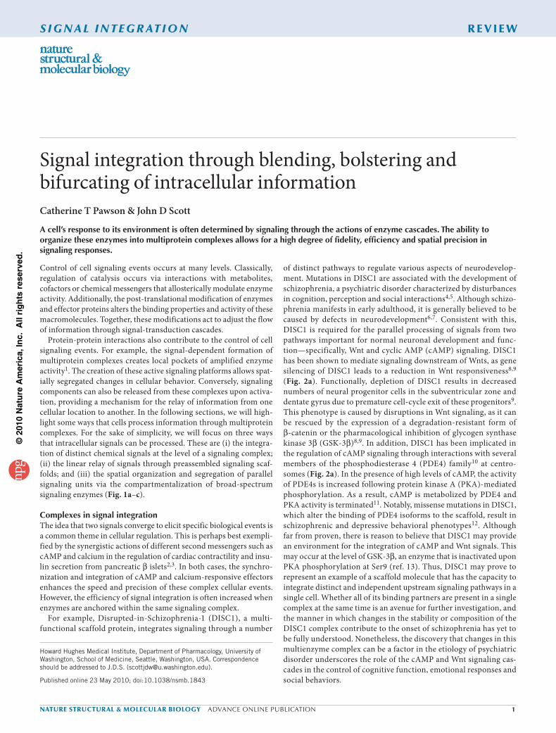
Figure 1 Schematic representation of mechanisms of signal transduction. (a) In signal integration, two independent inputs act on the same pathway to elicit a common outcome. (b) Scaffolding proteins allow the efficient relay of signals through successive enzymes in a pathway. (c) Specificity of signaling through broad-specificity enzymes is often assured by the spatial segregation of enzyme complexes.
P
PKA
a
b
Wnt
β-cat PDE4 cAMP
GSK-3β
Progenitor-cell proliferation Cell migration
DISC1
c
β-cat
IKKα
IKKβ
NEMO
NF-κB
IκBαIKKα
IKKβ
NEMO
NF-κB
NF-κB Transcriptionalactivation
IκBα
UB
SCF
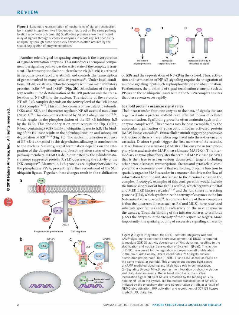
Figure 2 Signal integration: the DISC1 scaffold integrates Wnt and cAMP signaling to coordinate neurodevelopment. (a) DISC1 is required to regulate GSK-3β activity downstream of Wnt signaling, resulting in the stabilization and nuclear translocation of β-catenin (β-cat). This action of DISC1 is required for the regulation of progenitor-cell proliferation in the brain. Additionally, DISC1 coordinates PKA targets nuclear distribution protein nudE–like 1 (NDEL1) and LIS1 as well as PDE4 on the same molecular scaffold. This arrangement ensures tight control of cAMP-mediated signaling and likely has a role in cell migration. (b) Signaling through NF-κB requires the integration of phosphorylation and ubiquitination events. Under basal conditions, the nuclear localization signal (NLS) of NF-κB is masked by the binding of IκBs, holding NF-κB in the cytosol. (c) The nuclear translocation of NF-κB is initiated by the phosphorylation and ubiquitination of IκBs as a result of NEMO ubiquitination, IKK activation and recruitment of SCF E3 ligases to pIκB. UB, ubiquitin.
©20
10 N
atu
re A
mer
ica,
Inc.
All
rig
hts
res
erve
d.
nature structural & molecular biology advance online publication �
r e V i e W
scaffolds places them in a context that facilitates the preferential relay of signals to the terminal enzyme in the chain.
Another useful property of enzyme scaffolding is to segregate sig-nals in a manner that prevents indiscriminate cross-talk between pathways. This concept is particularly important in unicellular organisms such as yeast, where a variety of cytoplasmic processes can be simultaneously modulated by different kinase scaffolds. In yeast, mating, invasive growth and responses to high osmolarity are all regulated by distinct MAP kinase pathways that share a com-mon MAP3K called Sterile 11 (Ste11)35–37. Segregation of Ste11 activity involves binding to scaffolding proteins such as Pbs2 and Ste5. Recruitment of Ste11 into the osmosensing pathway requires interaction with Pbs2 (ref. 37). This protein not only scaffolds Ste11 but also acts as the MAP2K in this pathway. In contrast, Ste5 organ-izes Ste11 and the kinases Ste7 and Fus3 to direct signals through the yeast mating pathway (Fig. 3a). Recent evidence suggests that Ste5 also facilitates the activation of its kinase-binding partners38. Synthetic biology approaches have identified a regulatory domain in Ste5 that catalytically unlocks the Fus3 kinase for phosphoryla-tion by Ste7 (ref. 39). This finding broadens the role of scaffolding proteins, as it suggests that Ste5 not only functions to organize suc-cessive components of a yeast MAPK cascade but also allosterically modifies the conformation of its bound kinases, making them more amenable to activation.
In other contexts, scaffolding proteins can participate in the transfer of signals from one region of the cell to the next. The A kinase anchor-ing protein AKAP-Lbc forms a multiprotein complex that relays information from the plasma membrane to the nucleus in response to hypertrophic signals40,41. These signals, which include elevated adrenergic activity, lead to the reprogramming of cardiomyocyte gene expression by myocyte enhancer factor 2 (MEF2), known as the fetal
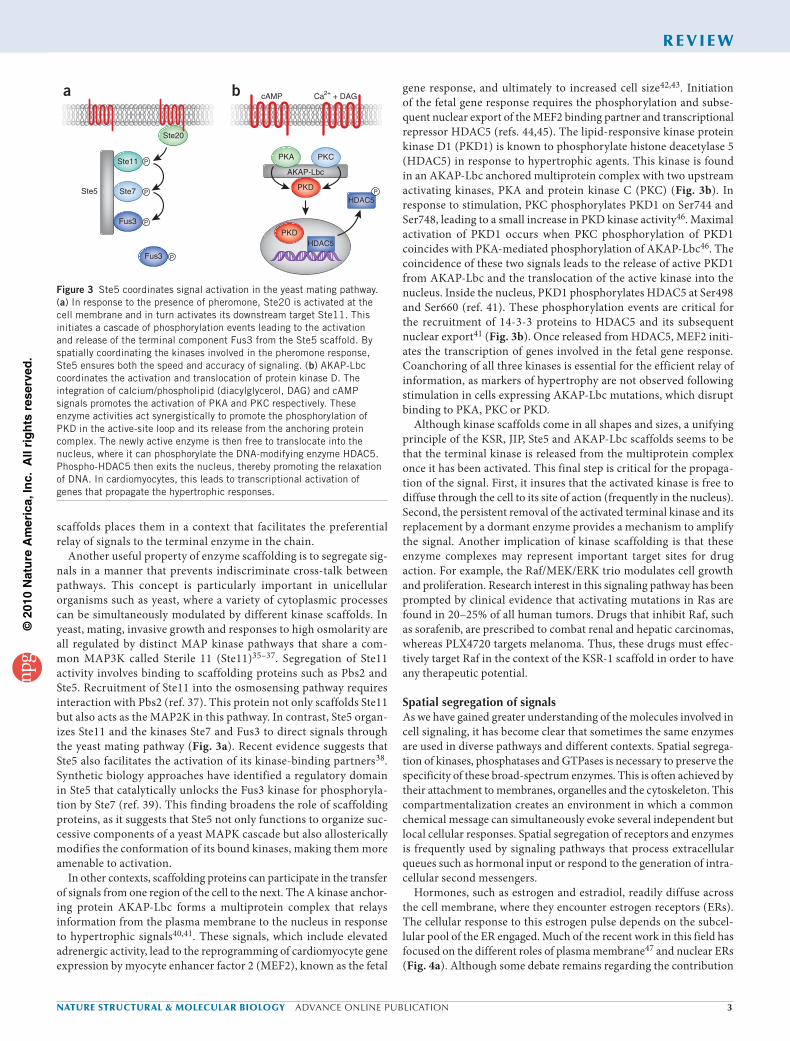
gene response, and ultimately to increased cell size42,43. Initiation of the fetal gene response requires the phosphorylation and subse-quent nuclear export of the MEF2 binding partner and transcriptional repressor HDAC5 (refs. 44,45). The lipid-responsive kinase protein kinase D1 (PKD1) is known to phosphorylate histone deacetylase 5 (HDAC5) in response to hypertrophic agents. This kinase is found in an AKAP-Lbc anchored multiprotein complex with two upstream activating kinases, PKA and protein kinase C (PKC) (Fig. 3b). In response to stimulation, PKC phosphorylates PKD1 on Ser744 and Ser748, leading to a small increase in PKD kinase activity46. Maximal activation of PKD1 occurs when PKC phosphorylation of PKD1 coincides with PKA-mediated phosphorylation of AKAP-Lbc46. The coincidence of these two signals leads to the release of active PKD1 from AKAP-Lbc and the translocation of the active kinase into the nucleus. Inside the nucleus, PKD1 phosphorylates HDAC5 at Ser498 and Ser660 (ref. 41). These phosphorylation events are critical for the recruitment of 14-3-3 proteins to HDAC5 and its subsequent nuclear export41 (Fig. 3b). Once released from HDAC5, MEF2 initi-ates the transcription of genes involved in the fetal gene response. Coanchoring of all three kinases is essential for the efficient relay of information, as markers of hypertrophy are not observed following stimulation in cells expressing AKAP-Lbc mutations, which disrupt binding to PKA, PKC or PKD.
Although kinase scaffolds come in all shapes and sizes, a unifying principle of the KSR, JIP, Ste5 and AKAP-Lbc scaffolds seems to be that the terminal kinase is released from the multiprotein complex once it has been activated. This final step is critical for the propaga-tion of the signal. First, it insures that the activated kinase is free to diffuse through the cell to its site of action (frequently in the nucleus). Second, the persistent removal of the activated terminal kinase and its replacement by a dormant enzyme provides a mechanism to amplify the signal. Another implication of kinase scaffolding is that these enzyme complexes may represent important target sites for drug action. For example, the Raf/MEK/ERK trio modulates cell growth and proliferation. Research interest in this signaling pathway has been prompted by clinical evidence that activating mutations in Ras are found in 20–25% of all human tumors. Drugs that inhibit Raf, such as sorafenib, are prescribed to combat renal and hepatic carcinomas, whereas PLX4720 targets melanoma. Thus, these drugs must effec-tively target Raf in the context of the KSR-1 scaffold in order to have any therapeutic potential.
Spatial segregation of signalsAs we have gained greater understanding of the molecules involved in cell signaling, it has become clear that sometimes the same enzymes are used in diverse pathways and different contexts. Spatial segrega-tion of kinases, phosphatases and GTPases is necessary to preserve the specificity of these broad-spectrum enzymes. This is often achieved by their attachment to membranes, organelles and the cytoskeleton. This compartmentalization creates an environment in which a common chemical message can simultaneously evoke several independent but local cellular responses. Spatial segregation of receptors and enzymes is frequently used by signaling pathways that process extracellular queues such as hormonal input or respond to the generation of intra-cellular second messengers.
Hormones, such as estrogen and estradiol, readily diffuse across the cell membrane, where they encounter estrogen receptors (ERs). The cellular response to this estrogen pulse depends on the subcel-lular pool of the ER engaged. Much of the recent work in this field has focused on the different roles of plasma membrane47 and nuclear ERs (Fig. 4a). Although some debate remains regarding the contribution
Figure 3 Ste5 coordinates signal activation in the yeast mating pathway. (a) In response to the presence of pheromone, Ste20 is activated at the cell membrane and in turn activates its downstream target Ste11. This initiates a cascade of phosphorylation events leading to the activation and release of the terminal component Fus3 from the Ste5 scaffold. By spatially coordinating the kinases involved in the pheromone response, Ste5 ensures both the speed and accuracy of signaling. (b) AKAP-Lbc coordinates the activation and translocation of protein kinase D. The integration of calcium/phospholipid (diacylglycerol, DAG) and cAMP signals promotes the activation of PKA and PKC respectively. These enzyme activities act synergistically to promote the phosphorylation of PKD in the active-site loop and its release from the anchoring protein complex. The newly active enzyme is then free to translocate into the nucleus, where it can phosphorylate the DNA-modifying enzyme HDAC5. Phospho-HDAC5 then exits the nucleus, thereby promoting the relaxation of DNA. In cardiomyocytes, this leads to transcriptional activation of genes that propagate the hypertrophic responses.
P
P
P
P
P
a b
AKAP-Lbc
PKD
HDAC5
Ste20
cAMP Ca2+ + DAG
Ste11
PKDHDAC5
PKC
Ste7Ste5
Fus3
Fus3
PKA
©20
10 N
atu
re A
mer
ica,
Inc.
All
rig
hts
res
erve
d.
� advance online publication nature structural & molecular biology
r e V i e W
of low-affinity ERs such as GPR30, it is clear that the classical nuclear receptors ERα and ERβ are localized to and crucial for estrogen sig-naling at the plasma membrane48–50. Palmitoylation and subsequent interaction with caveolin-1 appear to promote the translocation of ERα and ERβ to the plasma membrane; however, the precise regula-tion of these events is currently unclear51,52. Nevertheless, the identi-fication of the same subset of receptors at both the plasma membrane and the nucleus implies that cellular responses to estrogen are not solely determined by the receptor subtype but also by the subcellular location of the hormone–ER complex. For example, rapid activation of RAF/MEK/ERK and phosphoinositide 3 kinase (PI 3-kinase) is known to occur downstream of estrogen signaling53–57, leading to changes in signaling, cell motility and cell survival58,59. These effects require plasma-membrane rather than nuclear ERs50. Conversely, membrane-localized ERs are not sufficient to mediate the prolonged effects of estrogen, such as the upregulation of developmental pro-grams and transcriptional control by nuclear receptors50. Thus, these two pools of receptors have distinct functions that are crucial for nor-mal cellular behavior. Additionally, it has emerged that membrane ER signaling may synergize with nuclear receptors. In one example, cross-talk between membrane ERs and epidermal growth factor or insulin-like growth factor 1 leads to activation of downstream ERK signaling cascades60,61. In some circumstances, the upregulation of MAPK sig-naling leads to the phosphorylation of nuclear ERs on Ser305, trigger-ing ligand-independent transcriptional activation62,63. Activation of PI 3-kinase signaling by membrane ERs upregulates transcriptional regulation via a different mechanism. Under resting conditions, the nuclear ERα is inactivated through a chronic, GSK-3β–mediated phosphorylation of Ser118 (ref. 64). However, the cross-activation of PI 3-kinase and AKT upon estrogen binding to membrane ERα leads to the phosphorylation of GSK-3β. This results in the inactivation of GSK-3β65 and the derepression of the nuclear ERα. These examples highlight how spatial segregation of similar effector proteins can be used to impose higher levels of cellular organization.
Signal segregation is evident in the action of the second messen-ger cAMP. This cyclic nucleotide freely diffuses from its site of syn-thesis at the plasma membrane to discrete compartments where it activates cAMP responsive kinases (PKA), ion channels and guanine nucleotide exchange factors (EPACs). Subcellular targeting of these ‘cAMP receptors’ is mediated by a family of AKAPs that tether PKA and EPACs in proximity to selected substrates66,67. However, another notable feature of AKAPs is the ability to organize groups of second messenger–regulated enzymes. For example, the neuronal anchoring
protein AKAP79/150 anchors the cAMP-dependent kinase PKA, the Ca2+- and lipid-regulated PKC and the calmodulin-stimulated protein phosphatase PP2B/calcineurin68,69. A combination of biochemical and electrophysiological approaches has shown that distinct AKAP79/150 signaling complexes modulate the activity of α-amino-3-hydroxy-5-methyl-4-isoxazolepropionic acid receptor (AMPA) and M/KCNQ ion channels70–73. Notably, the regulation of these channels requires a nonoverlapping subset of anchored enzymes.
The AMPA current propagates excitatory synaptic responses74,75. This ligand-gated ion channel requires the anchoring of PP2B and PKA for full activity76–78. Specifically, AKAP79/150–anchored PP2B is required for the attenuation of the AMPA current in response to tonic stimulation, whereas PKA is required for the maintenance of AMPA currents and channels at the cell surface71. Although the AKAP79/150–AMPA channel complex has the capacity to recruit PKC, this enzyme does not seem to be required for the control of this subset of excitatory synaptic responses. However, in the sympathetic nervous system, anchoring of PKC by AKAP79/150 is required for the attenuation of the hyperpolarizing K+ current (M current) through the M/KCNQ channel79. AKAP79/150 targets PKC where it can opti-mally respond to activating signals from the m1 muscarinic recep-tor and preferentially phosphorylate the KCNQ2 subunit to suppress the M current71,72. Whereas anchoring of PKA and PP2B is dispens-able for the regulation of the M current, the PKC binding region of AKAP79/150 is both necessary and sufficient for M-current suppression71 (Fig. 4b). Another interesting feature of the M-current complex is that AKAP79/150 protects PKC from certain ATP-competitive inhibitors80. This implies that anchoring proteins such as AKAP79/150 not only segregate individual cell-signaling processes but also can change the pharmacological profile of certain protein kinases (Fig. 4c). Together, these data raise the possibility that AKAP79/150 segre-gates distinct second-messenger signals within a cell by tethering unique combinations of binding partners with different ion channels. An added level of regulation may be introduced if interaction with AKAPs or other interacting proteins can modify kinase susceptibility to pharmacological agents. This finding should have important con-sequences for drug discovery and research projects predicated on the selectivity of pharmacological protein kinase inhibitors.
ConclusionAs our appreciation of cellular architecture grows, it has become apparent that cells are not just bags of enzymes. Rather, eukaryotic cells require a high degree of molecular organization to integrate,
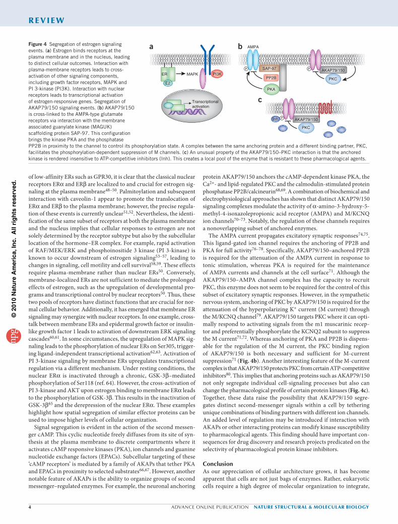
Figure 4 Segregation of estrogen signaling events. (a) Estrogen binds receptors at the plasma membrane and in the nucleus, leading to distinct cellular outcomes. Interaction with plasma-membrane receptors leads to cross-activation of other signaling components, including growth factor receptors, MAPK and PI 3-kinase (PI3K). Interaction with nuclear receptors leads to transcriptional activation of estrogen-responsive genes. Segregation of AKAP79/150 signaling events. (b) AKAP79/150 is cross-linked to the AMPA-type glutamate receptors via interaction with the membrane associated guanylate kinase (MAGUK) scaffolding protein SAP-97. This configuration brings the kinase PKA and the phosphatase PP2B in proximity to the channel to control its phosphorylation state. A complex between the same anchoring protein and a different binding partner, PKC, facilitates the phosphorylation-dependent suppression of M channels. (c) An unusual property of the AKAP79/150–PKC interaction is that the anchored kinase is rendered insensitive to ATP-competitive inhibitors (Inh). This creates a local pool of the enzyme that is resistant to these pharmacological agents.
P
P
a
AKAP79/150
Transcriptionalactivation
ER PI3KMAPK
E2
b
Inh
PKC
P
PKA
PP2B
SAP-97
AMPA
PKC
c
AKAP79/150
©20
10 N
atu
re A
mer
ica,
Inc.
All
rig
hts
res
erve
d.
nature structural & molecular biology advance online publication �
r e V i e W
relay or segregate the chemical signals that control all aspects of cel-lular behavior. Although the three modes of molecular organization discussed in this article offer unique advantages for efficient informa-tion transfer, it should be noted that these processes are not mutually exclusive. In fact, many signaling pathways use two or more of these mechanisms. For example, both DISC1 and NF-kB make use of the spatial segregation and the integration of multiple signaling inputs to fine tune cellular responses. Although DISC1 may integrate Wnt and cAMP signals, it is not yet clear that these messages influence the same biological event. In contrast, components of NF-kB signaling pathway use cytoplasmic phosphorylation and ubiquitination events to enact transcriptional activation in the nucleus. Certainly, the prox-imity of the phosphorylation and ubiquitination machinery ensures that changes in the stability of the NF-kB complex can be rapidly modulated. Although working in a different way, signaling from the membrane to the nucleus is a characteristic shared by AKAP-Lbc complexes. However, their mechanism of action shows hallmarks of all three modes of molecular organization. As a scaffold, AKAP-Lbc segregates protein kinase D from its nuclear targets by anchoring it in the cytosol. However, upon adrenergic stimulation, AKAP-Lbc inte-grates cAMP, calcium and phospholipid signals to mobilize successive enzymes in the PKD activation cascade. This results in the release and nuclear translocation of PKD, ultimately triggering transcriptional activation. In fact, translocation of active PKD from the cytoplasm to the nucleus is necessary to trigger the next phase in this transcrip-tional activation process. Thus, through the amalgamation of these simple regulatory steps, it is possible to coordinate and synchronize sophisticated cellular events in both space and time.
All in all, we have just begun to glimpse the complexity of the protein-protein interactions that underlie the cellular organization of cell-signaling cascades. Therefore, we would like to speculate on what the future might be for this field. Recent advances in technol-ogy hold promise for the future. Most signaling responses represent the coupling of individual chemical, physical or electrical events. Therefore, the simultaneous monitoring of more than one event will undoubtedly give more information about the order and dynamics of a given cellular response. For example, real-time visualization of AKAP79-anchored PKC phosphorylation with a fluorescent kinase activity reporter called CKAR can now be performed concurrently with measuring electrophysiological changes in muscarine-sensitive ion channels. The coincident detection of both steps suggests that AKAP79 not only directs PKC toward the ion channel but also syn-chronizes kinase activation to instantaneously reduce ion flow80,81. The advent, some time ago, of genetically encoded fluorescence reporters developed by Tsien and colleagues opens many more pos-sibilities for this type of study82,83. In addition, the ability to monitor the impact of a signal input using systems biology approaches should provide deeper insight as to how a cell manages to simultaneously coordinate several distinct events. Mathamatical modeling of these signaling pathways may go hand in hand with these system-wide approaches by providing an in silico framework to test cell-based hypotheses. Together, these approaches may shed new light on the dynamics and flux of signaling cascades and point the way toward new drug targets.
ACknowleDgmenTSWe would like to thank members of the Scott lab for their critical review of this manuscript. This work was supported by the Foundation Leducq and by NIH grant DK54441 to J.D.S.
ComPeTIng FInAnCIAl InTeReSTSThe authors declare no competing financial interests.
Published online at http://www.nature.com/nsmb/. Reprints and permissions information is available online at http://npg.nature.com/reprintsandpermissions/.
1. Scott, J.D. & Pawson, T. Cell signaling in space and time: where proteins come together and when they’re apart. Science 326, 1220–1224 (2009).
2. Saltiel, A.R. & Kahn, C.R. Insulin signalling and the regulation of glucose and lipid metabolism. Nature 414, 799–806 (2001).
3. Bers, D.M. Calcium cycling and signaling in cardiac myocytes. Annu. Rev. Physiol. 70, 23–49 (2008).
4. Millar, J.K. et al. Disruption of two novel genes by a translocation co-segregating with schizophrenia. Hum. Mol. Genet. 9, 1415–1423 (2000).
5. St. Clair, D. et al. Association within a family of a balanced autosomal translocation with major mental illness. Lancet 336, 13–16 (1990).
6. Rapoport, J.L., Addington, A.M., Frangou, S. & Psych, M.R. The neurodevelopmental model of schizophrenia: update 2005. Mol. Psychiatry 10, 434–449 (2005).
7. Lewis, D.A. & Levitt, P. Schizophrenia as a disorder of neurodevelopment. Annu. Rev. Neurosci. 25, 409–432 (2002).
8. Brandon, N.J. et al. Understanding the role of DISC1 in psychiatric disease and during normal development. J. Neurosci. 29, 12768–12775 (2009).
9. Mao, Y. et al. Disrupted in schizophrenia 1 regulates neuronal progenitor proliferation via modulation of GSK3β/β-catenin signaling. Cell 136, 1017–1031 (2009).
10. Millar, J.K. et al. DISC1 and PDE4B are interacting genetic factors in schizophrenia that regulate cAMP signaling. Science 310, 1187–1191 (2005).
11. Murdoch, H. et al. Isoform-selective susceptibility of DISC1/phosphodiesterase-4 complexes to dissociation by elevated intracellular cAMP levels. J. Neurosci. 27, 9513–9524 (2007).
12. Clapcote, S.J. et al. Behavioral phenotypes of Disc1 missense mutations in mice. Neuron 54, 387–402 (2007).
13. Fang, X. et al. Phosphorylation and inactivation of glycogen synthase kinase 3 by protein kinase A. Proc. Natl. Acad. Sci. USA 97, 11960–11965 (2000).
14. Wan, F. & Lenardo, M.J. The nuclear signaling of NF-κB: current knowledge, new insights, and future perspectives. Cell Res. 20, 24–33 (2010).
15. Beg, A.A. et al. IκB interacts with the nuclear localization sequences of the subunits of NF-κB: a mechanism for cytoplasmic retention. Genes Dev. 6, 1899–1913 (1992).
16. Baeuerle, P.A. & Baltimore, D. Activation of DNA-binding activity in an apparently cytoplasmic precursor of the NF-κB transcription factor. Cell 53, 211–217 (1988).
17. Thompson, J.E., Phillips, R.J., Erdjument-Bromage, H., Tempst, P. & Ghosh, S. IκB-β regulates the persistent response in a biphasic activation of NF-κB. Cell 80, 573–582 (1995).
18. Regnier, C.H. et al. Identification and characterization of an IκB kinase. Cell 90, 373–383 (1997).
19. Woronicz, J.D., Gao, X., Cao, Z., Rothe, M. & Goeddel, D.V. IκB kinase-β: NF-κB activation and complex formation with IκB kinase-α and NIK. Science 278, 866–869 (1997).
20. DiDonato, J.A., Hayakawa, M., Rothwarf, D.M., Zandi, E. & Karin, M. A cytokine-responsive IκB kinase that activates the transcription factor NF-κB. Nature 388, 548–554 (1997).
21. Zandi, E., Rothwarf, D.M., Delhase, M., Hayakawa, M. & Karin, M. The IκB kinase complex (IKK) contains two kinase subunits, IKKα and IKKβ, necessary for IκB phosphorylation and NF-κB activation. Cell 91, 243–252 (1997).
22. Yamaoka, S. et al. Complementation cloning of NEMO, a component of the IκB kinase complex essential for NF-κB activation. Cell 93, 1231–1240 (1998).
23. Tang, E.D., Wang, C.Y., Xiong, Y. & Guan, K.L. A role for NF-κB essential modifier/IκB kinase-γ (NEMO/IKKγ) ubiquitination in the activation of the IκB kinase complex by tumor necrosis factor-α. J. Biol. Chem. 278, 37297–37305 (2003).
24. Zhou, H. et al. Bcl10 activates the NF-κB pathway through ubiquitination of NEMO. Nature 427, 167–171 (2004).
25. Scherer, D.C., Brockman, J.A., Chen, Z., Maniatis, T. & Ballard, D.W. Signal-induced degradation of IκB alpha requires site-specific ubiquitination. Proc. Natl. Acad. Sci. USA 92, 11259–11263 (1995).
26. DiDonato, J. et al. Mapping of the inducible IκB phosphorylation sites that signal its ubiquitination and degradation. Mol. Cell. Biol. 16, 1295–1304 (1996).
27. Wu, C. & Ghosh, S. β-TrCP mediates the signal-induced ubiquitination of IκBβ. J. Biol. Chem. 274, 29591–29594 (1999).
28. Yaron, A. et al. Identification of the receptor component of the IκBα-ubiquitin ligase. Nature 396, 590–594 (1998).
29. Kovalenko, A. et al. The tumour suppressor CYLD negatively regulates NF-κB signalling by deubiquitination. Nature 424, 801–805 (2003).
30. Zeke, A., Lukacs, M., Lim, W.A. & Remenyi, A. Scaffolds: interaction platforms for cellular signalling circuits. Trends Cell Biol. 19, 364–374 (2009).
31. Morrison, D.K. & Davis, R.J. Regulation of MAP kinase signaling modules by scaffold proteins in mammals. Annu. Rev. Cell Dev. Biol. 19, 91–118 (2003).
32. Kolch, W. Coordinating ERK/MAPK signalling through scaffolds and inhibitors. Nat. Rev. Mol. Cell Biol. 6, 827–837 (2005).
33. Brown, M.D. & Sacks, D.B. Protein scaffolds in MAP kinase signalling. Cell. Signal. 21, 462–469 (2009).
34. Weston, C.R. & Davis, R.J. The JNK signal transduction pathway. Curr. Opin. Cell Biol. 19, 142–149 (2007).
©20
10 N
atu
re A
mer
ica,
Inc.
All
rig
hts
res
erve
d.
� advance online publication nature structural & molecular biology
35. Elion, E.A. Pheromone response, mating and cell biology. Curr. Opin. Microbiol. 3, 573–581 (2000).
36. Liu, H., Styles, C.A. & Fink, G.R. Elements of the yeast pheromone response pathway required for filamentous growth of diploids. Science 262, 1741–1744 (1993).
37. Posas, F. & Saito, H. Osmotic activation of the HOG MAPK pathway via Ste11p MAPKKK: scaffold role of Pbs2p MAPKK. Science 276, 1702–1705 (1997).
38. Feng, Y., Song, L.Y., Kincaid, E., Mahanty, S.K. & Elion, E.A. Functional binding between Gβ and the LIM domain of Ste5 is required to activate the MEKK Ste11. Curr. Biol. 8, 267–278 (1998).
39. Park, S.H., Zarrinpar, A. & Lim, W.A. Rewiring MAP kinase pathways using alternative scaffold assembly mechanisms. Science 299, 1061–1064 (2003).
40. Appert-Collin, A., Cotecchia, S., Nenniger-Tosato, M., Pedrazzini, T. & Diviani, D. The A-kinase anchoring protein (AKAP)-Lbc-signaling complex mediates α1 adrenergic receptor-induced cardiomyocyte hypertrophy. Proc. Natl. Acad. Sci. USA 104, 10140–10145 (2007).
41. Carnegie, G.K. et al. AKAP-Lbc mobilizes a cardiac hypertrophy signaling pathway. Mol. Cell 32, 169–179 (2008).
42. Molkentin, J.D. & Dorn, G.W. II. Cytoplasmic signaling pathways that regulate cardiac hypertrophy. Annu. Rev. Physiol. 63, 391–426 (2001).
43. Black, B.L. & Olson, E.N. Transcriptional control of muscle development by myocyte enhancer factor-2 (MEF2) proteins. Annu. Rev. Cell Dev. Biol. 14, 167–196 (1998).
44. Vega, R.B. et al. Protein kinases C and D mediate agonist-dependent cardiac hypertrophy through nuclear export of histone deacetylase 5. Mol. Cell. Biol. 24, 8374–8385 (2004).
45. Sucharov, C.C., Langer, S., Bristow, M. & Leinwand, L. Shuttling of HDAC5 in H9C2 cells regulates YY1 function through CaMKIV/PKD and PP2A. Am. J. Physiol. Cell Physiol. 291, C1029–C1037 (2006).
46. Carnegie, G.K., Smith, F.D., McConnachie, G., Langeberg, L.K. & Scott, J.D. AKAP-Lbc nucleates a protein kinase D activation scaffold. Mol. Cell 15, 889–899 (2004).
47. Pietras, R.J. & Szego, C.M. Specific binding sites for oestrogen at the outer surfaces of isolated endometrial cells. Nature 265, 69–72 (1977).
48. Norfleet, A.M., Thomas, M.L., Gametchu, B. & Watson, C.S. Estrogen receptor-α detected on the plasma membrane of aldehyde-fixed GH3/B6/F10 rat pituitary tumor cells by enzyme-linked immunocytochemistry. Endocrinology 140, 3805–3814 (1999).
49. Pedram, A., Razandi, M. & Levin, E.R. Nature of functional estrogen receptors at the plasma membrane. Mol. Endocrinol. 20, 1996–2009 (2006).
50. Pedram, A. et al. Developmental phenotype of a membrane only estrogen receptor α (MOER) mouse. J. Biol. Chem. 284, 3488–3495 (2009).
51. Acconcia, F. et al. Palmitoylation-dependent estrogen receptor alpha membrane localization: regulation by 17β-estradiol. Mol. Biol. Cell 16, 231–237 (2005).
52. Razandi, M. et al. Identification of a structural determinant necessary for the localization and function of estrogen receptor α at the plasma membrane. Mol. Cell. Biol. 23, 1633–1646 (2003).
53. Watters, J.J., Campbell, J.S., Cunningham, M.J., Krebs, E.G. & Dorsa, D.M. Rapid membrane effects of steroids in neuroblastoma cells: effects of estrogen on mitogen activated protein kinase signalling cascade and c-fos immediate early gene transcription. Endocrinology 138, 4030–4033 (1997).
54. Migliaccio, A. et al. Tyrosine kinase/p21ras/MAP-kinase pathway activation by estradiol-receptor complex in MCF-7 cells. EMBO J. 15, 1292–1300 (1996).
55. Migliaccio, A. et al. Steroid-induced androgen receptor-oestradiol receptor β-Src complex triggers prostate cancer cell proliferation. EMBO J. 19, 5406–5417 (2000).
56. Simoncini, T. et al. Interaction of oestrogen receptor with the regulatory subunit of phosphatidylinositol-3-OH kinase. Nature 407, 538–541 (2000).
57. Castoria, G. et al. PI3-kinase in concert with Src promotes the S-phase entry of oestradiol-stimulated MCF-7 cells. EMBO J. 20, 6050–6059 (2001).
58. Bartucci, M., Morelli, C., Mauro, L., Ando, S. & Surmacz, E. Differential insulin-like growth factor I receptor signaling and function in estrogen receptor (ER)-positive
MCF-7 and ER-negative MDA-MB-231 breast cancer cells. Cancer Res. 61, 6747–6754 (2001).
59. Levin, E.R. Plasma membrane estrogen receptors. Trends Endocrinol. Metab. 20, 477–482 (2009).
60. Kahlert, S. et al. Estrogen receptor α rapidly activates the IGF-1 receptor pathway. J. Biol. Chem. 275, 18447–18453 (2000).
61. Filardo, E.J., Quinn, J.A., Frackelton, A.R. Jr. & Bland, K.I. Estrogen action via the G protein-coupled receptor, GPR30: stimulation of adenylyl cyclase and cAMP-mediated attenuation of the epidermal growth factor receptor-to-MAPK signaling axis. Mol. Endocrinol. 16, 70–84 (2002).
62. Balasenthil, S., Barnes, C.J., Rayala, S.K. & Kumar, R. Estrogen receptor activation at serine 305 is sufficient to upregulate cyclin D1 in breast cancer cells. FEBS Lett. 567, 243–247 (2004).
63. Balasenthil, S. et al. p21-activated kinase-1 signaling mediates cyclin D1 expression in mammary epithelial and cancer cells. J. Biol. Chem. 279, 1422–1428 (2004).
64. Medunjanin, S. et al. Glycogen synthase kinase-3 interacts with and phosphorylates estrogen receptor α and is involved in the regulation of receptor activity. J. Biol. Chem. 280, 33006–33014 (2005).
65. Brunet, A. et al. Akt promotes cell survival by phosphorylating and inhibiting a Forkhead transcription factor. Cell 96, 857–868 (1999).
66. Wong, W. & Scott, J.D. AKAP signalling complexes: focal points in space and time. Nat. Rev. Mol. Cell Biol. 5, 959–970 (2004).
67. Dodge-Kafka, K.L. et al. The protein kinase A anchoring protein mAKAP coordinates two integrated cAMP effector pathways. Nature 437, 574–578 (2005).
68. Coghlan, V.M. et al. Association of protein kinase A and protein phosphatase 2B with a common anchoring protein. Science 267, 108–111 (1995).
69. Klauck, T.M. et al. Coordination of three signaling enzymes by AKAP79, a mammalian scaffold protein. Science 271, 1589–1592 (1996).
70. Bhattacharyya, S., Biou, V., Xu, W., Schluter, O. & Malenka, R.C. A critical role for PSD-95/AKAP interactions in endocytosis of synaptic AMPA receptors. Nat. Neurosci. 12, 172–181 (2009).
71. Hoshi, N., Langeberg, L.K. & Scott, J.D. Distinct enzyme combinations in AKAP signalling complexes permit functional diversity. Nat. Cell Biol. 7, 1066–1073 (2005).
72. Bal, M., Zhang, J., Hernandez, C.C., Zaika, O. & Shapiro, M.S. Ca2+/calmodulin disrupts AKAP79/150 interactions with KCNQ (M-Type) K+ channels. J. Neurosci. 30, 2311–2323 (2010).
73. Lu, Y. et al. Age-dependent requirement of AKAP150-anchored PKA and GluR2-lacking AMPA receptors in LTP. EMBO J. 26, 4879–4890 (2007).
74. Scannevin, R.H. & Huganir, R.L. Postsynaptic organization and regulation of excitatory synapses. Nat. Rev. Neurosci. 1, 133–141 (2000).
75. Bredt, D.S. & Nicoll, R.A. AMPA receptor trafficking at excitatory synapses. Neuron 40, 361–379 (2003).
76. Tavalin, S.J. et al. Regulation of GluR1 by the A-kinase anchoring protein 79 (AKAP79) signaling complex shares properties with long-term depression. J. Neurosci. 22, 3044–3051 (2002).
77. Banke, T.G. et al. Control of GluR1 AMPA receptor function by cAMP-dependent protein kinase. J. Neurosci. 20, 89–102 (2000).
78. Esteban, J.A. et al. PKA phosphorylation of AMPA receptor subunits controls synaptic trafficking underlying plasticity. Nat. Neurosci. 6, 136–143 (2003).
79. Hoshi, N. et al. AKAP150 signaling complex promotes suppression of the M-current by muscarinic agonists. Nat. Neurosci. 6, 564–571 (2003).
80. Hoshi, N., Langeberg, L.K., Gould, C.M., Newton, A.C. & Scott, J.D. Interaction with AKAP79 modifies the cellular pharmacology of PKC. Mol. Cell 37, 541–550 (2010).
81. Prince, J. & Ahn, N. The case of the disapprearing drug target. Mol. Cell 37, 455–456 (2010).
82. Giepmans, B.N., Adams, S.R., Ellisman, M.H. & Tsien, R.Y. The fluorescent toolbox for assessing protein location and function. Science 312, 217–224 (2006).
83. Tsien, R.Y. Constructing and exploiting the fluorescent protein paintbox (Nobel Lecture). Angew. Chem. Int. Edn Engl. 48, 5612–5626 (2009).
r e V i e W