Embed Size (px)
Citation preview
Signal Transduction Pathways
Chapter 13, Stryer Short Course
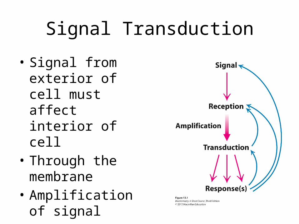
Signal Transduction
• Signal from exterior of cell must affect interior of cell
• Through the membrane
• Amplification of signal
• Ability to turn off
A few pathways…
• Example pathways– a-adrenergic receptor (epinephrine)– b-adrenergic receptor (epinephrine)– Epidermal growth factor receptor– Insulin receptor
• Same hormone can elicit different responses in different tissues
• Cross-talk: different hormones elicit same response (fine tuning)
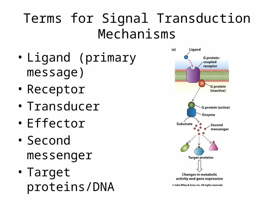
Terms for Signal Transduction Mechanisms
• Ligand (primary message)
• Receptor• Transducer• Effector• Second messenger• Target proteins/DNA
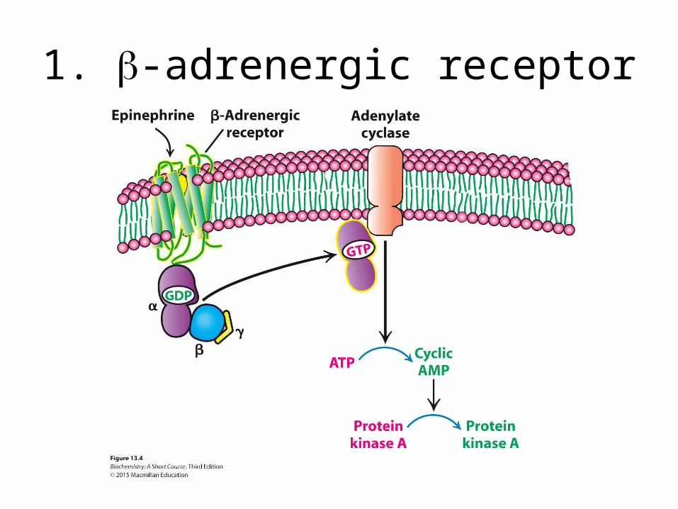
1. b-adrenergic receptor
G-Protein Signaling Pathways
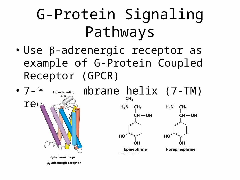
• Use b-adrenergic receptor as example of G-Protein Coupled Receptor (GPCR)
• 7-transmembrane helix (7-TM) receptor
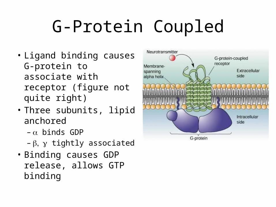
G-Protein Coupled
• Ligand binding causes G-protein to associate with receptor (figure not quite right)
• Three subunits, lipid anchored– a binds GDP– , b g tightly associated
• Binding causes GDP release, allows GTP binding
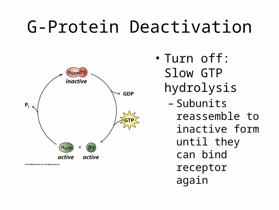
G-Protein Deactivation
• Turn off: Slow GTP hydrolysis– Subunits
reassemble to inactive form until they can bind receptor again
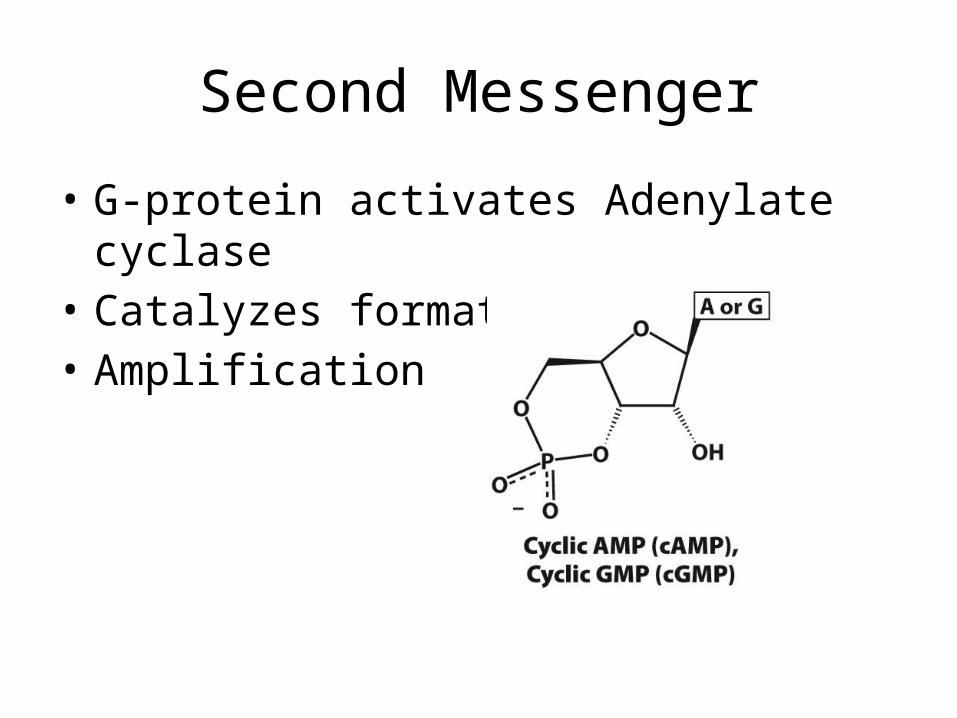
Second Messenger
• G-protein activates Adenylate cyclase • Catalyzes formation of cAMP • Amplification
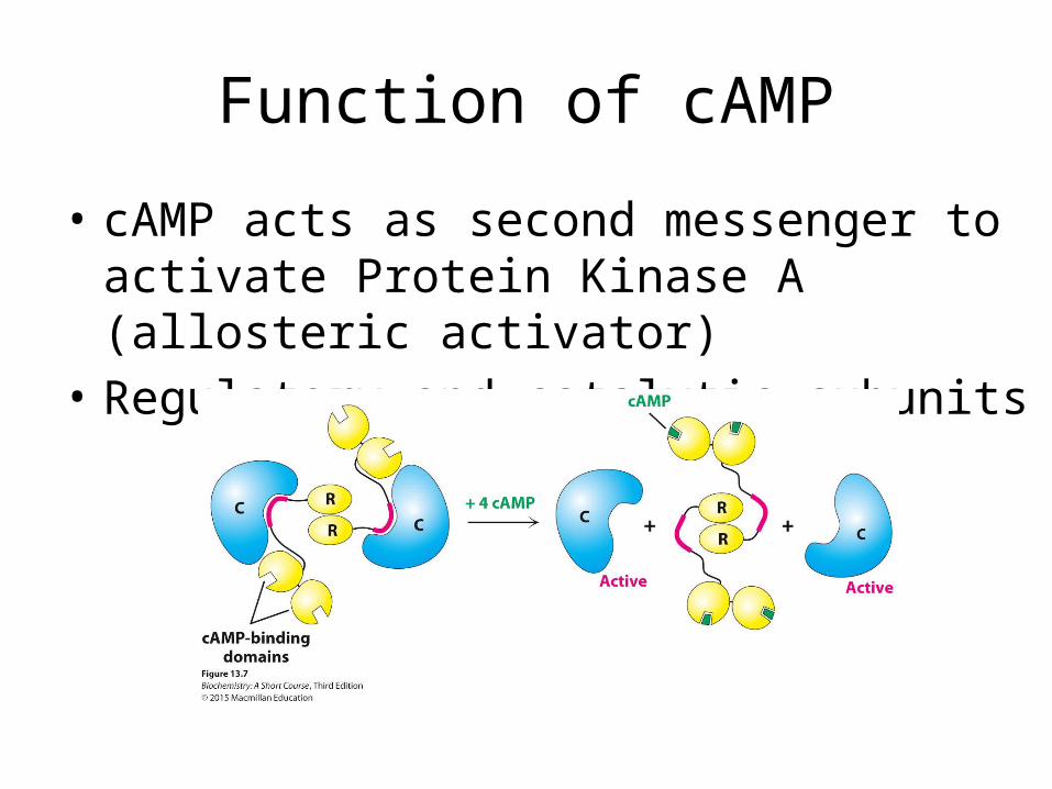
Function of cAMP
• cAMP acts as second messenger to activate Protein Kinase A (allosteric activator)
• Regulatory and catalytic subunits
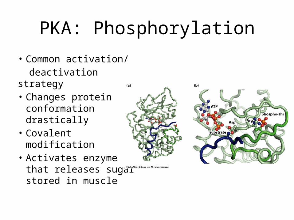
PKA: Phosphorylation
• Common activation/ deactivation strategy• Changes protein
conformation drastically
• Covalent modification• Activates enzyme
that releases sugar stored in muscle
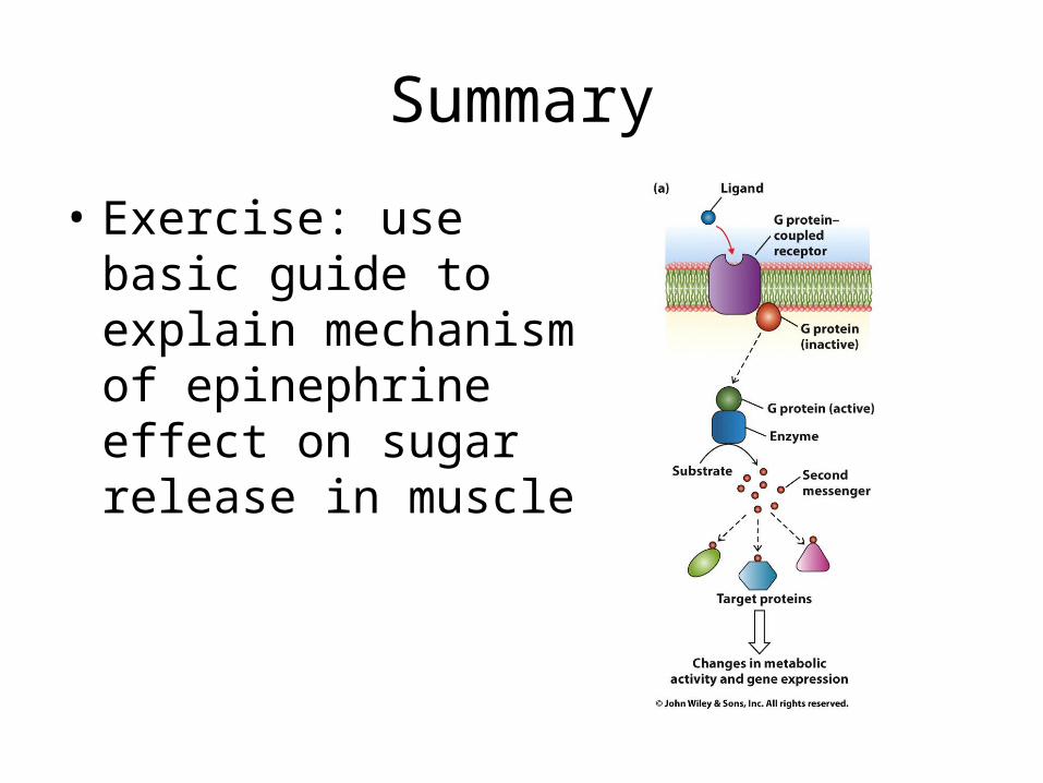
Summary
• Exercise: use basic guide to explain mechanism of epinephrine effect on sugar release in muscle
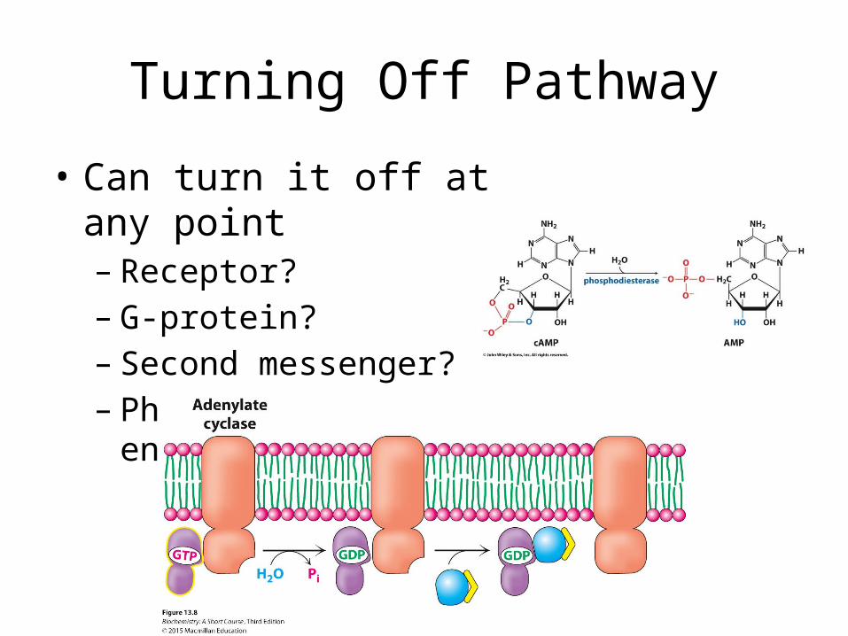
Turning Off Pathway
• Can turn it off at any point– Receptor?– G-protein?– Second messenger?– Phosphorylated enzyme?
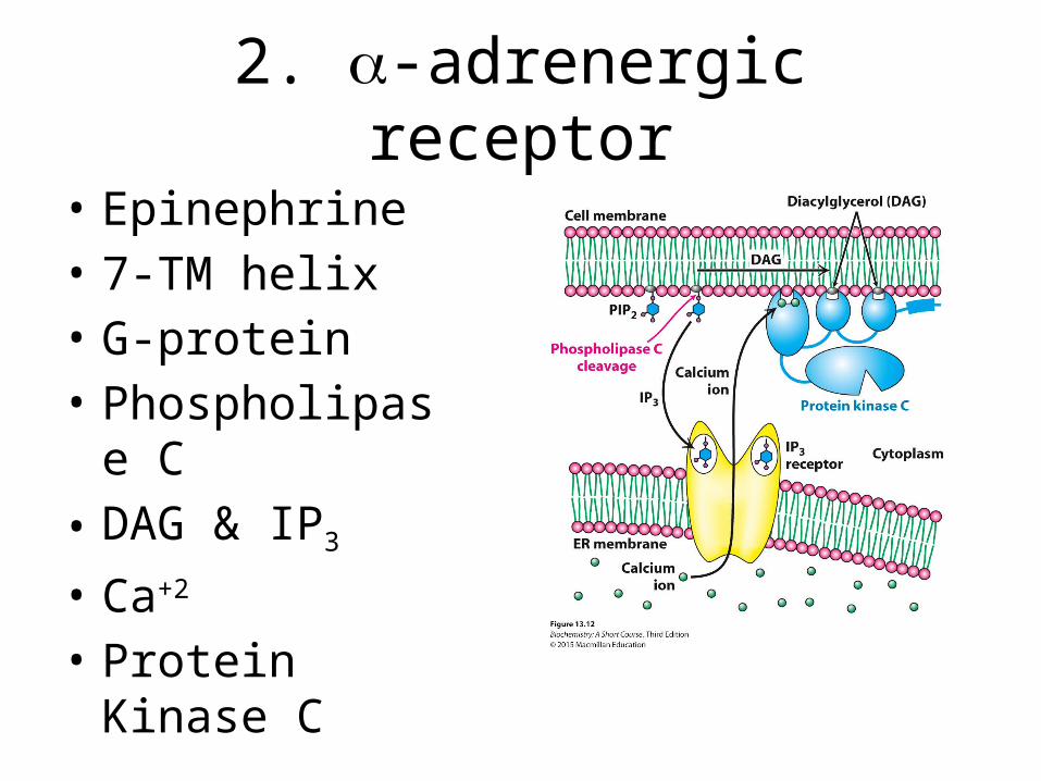
2. a-adrenergic receptor
• Epinephrine• 7-TM helix• G-protein• Phospholipase C• DAG & IP3
• Ca+2
• Protein Kinase C
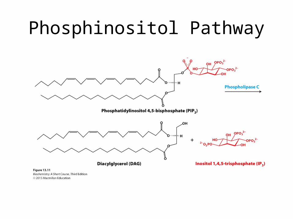
Phosphinositol Pathway
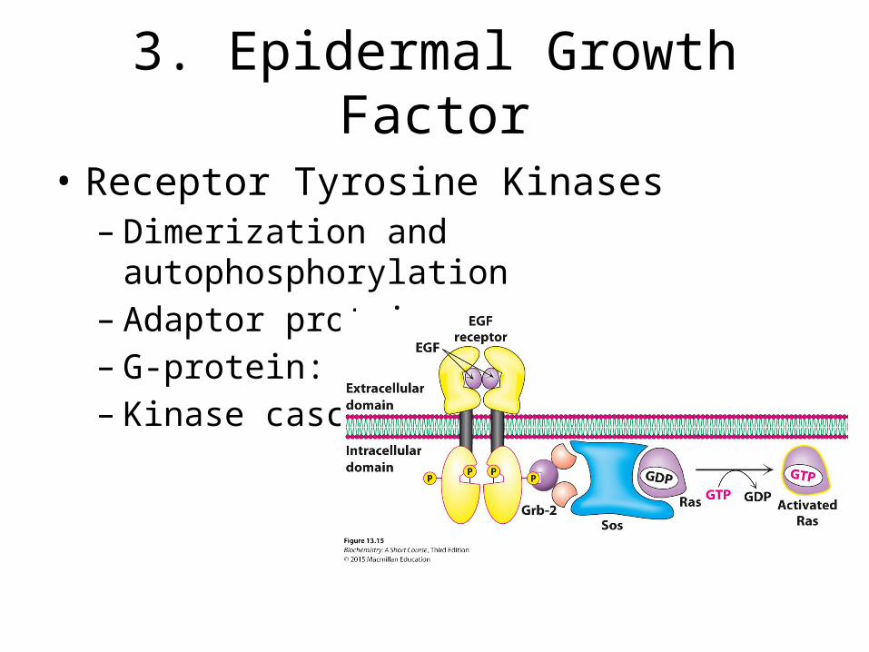
3. Epidermal Growth Factor
• Receptor Tyrosine Kinases– Dimerization and autophosphorylation– Adaptor proteins– G-protein: Ras– Kinase cascade
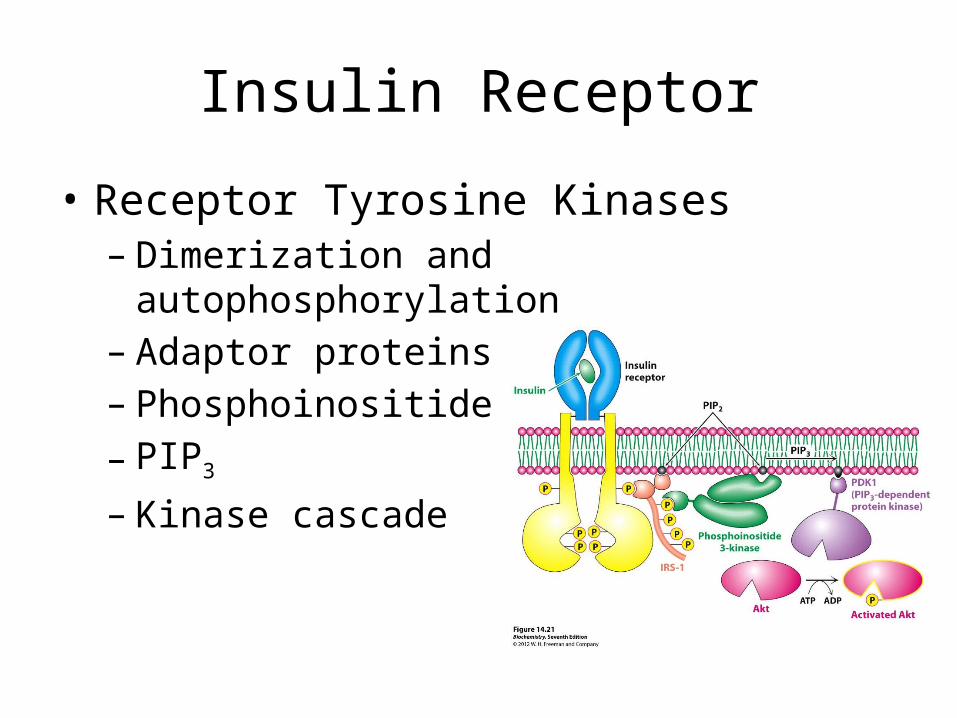
Insulin Receptor
• Receptor Tyrosine Kinases– Dimerization and autophosphorylation– Adaptor proteins– Phosphoinositide kinase– PIP3
– Kinase cascade
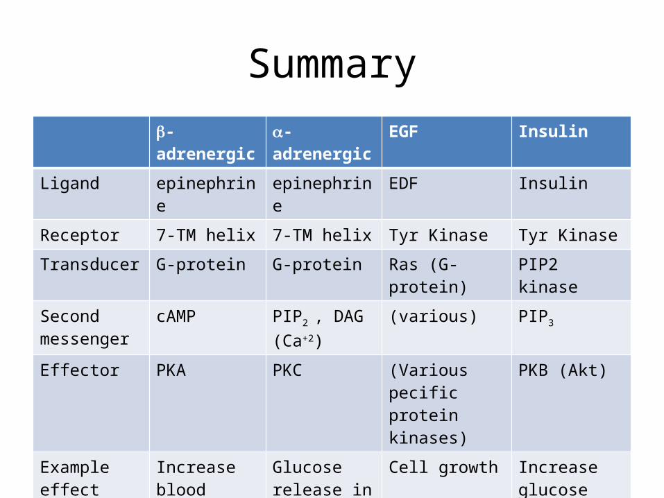
Summaryb-adrenergic a-adrenergic EGF Insulin
Ligand epinephrine epinephrine EDF Insulin
Receptor 7-TM helix 7-TM helix Tyr Kinase Tyr Kinase
Transducer G-protein G-protein Ras (G-protein) PIP2 kinase
Second messenger
cAMP PIP2 , DAG (Ca+2)
(various) PIP3
Effector PKA PKC (Various pecific protein kinases)
PKB (Akt)
Example effect Increase blood pressure
Glucose release in liver
Cell growth Increase glucose uptake from blood
Pathology
• Cholera– Covalent modification of a G-protein– Constitutively active– Opens chloride channel; leads to severe diarrhea
• Whooping cough– Toxin turns off an inhibitory G-protein– Adenylate cyclase remains active
Cancer
• Proto-oncogenes and oncogenes• Ras targets nuclear proteins; Key signal in cell
growth• Mutant Ras proteins have been found to be
associated with various types of cancer. What is the effect on a cell if the mutant Ras is able to bind GTP but is unable to hydrolyze it?


![Signal transduction and information processing in …...signal-transduction pathways [5, 6], but this remains to be widely accepted. After a discussion of transduction, I will analyze](https://img.pdfslide.net/doc/110x75/5f0a9dd27e708231d42c824e/signal-transduction-and-information-processing-in-signal-transduction-pathways.jpg)