Embed Size (px)
Citation preview

Copyright: © 2021 The Author(s). This article has been published under the terms of Creative Commons Attribution-Noncommercial 4.0 International License (CC BY-NC 4.0), which permits noncommercial unrestricted use, distribution, and reproduction in any medium, provided that the following statement is provided.
“This article has been published in Journal of Clinical and Translational Hepatology at https://doi.org/10.14218/JCTH.2020.00136 and can also be viewed on the Journal’s website at http://www.jcthnet.com ”.
Original Article
Journal of Clinical and Translational Hepatology 2021 vol. 9(5) | 615–625 DOI: 10.14218/JCTH.2020.00136
Significant Histologic Changes Are Not Rare in Treatment-naive Hepatitis B Patients with Normal Alanine Aminotransferase Level: A Meta-analysisChi Zhang1, Jia-Wen Li1, Zhao Wu1, Hong Zhao1,3* and Gui-Qiang Wang1,2,3*
1Department of Infectious Disease, Center for Liver Disease, Peking University First Hospital, Xicheng District, Beijing, China; 2The Collaborative Innovation Center for Diagnosis and Treatment of Infectious Diseases, Zhejiang University, Hang-zhou, Zhejiang, China; 3Peking University International Hospital, Beijing, China
Received: 30 November 2020 | Revised: 19 March 2021 | Accepted: 22 March 2021 | Published: 6 May 2021
Abstract
Background and Aims: Chronic hepatitis B is the main cause of liver cancer. However, the most neglected group has been treatment-naive chronic hepatitis B patients with normal alanine aminotransferase (ALT). People have tended to subjectively assume that the liver lesions of these pa-tients are not serious and do not need antiviral treatment. However, the truth is not as optimistic as we thought. We aimed in this study to analyze the proportion of significant inflammation or fibrosis in aforementioned patients. Meth-ods: Medline, Embase, and Cochrane Library were searched up to January 10th 2020, to identify studies of these pa-tients with liver biopsy. The double arcsine method was used with a random-effect model to combine the proportion of significant inflammation or fibrosis. Potential heterogene-ity was explored by subgroup analysis and meta-regression. Outcome of interests included the proportion of significant inflammation or fibrosis and cirrhosis. The secondary out-come was to find the risk factors of significant histologi-cal changes. Results: Nineteen eligible studies, with 2,771 participants, were included. The pooled proportion of sig-nificant inflammation or fibrosis was 35% [95% confidence interval (CI): 27 to 43] and 30% (95% CI: 25 to 36), re-spectively. The pooled proportion of cirrhosis was 3% [95% CI: 1 to 5, (12 studies; 1,755 participants)]. In subgroup analysis, old age [vs. young (<40 years-old), 44% vs. 26%, p=0.012] was significantly associated with higher fibrosis stage as well as cirrhosis [vs. young (<40 years-old), 4.8% vs. 1.8%, p<0.001]. Conclusions: About 1/3 of the treat-ment-naive chronic hepatitis B patients with normal ALT
show significant histological changes, and some even have cirrhosis.
Citation of this article: Zhang C, Li JW, Wu Z, Zhao H, Wang GQ. Significant histologic changes are not rare in treatment-naive hepatitis B patients with normal alanine aminotransferase level: A meta-analysis. J Clin Transl Hepa-tol 2021;9(5):615–625. doi: 10.14218/JCTH.2020.00136.
Introduction
Chronic hepatitis B CHB) infection remains an important global public health problem. Hepatitis B surface antigen (HBsAg) seroprevalence is about 3.61% all over the world, of which about 240 million people are chronically infected.1
Current CHB practice guidelines from the American As-sociation for the Study of Liver Diseases (AASLD), European Association for the Study of the Liver (EASL) and Asian Pa-cific Association for the Study of the Liver (commonly known as the APASL) stratify patients using serum tests for alanine aminotransferase (ALT), HBV DNA and hepatitis B e antigen (HBeAg) to evaluate the need for liver biopsy or antiviral therapy.2–4 According to the current recommendations of the aforementioned guidelines,2–4 treatment and liver bi-opsy are not recommended in CHB patients with normal ALT (except for special cases, such as liver cirrhosis, hepatitis C or human immunodeficiency virus infection, tumor chemo-therapy, etc.), regardless of HBeAg status and HBV DNA level. However, recently, numerous studies have shown that there are varying degrees of moderate and severe inflam-mation or significant fibrosis, and even liver cirrhosis in pa-tients with CHB whose ALT remains normal. The proportion of severe inflammation ranges from 4% (6/140)5 to 63% (60/95),6 while the proportion of significant fibrosis ranges from 9% (10/113)7 to 56% (63/113)8 and the proportion of liver cirrhosis ranges from 0% (0/140)5 to 19% (22/113).8
All aforementioned guidelines suggest the need for an-tiviral treatment for moderate and severe inflammation or fibrosis. Therefore, it is necessary to summarize the pro-portion of significant histological changes in CHB patients with normal ALT, so as to adjust the indications for antiviral therapy and liver biopsy. In addition, an American popu-lation-based study (including 39,206 people)9 found that
Keywords: Significant histologic changes; Chronic hepatitis B; Normal ALT; Meta-analysis.Abbreviations: AASLD, American Association for the Study of Liver Diseases; ALT, alanine aminotransferase; APASL, Asian Pacific Association for the Study of the Liver; AST, aspartate aminotransferase; BMI, body mass index; CHB, chronic hepatitis B; CI, confidence interval; EASL, European Association for the Study of the Liver; HBeAg, hepatitis B e antigen; HBsAg, hepatitis B surface antigen; HBV, hepatitis B virus; NOS, Newcastle–Ottawa scale; PLT, platelet; Tbil, total bilirubin; ULN, upper limit of normal.*Correspondence to: Gui-Qiang Wang and Hong Zhao, Department of Infectious Diseases and Center for Liver Diseases, Peking University First Hospital, No. 8 Xishiku Street, Xicheng District, Beijing 100034, China. Tel: +86-13911405123, Fax: +86-10-66551680, E-mail: [email protected], [email protected] (GQW); Tel: +86-13810765943, Fax: +86-10-66551680, E-mail: [email protected] (HZ)
Journal of Clinical and Translational Hepatology 2021 vol. 9 | 615–625616
Zhang C. et al: Histologic changes in CHB patients
the mortality of adults with CHB was still higher than that of uninfected patients, despite improved treatment. Those with chronic infection had 1.9-fold [95% confidence interval (CI): 1.1 to 3.3] and 13.3-fold (95% CI: 3.9 to 45.5) in-creased hazard of all-cause mortality and liver-related mor-tality compared to uninfected patients. In order to improve the survival rate of patients with CHB, it is necessary to start antiviral therapy in eligible patients.
The primary goal of this study was to identify the pro-portion of significant hepatic inflammation or fibrosis and cirrhosis in CHB patients with normal serum ALT levels. The secondary goal was to identify possible indications of signifi-cant histological changes.
Methods
This systematic review and meta-analysis was reported in accordance with the Preferred Reporting Items for System-atic Reviews and Meta-Analyses (PRISMA) statement10 and MOOSE check list (Supplementary Tables 7, 8), and was registered at International Prospective Register of System-atic Reviews (PROSPERO number: CRD42020164923).
Search strategy and selection criteria
Medline, Embase, and the Cochrane Central Register of Controlled Trials databases were searched from inception to January 10th, 2020 using the following keywords: “chronic hepatitis B”, “liver biopsy”, and “alanine aminotransferase”. Two reviewers independently screened the potential publi-cation titles and abstracts, and reviewed the full-text of the eligible articles. In addition, if two or more studies were published based on the same data, the article with the high-est quality was included.
The selected studies met the following inclusion and ex-clusion criteria:
Inclusion criteria were definite diagnosis of treatment-naive chronic hepatitis B. CHB patients with normal ALT, and available liver biopsy data (inflammation grade or fibrosis stage).
Exclusion criteria were sample size less than 50 CHB pa-tients, patients with other forms of chronic viral hepatitis (hepatitis C virus, hepatitis D virus, or human immunode-ficiency virus co-infection) and other chronic liver diseases (autoimmune, genetic, drug-induced etc.), patients with liver cancer or liver transplantation, reviews, editorials, let-ters, guidelines, and protocol type publications, or language other than English (Supplementary Table 1).
Data extraction
Two authors (CZ and ZW) independently reviewed each in-cluded paper using a standardized form for extraction of data including basic patient information [e.g., author’s name, pub-lication year, study design, country, age, sex, sample size, body mass index (BMI)], clinical data [e.g., HBV DNA, HBeAg status, ALT, aspartate aminotransferase (AST), γ-glutamyl transpeptidase, albumin, total bilirubin (Tbil), platelet (PLT)], and pathological data (e.g., inflammation grade, fibrosis stage, pathological scoring system). Any discrepancies were resolved by discussion by the senior investigators (HZ, GQW).
Quantitative variables were expressed as the mean±standard deviation and categorical variables were demonstrated with number and percentage. If the quantita-tive variables in the original study were expressed as me-dian and interquartile range or median with maximum and minimum, they were converted to mean±standard devia-
tion by means of mathematical statistics.11–13 Furthermore, singularities were handled by adding one to all cell frequen-cies of studies with a zero cell count.
According to the standards of the EASL2017 guidelines,3,4 we defined 40 U/L as the normal ALT upper limit of normal (commonly referred to as ULN). The pathological scoring system was converted to Scheuer’s scoring system.14 In the Scheuer’s score system, the inflammation or fibrosis score was more than 2 points, which was considered as moder-ate to severe inflammation (G ≥2) or significant fibrosis (S ≥2). According to Zachary D. Goodman’s liver puncture pa-thology score conversion method, if using the other scor-ing system of inflammation, histological activity index (HAI) ≥5 (Ishak15 or Knodell16 scoring system) or A ≥2 (Metavir scoring system)17 were also defined as moderate to severe inflammation.18 The fibrosis scoring system used >2 points to indicate significant fibrosis. Scheuer’s or Metavir fibro-sis scoring system score of 4 (G4 or F4) and Ishak fibrosis scoring system score of 5 to 6 (F5–6) were considered to indicate liver cirrhosis.
Quality assessment
Two independent investigators (CZ, ZW) assessed study quality using the Newcastle-Ottawa scale (commonly re-ferred to as NOS)19 for all the prospective and retrospec-tive studies, including eight items (Supplementary Table 3). Studies with a score of ≤4, 5–6, and >6 were considered as having high, moderate, and low risk of bias, respectively.
Outcome measure
The primary outcome of interests were the proportion of significant histological changes (moderate to server inflam-mation or significant fibrosis) and the proportion of cirrhosis in CHB patients with normal ALT. The secondary outcome of interest was to find the risk factors of significant histological changes.
Statistical analysis
Considering the low incidence of interest events, the double arcsine transformation was used to calculate the proportion of significant histological changes and cirrhosis.20 Q-statis-tics and Cochrane Q-test were used to assess heterogeneity between studies, where p<0.10 was regarded as statistical-ly significant.21,22 The I2 statistic was calculated to describe the percent of observed variation across studies caused by heterogeneity, with an I2 statistic of >75%, 25–75%, and <25% considered as high, moderate, and low heterogenei-ty, respectively.21 Heterogeneity was expected, so all analy-ses were performed with a random-effects model. Subgroup analysis and meta-regression analysis were performed to explore potential sources of heterogeneity. Factors exam-ined included study design (prospective vs. retrospective), region (Asian vs. Europe vs. Middle East vs. North America), age (<40 years vs. ≥40 years), BMI (<24 kg/m2 vs. ≥24 kg/m2), HBV DNA (<6 log10 IU/mL vs. ≥6 log10 IU/mL), Tbil (<17.1 μmol/L vs. ≥17.1 μmol/L), PLT (<200 ×109/L vs. ≥200 ×109/L), ALT (<25 U/L vs. ≥25 U/L), and AST (<25 U/L vs. ≥25 U/L). In subgroup analyses, we examined differences between groups with the chi-square test. In ad-dition, to examine the impact of a single study on total ef-fect, sensitivity analysis was carried out by leaving out one study each time.
Funnel plot (and trim-and-fill analysis,23 which yields an effect adjusted for funnel plot asymmetry), Begg’s test and

Journal of Clinical and Translational Hepatology 2021 vol. 9 | 615–625 617
Zhang C. et al: Histologic changes in CHB patients
Egger’s test were used to examine potential publication bias. A p-value of <0.05 was considered to be statistically significant. Analyses were done with Stata 15.0 (StataCorp LLC, College Station, TX, USA) and R version 3.6.2 using the meta and metafor packages.
Results
Search results and study characteristics
A total of 1,907 citations were retrieved from the Med-line, Embase, and Cochrane Library database search. Af-ter screening of titles and abstracts for relevant publica-tions and removal of duplicates, 149 potential articles were eligible for full-text screening, of which 19 studies5–8,24–38 (including 2,771 participants; Supplementary Table 2) met our inclusion criteria and were included in the meta-analysis (Fig. 1).
The characteristics of the included studies are summa-rized in Table 1,5–8,24–38 Supplementary Tables 4 and 5. Among the 19 studies published from 2007 to 2018, there were 7 prospective studies and 13 studies from the Asian region. The total number of people included in each study was quite different, with a median of 120 (ranging from 59 to 455). The youngest mean age was 23.8±6.7 years-old and the oldest was 50.0±15.0 years-old. The median male-to-female ratio was 1.9. Only one study did not report HBV DNA data, and 72.2% (13/18) of the remaining 18 studies had HBV DNA average of >6 log10 IU/mL. For other clini-
cal data (such as ALT, AST, HBeAg status, etc.), please see Supplementary Table 4.
Four different scoring systems were used in the evalua-tion of liver pathology, including Scheuer’s, Ishak, Knodell and Metavir scoring systems. The proportion of moderate to severe inflammation ranged from 4% (6/140) to 63% (60/95), with a median of 36%. The proportion of signifi-cant fibrosis ranged from 9% (10/113) to 56% (63/113), with a median of 30%. Twelve studies reported on cirrhosis; in most (11/12), the proportion of cirrhosis was <5%, but in one study, the proportion of liver cirrhosis was as high as 19% (22/113).
Methodological quality assessment
All of the selected studies were assessed for methodological quality by NOS. The NOS score of each study is presented in Supplementary Table 3. Ten studies5,6,8,24,30,31,33,34,38 were of high quality and 9 studies7,25–29,32,35–37 were of moderate quality. There were no studies with low quality.
Proportion of moderate to severe inflammation, sig-nificant fibrosis and liver cirrhosis
As shown in Figure 2A, the pooled proportion of moderate to severe inflammation was 35% (95% CI: 27 to 43). In the HBeAg-positive patients and the HBeAg-negative patients (Supplementary Fig. 2A) the rate of severe inflammation
Fig. 1. Flowchart for study selection in the meta-analysis.

Journal of Clinical and Translational Hepatology 2021 vol. 9 | 615–625618
Zhang C. et al: Histologic changes in CHB patients
Tab
le 1
. C
har
acte
rist
ics
of s
tud
ies
incl
ud
ed in
th
e m
eta-
anal
ysis
Firs
t au
thor
(Y
ear)
Stu
dy
des
ign
Cou
ntr
yA
ge
Tota
lM
ale/
Fem
ale
Mod
erat
e to
sev
ere
infl
amm
atio
n
Sig
-n
ific
ant
fib
rosi
sC
ir-
rhos
isH
isto
log
y as
sess
men
t
Lai M
32 (
2007
)Re
tros
pect
ive
USA
36.7
±5.
359
24/3
520
112
Sch
euer
’s
Papa
theo
doridi
s G
8 (2
008)
Pros
pect
ive
Gre
ece
50.0
±15
.011
374
/39
6163
21Is
hak
Kum
ar M
33 (
2008
)Pr
ospe
ctiv
eIn
dia
27.7
±15
.3*
/34.
6±14
.5#
131
102/
2969
372
Kno
dell
and
Met
avir
Ngu
yen
MH
30 (
2009
)Re
tros
pect
ive
USA
44.8
±11
.410
152
/49
2230
0Sch
euer
’s
Che
n EQ
36 (
2010
)Re
tros
pect
ive
Chi
na33
.0±
10.1
141
82/5
967
47N
ASch
euer
’s
Gui
HL3
4 (2
010)
Retr
ospe
ctiv
eChi
na33
.6±
10.4
252
176/
7655
40N
AIs
hak
Mon
taze
ri G
38 (
2010
)Pr
ospe
ctiv
eIr
an36
.7±
12.0
132
80/5
253
40N
AKno
dell
and
met
avir
San
ai F
M28
(20
11)
Pros
pect
ive
KSA
35.0
±11
.510
869
/39
3732
1M
etav
ir
Lesm
ana
CR
31 (
2011
)Pr
ospe
ctiv
eIn
done
sia
41.5
±10
.710
358
/45
5756
NA
Met
avir
Ala
m S
37 (
2011
)Re
tros
pect
ive
Ban
glad
esh
26.8
±7.
918
115
1/30
9536
2Kno
dell
and
met
avir
Liao
B5
(201
3)Re
tros
pect
ive
Chi
na23
.8±
6.7*
/3
5.4±
7.2#
140
73/6
76
590
Met
avir
Wan
R27
(20
15)
Retr
ospe
ctiv
eChi
na33
.8±
8.9
125
82/4
346
383
Sch
euer
’s
Gon
g X
35 (
2015
)Re
tros
pect
ive
Chi
na32
.0±
12.2
* /4
1.8±
9.6
100
70/3
013
37N
ASch
euer
’s
Tan
Y7 (
2015
)Re
tros
pect
ive
Chi
na32
.4±
13.2
113
77/3
666
100
Kno
dell
Orm
eci A
29 (
2016
)Re
tros
pect
ive
Turk
ey42
.8±
11.3
212
058
/62
1843
0Is
hak
Zho
u J2
4 (2
017)
Pros
pect
ive
Chi
na37
.6±
10.1
* /4
2.3±
10.6
#19
313
4/59
7063
NA
Isha
k
Tan
YW6
(201
7)Re
tros
pect
ive
Chi
na34
.5±
11.2
9570
/25
6023
NA
Kno
dell
Xin
g YF
26 (
2018
)Pr
ospe
ctiv
eChi
na34
.9±
6.4
455
287/
168
137
182
6Is
hak
Xu Z
25 (
2018
)Re
tros
pect
ive
Chi
na33
.3±
8.3
109
91/1
813
233
Sch
euer
’s
*HBeA
g-po
sitiv
e; #
HBeA
g-ne
gativ
e. B
ased
on
the
Isha
k sc
orin
g sy
stem
, th
e de
finiti
on o
f m
oder
ate
to s
ever
e in
flam
mat
ion
by G
ui H
L (2
010)
was
HAI
≥4,
by
Papa
theo
doridi
s G
(20
08),
Zho
u J
(201
7),
and
Xin
g YF
(20
18)
was
HAI
≥5,
and
by
Orm
eci A
(20
16)
was
HAI
≥6.
Bas
ed o
n th
e Kno
dell
scor
ing
syst
em,
Kum
ar M
(20
08),
Mon
taze
ri G
(20
10),
Ala
m S
(20
11),
Tan
Y (
2015
) an
d Ta
n YW
(20
17)
defin
ed m
oder
ate
to
seve
re in
flam
mat
ion
as H
AI
≥4.

Journal of Clinical and Translational Hepatology 2021 vol. 9 | 615–625 619
Zhang C. et al: Histologic changes in CHB patients
Fig. 2. Proportion of significant pathological changes in patients with CHB and normal ALT. (A) Inflammation grade ≥2. (B) Fibrosis stage ≥2. (C) Cirrhosis.

Journal of Clinical and Translational Hepatology 2021 vol. 9 | 615–625620
Zhang C. et al: Histologic changes in CHB patients
was 34% (95% CI: 19 to 50) and 32% (95% CI: 21 to 43), respectively, but the difference between the two was not statistically significant (p=0.806). The pooled proportion of significant fibrosis (Fig. 2B) was 30% (95% CI: 25 to 36), 27% (95% CI: 18 to 36) in the HBeAg-positive patients and 34% (95% CI: 26 to 42) in the HBeAg-negative patients; again, the between-group difference was not statistically significant (p=0.255; Supplementary Fig. 2B). The propor-tion of liver cirrhosis (Fig. 2C) accounted for 3% (95% CI: 1 to 5), and there was no significant difference between the HBeAg-positive and HBeAg-negative patients [2% (95% CI: 1 to 4) vs. 3% (95% CI: 0 to 8), p=0.571; Supplementary Fig. 2C].
Subgroup analysis and meta-regression
Proportion of moderate to severe inflammation: Fig-ure 3 and Supplementary Table 6 shows the proportion of moderate to severe inflammation in different subgroups and meta-regression results. Prospective studies (n=7) seemed to have a higher proportion of moderate to severe inflam-mation than retrospective studies (n=12), but the differ-ence was not statistically significant [43% (95% CI: 35 to 51) vs. 30% (95% CI: 19 to 42), p=0.087] nor by meta-regression (p=0.126). There was no statistical difference in age (<40 years vs. ≥40 years), BMI (<24 kg/m2 vs. ≥24 kg/m2), HBV DNA (<6 log10 IU/mL vs. ≥6 log10 IU/mL), Tbil (<17.1 μmol/L vs. ≥17.1 μmol/L), PLT (<200×109/L vs. ≥200×109/L), ALT (<25 U/L vs. ≥25 U/L) and AST (<25 U/L
vs. ≥25 U/L). Similarly, there was no statistical difference by meta-regression.
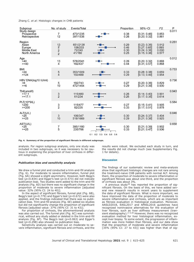
Proportion of significant fibrosis: The results of sub-group analysis and meta-regression of significant fibrosis ratio are shown in Figure 4 and Supplementary Table 6. Similar to the proportion of moderate to severe inflamma-tion, the proportion of significant fibrosis in prospective studies was higher than that in retrospective studies, and the difference was statistically significant [38% (95% CI: 31 to 46) vs. 26% (95% CI: 20 to 32), p=0.011]. The re-sult by meta-regression was also significant (p=0.013). The proportion of significant fibrosis in people >40 years-old [44% (95% CI: 31 to 57)] was almost twice as high as that in people <40 years-old [26% (95% CI: 20 to 32)]. There were significant differences in subgroup analysis (p=0.012) and meta regression (p=0.009). The remaining seven sub-groups (region, BMI, HBV DNA, Tbil, PLT, ALT, and AST) were also analyzed, and no statistical difference was found in either subgroup analysis or meta-regression.
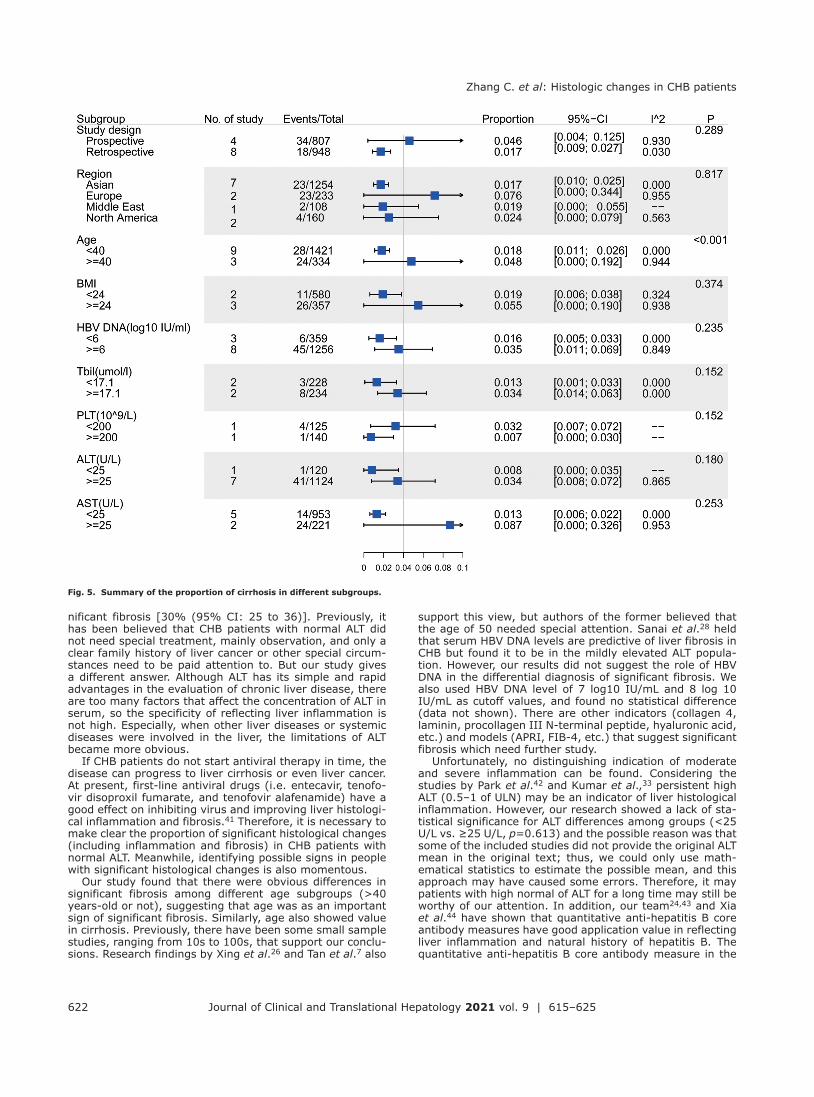
Proportion of liver cirrhosis: Figure 5 and Supple-mentary Table 6 show the proportion of liver cirrhosis. In the subgroup analysis, only the factor of age (<40 years or ≥40 years) showed statistically significant difference [1.8% (95% CI: 1.1 to 2.6) vs. 4.8% (95% CI: 0 to 19.2), p<0.001]. No statistical difference was found in the other nine subgroups (study design, region, BMI, HBV DNA, Tbil, PLT, ALT, and AST). However, there was statistical signifi-cance in AST and region by meta-regression, probably be-cause the range of 95% CI in subgroups with AST was so large that there was no statistical difference in subgroup
Fig. 3. Summary of the proportion of moderate to severe inflammation in different subgroups.
Journal of Clinical and Translational Hepatology 2021 vol. 9 | 615–625 621
Zhang C. et al: Histologic changes in CHB patients
analysis. For region subgroup analysis, only one study was included in two subgroups, so it was necessary to be cau-tious in explaining the proportion of liver cirrhosis in differ-ent subgroups.
Publication bias and sensitivity analysis
We drew a funnel plot and conducted a trim-and-fill analysis (Fig. 6). For moderate to severe inflammation, funnel plot (Fig. 6A) showed a slight asymmetry. However, both Begg’s test (p=0.834) and Egger’s test (p=0.573) did not indicate publication bias. Two studies were added to the trim-and-fill analysis (Fig. 6D) but there was no significant change in the proportion of moderate to severe inflammation [adjusted value: 32% (95% CI: 24 to 40)].
In the aspect of significant fibrosis, funnel plot (Fig. 6B), Begg’s test (p=0.779) and Egger’s test (p=0.672) were also applied, and the findings indicated that there was no publi-cation bias. Trim-and-fill analysis (Fig. 6E) added six studies but did not significantly change the proportion of significant fibrosis [adjusted value: 37% (95% CI: 31 to 43)].
For proportion of cirrhosis, the aforementioned analysis was also carried out. The funnel plot (Fig. 6C) was symmet-rical, without any study added or deleted in the trim-and-fill analysis (Fig. 6F). The Begg’s test (p=0.063) and Egger’s test (p=0.298) also showed no publication bias.
Sensitivity analysis was carried out on moderate to se-vere inflammation, significant fibrosis and cirrhosis, and the
results were robust. We excluded each study in turn, and the results did not change much (see Supplementary Fig. 1 for details).
Discussion
The findings of our systematic review and meta-analysis show that significant histologic changes are not rare among the treatment-naive CHB patients with normal ALT. Among them, the proportion of moderate to severe inflammation or significant fibrosis was about one-third, and the proportion of cirrhosis was about 3%.
A previous study39 has reported the proportion of sig-nificant fibrosis. On the basis of this, we have added sev-eral new research results in recent years to supplement the data of significant fibrosis. What is more important, we have improved the data of the proportion of moderate to severe inflammation and cirrhosis, which are as important as fibrosis evaluation in histological evaluation. Moreover, AASLD2018, EASL2017 and APASL2016 guidelines have recognized noninvasive alternatives for the evaluation of liver fibrosis, such as liver stiffness measurement (tran-sient elastography).2–4,40 However, there was no recognized evaluation method for liver histological inflammation, ex-cept liver biopsy. To some extent, the progression of inflam-mation was more hidden than fibrosis, and our data show that the proportion of moderate and severe inflammation [35% (95% CI: 27 to 43)] was higher than that of sig-
Fig. 4. Summary of the proportion of significant fibrosis in different subgroups.
Journal of Clinical and Translational Hepatology 2021 vol. 9 | 615–625622
Zhang C. et al: Histologic changes in CHB patients
nificant fibrosis [30% (95% CI: 25 to 36)]. Previously, it has been believed that CHB patients with normal ALT did not need special treatment, mainly observation, and only a clear family history of liver cancer or other special circum-stances need to be paid attention to. But our study gives a different answer. Although ALT has its simple and rapid advantages in the evaluation of chronic liver disease, there are too many factors that affect the concentration of ALT in serum, so the specificity of reflecting liver inflammation is not high. Especially, when other liver diseases or systemic diseases were involved in the liver, the limitations of ALT became more obvious.
If CHB patients do not start antiviral therapy in time, the disease can progress to liver cirrhosis or even liver cancer. At present, first-line antiviral drugs (i.e. entecavir, tenofo-vir disoproxil fumarate, and tenofovir alafenamide) have a good effect on inhibiting virus and improving liver histologi-cal inflammation and fibrosis.41 Therefore, it is necessary to make clear the proportion of significant histological changes (including inflammation and fibrosis) in CHB patients with normal ALT. Meanwhile, identifying possible signs in people with significant histological changes is also momentous.
Our study found that there were obvious differences in significant fibrosis among different age subgroups (>40 years-old or not), suggesting that age was as an important sign of significant fibrosis. Similarly, age also showed value in cirrhosis. Previously, there have been some small sample studies, ranging from 10s to 100s, that support our conclu-sions. Research findings by Xing et al.26 and Tan et al.7 also
support this view, but authors of the former believed that the age of 50 needed special attention. Sanai et al.28 held that serum HBV DNA levels are predictive of liver fibrosis in CHB but found it to be in the mildly elevated ALT popula-tion. However, our results did not suggest the role of HBV DNA in the differential diagnosis of significant fibrosis. We also used HBV DNA level of 7 log10 IU/mL and 8 log 10 IU/mL as cutoff values, and found no statistical difference (data not shown). There are other indicators (collagen 4, laminin, procollagen III N-terminal peptide, hyaluronic acid, etc.) and models (APRI, FIB-4, etc.) that suggest significant fibrosis which need further study.
Unfortunately, no distinguishing indication of moderate and severe inflammation can be found. Considering the studies by Park et al.42 and Kumar et al.,33 persistent high ALT (0.5–1 of ULN) may be an indicator of liver histological inflammation. However, our research showed a lack of sta-tistical significance for ALT differences among groups (<25 U/L vs. ≥25 U/L, p=0.613) and the possible reason was that some of the included studies did not provide the original ALT mean in the original text; thus, we could only use math-ematical statistics to estimate the possible mean, and this approach may have caused some errors. Therefore, it may patients with high normal of ALT for a long time may still be worthy of our attention. In addition, our team24,43 and Xia et al.44 have shown that quantitative anti-hepatitis B core antibody measures have good application value in reflecting liver inflammation and natural history of hepatitis B. The quantitative anti-hepatitis B core antibody measure in the
Fig. 5. Summary of the proportion of cirrhosis in different subgroups.

Journal of Clinical and Translational Hepatology 2021 vol. 9 | 615–625 623
Zhang C. et al: Histologic changes in CHB patients
immune tolerance stage was significantly lower than that in the immune clearance stage.
Although subgroup analysis and meta-regression were carried out as far as possible, there was still some hetero-geneity implication for outcomes. According to the results of proportion of significant fibrosis subgroup analysis, dif-ferent ALT levels may represent the main source of het-erogeneity (I2: 40.4% vs. 92.5% in ALT <25 U/L and ≥25 U/L, respectively). Several factors can explain the source of heterogeneity in proportion of cirrhosis. Among them, pro-spective studies had greater heterogeneity than regression
studies (I2: 93.0% vs. 3.0%), and older age had greater heterogeneity than younger age (I2: 94.4% vs. 0.0%). Un-fortunately, the source of moderate and severe inflamma-tory heterogeneity has not been found. We speculate that the first reason may be that there was no recognized value for the normal upper limit of ALT, which was considered by the APASL and EASL guidelines as 40 U/L but by the AASLD guidelines as 35 U/L for male and 25 U/L for female. Sec-ond, compared with the pathological evaluation of fibrosis, the evaluation of inflammation was more easily affected by the scoring system and pathologists, especially upon the
Fig. 6. Funnel plot and trim-and-fill analysis plot. (A) Funnel plot of the proportion of moderate to severe inflammation. (B) Funnel plot of the proportion of sig-nificant fibrosis. (C) Funnel plot of the proportion of cirrhosis. (D) Trim-and-fill plot of the proportion of moderate to severe inflammation (two studies were added, as shown by the red points in the figure). (E) Trim-and-fill plot of the proportion of significant fibrosis (six studies were added, as shown by the red points in the figure). (F) Trim-and-fill plot of the proportion of cirrhosis (no studies were added).

Journal of Clinical and Translational Hepatology 2021 vol. 9 | 615–625624
Zhang C. et al: Histologic changes in CHB patients
application of Ishak and Knodell scoring systems, as the items were too detailed to form a unified consensus.
Our study has several limitations. First, there may be a patient selection bias in this study. For the CHB patients with normal ALT, both the patients and doctors were reluc-tant to carry out invasive liver biopsy due to its inherent risks, which reduced the implementation of liver biopsy to a certain extent. Therefore, the proportion of significant his-tological changes may be higher in actuality than this study found. Second, there were non-randomized controlled trials among the included studies. Although the results of publica-tion bias were negative, their inclusion inevitably reduced the overall quality of the study.
Conclusions
In summary, significant histologic changes present in ap-proximately one-third of treatment-naive CHB patients with normal ALT levels, and about 3% of patients even pro-gressed to cirrhosis. It is worth noting that the proportion of significant fibrosis and cirrhosis in people >40 years-old are more than twice as high as those in younger people. The management of treatment-naive CHB patients with normal ALT remains a challenge and requires an individualized ap-proach, in addition to the standardized paradigms recom-mended by current guidelines.
Funding
This study was supported by the China Mega-Project for Infectious Diseases (Grant Nos. 2017ZX10203202 and 2013ZX10002005) and the China Mega-Project for Innova-tive Drugs (Grant No. 2016ZX09101065).
Conflict of interest
The authors have no conflict of interests related to this pub-lication.
Author contributions
Search of the literature and data extraction (CZ, ZW), draft-ed the manuscript (CZ), creation of figures and table (CZ, ZW, JWL), methodological guidance (HZ), and provision of the overall principle and oversight of the direction of the study (HZ, GQW).
Data sharing statement
All data are available upon request.
References
[1] Schweitzer A, Horn J, Mikolajczyk RT, Krause G, Ott JJ. Estimations of worldwide prevalence of chronic hepatitis B virus infection: a system-atic review of data published between 1965 and 2013. Lancet 2015; 386(10003):1546–1555. doi:10.1016/S0140-6736(15)61412-X.
[2] Terrault NA, Lok ASF, McMahon BJ, Chang KM, Hwang JP, Jonas MM, et al. Update on prevention, diagnosis, and treatment of chronic hepatitis B: AASLD 2018 hepatitis B guidance. Hepatology 2018;67(4):1560–1599. doi:10.1002/hep.29800.
[3] EASL 2017 Clinical Practice Guidelines on the management of hepatitis B virus infection. J Hepatol 2017;67(2):370–398. doi:10.1016/j.jhep.2017. 03.021.
[4] Sarin SK, Kumar M, Lau GK, Abbas Z, Chan HL, Chen CJ, et al. Asian-Pacific
clinical practice guidelines on the management of hepatitis B: a 2015 up-date. Hepatol Int 2016;10(1):1–98. doi:10.1007/s12072-015-9675-4.
[5] Liao B, Wang Z, Lin S, Xu Y, Yi J, Xu M, et al. Significant fibrosis is not rare in Chinese chronic hepatitis B patients with persistent normal ALT. PLoS One 2013;8(10):e78672. doi:10.1371/journal.pone.0078672.
[6] Tan YW, Zhou XB, Ye Y, He C, Ge GH. Diagnostic value of FIB-4, aspar-tate aminotransferase-to-platelet ratio index and liver stiffness measure-ment in hepatitis B virus-infected patients with persistently normal ala-nine aminotransferase. World J Gastroenterol 2017;23(31):5746–5754. doi:10.3748/wjg.v23.i31.5746.
[7] Tan Y, Ye Y, Zhou X, Chen L, Wen D. Age as a predictor of significant fibrosis features in HBeAg-negative chronic hepatitis B virus infection with persis-tently normal alanine aminotransferase. PLoS One 2015;10(4):e0123452. doi:10.1371/journal.pone.0123452.
[8] Papatheodoridis GV, Manesis EK, Manolakopoulos S, Elefsiniotis IS, Goulis J, Giannousis J, et al. Is there a meaningful serum hepatitis B virus DNA cutoff level for therapeutic decisions in hepatitis B e antigen-negative chronic hepatitis B virus infection? Hepatology 2008;48(5):1451–1459. doi:10.1002/hep.22518.
[9] Zhou K, Dodge JL, Grab J, Poltavskiy E, Terrault NA. Mortality in adults with chronic hepatitis B infection in the United States: a population-based study. Aliment Pharmacol Ther 2020;52(2):382–389. doi:10.1111/apt.15803.
[10] Liberati A, Altman DG, Tetzlaff J, Mulrow C, Gøtzsche PC, Ioannidis JP, et al. The PRISMA statement for reporting systematic reviews and meta-anal-yses of studies that evaluate health care interventions: explanation and elaboration. Ann Intern Med 2009;151(4):W65–W94. doi:10.7326/0003-4819-151-4-200908180-00136.
[11] Hozo SP, Djulbegovic B, Hozo I. Estimating the mean and variance from the median, range, and the size of a sample. BMC Med Res Methodol 2005;5:13. doi:10.1186/1471-2288-5-13.
[12] Luo D, Wan X, Liu J, Tong T. Optimally estimating the sample mean from the sample size, median, mid-range, and/or mid-quartile range. Stat Meth-ods Med Res 2018;27(6):1785–1805. doi:10.1177/0962280216669183.
[13] Wan X, Wang W, Liu J, Tong T. Estimating the sample mean and standard deviation from the sample size, median, range and/or interquartile range. BMC Med Res Methodol 2014;14:135. doi:10.1186/1471-2288-14-135.
[14] Desmet VJ, Gerber M, Hoofnagle JH, Manns M, Scheuer PJ. Classification of chronic hepatitis: diagnosis, grading and staging. Hepatology 1994; 19(6):1513–1520. doi:10.1002/hep.1840190629.
[15] Ishak K, Baptista A, Bianchi L, Callea F, De Groote J, Gudat F, et al. Histo-logical grading and staging of chronic hepatitis. J Hepatol 1995;22(6):696–699. doi:10.1016/0168-8278(95)80226-6.
[16] Knodell RG, Ishak KG, Black WC, Chen TS, Craig R, Kaplowitz N, et al. Formulation and application of a numerical scoring system for assessing histological activity in asymptomatic chronic active hepatitis. Hepatology 1981;1(5):431–435. doi:10.1002/hep.1840010511.
[17] Bedossa P, Poynard T. An algorithm for the grading of activity in chronic hepatitis C. The METAVIR Cooperative Study Group. Hepatology 1996; 24(2):289–293. doi:10.1002/hep.510240201.
[18] Goodman ZD. Grading and staging systems for inflammation and fibrosis in chronic liver diseases. J Hepatol 2007;47(4):598–607. doi:10.1016/j.jhep.2007.07.006.
[19] Wells G, Shea B, O’Connell D, Robertson J, Peterson J, Welch V, et al. The New-castle-Ottawa Scale (NOS) for assessing the quality of nonrandomised studies in meta-analyses. Available from: http://www3.med.unipmn.it/dispense_ebm/2009-2010/Corso%20Perfezionamento%20EBM_Faggiano/NOS_ oxford.pdf.
[20] Barendregt JJ, Doi SA, Lee YY, Norman RE, Vos T. Meta-analysis of preva-lence. J Epidemiol Community Health 2013;67(11):974–978. doi:10.1136/jech-2013-203104.
[21] Higgins JP, Thompson SG, Deeks JJ, Altman DG. Measuring inconsist-ency in meta-analyses. BMJ 2003;327(7414):557–560. doi:10.1136/bmj. 327.7414.557.
[22] Melsen WG, Bootsma MC, Rovers MM, Bonten MJ. The effects of clinical and statistical heterogeneity on the predictive values of results from meta-analyses. Clin Microbiol Infect 2014;20(2):123–129. doi:10.1111/1469-0691.12494.
[23] Duval S, Tweedie R. Trim and fill: A simple funnel-plot-based method of testing and adjusting for publication bias in meta-analysis. Biometrics 2000;56(2):455–463. doi:10.1111/j.0006-341x.2000.00455.x.
[24] Zhou J, Song L, Zhao H, Yan L, Ma A, Xie S, et al. Serum hepatitis B core antibody as a biomarker of hepatic inflammation in chronic hepatitis B patients with normal alanine aminotransferase. Sci Rep 2017;7(1):2747. doi:10.1038/s41598-017-03102-3.
[25] Xu Z, Shen J, Pan X, Wei M, Liu L, Wei K, et al. Predictive value of serum Golgi protein 73 for prominent hepatic necroinflammation in chronic HBV infection. J Med Virol 2018;90(6):1053–1062. doi:10.1002/jmv.25045.
[26] Xing YF, Zhou DQ, He JS, Wei CS, Zhong WC, Han ZY, et al. Clinical and histopathological features of chronic hepatitis B virus infected patients with high HBV-DNA viral load and normal alanine aminotransferase level: A multicentre-based study in China. PLoS One 2018;13(9):e0203220. doi:10.1371/journal.pone.0203220.
[27] Wan R, Liu H, Wang X, Wan G, Wang X, Zhou G, et al. Noninvasive predic-tive models of liver fibrosis in patients with chronic hepatitis B. Int J Clin Exp Med 2015;8(1):961–971.
[28] Sanai FM, Helmy A, Bzeizi KI, Babatin MA, Al-Qahtani A, Al-Ashgar HA, et al. Discriminant value of serum HBV DNA levels as predictors of liv-er fibrosis in chronic hepatitis B. J Viral Hepat 2011;18(7):e217–e225. doi:10.1111/j.1365-2893.2011.01437.x.
[29] Ormeci A, Aydın Y, Sumnu A, Baran B, Soyer OM, Pınarbasi B, et al. Pre-dictors of treatment requirement in HBeAg-negative chronic hepatitis B
Journal of Clinical and Translational Hepatology 2021 vol. 9 | 615–625 625
Zhang C. et al: Histologic changes in CHB patients
patients with persistently normal alanine aminotransferase and high se-rum HBV DNA levels. Int J Infect Dis 2016;52:68–73. doi:10.1016/j.ijid. 2016.09.007.
[30] Nguyen MH, Garcia RT, Trinh HN, Lam KD, Weiss G, Nguyen HA, et al. His-tological disease in Asian-Americans with chronic hepatitis B, high hepatitis B virus DNA, and normal alanine aminotransferase levels. Am J Gastroen-terol 2009;104(9):2206–2213. doi:10.1038/ajg.2009.248.
[31] Lesmana CR, Gani RA, Hasan I, Simadibrata M, Sulaiman AS, Pakasi LS, et al. Significant hepatic histopathology in chronic hepatitis B patients with serum ALT less than twice ULN and high HBV-DNA levels in Indonesia. J Dig Dis 2011;12(6):476–480. doi:10.1111/j.1751-2980.2011.00540.x.
[32] Lai M, Hyatt BJ, Nasser I, Curry M, Afdhal NH. The clinical significance of persistently normal ALT in chronic hepatitis B infection. J Hepatol 2007;47(6):760–767. doi:10.1016/j.jhep.2007.07.022.
[33] Kumar M, Sarin SK, Hissar S, Pande C, Sakhuja P, Sharma BC, et al. Viro-logic and histologic features of chronic hepatitis B virus-infected asymp-tomatic patients with persistently normal ALT. Gastroenterology 2008; 134(5):1376–1384. doi:10.1053/j.gastro.2008.02.075.
[34] Gui HL, Wang H, Yang YH, Wu YW, Zhou HJ, Guo SM, et al. Significant his-topathology in Chinese chronic hepatitis B patients with persistently high-normal alanine aminotransferase. J Viral Hepat 2010;17(Suppl 1):44–50. doi:10.1111/j.1365-2893.2010.01270.x.
[35] Gong X, Yang J, Tang J, Gu C, Huang L, Zheng Y, et al. A mechanistic as-sessment of the discordance between normal serum alanine aminotrans-ferase levels and altered liver histology in chronic hepatitis B. PLoS One 2015;10(7):e0134532. doi:10.1371/journal.pone.0134532.
[36] Chen EQ, Huang FJ, He LL, Bai L, Wang LC, Zhou TY, et al. Histological chang-es in chinese chronic hepatitis B patients with ALT lower than two times upper limits of normal. Dig Dis Sci 2010;55(2):432–437. doi:10.1007/ s10620-009-0724-5.
[37] Alam S, Ahmad N, Mustafa G, Shrestha A, Alam AK, Khan M. Evaluation of normal or minimally elevated alanine transaminase, age and DNA lev-el in predicting liver histological changes in chronic hepatitis B. Liver Int 2011;31(6):824–830. doi:10.1111/j.1478-3231.2011.02491.x.
[38] Montazeri G, Rahban M, Mohamadnejad M, Zamani F, Hooshyar A, Fazlolahi A, et al. Liver histology and HBV DNA levels in chronically HBV infected patients with persistently normal alanine aminotransferase. Arch Iran Med 2010;13(3):193–202.
[39] Chao DT, Lim JK, Ayoub WS, Nguyen LH, Nguyen MH. Systematic review with meta-analysis: the proportion of chronic hepatitis B patients with nor-mal alanine transaminase ≤ 40 IU/L and significant hepatic fibrosis. Ali-ment Pharmacol Ther 2014;39(4):349–358. doi:10.1111/apt.12590.
[40] Li Y, Huang YS, Wang ZZ, Yang ZR, Sun F, Zhan SY, et al. Systematic review with meta-analysis: the diagnostic accuracy of transient elastography for the staging of liver fibrosis in patients with chronic hepatitis B. Aliment Pharmacol Ther 2016;43(4):458–469. doi:10.1111/apt.13488.
[41] Tang LSY, Covert E, Wilson E, Kottilil S. Chronic hepatitis B infection: A review. JAMA 2018;319(17):1802–1813. doi:10.1001/jama.2018.3795.
[42] Park JY, Park YN, Kim DY, Paik YH, Lee KS, Moon BS, et al. High preva-lence of significant histology in asymptomatic chronic hepatitis B pa-tients with genotype C and high serum HBV DNA levels. J Viral Hepat 2008;15(8):615–621. doi:10.1111/j.1365-2893.2008.00989.x.
[43] Jia W, Song LW, Fang YQ, Wu XF, Liu DY, Xu C, et al. Antibody to hepa-titis B core antigen levels in the natural history of chronic hepatitis B: a prospective observational study. Medicine (Baltimore) 2014;93(29):e322. doi:10.1097/MD.0000000000000322.
[44] Song LW, Liu PG, Liu CJ, Zhang TY, Cheng XD, Wu HL, et al. Quantita-tive hepatitis B core antibody levels in the natural history of hepatitis B virus infection. Clin Microbiol Infect 2015;21(2):197–203. doi:10.1016/j.cmi.2014.10.002.