Embed Size (px)
Citation preview
Signs and Symptoms of Parasitic Diseases

Abdominal pain
• Entamoeba histolytica (amebic colitis, liver infection)
• Giardia lamblia• Cryptosporidium• Intestinal helminths (Ascaris, Strongyloides)• Anisakis (symptoms suggestive of gustric or
duodenal ulcer or appendicitis)

Abscess, Amebic (liver)Abscess, Amebic (liver)
• Entamoeba histolytica
Abscess, FilarialAbscess, Filarial
• occur along the course of lymphatics or at lymph nodes (generally sterile)

Anemia• Malaria (Plasmodium spp.)2.5 – 4.0 mil/mm3 – average
severity<1 mil/mm3 – severe infections)
• Kala-azar
• Ancylostoma duodenale (microcytic hypochromic)
• Diphyllobothrium latum (macrocytic hyperchromic)
• Trichuris trichiura

Appendicitis
• Ascaris lumbricoides• Trichuris trichiura• Enterobius
vermicularis
Ascites
• Schistosoma spp. (fibrosis of the liver)• Kala-azara (chronic
inf.)

Asthma, Bronchial• Ascaris – visceral migration• VLM
Calabar Swellings• Loa loa – circumscribed subcutaneous swellings (they are pruritic and may be quite painful)

Calcifications, Cerebral
• Congenital toxoplasmosis
• Cysticercosis
Chagoma
• Trypanosoma cruzi

Chyluria
• Lymphatic filariasisWuchereria bancroftiBrugia spp.
Coma
• P. falciparum• Sleeping sickness• PAM• Cysticercosis

Conjunctivitis
• Ochocerca volvulus• Miyasis
Convulsions
• Schistosoma, Trichinella, Echinococcus, Cysticercus
• Malaria• Acute toxoplasmosis• Ascaris infection in
children

Dermatitis• Leishmania spp.• Schistosomiasis• Strongyloides stercoralis• Onchocerca volvulus• KLM (Ancylostoma spp.)• Sarcoptes scabiei & other mites• Miyasis• Pediculosis

Diarrhea
• Entamoeba histolytica• Balantidium coli• Giardia lamblia• Intestinal coccidia(Isospora, Cryptospridium,
Cyclospora)
• Microsporidia• Blastocystis hominis• Intestinal helminths• Trichinella spiralis
Dysentery
• Entamoeba histolytica• Balantidium coli
• Kala-azar• Falciparum malaria• Strongyloidiasis• Schistosomiasis

Edema
• LocalizedLoa loaTrypanosoma cruzi
(Romaña sign)
• Other types Sleeping sicknessMalnutrition caused by
intestinal helminths (Ancylostoma,
Fasciolopsis)

Elephantiasis
• Filariae (Wuchereria bancrofti, Brugia timori, Brugia malayi)
Epididymitis, funiculitis, orchitis
• Filariae

FEVER
• Malaria• Kala-azar• Amebic hepatitis, fascioliasis, clonorchiasis, opistorchiasis• Schistosomiasis• African and american trypanosomiasis• Trichinellosis• Filariasis

Hematuria
• Schistosoma haematobium
Hemoglobinuria
• Malaria (blackwater fever
/P. falciparum)

Hepatomegaly
• Amebic hepatitis• Falciparum malaria• Kala-azar• VLM (Toxocara)• Infections caused by
liver flukes, including schistosomiasis
Splenomegaly
• Trypanosomiasis• Kala-azar• Malaria• Schistosomiasis• Tropical splenomegaly
syndrome (in areas where malaria, schistosomiasis and kala-azar are common)

Hydrocephalus i microcephalus
• Congenital T. gondii infection
• Cysticercosis
Hyperpigmentation
• Kala-azar• Onchocerca volvulus• Pediculosis (chronic
infection with body louse)

Kerandel’s sign
• Sleeping sickness
(may be elicted by pressure on the palm of the hand or over the ulnar nerve and consists of severe pain that occurs shortly after the pressure has been relieved)
Keratitis
• Onchocerca volvulus• Acanthamoeba
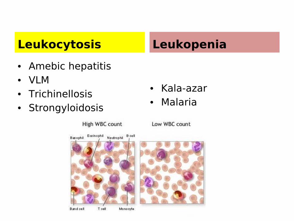
Leukocytosis
• Amebic hepatitis • VLM• Trichinellosis• Strongyloidosis
Leukopenia
• Kala-azar • Malaria

Lymphadenitis/lymphangitis
• Filariae• T. gondii• Sleeping sickness• Chagas’ disease
Lymphocytosis
• Chagas’ disease• Sleeping sickness

Melena
• Strongyloides
Meningoencephalitis
• Sleeping sickness• Chagas’ disease• Malaria• PAM• GAM• Balamuthia
mandrillaris• Angiostrongylus
cantonensis

Myocarditis
• Chagas’ disease• Sleeping sickness (T. b. rhodesiense)• Trichinellosis• Acute T. gondii
infection
Myositis
• Trichinellosis• Sarcocystis

Subcutaneous nodules
• Onchocerca volvulus (“lipoma like”)• Cysticercosis• Echinococcus granulosus• Sparganosis• Miyasis• Dirofilaria spp. (D. repens in Europe)

Intestinal obstruction/Peritonitis
• Ascaris lumbricoides• Angyostrongylus
costaricensis
Pneumonitis
• Ascaris lumbricoides• Strongyloides
stercoralis• Ancylostoma
duodenale• Schistosomiasis

Pruritus ani
• Enterobius vermicularis
• Taenia saginata
Retinochoroiditis
• Toxoplasma gondii• Onchocerca volvulus• Toxocara• Angiostrongylus
cantonensis

Urethritis /Vaginitis
• Trichomonas vaginalis
Proteinuria
• Malaria

Eosinophilia and Parasitic Infections
• Eosinophils (Eo) are type of polymorphonuclear leukocytes found in human blood and tissue.
• They have large intracytoplasmic granules that stain red with the dye eosin.
• Eo originate in the bone marrow and circulate in blood before entering the tissues – skin, GI tract, and lungs are major sites of eosinophil migration.
• The surface of the Eo has receptors for IgG, IgE, and complement.
• Elevation of the Eo count above the normal range is called eosinophilia.
• Clinicians frequently conclude that a significant eosinophilia is present when the eosinophils exceed 5% of the total WBCs (normal range 2-4%).

• Within the Eo granule toxic proteins (major basic protein, neurotoxin) are found. They are toxic for larval helminth parasites.
• Eo bind to certain parasites and following adherence, the Eo degranulates, and the cytotoxic effects are observed.
• The highest levels of eosinophilia occur in helminth infections in which larvae migrate in the tissues. Moderate to high levels of eosinophilia occur in the early stages of intestinal nematode infections that have an extraintestinal migration phase, filarial infections, and infections with blood stream or tissue dwelling trematodes. Higher levels of eosinophilia are seen in the early stages of infection.
• The only occurence of significant eosinophilia with protozoan infections is seen with Isospora belli.