Embed Size (px)
Citation preview

SIIM 2007 Hot Topic 7 DICOM Update
David Clunie CTO, RadPharm, Inc.

The Medicine Behind the Image
Overview
New objects & services (David Clunie)
Network Configuration (Rob Horn)
Application Hosting (Lawrence Tarbox)

The Medicine Behind the Image
DICOM Update More enhanced & new technology image objects Additional dose encoding objects More SR-based results & CAD objects More 3D work on registration & segmentation Structured display Communication of display parameters Document encapsulation Specimen identification Substance administration query/verify Unified worklist Frame level retrieval

The Medicine Behind the Image
Enhanced Image Objects Initially MR, then CT, then XRA/RF Sup 43 (WIP) - 3D Ultrasound Sup 110 (LB) - Ophthalmic Tomography Sup 116 (FT 2007/01) - 3D X-Ray
• Cone beam CT & tomosynthesis • General purpose & dentistry
Sup 117 (DLB) - Enhanced PET • Harmonize cardiac/respiratory gating with CT/MR
Sup 125 (PC) - Breast Tomosynthesis

The Medicine Behind the Image
Enhanced Image Objects “Old” objects
• Single frame • Not up to date with technology changes (MDCT) • Too much optional, ambiguous, or proprietary
“New” (enhanced) objects • Multi-frame (faster performance, better compression) • Better organized (volumes, dynamic contrast) • Encode advanced acquisition technique • Mandatory rather than optional terms & attributes

The Medicine Behind the Image
Dose Encoding Increasing international public and regulatory
scrutiny of radiation dose from imaging Existing encoding in images & PPS inadequate Need persistent object related to irradiation
events SR-based encoding Sup 94 (FT 2005/11) - Radiation Dose Report Sup 127 (PC) - CT Radiation Dose Report CP 687 - Dose Reporting for Mammography

The Medicine Behind the Image
CT Radiation Dose Reporting Significant concern about radiation dose of screening
MDCT exams Difficult to estimate/monitor from images alone Acquire, store and analyze information about
“irradiation events” separately from images IEC defines metrics DICOM defines encoding in Sup 127 (as SR objects) ACR and FDA “encourage” adoption NEMA (vendors) commit to timely implementation

The Medicine Behind the Image
Results SR and CAD
CAD • Sup 126 (WIP) - Colonoscopy CAD
Results reporting • Sup 128 (PC) - Cardiac stress testing • Sup 129 (WIP) - Electrophysiology • Sup 130 (WIP) - Ophthalmic refraction

The Medicine Behind the Image
3D-related Objects
Registration • Sup 73 (FT 2004/01) - Rigid & Fiducials • Sup 112 (FT 2006/08) - Deformable
Segmentation • Sup 111 (FT 2006/08) - Raster • Sup 132 (WIP) - Surface
• Sup 131 (WIP) - Implant Description

The Medicine Behind the Image
Display & Presentation
Sup 123 (WIP) - Structured Display • How to layout specific images • As opposed to hanging protocols, which are
rules for a class of images • Dentistry initiative, general mechanism
Sup 124 (WIP) - Communication of Display Parameters • For managing display device calibration • Centralized storage of QC results
The Medicine Behind the Image
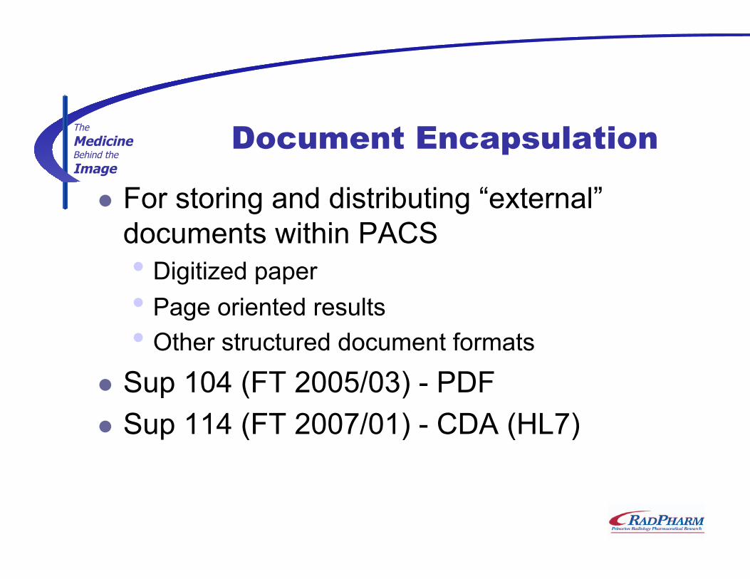
Document Encapsulation
For storing and distributing “external” documents within PACS • Digitized paper • Page oriented results • Other structured document formats
Sup 104 (FT 2005/03) - PDF Sup 114 (FT 2007/01) - CDA (HL7)

The Medicine Behind the Image
Integration of Images and LIS in Anatomic Pathology
In the normal clinical environment, an image can be associated with a Part, a Block or a Slide
In some situations, an image can be further associated with an area of a Slide, for example, one can specify an x,y,z location on a slide (see coordinate microscopy IOD)
One can always image a small region of a gross specimen. This would be associated with a Part and with a comment describing the field (i.e. “tumor ”)
One could imagine an image of material from two Parts in the clinical environment, this image would probably be associated with the Accession.
Patient
Operation
Accession
A patient may have 0 or more operations
Part
Block
Slide
Image
Workflow objects in LIS
Location within Slide
Data in Imaging System
Specimen Identification
Sup 122 (WIP) - Specimen Identification Renewed interest by pathology group Original attempt was too simplistic

The Medicine Behind the Image
Other work …
Substance administration query/verify • E.g., for modality to check contrast sensitivity
Unified worklist • Re-visit use cases for General Purpose Worklist • 1:1 scheduled:performed steps • Push (notify) & pull (query) models for tasks
Frame level retrieval • For large (enhanced) multi-frame images • E.g., to view an SR reference to a subset of frames

The Medicine Behind the Image
Conclusion
DICOM continues to track modality technology advances
Revisiting outmoded objects Increasing diversity of SR for results Greater 3D emphasis as registration,
segmentation and fusion become routine Other innovative work in new areas …