Embed Size (px)
Citation preview

Virology 421 (2011) 129–140
Contents lists available at SciVerse ScienceDirect
Virology
j ourna l homepage: www.e lsev ie r .com/ locate /yv i ro
Simian hemorrhagic fever virus infection of rhesus macaques as a model of viralhemorrhagic fever: Clinical characterization and risk factors for severe disease
Reed F. Johnson a,⁎, Lori E. Dodd b, Srikanth Yellayi c, Wenjuan Gu d, Jennifer A. Cann c, Catherine Jett c,John G. Bernbaum c, Dan R. Ragland c, Marisa St. Claire c, Russell Byrum c, Jason Paragas c,Joseph E. Blaney a, Peter B. Jahrling a,c
a Emerging Viral Pathogens Section , National Institute of Allergy and Infectious Diseases, National Institutes of Health, Bethesda, MD 20892, USAb Biostatistics Research Branch, National Institute of Allergy and Infectious Diseases, National Institutes of Health, Frederick, MD 21702, USAc Integrated Research Facility, National Institute of Allergy and Infectious Diseases, National Institutes of Health, Frederick, MD 21702, USAd Biostatistics Research Branch, SAIC-Frederick, Inc., National Cancer Institute NCI-Frederick, Frederick, MD 21702, USA
⁎ Corresponding author at: National Institutes ofHealth33 North Drive Bethesda MD, USA. Fax: +1 301 480 3322.
E-mail address: [email protected] (R.F. John
0042-6822/$ – see front matter. Published by Elsevier Idoi:10.1016/j.virol.2011.09.016
a b s t r a c t
a r t i c l e i n f oArticle history:Received 14 July 2011Returned to author for revision29 August 2011Accepted 13 September 2011Available online 19 October 2011
Keywords:Hemorrhagic fever virusAnimal modelPathogenesisSimian hemorrhagic fever virusArterivirusCoagulopathyEmerging pathogensVirusHemorrhagic disease
Simian Hemorrhagic Fever Virus (SHFV) has caused sporadic outbreaks of hemorrhagic fevers in macaques at pri-mate research facilities. SHFV is a BSL-2 pathogen that has not been linked to human disease; as such, investigationof SHFV pathogenesis in non-human primates (NHPs) could serve as amodel for hemorrhagic fever viruses such asEbola, Marburg, and Lassa viruses. Here we describe the pathogenesis of SHFV in rhesus macaques inoculated withdoses ranging from50 PFU to 500,000 PFU. Disease severitywas independent of dosewith an overall mortality rateof 64%with signs of hemorrhagic fever andmultiple organ system involvement. Analyses comparing survivors andnon-survivorswere performed to identify factors associatedwith survival revealing differences in the kinetics of vi-remia, immunosuppression, and regulation of hemostasis. Notable similarities between the pathogenesis of SHFV inNHPs and hemorrhagic fever viruses in humans suggest that SHFV may serve as a suitable model of BSL-4pathogens.
, NIAID/EVPS, Bldg 33Rm2E19A,
son).
nc.
Published by Elsevier Inc.
Introduction
The causative agents of viral hemorrhagic fevers (VHF) that affecthumans are RNA viruses from the families Filoviridae, Arenaviridae,Bunyaviridae, and Flaviviridae including Ebola, Marburg, Lassa, Rift ValleyFever, Crimean–Congo Hemorrhagic Fever, and Omsk HemorrhagicFever viruses (Feldmann and Geisbert, 2011; Keshtkar-Jahromi et al.,2011; Paragas and Geisbert, 2006; Peters et al., 1989; Ruzek et al.,2010). Because of the extrememorbidity associatedwith these emergingviruses and the concern that one or more may be used as bioterrorismagents, efforts to further our understanding of disease pathogenesisand to identify countermeasures have intensified.While numerous stud-ies have defined the clinical, virological, immunological, and pathologicalmanifestations of hemorrhagic fever viruses using non-human primate(NHP) models (Geisbert et al., 2003a; Jaax et al., 1995; Johnson et al.,
1995; Paragas and Geisbert, 2006; Peters et al., 1989), the viral andhost molecular mechanisms that control disease severity and outcomeremain largely unknown. Furthermore, no licensed therapeutic treat-ments exist for any VHF. A better understanding of the mechanisms as-sociated with VHF outcome would facilitate the investigation oftherapeutic agents. Identification of broad-spectrum treatments target-ing common viral or host factors is most desirable because the develop-ment of individual therapies for each VHF is hindered by the sporadicnature of the outbreaks and the limited commercial viability of suchproducts.
The necessity for high containment laboratories, for instance, bio-safety level- (BSL-) 3 or 4, complicates the investigation of these VHFpathogens. Alternatively, a virus that produces similar disease inNHPs that can be studied under BSL-2 conditions would facilitatestudies of VHF viruses by virtue of broader access to the scientificcommunity. SHFV in NHPs might serve as an ideal model for humanviral hemorrhagic fevers because SHFV 1) has never been associatedwith human disease, 2) is a biosafety level BSL-2 pathogen, and 3)has clinical manifestations similar to other hemorrhagic fever viruses.
SHFV is an arterivirus that was first identified in 1964 as the causa-tive agent during an outbreak of hemorrhagic disease in Asian origin

Table 1Survival, viremia, and incidence of secondary bacterial infection by dose.
Dose(PFU)
% Moribund(no. moribund/total no.)
Mean day ofmoribundendpoint(range)
Mean peakviremia
Proportion ofnon-survivorsthat developedbacteremiaa
(log10PFU/ml)(range)
50 66 (2/3) 10.5 (5–16) 5.9 (0.0–6.1) 1/2500 66 (2/3) 17.0 (15–19) 5.8 (4.7–6.2) 2/25000 66 (4/6) 13.5 (9–16) 5.7 (2.8–6.4) 2/450,000 75 (6/8) 9.3 (9–16) 5.5 (2.8–6.1) 5/6500,000 40 (2/5) 12.0 (8–16) 4.7 (2.5–5.0) 2/2
a Bacteremia was defined as positive by blood culture, presence of abscesses at nec-ropsy, or presence of bacteria in multiple organs during histopathological examination.
130 R.F. Johnson et al. / Virology 421 (2011) 129–140
macaques that occurred at both the National Institutes of Health (NIH,Bethesda, MD) (Allen et al., 1968; Palmer et al., 1968; Tauraso et al.,1968) and the Sukhumi Institute of Experimental Pathology and Thera-py in the former USSR (Lapin and Shevtsova, 1971; Shevtsova, 1969b;Shevtsova and Krylova, 1971b). Macaques from both institutes were ac-quired from the same region of India andhousedwith African origin pri-mates including patas monkeys, baboons, and African green monkeys(Palmer et al., 1968; Shevtsova, 1969b). During the Sukhumi outbreak,the case fatality rate was 100% over 2 months (Lapin and Shevtsova,1971; Shevtosova et al., 1975) with disease presenting as a hemorrhagicdiathesis and acute diffuse encephalomyelitis (Shevtsova and Krylova,1971b). During theNIH outbreak, the route of transmissionwas thoughtto be iatrogenic: needles that were used for tattooing and tuberculosistesting were shared between the African origin primates and themacaques (Allen et al., 1968; Palmer et al., 1968; Tauraso et al., 1968).Macaques developed high fevers and hemorrhagic diathesis but notacute diffuse encephalomyelitis that was observed at Sukhumi (Allenet al., 1968; Shevtsova and Krylova, 1971a). Mortality occurred in 233of 1029macaques in affected rooms over a 2 month period. Initial char-acterization suggested that all infected NHPs succumbed to disease.However, follow up experiments indicated that macaques can developasymptomatic infection. Specifically, blood and tissue from an asymp-tomatic survivor successfully induced a viral hemorrhagic fever inmacaques not associated with the initial outbreak (Palmer et al., 1968).
Sporadic SHFV outbreaks of iatrogenic origin have occurred since1964 with mortality rates reported varying from 11% to as high as100% (Gravell et al., 1986; London, 1977; Palmer et al., 1968; Taurasoet al., 1970). During SHFV outbreaks in 1972 and 1989 the virus wasthought to be spread by both direct and indirect contact betweenmacaques (London, 1977; Renquist, 1990). In the 1989 Ebola-Restonoutbreak, SHFV was found in 19 of 49 Ebola-Reston positive macaquesthat succumbed to hemorrhagic fever (Dalgard et al., 1992).
Analysis of SHFV outbreaks and limited experimental infection of ma-caques identified common clinical signs including fever, mild facial ery-thema, and edema as early as 48–72 h post-infection (Abildgaard et al.,1975; Gravell et al., 1986; London, 1977; Palmer et al., 1968). Clinicalsigns indicative of initial infection developedwithin 72 h post-inoculationand included depression and petechial rash (Palmer et al., 1968). As thedisease progressed,macaques developed facial edema, cyanosis, anorexia,adipsia, epistaxis, emesis, dehydration, melena, hematomata, retrobulbarhemorrhage and hematologic signs of coagulopathy (Abildgaard et al.,1975; Allen et al., 1968; Gravell et al., 1986; London, 1977; Palmer et al.,1968; Shevtsova, 1969a; Shevtsova and Krylova, 1971a; Tauraso et al.,1968). Clinically, SHFV-infected macaques developed increased activatedpartial thromboplastin time (aPTT) and prothrombin time (PT), de-creased hematocrit, variations in both complete blood count (CBC) pa-rameters and degrees of thrombocytopenia (Palmer et al., 1968). Mostanimals succumbed to infection within 10 to 15 days after initial onsetof disease.
The primary purpose of this study was to further investigate SHFVas a BSL-2 model of viral hemorrhagic fever, and a secondary goal wasto identify factors that were associated with lethal disease. We dis-covered that disease severity was not associated with dose, with anoverall mortality rate of 64%, although statistical power was limiteddue to group size. Infected NHPs developed disease involving multi-ple organ systems including the mononuclear phagocyte, circulatory,lymphoid, renal, and hepatic systems. We compared survivors to non-survivors to help identify clinical features of lethal disease andmarkers that may predict outcome and provide targets for clinicaltreatment and developing therapeutic options for other VHFs. Ourcomparison of survivors and non-survivors revealed different kineticsof viremia, varying severity and kinetics of immunosuppression, anddysregulation of hemostasis. Backwards matched longitudinal analy-sis (Dodd et al., in preparation) associated increased AST, ALP, ALT,MCP-1, aPTT and IL-6 concentrations, decreases in ALB, and increasedaPTT with lethal disease.
Results
Clinical outcome of SHFV infection
Five groups of rhesus macaques were inoculated intramuscularlywith increasing doses of SHFV from 50 to 500,000 PFU and were moni-tored daily for clinical signs and periodically for physiological, virologi-cal, and immunological parameters. The initial goal of these studies wasto identify a uniformly lethal dose for SHFV in rhesus macaques. Threeindependent studies were performed. The first study was a pilot studyconsisting of 3 NHPs that were given 5000 PFU of SHFV, and the secondstudy was a dose ranging study with 4 groups of 3 NHPs which weregiven 50, 500, 5000, or 50,000 PFU of SHFV. No dose response wasobserved in the second study, so a third study was performed with 2groups of 5 NHPs at 50,000 PFU and 500,000 PFU. Table 1 describesmortality rates and viremia by dose; no differences in mortality bydose or gender were observed, therefore comparisons between survi-vors and non-survivors were used to evaluate factors that may affectlethal outcome. All NHPs developed clinical signs of severe disease; six-teen of the 25 challenged NHPs progressed to established endpoint cri-teria (see Materials and methods) and were euthanized.
The most common clinical signs of disease were weight loss (asdefined by 10% or greater decrease in bodyweight fromD0), dehydration,edema, lymphadenopathy, petechial rash, and splenomegaly (Table 2).Less common or transient signs were diarrhea, melena, epistaxis, weak-ness, depression, gingival hemorrhage, and dyspnea. Hematuria was fre-quently observed at necropsy (15 of 25 animals), and proteinuria wasobserved in all non-survivors. Hematuria and proteinuria were the onlyclinical signs associated with mortality (p=0.0081, and pb0.0001respectively by Fisher's Exact Test). Common gross necropsy findingsincluded myocardial, pyloric junction, and terminal colon hemorrhage,fibrinous exudates in the lungs and heart, generalized edema, hepaticnecrosis, renal necrosis, splenomegaly and bacterial abscesses (Table 3).
SHFV induces a consumption coagulopathy that is consistent with ahemorrhagic disease
Hematology supported a consumption coagulopathy as evidencedby increases in activated partial thromboplastin time (aPTT) and pro-thrombin time (PT), as well as decreases in hematocrit (HCT), hemoglo-bin (HGB), and platelet counts (Fig. 1). For non-survivors, mean peakelevations for aPTT and PT, and decreases for HCT, HGB, and plateletcounts occurred at days 9 (77.1 s), 9 (18.7 s), 11 (25.2%), 12 (93 g/L),and 9 (119 cells×103/ml), respectively. For survivors, mean elevationpeaks for aPTT, PT, and decreases for HCT, HGB, and platelet countsoccurred at days 9 (66.1 s), 9 (18.9 s), 15 (23.8%), 15 (89 g/L), and 17(134 cells×103/mL), respectively. The presence of fibrin degradationproducts (FDP), a characteristic of consumption coagulopathy, wasobserved in 10/10 NHPs (4 survivors and 6 non-survivors) that com-prised the third experiment (data not shown). FDP was detectable insurviving NHPs by day 6 post inoculation and by day 4 post inoculationin non-surviving NHPs. The concentrations of FDP were higher in non-

Table 2Summary of clinical findings.a
Clinical sign Survivors(incidence (%))
Mean daysobserved
Non-survivors(incidence (%))
Mean daysobserved
Significant difference inincidence by Fisher'sExact Test (p-value)
Dehydration 9/9 (100%) 6–16 16/16 (100%) 6.7–11.5 1.0000Facial and/or scrotal edema 5/9 (56%) 7.5–16.5 11/16 (69%) 9.8–9.6 0.6707Hematuriab 4/9 (44%) 11/11 (100%) 0.0081Lymphadenopathy 7/8 (88%) 3.8–26 15/16 (94%) 4.3–11.5 1.000Nares and perineumhemorrhagec
2/9 (22%) 3/16 (19%) 1.000
Petechiation of skin 5/9 (56%) 7.6–11.6 10/16 (63%) 9.8–10.8 1.000Proteinuriab 0/9 (0%) 11/11 (100%) b0.0001Splenomegaly 3/9 (33%) 15.6–17.3 8/16 (50%) 7.6–13.1 0.6766Weight Lossd 1/9 (11%) 21–36 10/16 (63%) 5–11.4 0.0330
a Criteria evaluated at each physical examunless otherwise noted andwas included if observed at any physical exam. For a fewNHPs, sample collectionmay have been incomplete andwas excluded for analysis as indicated by a lower denominator.
b Determined at necropsy.c Not continually sustained.d Weight loss was defined as 10% or greater decrease from Day 0.
131R.F. Johnson et al. / Virology 421 (2011) 129–140
survivors than survivors. Backwards matched longitudinal analyses(BMLA) comparing survivors and non-survivors indicated that increasedAUC and rate of change of aPTT was associated with lethality (AUCp=0.0020, rate of change p=0.0249, and peak value p=0.0744).Changes in PT, HCT, HGB and platelet concentration did not differ signif-icantly between survivors and non-survivors.
Increased serum concentrations of AST, ALP, and ALT and decreases inALB were associated with lethal disease
Elevations in the concentrationsof AST, ALP, andALT anddecreases inALB concentrationswere observed between survivors and non-survivors(Fig. 2) and statistically associated with lethal disease by backwardsmatched longitudinal analysis. For non-survivors, mean peak concentra-tions of AST, ALP, and ALT and peak decrease in ALB occurred at days 10(1127 g/L), 10 (652 g/L), 9 (156 g/L), and 13 (1.7 g/L), respectively. Forsurvivors, mean peak concentrations of AST, ALP, ALT and peak decreasein ALB occurred at days 8 (408 g/L), 10 (543 g/L), 15 (92 g/L), and 13(2.3 g/L), respectively. Comparisons of concentrations of AST betweensurvivors and non-survivors using BMLA indicated that increased AUC(p=0.0014), rate of change (p=0.0012), and peak value (p=0.0002),were associated with lethal disease. The AUC analysis of ALP indicatedthat increasedALPwas associatedwith lethal disease (p=0.0225). Addi-tionally, rate of change of ALT (p=0.0190) and ALB (p=0.0073) wereassociated with lethal disease.
Table 3Summary of gross pathological findings.a
Clinical sign Survivors(incidence (%))
Non-survivors(incidence (%))
Significant difference inincidence by Fisher'sExact Test (p-value)
Bacterial Abscesses 1/9 (11%) 3/16 (19%) 1.0000Myocardialhemorrhage
1/9 (11%) 8/16 (50%) 0.0875
Pyloric junctionhemorrhage
0/9 (0%) 4/16 (25%) 0.2601
Splenomegalyb 2/9 (22%) 7/12 (58%) 0.1842Terminal colonhemorrhage
1/9 (11%) 9/16 (56%) 0.0405
a Criteria evaluated at gross necropsy of all subjects. Survivors were necropsied atend of study on day 36.
b Splenomegaly based upon spleen weight as a twofold change in spleen mass as apercentage of body weight at necropsy using the standard of Davies and Morris(Davies and Morris, 1993).
Immunosuppression is common in SHFV-infected NHPs
Immunosuppression was defined as a 20% or greater decrease in cellcount based on CBC/Diff analysis. Results, summarized in Table 4, dem-onstrate that leukopenia, lymphocytopenia,monocytopenia, and neutro-peniawere prevalent in both survivors and non-survivors, and therewasno indication of statistically relevant differences. However, monocytope-nia occurred with twofold greater incidence (88%) in non-survivorswhen compared to survivors (44%) but was not supported by Fisher'sExact Test (p=0.0581). Monocytopenia was also observed earlier in in-fection with a 7 day duration frommedian days 2 to 9 for non-survivors.41.7% of surviving NHPs demonstrated a monocytopenia with a lateronset and duration occurring between days 4 and 16. Surprisingly, neu-tropeniawasmore common and severe in survivingNHPs, but the differ-ence in incidence and severity was not supported by Fisher's Exact Test(p=0.2077). Neutropenia in survivors lasted for 18 days with reduc-tions from 60.1 to 87.1%. Fewer non-surviving NHPs developed neutro-penia with a mean duration of 7 days and ranges from 42.3 to 94.0%reduction. Leukopenia and lymphocytopenia demonstrated little differ-ence between survivors and non-survivors.
Plaque reduction neutralizing titer50 (PRNT50) indicates that NHPsdeveloped a variable antibody response
Although, NHPs became immunosuppressed they were able todevelop a neutralizing antibody response as shown in Fig. 3. PRNT50indicated that 13 of 16 non-surviving NHPs developed neutralizingantibody titers of 1:160 or greater by average day 9.5 post-inoculation.Eight of 9 surviving NHPs developed neutralizing antibody titers of1:160 or greater by average day 9.8. Changes in the peripheral bloodmononuclear cell populations and specific T-cell responses were notmeasured.
MCP-1 and IL-6 are elevated in non-surviving NHPs
Simian hemorrhagic fever virus-infected NHPs typically mounted apro-inflammatory cytokine response. Of the 24 cytokines and chemo-kines measured, only IL-1ra, IL-6, IL-8, IL-18, IFNγ, RANTES, MCP-1, andVEGF increased 2-fold above baseline values andwere included for anal-ysis. These data are summarized in Table 5. Backwardsmatched longitu-dinal analyseswere performed on these selected cytokines and indicatedthat MCP-1 and IL-6 were associated with lethal disease. MCP-1 profileswere elevated for non-survivors over survivors for all 3 comparisons butwas suggestive of statistically significant differences for only AUC (AUCp=0.0285, rate of change p=0.1450, and peak value p=0.0538). IL-6

A BaPTT PT
C DHCT
E
HGB
Platelets
Fig. 1. Hematology supports an SHFV induced consumption coagulopathy. Longitudinal analysis of the daily averages from time of inoculation to study end. The red line representsthe daily average of survivors, the black line represents the daily average of non-survivors, data points represent individual NHPs.
132 R.F. Johnson et al. / Virology 421 (2011) 129–140
profiles were also increased for all 3 comparisons but was suggestive ofstatistically significant differences for only AUC (AUC p=0.0485, rateof change p=0.0577, and peak value p=0.1074).
Histopathological analysis supports coagulopathy in SHFV infectedanimals
Major histopathological findings and incidence within groups areshown in Table 6. Histopathological examination of tissues fromnon-surviving NHPs (16/25) demonstrated myocarditis (Fig. 4A),necrotizing hepatitis (Fig. 4B), interstitial nephritis (Fig. 4C), pulmo-nary edema and fibrin thrombi (Fig. 4D), thymic necrosis with dystro-phic mineralization (Fig. 5A), lymphadenitis (Fig. 5B), and necrotizingsplenitis (Fig. 5C). Twelve of 16 non-survivors developed septicemiacharacterized by suppurative hepatitis, thyroiditis, orchitis (Fig. 4E),encephalitis, pleuropneumonia with intralesional bacteria, and bone
marrow atrophy and necrosis. Septicemia was further confirmed byculturing Streptococcus. sp and Staphylococcus. sp from the blood ofnon-survivors (Table 1). Major histopathologic changes in the survi-vors (9/25) included interstitial pneumonia with fibrosis, lymphoidhyperplasia, interstitial nephritis, non-suppurative meningoencepha-litis and myelitis (Fig. 6A). Immunohistochemical analysis of thespleen and brain revealed positive staining for viral antigen in splenicmacrophages (Fig. 5D) and neuronal cell bodies, astrocytes, glial cells,and encephalitic lesions (Fig. 6B). The incidence of bacteremia,lymphadenitis, lymphoid depletion and lymphocytolysis of lymphnodes, splenitis with lymphoid depletion, and thymocyte depletionwith necrosis and dystrophic mineralization was more common innon-survivors than survivors (Fisher's Exact Test; p=0.0036,pb0.0001, pb0.0001, and pb0.0001 respectively). Lymphoid hyper-plasia within the spleen and lymph nodes occurred with greater inci-dence in survivors (pb0.0001 and pb0.0001) (Table 6).

C D
A BAST ALP
ALBALT
Fig. 2. Serum chemistry values associated with lethality. Longitudinal analysis of the daily averages from time of inoculation to study end. Data points represent individual NHPs, thered line represents the daily average of survivors; the black line represents the daily average of the non-survivors.
Table 4Immunosuppression in SHFV-infected macaques.
Immunosuppressiona Survivors Non-survivors Significant difference inincidence by Fisher'sExact Test (p-value)
Incidence Mean days of suppression(days post inoculation)
% reduction(range)
Incidence Mean days of suppression(days post inoculation)
% reduction(range)
Leukopenia 7/9 (78%) 4–12 60.2 11/16 (69%) 4–8 47.1 1.0000(36.4–79.0) (20–79)
Lymphocytopenia 7/9 (78%) 2–10 62.8 12/16 (75%) 2–11 67.1 1.0000(21–79) (50–82)
Monocytopenia 4/9 (44%) 4–16 67.7 14/16 (88%) 2–9 71.6 0.0581(31.3–87.5) (47.1–90.3)
Neutropenia 7/9 (78%) 4–16 71.9 7/16 (44%) 4–16 65.5 0.2077(60.1–87.1) (42.3–94.0)
a Immunosuppression defined by 20% or greater reduction in cell number post-inoculation.
133R.F. Johnson et al. / Virology 421 (2011) 129–140
Transmission electron microscopy suggests endothelial cells andmacrophages are targeted by SHFV
Evaluation of the spleen revealed endothelial cell degeneration andnecrosis as evidenced by marked cytoplasmic vacuolation, high ampli-tude mitochondrial swelling, loss of organelles, irregular chromatinclumping and fragmentation, and disintegration of the nuclear and plas-ma membranes (Fig. 7A). Medium electron dense paracrystalline arraysof viral proteinwere commonly foundwithin dilated endoplasmic reticu-lum of degenerate endothelial cells and macrophages (Fig. 7B). Rarely,viral particles were seen within macrophages (Fig. 7C). Viral proteinwas found within degenerate sinusoidal endothelial cells and macro-phages in the liver (data not shown). In many areas throughout theliver, endothelial cells were detached from the basement membrane, cel-lular debrisfilled the lumina, and theperivascular adventitiawasmarked-ly expanded by abundant electron lucent finely granular acellularmaterial (edema). Lymph node examination further supported viral rep-licationwithinmacrophages as large paracrystalline arrays of viral protein
were commonly found intracytoplasmically (data not shown). Evaluationof the cerebrum also supports endothelial cells as a target cell type be-cause all other cell types present appeared normal with only endothelialcells demonstrating paracrystalline arrays of viral protein (Fig. 7D).
Viral load in tissues supports that SHFV targets the lymphoid, immune,circulatory and hematopoietic systems
Viremia was higher and was present longer in non-surviving NHPswhen compared to surviving NHPs (Fig. 8A). Viremia could not bedetected past day 10 post-inoculation in surviving NHPs, and survivorshad a mean peak viremia of 4.79 log10 PFU/ml that occurred on day 6post inoculation. Mean peak viremia for non-survivors was 5.66 log10PFU/ml and occurred on day 7. There was no statistical evidence of anassociation between increased viremia and disease outcome.
Plaque assays were performed to determine the concentration ofinfectious virus for 65 tissues. Viral load was observed consistentlyin 19 tissues from various organ systems. No virus could be detected

Fig. 3. Plaque reduction neutralizing titer 50%. Longitudinal analysis of the averages ofthe PRNT50. The red line represents the daily average of survivors; the black line repre-sents the daily average of the non-survivors. The bars indicate the standard deviation.
134 R.F. Johnson et al. / Virology 421 (2011) 129–140
in surviving NHPs at 36 days post inoculation. The organ systems thatdeveloped the highest viral titers were the lymphoid, hematopoietic,circulatory, renal, endocrine, and gastrointestinal systems (Fig. 8B).Analysis of the data suggested a temporal change in virus distributionwith NHPs that succumbed prior to and including day 12 post inocu-lation (7/14 NHPs) demonstrating higher concentrations of live virusin more tissues than NHPs that succumbed past day 12 which onlyhad detectable virus in lung, duodenum, brain and bone marrow; in-dicating that NHPs were able to reduce viral load in the tissues, butdeveloped other complications induced by SHFV. One such complica-tion is the onset of bacteremia which was statistically associated withlethal disease (p=0.0036) by Fisher's Exact Test. Four of eight NHPsthat succumbed prior to and including day 12 post inoculation and6/6 that succumbed after day 12 were bacteremic based on blood cul-ture at necropsy and histopathological evaluation suggesting a coin-fection that may have exacerbated SHFV induced disease.
Discussion
The main purpose of this study was to establish SHFV infection ofNHPs as a suitable BSL-2 model of viral hemorrhagic fevers in humans
Table 5Cytokines and chemokines.
Cytokine/chemokine Group Mean day of cytokine peak (range) Mea
IL-1-raSurvivor 3.78 (2–8)
Non-survivor 8.06 (2–16)
IL-6Survivor 3.56 (2–16)
Non-survivor 9.69 (2–19)
IL-8Survivor 9 (2–21)
Non-survivor 4.88 (0–15)
IL-18Survivor 3.3 (2–4)
Non-survivor 3 (2–4)
IFN-γSurvivor 5.56 (2–6)
Non-survivor 4.06 (2–8)
MCP-1Survivor 6.4 (2–12)
Non-survivor 5.75 (2–16)
RANTESSurvivor 11.11 (6–21)
Non-survivor 5.75 (0–10)
VEGFSurvivor 11.56 (4–36)
Non-survivor 5.75 (0–15)
a Out of range.
and define factors associated with disease outcome. Our data supportthat SHFV LVR induces a viral hemorrhagic feverwith similar characteris-tics as other hemorrhagic fever viruses. Clinical signs of SHFV infectionsuch as edema, petechial rash and coagulopathywere similar to those ob-served in both human cases andNHPmodels of EBOV,MARV, CCHFV, andLASV (Carneiro et al., 2007; Cummins, 1991; Feldmann and Geisbert,2011; Leblebicioglu, 2010). As in filovirus and arenavirus infections, en-dothelial cells appear to be infected and may play a role in the develop-ment of coagulopathy (Hensley and Geisbert, 2005; Kunz, 2009).Monocytes/macrophages also appear to be infected by SHFV, similar toEBOV, MARV, and LASV infections (Lewis et al., 1989; Lukashevich et al.,1999; Stroher et al., 2001). Additionally, the encephalitis andmyelitis ob-served is similar to cases of convalescent human hemorrhagic viral dis-ease (Solbrig and Naviaux, 1997; Walker et al., 1982).
SHFV-induced disease was characterized clinically by petechial rash,edema, perineum hemorrhage, epistaxis, weight loss, and splenomegaly.Hematology indicated evidence of DIC with increases in aPTT, PT, FDP,and decreases in platelet counts, hematocrits, and hemoglobin concen-trations. Gross necropsy findings included myocardial, pyloric junction,and terminal colon hemorrhage, splenomegaly, and lymphadenopathy.Histopathologic analysis supported clinical findings with lesions thatwere indicative of active disease in the heart, kidneys, liver, lung,lymph nodes, spleen, thymus, and CNS. The incidence of lymph nodeswith lymphadenitis, lymphoid depletion, and lymphocytolysis, splenitiswith lymphoid depletion, and thymocyte depletion with necrosis andmineralization, was significantly increased in non-survivors comparedto survivors indicating that disease in these organs is associated with afatal outcome. Conversely, the incidence of lymphoid hyperplasia oc-curred with a significantly greater incidence in survivors comparedto non-survivors indicates that survivors ultimately developed aneffective immune response to viral infection. This finding indicatesthat intervention aimed at supporting the immune response mayhelp aid survival after infection of VHFs. Assessment of viral load fur-ther supported the role of the lymphoid and circulatory systems indisease progression. Based on clinical and histopathological diseasepresentation and organ systems affected we feel that further investi-gation of SHFV and comparison to other VHFs may identify mecha-nisms of disease shared with other VHFs that may lead to broadspectrum therapeutics.
n peak cytokine concentration (pg/ml) (range) Peak fold change from day zero
1309.59 (379.73–4994.85) 329.752409.07 (337.98–7386.03) 20419.68
97.85 (20.13–434.29) 80.931519.51 (72.19–9101.49) 1149.05
708.06 (135.22–1969.26) 9.84847.25 (185.27–5338.66) 9.84
2681.25 (418.17–8541.02) 881.834004.654 (985.98–10742.16) 2303.45
106.63 (49.39–217.92) 46.28169.57 (37.32–588.23) 70.81
2797.70 (526.29–4826.65) 31.794140.10 (1581.9–5689.92) 40.07
150095.5 (25402.41–150095.5) 30.4066696.84 (6222.8–OORa High) 16.64
288.523 (96.72–1355.71) 63.68170.96 (11.35–500.43) 46.79

Table 6Summary of histopathological findings.a
Organ Major finding Survivor(incidence (%))
Non-survivor(incidence (%))
Significant difference inincidence by Fisher'sExact Test (p-value)
Lung, kidney, liver, heart, GI tract Bacteremia 1/9 (11%) 12/16 (75%) 0.0036CNS Meningitis/encephalitis/myelitis 4/9 (44%) 9/16 (56%) 0.6882Heart Myocarditis 9/9 (100%) 15/16 (94%) 1.0000Kidney Interstitial nephritis 9/9 (100%) 11/16 (69%) 0.1225Liver Necrotizing hepatitis 3/9 (33%) 6/16 (38%) 1.0000Lung Interstitial pneumonia fibrin, edema 9/9 (100%) 11/16 (69%) 0.1225Lymph node Lymphoid hyperplasia 9/9 (100%) 2/16 (13%) b0.0001Lymph node Lymphadenitis/lymphoid 0/9 (0%) 12/16 (75%) b0.0001
Depletion/lymphocytolysisSpleen Lymphoid hyperplasia 7/9 (78%) 1/16 (6%) b0.0001Spleen Splenitis/lymphoid depletion 0/9 (0%) 14/16 (88%) b0.0001Thymus Thymocyte depletion with necrosis
and mineralization0/9 (0%) 14/16 (88%) b0.0001
a Criteria evaluated at gross necropsy of all subjects. Survivors were necropsied at end of study on day 36.
Fig. 4. Select H&E and IHC from non-surviving SHFV infected NHPs. A. Heart: Non-suppurative myocarditis (20×) B. Necrotizing hepatitis. C. Interstitial Nephritis. D. Interstitialpneumonia with thrombus (arrow). E. Abscess of the testis (arrow).
135R.F. Johnson et al. / Virology 421 (2011) 129–140
A second goal of this study was to identify host factors statistically as-sociated with lethal disease. Backwards matched longitudinal analysesimplicated increases inAST, ALT, ALP,MCP-1, IL-6 and aPTT, anddecreasesin ALB concentrations associated with progression to lethal disease. Al-though the serum chemistry analyteswere associatedwith lethal disease,these analyte changes are not specific to one organ or organ system. Assuch, these datamay serve as a predictor formore severe disease and pro-vide a clinical measure that could be easily assessed to evaluate the effec-tiveness of treatment strategies in real time. Cytokines and otherbiomarkers such as aPTT that are associated with lethal disease and arealso associated with specific host processes (inflammation and clotting)provide targets to further identify host responses leading to severe dis-ease and possibly aid in survival. For example, treatments targeting thecoagulation cascademayhelp improve survival as has beendemonstratedfor activated protein C treatment of Ebola virus infections of non-humanprimates (Hensley et al., 2007).
Similar to a recent study of human survivors and non-survivorsinfected with Ebola virus Zaire, MCP-1 and IL-6 were also associated
with lethal outcome (Wauquier et al., 2010) indicating the utility of theSHFV model for studying the involvement of the cytokine response inVHF pathogenesis. In fact, concentrations and combinations of IL-6, IL-8,IL-18, IFNγ, RANTES, andMCP-1may support a pro-coagulative state. Al-though high MCP-1 concentrations have been shown to result in endo-thelial cell contraction via RhoA signaling (Deshmane et al., 2009), noevidence of endothelial contraction could be found by TEM in ourstudy, perhaps because the samples were collected at necropsy insteadof periodically through disease progression. Another possibility is thatDIC and infarction are responsible for the observed endothelial cell necro-sis. IL-6 has been demonstrated to increase tissue factor expression onmonocytes, macrophages, and endothelial cells, (Levi, 2010) thereby ini-tiating coagulation. We hypothesize that in conjunction with endothelialcell disruption, the synergistic effect of IL-6, IL-8, IL-18, IFNγ, RANTES, andMCP-1 could trigger and/or perpetuate the clotting cascade resulting inthe observed consumptive coagulopathy. For example, once the clottingcascade is initiated by viral induced endothelial cell death the clottingcascade expands due to increased endothelial cell death as virus

Fig. 6. Select H&E and IHC of cerebrum from SHFV infected NHPs that survived. A. Encephalitis with perivascular cuffing (arrow) B. IHC demonstrating SHFV positive endothelialcells (arrow) with encephalitis.
Fig. 5. Select H&E and IHC of lymphoid tissue from non-surviving SHFV infected NHPs. A. Thymocyte depletion with necrosis and mineralization B. Lymphadenitis with lymphoiddepletion and lymphocytolysis C. Splenitis with lymphoid depletion D. IHC demonstrating SHFV antigen positive macrophages.
136 R.F. Johnson et al. / Virology 421 (2011) 129–140
disseminates causing an increase in the concentrations of the pro-coagu-lative cytokines. The expansion of the clotting cascade causes thrombinconcentrations to increase, resulting in upregulation of MCP-1, IL-6, andother pro-inflammatory cytokines via PARs signaling within endothelialcells and macrophages (Charo and Taubman, 2004; Huerta-Zepeda etal., 2008; van der Poll et al., 2011). The end result is an exacerbation ofthe coagulopathy. Further experimentation is necessary to delineate therole of MCP-1, IL-6, other cytokines, thrombin, and PARS signaling inthe development of hemorrhagic disease.
Previously, Shevtsova et al. were able to demonstrate SHFV antigenin vascular endothelial cells by immunofluorescence (Shevtosova et al.,1975), a finding supported by our data. Infection of endothelial cells andmonocytes/macrophages as targets for infection by SHFV was sup-ported by TEMand IHCfindings and is similar to that seen in other hem-orrhagic fever virus infections (Geisbert et al., 2003b; Kunz, 2009;Lukashevich et al., 1999; Wahl-Jensen et al., 2005). It has been hypoth-esized that lytic endothelial cell infection contributes to hemorrhagicdisease by triggering coagulation via exposure of endothelial collagenas endothelial cells are destroyed (Feldmann and Geisbert, 2011). Our
data suggest that the infection and destruction of endothelial cells initi-ated a coagulopathy, possibly by exposure of the endothelial collagenand induction of IL-6,MCP-1, and other cytokines. Changes in the vascu-lar permeability would aid the dissemination of SHFV via infectedmonocytes and macrophages. Immunohistochemistry demonstratedthe presence of SHFV antigen, and TEM demonstrated intracytoplasmicparacrystalline arrays of viral protein in endothelial cells. TEM also indi-cated that hepatic and splenic macrophages were infected by SHFV.SHFV targeting of these cells suggests that infection in both the spleenand liver could result in a release of pro-inflammatory cytokines thatwould likely alter organ function and vascular permeability.
Another major finding from our study was the high incidence of bac-teremia that was significantly associated with lethal disease. Of the 16NHPs that succumbed to infection, 12 developed secondary bacterial in-fections and 1/9 non-survivors were found to be bacteremic by the endof the study. The secondary bacterial infection findings in conjunctionwith the observed immunosuppression and histopathological analysissuggest that study animals developed initial experimental simian hemor-rhagic fever followed by onset of bacteremia which exacerbated disease.

2µm
*
A B
C D500nm
500nm100nm
Fig. 7. Transmission electron microscopy of non-surviving SHFV-infected NHPs. A) Sinusoidal endothelial cell degeneration (*) in the spleen. B) Intracytoplasmic paracrystallinearrays of viral protein within a hepatic macrophage. C) Viral particles within the cytoplasm of a splenic macrophage (white arrow). D) Intracytoplasmic paracrystalline arrays ofviral protein within an endothelial cell in the brain (white arrow).
137R.F. Johnson et al. / Virology 421 (2011) 129–140
Bacterial infections concomitant with SHFV infections have beenreported previously (Allen et al., 1968; Renquist, 1990) but systematicstudy of the role of co-infection during SHFV or any other VHF has notbeen reported. Given the similar clinical presentation and tissue andcell tropism that SHFV shares with other hemorrhagic fever viruses, itis possible that the immunosuppression of the host during the courseof the disease predisposes the subject to opportunistic pathogens. Asimilar phenomenon could be occurring in human VHFs and case defi-nition possibly under-represents co-infections in the course of disease.Follow up studies in animal models may suffer a similar same fate be-cause the onset of bacteremia may provide grounds for exclusion ofstudy data and the co-infection relegated to an unintended break inprocedure. Bacterial infections as a normal, complicating occurrenceof human VHF disease is supported by secondary bacterial infec-tions for Junín, Ebola, and hemorrhagic orthopoxvirus infections(Beer et al., 1999; Green et al., 1987; Kempton and Parsons, 1920;Johnson et al., 2011). Further study of SHFV may yield a better un-derstanding of the contribution of secondary bacterial infectionsto VHF pathogenesis.
SHFV infection results in a disease similar to other viral hemor-rhagic fevers. As such, this BSL-2 pathogen provides a model withwhich to study viral hemorrhagic disease without the constraints ofBSL-4 containment and Select Agent restrictions. Key similarities be-tween SHFV and other hemorrhagic fever infections include coagulo-pathy, upregulation of pro-inflammatory cytokines, involvement ofthe bone marrow, spleen, lymphoid, and hepatic systems and highmortality rates (64%). Further development of SHFV as a model forhemorrhagic disease may provide insights into the pathogenesis ofmany other hemorrhagic fever viruses.
Materials and methods
Cells and virus
SHFV strain LVRwas initially obtained fromATCC by P.B. Jahrling andpassaged 3 times on BSC-1 cells, passaged 1 additional time on MA104cells followed by propagation in MA104 cells for stock generation:virus was isolated by three rounds of freeze–thaw cycles followed bylow speed centrifugation and titration on BSC-1 cells. Virus stocks weretested for sterility by blood agar streak, mycoplasma contaminationusing Mycosensor (Agilent Technologies Santa Clara CA), endotoxinlevels by limulus test (Endosafe-PTS Charles River, Wilmington MA01887)), and cross contamination with other laboratory viruses by PCR(vaccinia and cowpox). Only stocks thatwere negative by the above test-ing were used for NHP studies.
Inoculation of NHPs
Twenty-five Rhesus macaques of Chinese and Indian origin were in-cluded in the study: 8 females and 17males with weights ranging from3.97 kg to 7.91 kg. Prior to study inclusion, NHPswere given a completephysical and screened for antibodies to SHFV, simian retrovirus (SRV),and simian T-lymphotrophic virus (STLV) and only negative NHPswere included. Inoculawere diluted in sterile PBS and injected intramus-cularly in the quadriceps of the right leg. This study was a compilation of3 independent experiments. The first study was a pilot with n=3 at5000 PFU of SHFV, the second study included 4 groups of 3 NHPs withdoses of SHFV ranging from50 PFU to 50,000 PFU, the third study includ-ed 2 groups of 5 with doses of 50,000 and 500,000 PFU. NHPs were
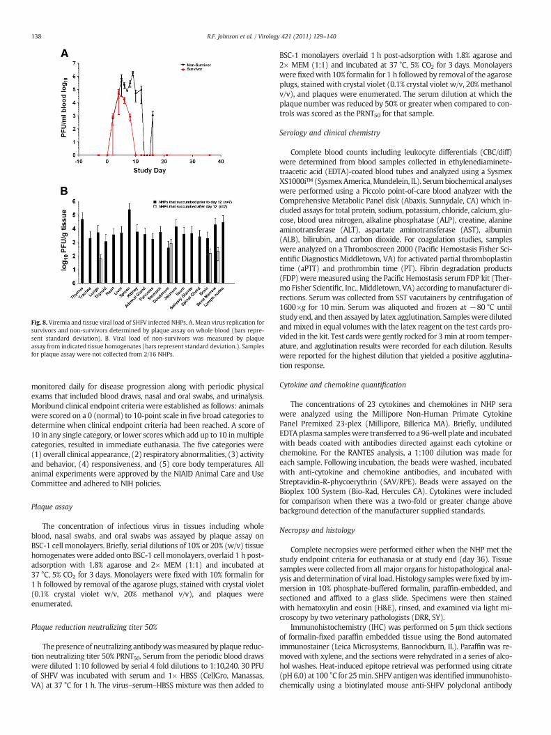
Fig. 8. Viremia and tissue viral load of SHFV infected NHPs. A. Mean virus replication forsurvivors and non-survivors determined by plaque assay on whole blood (bars repre-sent standard deviation). B. Viral load of non-survivors was measured by plaqueassay from indicated tissue homogenates (bars represent standard deviation.). Samplesfor plaque assay were not collected from 2/16 NHPs.
138 R.F. Johnson et al. / Virology 421 (2011) 129–140
monitored daily for disease progression along with periodic physicalexams that included blood draws, nasal and oral swabs, and urinalysis.Moribund clinical endpoint criteria were established as follows: animalswere scored on a 0 (normal) to 10-point scale in five broad categories todetermine when clinical endpoint criteria had been reached. A score of10 in any single category, or lower scores which add up to 10 inmultiplecategories, resulted in immediate euthanasia. The five categories were(1) overall clinical appearance, (2) respiratory abnormalities, (3) activityand behavior, (4) responsiveness, and (5) core body temperatures. Allanimal experiments were approved by the NIAID Animal Care and UseCommittee and adhered to NIH policies.
Plaque assay
The concentration of infectious virus in tissues including wholeblood, nasal swabs, and oral swabs was assayed by plaque assay onBSC-1 cell monolayers. Briefly, serial dilutions of 10% or 20% (w/v) tissuehomogenateswere added onto BSC-1 cell monolayers, overlaid 1 h post-adsorption with 1.8% agarose and 2× MEM (1:1) and incubated at37 °C, 5% CO2 for 3 days. Monolayers were fixed with 10% formalin for1 h followed by removal of the agarose plugs, stained with crystal violet(0.1% crystal violet w/v, 20% methanol v/v), and plaques wereenumerated.
Plaque reduction neutralizing titer 50%
The presence of neutralizing antibodywasmeasured by plaque reduc-tion neutralizing titer 50% PRNT50. Serum from the periodic blood drawswere diluted 1:10 followed by serial 4 fold dilutions to 1:10,240. 30 PFUof SHFV was incubated with serum and 1× HBSS (CellGro, Manassas,VA) at 37 °C for 1 h. The virus–serum–HBSS mixture was then added to
BSC-1 monolayers overlaid 1 h post-adsorption with 1.8% agarose and2× MEM (1:1) and incubated at 37 °C, 5% CO2 for 3 days. Monolayerswerefixedwith 10% formalin for 1 h followed by removal of the agaroseplugs, stained with crystal violet (0.1% crystal violet w/v, 20% methanolv/v), and plaques were enumerated. The serum dilution at which theplaque number was reduced by 50% or greater when compared to con-trols was scored as the PRNT50 for that sample.
Serology and clinical chemistry
Complete blood counts including leukocyte differentials (CBC/diff)were determined from blood samples collected in ethylenediaminete-traacetic acid (EDTA)-coated blood tubes and analyzed using a SysmexXS1000i™ (SysmexAmerica,Mundelein, IL). Serumbiochemical analyseswere performed using a Piccolo point-of-care blood analyzer with theComprehensive Metabolic Panel disk (Abaxis, Sunnydale, CA) which in-cluded assays for total protein, sodium, potassium, chloride, calcium, glu-cose, blood urea nitrogen, alkaline phosphatase (ALP), creatine, alanineaminotransferase (ALT), aspartate aminotransferase (AST), albumin(ALB), bilirubin, and carbon dioxide. For coagulation studies, sampleswere analyzed on a Thromboscreen 2000 (Pacific Hemostasis Fisher Sci-entific Diagnostics Middletown, VA) for activated partial thromboplastintime (aPTT) and prothrombin time (PT). Fibrin degradation products(FDP) were measured using the Pacific Hemostasis serum FDP kit (Ther-mo Fisher Scientific, Inc., Middletown, VA) according tomanufacturer di-rections. Serum was collected from SST vacutainers by centrifugation of1600×g for 10 min. Serum was aliquoted and frozen at −80 °C untilstudy end, and then assayed by latex agglutination. Sampleswere dilutedandmixed in equal volumes with the latex reagent on the test cards pro-vided in the kit. Test cards were gently rocked for 3 min at room temper-ature, and agglutination results were recorded for each dilution. Resultswere reported for the highest dilution that yielded a positive agglutina-tion response.
Cytokine and chemokine quantification
The concentrations of 23 cytokines and chemokines in NHP serawere analyzed using the Millipore Non-Human Primate CytokinePanel Premixed 23-plex (Millipore, Billerica MA). Briefly, undilutedEDTAplasma sampleswere transferred to a 96-well plate and incubatedwith beads coated with antibodies directed against each cytokine orchemokine. For the RANTES analysis, a 1:100 dilution was made foreach sample. Following incubation, the beads were washed, incubatedwith anti-cytokine and chemokine antibodies, and incubated withStreptavidin-R-phycoerythrin (SAV/RPE). Beads were assayed on theBioplex 100 System (Bio-Rad, Hercules CA). Cytokines were includedfor comparison when there was a two-fold or greater change abovebackground detection of the manufacturer supplied standards.
Necropsy and histology
Complete necropsies were performed either when the NHP met thestudy endpoint criteria for euthanasia or at study end (day 36). Tissuesamples were collected from all major organs for histopathological anal-ysis and determination of viral load. Histology sampleswere fixed by im-mersion in 10% phosphate-buffered formalin, paraffin-embedded, andsectioned and affixed to a glass slide. Specimens were then stainedwith hematoxylin and eosin (H&E), rinsed, and examined via light mi-croscopy by two veterinary pathologists (DRR, SY).
Immunohistochemistry (IHC) was performed on 5 μm thick sectionsof formalin-fixed paraffin embedded tissue using the Bond automatedimmunostainer (Leica Microsystems, Bannockburn, IL). Paraffin was re-moved with xylene, and the sections were rehydrated in a series of alco-hol washes. Heat-induced epitope retrieval was performed using citrate(pH 6.0) at 100 °C for 25 min. SHFV antigenwas identified immunohisto-chemically using a biotinylated mouse anti-SHFV polyclonal antibody

139R.F. Johnson et al. / Virology 421 (2011) 129–140
(1:100; raised in mice against the nucleocapsid protein sequenceCLVNLRKYGWQTKNK by Genscript (Piscataway NJ)) incubated for15 min at room temperature. Primary antibodywas localizedwith horse-radish peroxidase and diaminobenzidine substrate. Antibody specificityto SHFV was performed by IHC analysis using the SHFV polyclonal anti-body against tissues from historical, normal uninfected NHP controls. Incontrols to determine background IHC staining, buffer was used inplace of the primary antibody. Sections were counterstained with hema-toxylin and examined by light microscopy by veterinary pathologists.
Electron microscopyFor thin-section electron microscopic evaluation, dissected tissues
were immediately fixed in 2.5% glutaraldehyde and 2.0% paraformalde-hyde, in Millonig's sodium phosphate buffer (Tousimis Research, Rock-ville, MD), for 72 h. Fixed tissue samples were rinsed repeatedly inMillonig's buffer and post-fixed in 1.0% osmium tetroxide in the samebuffer. Following rinsing steps in ultrapure water and en bloc stainingwith 2.0% uranyl acetate, the samples were dehydrated in a series ofgraded ethanols, infiltrated, and embedded in DER-736 plastic resin(Tousimis Research, Rockville, MD). Embedded blocks were sectionedusing a Leica EM UC7 Ultramicrotome. Sections between 50 and 70 nmwere collected on 200 mesh copper grids and post-stained with Rey-nold's lead citrate. The tissue sectionswere examined by a veterinary pa-thologist (JAC) using a FEI Tecnai Spirit Twin transmission electronmicroscope operating at 80 kV.
Statistical analysis
Survival analyses comparing mortality by inoculation dose andgender were performed using the log-rank test. Hypothesis tests onincidence rates were conducted in Stata 9.0 using Fisher's Exact Testvalues were considered significant when p≤0.01.
A total of 60 parameterswere assessed during the course of the study.Of these 60, ten parameterswere considered subjective (relied on clinicalexperience i.e. spleen palpation) or did not generate numerical values,two yielded no usable data (nasal and oral swab titers), six were cyto-kines that did not increase over baseline for any NHP in any dose group,and seven were serum chemistry analytes that did not appreciablychange. The remaining 35 parameters (AST, aPTT, ALP, MCP-1, ALT, IL-6,neutrophil counts, platelet count, hematocrits, hemoglobin concentra-tions, IFNγ, viremia, IL-15, PT, VEGF, IL-5, creatinine, lymphocyte count,sCD40L, GM-CSF, IL-12, total bilirubin, TGFα, IL-2, G-CSF, total protein,IL-8, leukocyte count, monocyte count, IL-18, albumin, MIP-1α, TNFα,IL-1ra, and RANTES) were considered relevant to disease progressionfor statistical analysis. Because of themultiple comparisons, the followingp-values for test parameters were considered to be supportive of statisti-cal significance for an associationwith survival status when p≤0.01, andsuggestive of statistical significancewhen 0.01≤p≥0.05. Also, due to thepossible correlation between many of the factors, such as cytokines andclotting factors, the p-values was selected a priori rather than the Bonfer-roni method, which is known to be conservative.
To identify factors that may associate with disease severity, changesfrom baseline were evaluated according to BMLA. The trajectories for agiven subject were summarized by the area under the curve (AUC), asthis characterizes both the intensity and duration of exposure. Secondaryanalyses summarized trajectories according to the rate of change (asrepresented by the slope of a line, over a time period during whichchange was captured by a line), and the maximum observed value. Tocompare these summaries between survivors and non-survivors, BLAcompares summary measures over similar time intervals for survivorsand non-survivors. Survivors and non-survivors are matched and thesummary measures (e.g., AUC) are computed over the same time inter-val for each pair. The differences in the AUC are computed between thepairs and a paired t-test is performed. Because there are many possiblepairings, this process is repeated 1000 times, and a common p-value isobtained using the inverse normal p-value averaging (Dodd et al., 2011).
Acknowledgments
Thiswork, in part, was supported by the NIAID andNIAIDDivision ofIntramural Research. We are grateful to Cindy Allan, Krisztina Janosko,Abigail Lara, Erika Zommer, Rebecca Kurnat, Nicholas Oberlander, IsisAlexander, Bernardo Rosa, Oscar Rojas, Haifeng Song, and Kurt Cooperfor their contributions to these studies. We thank Sharon Altmann,Stacy Agar, James Lawler, Laura Bollinger, and Fabian De Kok Mercadofor their critical review and contribution to the preparation of this man-uscript. We would also like to thank Patrick Murray for his assistancewith blood culture and bacterial identification.
This project has been funded in whole or in part with federal fundsfrom the National Cancer Institute, National Institutes of Health, underContract No. HHSN261200800001E. The content of this publication doesnot necessarily reflect the views or policies of the Department of HealthandHuman Services, nor doesmention of trade names, commercial prod-ucts, or organizations imply endorsement by the U.S. Government.
References
Abildgaard, C., Harrison, J., Espana, C., Spangler, W., Gribble, D., 1975. Simian hemor-rhagic fever: studies of coagulation and pathology. Am. J. Trop. Med. Hyg. 24,537–544.
Allen, A.M., Palmer, A.E., Tauraso, N.M., Shelokov, A., 1968. Simian hemorrhagic fever.II. Studies in pathology. Am. J. Trop. Med. Hyg. 17, 413–421.
Beer, B., Kurth, R., Bukreyev, A., 1999. Characteristics of Filoviridae: Marburg and Ebolaviruses. Naturwissenschaften 86, 8–17.
Carneiro, S.C., Cestari, T., Allen, S.H., Ramos e-Silva, M., 2007. Viral exanthems in thetropics. Clin. Dermatol. 25, 212–220.
Charo, I.F., Taubman, M.B., 2004. Chemokines in the pathogenesis of vascular disease.Circ. Res. 95, 858–866.
Cummins, D., 1991. Arenaviral haemorrhagic fevers. Blood Rev. 5, 129–137.Dalgard, D.W., Hardy, R.J., Pearson, S.L., Pucak, G.J., Quander, R.V., Zack, P.M., Peters, C.J.,
Jahrling, P.B., 1992. Combined simian hemorrhagic fever and Ebola virus infectionin cynomolgus monkeys. Lab. Anim. Sci. 42, 152–157.
Davies, B., Morris, T., 1993. Physiological parameters in laboratory animals andhumans. Pharm. Res. 10, 1093–1095.
Deshmane, S.L., Kremlev, S., Amini, S., Sawaya, B.E., 2009. Monocyte chemoattractantprotein-1 (MCP-1): an overview. J. Interferon Cytokine Res. 29, 313–326.
Dodd, L.E., Johnson, R.F., Blaney, J.E., in preparation. Backward Matched LongitudinalAnalysis of Biomarkers Associated with Survival.
Feldmann, H., Geisbert, T.W., 2011. Ebola haemorrhagic fever. Lancet 377, 849–862.Geisbert, T.W., Hensley, L.E., Jahrling, P.B., Larsen, T., Geisbert, J.B., Paragas, J., Young, H.A.,
Fredeking, T.M., Rote, W.E., Vlasuk, G.P., 2003a. Treatment of Ebola virus infectionwith a recombinant inhibitor of factor VIIa/tissue factor: a study in rhesus monkeys.Lancet 362, 1953–1958.
Geisbert, T.W., Young, H.A., Jahrling, P.B., Davis, K.J., Larsen, T., Kagan, E., Hensley, L.E.,2003b. Pathogenesis of Ebola hemorrhagic fever in primate models: evidencethat hemorrhage is not a direct effect of virus-induced cytolysis of endothelialcells. Am. J. Pathol. 163, 2371–2382.
Gravell, M., London, W.T., Leon, M.E., Palmer, A.E., Hamilton, R.S., 1986. Differencesamong isolates of simian hemorrhagic fever (SHF) virus. Proceedings of the Societyfor Experimental Biology and Medicine: Society for Experimental Biology andMedicine, 181, pp. 112–119.
Green, D.E.,Mahlandt, B.G.,McKee Jr., K.T., 1987. Experimental Argentine hemorrhagic feverin rhesus macaques: virus-specific variations in pathology. J. Med. Virol. 22, 113–133.
Hensley, L.E., Geisbert, T.W., 2005. The contribution of the endothelium to the develop-ment of coagulation disorders that characterize Ebola hemorrhagic fever in pri-mates. Thromb. Haemost. 94, 254–261.
Hensley, L.E., Stevens, E.L., Yan, S.B., Geisbert, J.B.,Macias,W.L., Larsen, T., Daddario-DiCaprio,K.M., Cassell, G.H., Jahrling, P.B., Geisbert, T.W., 2007. Recombinant human activatedprotein C for the postexposure treatment of Ebola hemorrhagic fever. J. Infect. Dis.196 (Suppl. 2), S390–S399.
Huerta-Zepeda, A., Cabello-Gutierrez, C., Cime-Castillo, J., Monroy-Martinez, V., Manjarrez-Zavala, M.E., Gutierrez-Rodriguez,M., Izaguirre, R., Ruiz-Ordaz, B.H., 2008. Crosstalk be-tween coagulation and inflammation during Dengue virus infection. Thromb. Haemost.99, 936–943.
Jaax, N., Jahrling, P., Geisbert, T., Geisbert, J., Steele, K., McKee, K., Nagley, D., Johnson, E.,Jaax, G., Peters, C., 1995. Transmission of Ebola virus (Zaire strain) to uninfectedcontrol monkeys in a biocontainment laboratory. Lancet 346, 1669–1671.
Johnson, E., Jaax, N., White, J., Jahrling, P., 1995. Lethal experimental infections of rhe-sus monkeys by aerosolized Ebola virus. Int. J. Exp. Pathol. 76, 227–236.
Johnson, R.F., Yellayi, S., Cann, J.A., Johnson, A., Smith, A.L., Paragas, J., Jahrling, P.B., Blaney,J.E., 2011. Cowpox virus infection of cynomolgusmacaques as amodel of hemorrhagicsmallpox. Virology 418 (2), 102–112.
Kempton, R.M., Parsons, J.P., 1920. Report of a case of hemorrhagic smallpox: a consider-ation of the role played by thehemolytic Streptococcus. Arch. Intern.Med. 26, 594–600.
Keshtkar-Jahromi, M., Kuhn, J.H., Christova, I., Bradfute, S.B., Jahrling, P.B., Bavari, S.,2011. Crimean–Congo hemorrhagic fever: current and future prospects of vaccinesand therapies. Antiviral Res. 90, 85–92.

140 R.F. Johnson et al. / Virology 421 (2011) 129–140
Kunz, S., 2009. The role of the vascular endothelium in arenavirus haemorrhagic fevers.Thromb. Haemost. 102, 1024–1029.
Lapin, B.A., Shevtsova, Z.V., 1971. On the identity of two simian hemorrhagic fever virusstrains (Sukhumi and NIH). Z. Versuchstierkd. 13, 21–23.
Leblebicioglu, H., 2010. Crimean–Congo haemorrhagic fever in Eurasia. Int. J. Antimi-crob. Agents 36 (Suppl. 1), S43–S46.
Levi, M., 2010. The coagulant response in sepsis and inflammation. Hamostaseologie 30(10–12), 14–16.
Lewis, R.M., Morrill, J.C., Jahrling, P.B., Cosgriff, T.M., 1989. Replication of hemorrhagicfever viruses in monocytic cells. Rev. Infect. Dis. 11 (Suppl. 4), S736–S742.
London, W.T., 1977. Epizootiology, transmission and approach to prevention of fatalsimian haemorrhagic fever in rhesus monkeys. Nature 268, 344–345.
Lukashevich, I.S., Maryankova, R., Vladyko, A.S., Nashkevich, N., Koleda, S., Djavani, M.,Horejsh, D., Voitenok, N.N., Salvato, M.S., 1999. Lassa and Mopeia virus replicationin human monocytes/macrophages and in endothelial cells: different effects on IL-8 and TNF-alpha gene expression. J. Med. Virol. 59, 552–560.
Palmer, A.E., Allen, A.M., Tauraso, N.M., Shelokov, A., 1968. Simian hemorrhagic fever. I.Clinical and epizootiologic aspects of an outbreak among quarantined monkeys.Am. J. Trop. Med. Hyg. 17, 404–412.
Paragas, J., Geisbert, T.W., 2006. Development of treatment strategies to combat Ebolaand Marburg viruses. Expert Rev. Anti Infect. Ther. 4, 67–76.
Peters, C.J., Liu, C.T., Anderson Jr., G.W., Morrill, J.C., Jahrling, P.B., 1989. Pathogenesis ofviral hemorrhagic fevers: Rift Valley fever and Lassa fever contrasted. Rev. Infect.Dis. 11 (Suppl. 4), S743–S749.
Renquist, D., 1990. Outbreak of simian hemorrhagic fever. J. Med. Primatol. 19, 77–79.Ruzek, D., Yakimenko, V.V., Karan, L.S., Tkachev, S.E., 2010. Omsk haemorrhagic fever.
Lancet 376, 2104–2113.Shevtosova, Z.V., Karmysheva, V., Chumakov, M.P., 1975. Virological study of simian
hemorrhagic fever. Vopr. Virusol. 471–476.
Shevtsova, Z.V., 1969a. Further studies of the virus of simian hemorrhagic fever. Vopr.Neirokhir. 33, 604–607.
Shevtsova, Z.V., 1969b. A further study of simian hemorrhagic fever virus. Vopr. Viru-sol. 14, 604–607.
Shevtsova, Z.V., Krylova, R.I., 1971a. A comparative study of 2 strains of simian hemor-rhagic fever virus. Vopr. Virusol. 16, 686–688.
Shevtsova, Z.V., Krylova, R.I., 1971b. A comparative study of 2 strains of simian hemor-rhagic fever virus. Vopr. Virusol. 16, 686–688.
Solbrig, M.V., Naviaux, R.K., 1997. Review of the neurology and biology of Ebola andMarburg virus infections. Neurol. Infect. Epidemiol. 2, 5–12.
Stroher, U., West, E., Bugany, H., Klenk, H.D., Schnittler, H.J., Feldmann, H., 2001. Infectionand activation of monocytes byMarburg and Ebola viruses. J. Virol. 75, 11025–11033.
Tauraso, N.M., Shelokov, A., Palmer, A.E., Allen, A.M., 1968. Simian hemorrhagicfever. 3. Isolation and characterization of a viral agent. Am. J. Trop. Med. Hyg. 17,422–431.
Tauraso, N., Myers, M.G., McCarthy, K., Tribe, G.W., 1970. Simian hemorrhagic fever. In:Balner, H., Beveredge, W.I.B. (Eds.), Infection and Immunosuppression in Sub-Human Primates. Munksgaard, Copenhagen, Denmark, pp. 101–109.
van der Poll, T., de Boer, J.D., Levi, M., 2011. The effect of inflammation on coagulationand vice versa. Curr. Opin. Infect. Dis. 24, 273–278.
Wahl-Jensen, V.M., Afanasieva, T.A., Seebach, J., Stroher, U., Feldmann, H., Schnittler, H.J.,2005. Effects of Ebola virus glycoproteins on endothelial cell activation and barrierfunction. J. Virol. 79, 10442–10450.
Walker, D.H., Johnson, K.M., Lange, J.V., Gardner, J.J., Kiley, M.P., McCormick, J.B., 1982.Experimental infection of rhesus monkeys with Lassa virus and a closely related ar-enavirus, Mozambique virus. J. Infect. Dis. 146, 360–368.
Wauquier, N., Becquart, P., Padilla, C., Baize, S., Leroy, E.M., 2010. Human fatal Zaireebola virus infection is associated with an aberrant innate immunity and with mas-sive lymphocyte apoptosis. PLoS Negl. Trop. Dis. 4.


![Paulina black macaques [recovered]](https://img.pdfslide.net/doc/110x75/5559dee9d8b42a39498b4992/paulina-black-macaques-recovered.jpg)