Embed Size (px)
Citation preview
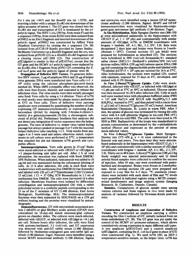
Proc. Natl. Acad. Sci. USAVol. 90, pp. 3655-3659, April 1993Neurobiology
Altering central nervous system physiology with a defective herpessimplex virus vector expressing the glucose transporter geneDoRA Y. Ho*, EDWARD S. MOCARSKIt, AND ROBERT M. SAPOLSKY*Departments of *Biological Sciences and tMicrobiology and Immunology, Stanford University, Stanford, CA 94305
Communicated by Seymour S. Kety, January 12, 1993 (receivedfor review November 1, 1992)
ABSTRACT Because of their postmitotic nature, neuronsare difficult subjects for gene transfer. To circumvent this, wehave used a defective herpes simplex virus vector to overexpressthe rat brain glucose transporter (GT) gene under the controlof the human cytomegalovirus iel promoter. This vector,designated vIElGT, was propagated using a herpes simplexvirus type 1 temperature-sensitive mutant, ts756. GT expressedfrom vIElGT was readily immunoprecipitated from mem-brane fractions of vIElGT-infected Vero cells. By using indi-rect double immunofluorescence techniques, vIElGT wasshown to be capable of enhancing GT expression in culturedhippocampal neurons and glia. Glucose transport in suchvIElGT-infected cultures was increased -2-fold relative tocontrols. The efficacy of this system in vivo was then tested bymicroinjection of vIElGT into adult rat hippocampus. Whenexamined 2 days later, GT expression from vIElGT wasdemonstrated in hippocampal neurons by in situ hybridization;a small but significant increase in glucose transport wasdetected in tissue immediately surrounding the injection site by2-deoxy[14C]glucose uptake and autoradiography. Such iinjec-tions did not cause marked cytopathology. Thus, this approachcan be used to alter central nervous system physiology in vitroand in vivo.
Because of the postmitotic nature of neurons, gene transferto these somatic nondividing cells requires innovative ap-proaches; use of herpes simplex virus (HSV) vectors mayrepresent such an approach. HSV-1 and HSV-2 are well-known neurotropic agents capable of replication and latencyin the peripheral nervous system. In addition, work inexperimental animals and in the natural human host hasshown that these viruses also interact with neurons in thecentral nervous system (CNS). Recombinant HSVs havebeen employed as vectors of foreign genes in cultured cells(1-5) and have been used to express an indicator gene(Escherichia coli lacZ gene) in sensory neurons or in CNSduring acute growth and latency (6-11).Recent development ofHSV vectors has capitalized on the
natural occurrence ofdefective interfering particles that ariseduring high-multiplicity propagation of HSV (12). Thesedefective viruses contain DNA molecules that are the samesize as standard virus (152 kbp) but consist of head-to-tailconcatemers of small regions ofthe viral genome including anorigin of DNA replication (oris or oriL) and a cleavage/packaging signal (a sequence). Defective viruses replicateand package only in the presence of a replication-competenthelper virus. This phenomenon was extended to the con-struction of cloning-amplifying vectors (termed amplicons)consisting of a prokaryotic plasmid for amplification in bac-terial host along with the important cis-acting HSV regulatorysequences for propagation in eukaryotic cells (13). Anyeukaryotic transcription unit may be included on ampliconsand propagated with helper virus to transfer genes into
eukaryotic cells. Such defective HSV vectors have been usedto express foreign genes in cultured cells, including neurons(14, 15). Although Kaplitt et al. (16) have used a defectiveHSV vector to express an indicator gene in adult rat brain, toour knowledge, no one has reported the use ofHSV vectors,albeit intact or defective, to alter the physiology of the CNSin adult animals. In this study, we have employed a defectiveHSV vector to deliver the brain-type glucose transporter(GT) into primary hippocampal neurons in vitro and into thehippocampus. This particular gene was chosen as the firststep in a strategy to deliver more glucose to hippocampalneurons, to buffer them from the neurotoxic effects of anumber of metabolically disruptive neurological insults (seeDiscussion). We find that in vitro and in vivo, expression ofthe newly introduced GT gene is associated with increasedglucose uptake.
MATERIALS AND METHODSCells and Virus. Mixed neuronal/glial cultures and en-
riched glial cultures were prepared from the hippocampi offetal Sprague-Dawley rats on the 18th day of gestation asdescribed (17). Mixed neuronal/glial cultures were main-tained in MEM-PAK (a modified minimal essential mediumpurchased from the cell culture facility of University ofCalifornia, San Francisco) supplemented with 10% (vol/vol)horse serum (HyClone) and were used at -10 days of agewhen the neuron/astrocyte ratio was z50:50, as determinedby immunocytochemical staining (data not shown). The glialcultures were maintained in Dulbecco's modified Eagle'smedium (DMEM; GIBCO/BRL) supplemented with 5%(vol/vol) fetal calf serum (HyClone). Vero cells (ATCCCCL81) were maintained in DMEM/10%o (vol/vol) NuSerum(Collaborative Research). All cells are maintained in a 5%C02/95% air atmosphere.
ts756 (HSV-1 KOS) has a temperature-sensitive mutationin an essential regulatory gene encoding ICP4 and wasprovided by R. G. Hughes, Jr. (Rosewell-Park Cancer Insti-tute, Buffalo, NY).
Construction of Amplicons. The amplicon pIElGTori wasconstructed as follows: The GT coding sequence was isolatedfrom prGT3 (kindly provided by Morris J. Bimbaum, Har-vard University) as an EcoRI-Bgl II fragment and cloned intopG310, an expression vector provided by Lidia Sambucetti(Stanford University). pG310 contains the promoter, exon 1(untranslated), and intron 1 (from nt -1021 to +947), and thepoly(A) signal (from nt +3270 to +3430) of the humancytomegalovirus (HCMV) iel gene in a pGEM-2 background.This plasmid was derived from pON308G (18) by first delet-ing the pGEM-2 sequences between EcoRI and BamHI,deleting the iel coding sequences (exons 2-4) between the
Abbreviations: HSV, herpes simplex virus; GT, glucose transporter;3-Gal, ,B-galactosidase; X-Gal, 5-bromo-4-chloro-3-indolyl P-D-galactopyranoside; pfu, plaque-forming unit; CNS, central nervoussystem; HCMV, human cytomegalovirus; MAP2, microtubule-associated protein 2; GFAP, glial fibrillary acidic protein.
3655
The publication costs of this article were defrayed in part by page chargepayment. This article must therefore be hereby marked "advertisement"in accordance with 18 U.S.C. §1734 solely to indicate this fact.
Proc. Natl. Acad. Sci. USA 90 (1993)
Pst I site (nt +947) and the BamHI site (nt +3270), andinserting a linker with a unique EcoRI site downstream of thesplice acceptor of intron 1. The GT gene was cloned into theEcoRI site and transcription of GT is terminated by the ielpoly(A) signal. The HSV-1 oris (295 bp, from strain F) and thea sequence (330 bp, from strain KOS) were then isolated frompON812 as anXba I fragment and inserted downstream ofthepoly(A) signal. pON812 was constructed by Jeffrey Viera(Stanford University) by cloning the a sequence (19, 20)isolated from pUC18-19 (kindly provided by James Smiley,McMaster University) as aBamHI fragment and the oris wasisolated from pON103 (21) as a HindIII-EcoRI fragment intoa cloning vector pIC-20H (22). The construction ofpIEl1,galori is similar to that of pIElGTori, except that theGT gene and the HCMV iel poly(A) signal were replaced byan EcoRI-Xba I fragment from pON3 (23) containing the E.coli lacZ gene and the poly(A) signal of simian virus 40.
Propagation of Defective HSV Vectors. To generate defec-tive HSV vectors, 3 ,ug of amplicon DNA and 10 ,g of helpervirus genomic DNA were transfected into Vero cells (1 x10⁶ cells) using the calcium phosphate/glycerol-shockmethod (6). When 100% cytopathic effect was observed, thecells were then frozen, thawed, and sonicated to release theinfectious virus. The virus stocks were then serially passagedonto fresh Vero cells by using a 1:4 dilution. Titers of ts756in viral stocks were determined by plaque assays performedat 33°C on Vero cells. Titers of defective virus carryingamplicons were estimated by quantitating the number of cellsexpressing GT immunocytochemically or expressing 3-ga-lactosidase (,S-Gal) by staining with 5-bromo-4-chloro-3-indolyl «-D galactopyranoside [X-Gal, a chromogenic sub-strate of ,B-Gal (6)]. Preliminary Southern blot analyses didnot detect any integration of vector sequences into the helpergenome. The ratio of defective vs. helper virus varied duringpassage but always peaked at passage 4 or 5 with the maximalhelper/defective ratio reaching 1:1. Viral stocks from pas-sages 4 or 5 were used and unless otherwise stated, experi-ments in cell culture were performed at 37°C, a temperaturethat retarded but did not eliminate ts756 growth and cyto-pathic effects.
Immunoprecipitation. Vero cells grown in 25-cm2 flaskswere mock-infected or infected with vIElGT or vIE1/3gal at=1 plaque-forming unit (pfu) per cell. After 1 h of viraladsorption, the inoculum was replaced with fresh DMEM/10% NuSerum. When indicated, tunicamycin was added to 10pg/ml and was maintained during the subsequent labeling ofcells. At 13 h after infection, the cells in each flask werewashed twice with methionine-free DMEM (Irvine Scientific)and labeled with 120 ,uCi of [35S]methionine (1102 Ci/mmol,15 mCi/ml; 1 Ci = 37 GBq; ICN Biomedicals) in 1.5 ml ofmethionine-free DMEM. The cells were harvested 16 h afterinfection. Membrane fractions were isolated by differentialcentrifugation and immunoprecipitated (24) with a rabbitpolyclonal serum to a synthetic peptide corresponding to the13 aa of the C terminus of GT (1:400 dilution; East AcresBiologicals, Southbridge, MA). The immunoprecipitatedfractions were then separated by SDS/PAGE on a 12% gelwithout heating and the proteins were visualized by autora-diography.
Immunofluorescence. GT with microtubule-associated pro-tein 2 (MAP2) or glial fibrillary acidic protein (GFAP) werecolocalized on 10-day-old mixed neuronal/glial culturesgrown on chamber slides. The cultures were mock-infected,infected with vIElGT, or infected with vIEl/3gal at -0.1 pfuper cell. Sixteen hours after infection, the cells were fixedwith acetone/methanol, 3:1 (vol/vol), and GT expressionwas detected with anti-GT rabbit serum (1:400 dilution),followed by rhodamine-conjugated goat anti-rabbit IgG an-tibody (1:80 dilution; Tago). Neurons were identified using amouse MAP2 monoclonal antibody (1:100 dilution; Sigma)
and astrocytes were identified using a mouse GFAP mono-clonal antibody (1:200 dilution; Sigma). MAP2 and GFAPimmunoreactivities were visualized by fluorescein-conju-gated goat anti-mouse IgG antibody (1:40 dilution; Tago).In Situ Hybridization. Male Sprague-Dawley rats (300-350
g) were microinfused unilaterally in the hippocampus withvIElGT (2 ,ul; 1 x 10⁵ pfu) and contralaterally with similaramounts of vIE13gal, ts756, or DMEM (coordinates withbregma = lambda: AP, 4.1; ML, 2.1; DV, 3.0). Rats weredecapitated 2 days later and brains were frozen in 2-meth-ylbutane (-20°C). Coronal sections (15 ,um thick) wereprepared for hybridization as described (25). The sectionswere hybridized in 50%o (vol/vol) formamide/4x standardsaline citrate (SSC)/1x Denhardt's solution/10% (wt/vol)dextran sulfate/tRNA (250 ,ug/ml)/salmon sperm DNA (500,ug/ml) containing the oligonucleotide probe end-labeled with35S-labeled dATP (5 x 10⁵ cpm per section) for 16 h at 37°C.After hybridization, the sections were washed (25), coatedwith emulsion, exposed for 15 days at 4°C, developed, andstained with 0.1% thionin.In Vitro 2-Deoxy[14C]glucose Uptake. Cells were mock-
infected, infected with vIElGT, or infected with vIEl/3gal at=1 pfu per cell at 37°C or 39°C as indicated. Glucose uptakewas then measured 16-20 h after infection (26). Cells in eachwell were washed twice with phosphate-buffered saline (PBS= 137 mM NaCl/4 mM KCl/8 mM Na2HPO4/1.5 mMKH2PO4), warmed to 37°C, and then pulsed with a tracer dose(1 ,uCi/ml) of 2-deoxy[14C]glucose (55 mCi/mmol; AmericanRadiolabeled Chemicals, St. Louis) in warm PBS. After 5min, the uptake of glucose was terminated by rinsing the cellstwice with 0.4 mM phloretin (Sigma) in ice-cold PBS (4°C)and twice with ice-cold PBS. The cells were then lysed in 1%SDS in PBS. Radioactivity of the lysate was standardized toprotein concentration as determined by the o-phthaldehydemethod (Pierce) and expressed as the percent of meanmock-infected values.In Vivo 2-Deoxy[14C]glucose Uptake. Male Sprague-
Dawley rats (375-450 g; n = 4) were catheterized in thefemoral vein and artery. Two days later they were microin-fused unilaterally in the hippocampus with vIElGT (2 ;1I; 1 x10⁵ pfu) and contralaterally with a similar amount ofvIEl/3gal(coordinates with bregma = lambda: AP, 4.1; ML, 2.1; DV,3.0). Thirty-six to 48 h later, 2-deoxy[14C]glucose [100 ,Ci/kg(body weight)] was injected intravenously and 12 timedarterial blood samples were collected to confirm the successof injection. After 45 min, rats were overdosed with pento-barbitol and decapitated. Brains were frozen in 2-methylbu-tane. Serial coronal sections (20 um) were prepared andexposed to x-ray film for 4-5 days. 14C standards (Amer-sham) were included with each sheet of film and 14C levelswere quantified in indicated regions using a MCID comput-erized densitometry and image analysis system (ImagingResearch, St. Catharines, Ontario, Canada).
Statistics. Comparisons of glucose uptake rates amongvarious cultures and brain regions in vivo were made byanalysis of variance followed by Newman-Keuls post-hoctests.
RESULTSConstruction of Amplicons and Generation of Defective
Vectors. We constructed an amplicon carrying a cDNAencoding the Glut-1 isoform of GT, initially isolated from ratbrain endothelium (27, 28). The GT gene was put under thecontrol of the HCMV iel promoter-enhancer, which allowsconstitutively strong expression in many different cell types.A test amplicon (pIElGTori) and a control amplicon(pIEl/3galori, containing the E. coli lacZ gene in place ofGT)were constructed (Fig. 1). We used ts756 (29), an HSV-1temperature-sensitive mutant, as the helper virus. ts756 has
3656 Neurobiology: Ho et al.

Proc. Natl. Acad. Sci. USA 90 (1993) 3657
Otis
FIG. 1. pIElGTori. The transcriptional unit is driven by theHCMV iel promoter-enhancer. Exon 1 of iel is untranslated andintron 1 is spliced. The poly(A) signal inserted downstream ofthe GTgene is derived from the HCMV iel gene. The oris and the asequences provide the necessary HSV replication signals. Theamplicillin-resistance gene of the parent plasmid pGEM-2 is alsoindicated. The structure of pIE1,8galori is similar to that ofpIElGTori, except that the GT gene and the iel polA signal arereplaced by lacZ terminated with the simian virus 40 poly(A) signal.
a mutation in ICP4, an essential regulatory a gene ofthe virusand is completely replication-defective at 39°C. Defectivevirus vectors were generated by cotransfecting pIElGTori orpIE1.8galori along with genomic DNA ofts756 into Vero cellsand then serially passaging the viral stocks at 33°C by usinga 1:4 dilution to encourage growth of defective virus. Thevirus stocks thus generated from pIElGTori and pIE13galoriare referred to as vIElGT and vIE1,8gal, respectively.
Immunoprecipitation of GT from vIElGT-Infected VeroCells. We first confirmed that vIElGT enhanced GT expres-sion in Vero cells. A new species of -41 kDa was immuno-precipitated from membrane fractions from vIEIGT-infectedcells (Fig. 2, lane 3) but not in mock-infected or vIEl18gal-infected controls (Fig. 2, lanes 1 and 4, respectively). In thepresence of tunicamycin, this product was reduced to =38kDa (Fig. 2, lane 2), the reported size of the primary trans-lation product of Glut-i (28). The endogenous GT in rat brainendothelium is known to be heterogeneously glycosylatedwith a mean molecular mass of -52 kDa (27), and alternativeGT species of 45-47 kDa also occur in rat brain (27, 28).Thus, the 41-kDa GT that we observed probably results fromdifferential glycosylation in Vero cells.
Efficacy of Infection with vIElGT in Cultured Neurons andAstrocytes. We next tested the ability of vIElGT to expressGT in primary cultures of hippocampal neurons and astro-cytes by using immunofluorescence. GT immunoreactivitywas observed in vIElGT-infected cultures (Fig. 3) but not invIEl3gal-infected or mock-infected cultures (data notshown). By using double immunofluorescence methods to
1 234kDa
69 - ;*.. .1i46
30 -
FIG. 2. Detection of GT expression byimmunoprecipitation. Vero cells were mock-infected or infected with the defective vec-tors, with (lane 2) or without (lanes 1, 3, and4) tunicamycin treatment, and were labeledwith [35S]methionine from 13 to 16 h afterinfection. Membrane fractions were thenprepared and immunoprecipitated with ananti-GT rabbit serum. The immune com-plexes were subjected to SDS/PAGE (12%gel) and visualized by autoradiography.Lanes: 1, infected with vIElgal; 2 and 3,infected with vIElGT; 4, mock-infected.The sizes of protein standards are shown.
FiG. 3. Colocaization of GT with MAP2 and GFAP antigens onmixed neuronal/glial cultures using double indirect immunofluores-cence. Cells were double-labeled with anti-GT serum (A) and anti-MAP2 monoclonal antibody (B) with anti-GT serum (C) and anti-GFAP monoclonal antibody (D). The secondary antibodies werelabeled with rhodamine (for anti-GT serum) and with fluorescein (foranti-MAP2 and anti-GFAP antibodies, respectively).
colocalize neuron-specific antigen MAP2 or glia-specific an-tigen GFAP with GT immunoreactivity, both neurons andastrocytes were found to be capable of expressing GT (Fig.3). Similarly, p3-Gal-expressing neurons or astrocytes wereobserved in vIE13gal-infected but not in vIElGT-infected ormock-infected cultures (data not shown).Given that our goal was to manipulate cellular function with
these interventions, we tested whether increased GT expres-sion was associated with an increased rate of glucose trans-port. Infection with vIElGT doubled the rate of2-deoxy['4C]glucose transport in Vero cells, mixed hippocam-pal cultures, and primary astrocyte cultures (Fig. 4). Mockinfection or infection with vIElfgal did not alter transport.
Efficacy ofInfection with vIElGT and vIElfiJa in Vivo. Wenext tested the ability of vIElGT to express GT wheninjected into the hippocampus of adult rats by using in situhybridization techniques. To distinguish the viral vector-
~.coO _
11)c i
Ca 200 - c _I
-
0 C\-0 - C ! S <--
0X100
Vero MH MH Glia(370C) (390C) (370C) (37°C)
FIG. 4. Effect of vIElGT infection on 2-deoxy[14C]glucose up-take in vitro. Vero cells, astrocytes, and mixed hippocampal (MH)cultures were mock-infected or infected with defective vectors at37°C or 39°C as indicated. Uptake in vIElGT-infected culturesdiffered significantly from mock- and vIEl,gal-infected cultures (P< 0.001 for all cases except astroglia, where P < 0.05; Newman-Keuls post-hoc test after analysis of variance; n averaged 10 amongthe various groups). Bars: darkly hatched, mock-infected; lightlyhatched, lacZ; shaded, GT. n, Number of culture wells.
Neurobiology: Ho et al.

Proc. Natl. Acad. Sci. USA 90 (1993)
derived GT mRNA from the endogenous version, a 75-ntoligonucleotide probe antisense to the iel promoter from nt-35 to +40 was used. This probe hybridized to the 5'untranslated end of both the GT transcripts from vIElGTand the lacZ transcripts from vIEl3gal. Hybridization sig-nals were readily detected in hippocampus microinfusedwith vIElGT (Fig. 5A) or vIElpgal (Fig. 5B); in contrast, nosuch signal was detected after injection with the helpervirus or with DMEM alone (data not shown). The signalswere clustered around the injection site and highly concen-trated along the dentate gyrus. The pattern of vIE1,Bgalexpression as visualized by in situ hybridization was similarto that visualized by histochemical staining with X-Gal (Fig.5C).
We further characterized the parameters of expressionwith the HSV system by injecting vIEl1,gal and staining withX-Gal. (Gal expression was analyzed 2, 4, and 7 days afterinjections; expression peaked at day 2. At that time, expres-sion was localized immediately surrounding the injection site,with most positive cells within a 150-,um radius. Within thatarea, :5% of cells expressed (-Gal. Of those cells, :'82%were found to be neuronal by morphological criteria. Thenumber of cells that expressed 3-Gal within the 150-,umradius of the injection site was -50% of the peak value by 4days after injection and declined to zero by 7 days.We then tested whether injection of vIElGT altered glu-
cose uptake in the CNS of adult rats. vIElGT was microin-fused unilaterally into the hippocampus and vIEl,Bgal wasmicroinfused contralaterally; 2-deoxy[14C]glucose uptakewas assessed 2 days later. Because of the data for the spatialdistribution of 13-Gal-positive cells, we analyzed glucoseuptake within a small sphere of tissue, with a radius of "'450Am, surrounding the injection site (corresponding to thedorsal blade of the dentate gyrus).Glucose transport was enhanced a significant 10o at the
injection site on the vIElGT side, relative to the contralateralside. This enhancement declined with increasing distancefrom the infusion site, and uptake was equivalent to that onthe contralateral side within "180 ,um of the injection site(Fig. 6). There were no differences in uptake in hippocampalregions more distant from the injection site (data not shown).
DISCUSSIONThe use of defective HSV vectors to deliver genes to neuronsand glia shows great potential, and a number of investigatorshave exploited this system in recent years. In some reports,HSV vectors were used with cultured cells (e.g., rat neuromacell lines, mouse ovary cells, Vero cells, or HEP-2 cells). Inall of these cases, efficacy of the vector was documented byenhanced levels of mRNA or protein for a reporter gene orenhanced activity of the reporter enzyme (2, 3, 14, 30). Insome instances, HSV vector systems have been used for invivo studies. Pallela et al. (3) overexpressed mRNAs for theHPRT gene in mouse brain by such an approach. Chiocca etal. (11), Kaplitt et al. (16), Huang et al. (8), and Fink et al. (9)delivered lacZ as a reporter gene to the rodent brain, and Hoand Mocarski (6, 7) and Dobson et al. (10) used a similar
a1)0Cc$
0-a1)a)
CO
0
I0-
..~~~~~~~~~~~~~~~~~~~~~~~~~~~~~~.. .:. ..... . ... ..,L:,,....jE'. '."7 .',''.,
:' .......~~~~~~~~~~~~~~. .. :..'........... '.'.......'':.:
-~ ~ ~~4*
,,.~~~~~~~~~~~~~~~~~~~~~~~~~~~~. ....',. ...... : ...
FIG. 5. Expression of vlElGT and vIE1/8gal in hippocampus as
detected by in situ hybridization and X-Gal staining. (A and B)
Dark-field microscopy of hybridization on the dorsal blade of the
dentate gymus from vIElGT-infected (A) and vIE1(3gal-infected (B)
animals. The ventral blade of the dentate gyrs is outlined with
arrows. (C) Bright-field microscopy showing P-Gal-expressing cells
as darkly stained cells on the dorsal blade of the dentate gyrus.
115-
110-
105
100 -
95.
-180 -120 -60 6 +60 +120 +180Distance from infusion site, ,um
FIG. 6. Effect of vIElGT infection on 2-deoxy[14C]glucose up-take in adult rat CNS. Anterior/posterior analysis showing that2-deoxy[14C]glucose uptake was enhanced nearest to the site ofinjection (arrow) of vIElGT. Uptake in hippocampus (relative to thecontralateral side) is shown as a function of distance from theinjection site in the anterior/posterior plane, with data pooled forevery three 20-/m section. Numbers in parentheses indicate thenumbers of sections from the four rats analyzed; numbers variedbecause, in some cases, sections corresponding to the stated distancefrom the infusion site were not available.
3658 Neurobiology: Ho et al.
11s
Proc. Natl. Acad. Sci. USA 90 (1993) 3659
approach with sensory neurons and hypoglossal motor neu-rons. In a recent advance in this approach, Federoffet al. (31)delivered the nerve growth factor gene to superior cervicalganglion neurons and were able to alter neuronal survivalafter axotomy. In the present report, we have used a defec-tive HSV vector, vIElGT, to enhance glucose transport inthe rat hippocampus. To our knowledge, this is the firstreport that such a system has been used to deliver a gene intothe brain itself that alters physiology (rather than servingmerely as a reporter gene).The magnitude of vIElGT's effect upon glucose transport
differed considerably between cell culture and intact host. Inepithelial or mixed hippocampal cells in culture, our bestvIElGT stock (with a defective/helper ratio 1:1) increased
2-deoxyglucose uptake 200-300%. In contrast, in vivo,vIElGT caused an -10% increase in 2-deoxyglucose uptakein a small sphere of tissue surrounding the injection site.However, the effect on uptake in individual cells might havebeen greater than that. The data regarding the number anddistribution of 3-Gal-positive cells demonstrated that -5% ofcells within such a sphere were positive. One can safelyassume very roughly equivalent efficiencies of expression invIElGT and vIEl,Bgal. Thus, if the glucose uptake rate at thecenter of the sphere of tissue was increased 10% due toinfection of only this small subset of cells, this implies thatuptake was enhanced considerably in those individual cells.
Such an increase might be physiologically meaningful withinthe tightly regulated realm of cerebral metabolism (32).As noted, our studies utilized the HCMV iel promoter-
enhancer and a temperature-sensitive mutant of HSV as
helper. Although the strength of the HCMV promoter-enhancer is well established, its long-term pattern of expres-
sion in neurons is unclear. We observed (-Gal-expressingcells in rat hippocampus only for the first few days afterinfection, in agreement with a report (16) that also used theHCMV iel promoter to express lacZ. We (7) and others (10)have demonstrated that the latency-associated transcripts(LAT) promoter from HSV-1 can cause long-termed gene
expression in latently infected animals. Thus, the LAT pro-
moter and other promoters from housekeeping genes may bemore effective at causing sustained changes in CNS function.With respect to the helper virus, the internal body temper-ature of the rat is not completely nonpermissive for ts756replication and we have observed some cytopathicity in thehippocampus by using ts756 (unpublished data). Virusescarrying deletion mutations in key regulatory genes such as
ICP4 and ICPO will potentially facilitate this technique (33).These studies were initiated to find a somatic gene therapy
to alter neuronal function and potentially protect neurons
from insult. The choices of attempting to enhance glucosetransport and using the hippocampus were not random. Thebrain is exquisitely dependent upon glucose; nervous tissuehas extremely high metabolic rates, utilizes little other thanglucose, and stores it poorly (34). Moreover, neurologicalinsults such as hypoxia-ischemia, hypoglycemia, and sus-
tained seizure disrupt energy charge in the brain and prefer-entially damage the hippocampus; in the case of sustainedseizure, glucose transport can become the rate-limiting stepdetermining whether there is a damaging mismatch betweenenergy delivery and utilization (32). Furthermore, glucocor-ticoids damage hippocampal neurons and enhance the tox-icity of these neurological diseases; this endangerment isenergetic in nature, as it can be lessened by supplementationof the hippocampus with excessive glucose (35). Finally,neuron death emerges slowly over the 48-72 h after theseneurological insults and a number of pharmacological inter-ventions during this delayed period can be neuroprotective(35); thus, were the rat hippocampus exposed to vIElGT
immediately after onset of the insult, glucose transport wouldbe enhanced during the time window of vulnerability. Asnoted, the enhancement of glucose transport in the small setof affected neurons was probably well in excess of 10%o abovecontrol values; there is reason to believe that with optimi-zation of the protocol described here, the percentage ofneurons so affected and/or the magnitude of the effect can beimproved. Our data suggest that this method of interventionmight have neuroprotective potential.
We thank Sheila Brooks, Maria Kirichenko, and Jeremy Tompkinsfor technical assistance; Lauren Jacobson, Michael Romero, andBecky Stein-Behrens for assistance with surgical procedures; andElaine Brown for helpful suggestions. This work was supported bythe American Paralysis Association and the Stanford Office ofTechnology Liscencing (R.M.S.) and by a Huntington's DiseaseSociety of America fellowship (D.Y.H.).
1. Shih, M. F., Arsenakis, M., Tiollais, P. & Roizman, B. (1984) Proc.Natl. Acad. Sci. USA 81, 5867-5870.
2. Smiley, J., Smibert, C. & Everett, R. (1987) J. Virol. 61,2368-2377.3. Pallela, T., Silverman, L., Schroll, C., Homa, F., Levine, M. &
Kelley, W. (1988) Mol. Cell. Biol. 8, 457-460.4. Rosen-Wolff, A., Raab, A., Zoller, L., Darai, G., Eberle, J. &
Deinhardt, F. (1990) Virus Genes 4, 325-337.5. Dormitzer, P. R., Ho, D. Y., Mackow, E. R., Mocarski, E. S. &
Greenberg, H. B. (1992) Virology 187, 18-32.6. Ho, D. Y. & Mocarski, E. S. (1988) Virology 167, 279-283.7. Ho, D. Y. & Mocarski, E. S. (1989) Proc. Natl. Acad. Sci. USA 86,
7596-7600.8. Huang, Q., Vonstattel, J., Schaffer, P., Martuza, R., Breakefield,
X. & Defiglia, M. (1992) Exp. Neurol. 115, 303-316.9. Fink, D., Lawrence, R., Stemnberg, L., Weber, P., Marina, M.,
Goins, W. & Giorioso, J. (1992) Hum. Gene Ther. 3, 11-19.10. Dobson, A. T., Margolis, T. P., Sedarati, F., Stevens, J. G. &
Feldman, L. T. (1990) Neuron 5, 353-360.11. Chiocca, E. A., Choi, B. B., Weizhong, N. A., DeLuca, N. A.,
Schaffer, P. A., DeFiglia, M., Breakefield, X. A. & Martuza, R. L.(1990) New Biol. 2, 739-746.
12. Frenkel, N., (1981) in The Human Herpesviruses: An Interdiscipli-nary Prospective, eds. Nahmias, A. J., Dowdle, W. R. & Schinazy,R. S. (Elsevier/North-Holland, New York), pp. 91-120.
13. Spaete, R. R. & Frenkel, N. (1982) Cell 30, 295-304.14. Kwong, A. D. & Frenkel, N. (1985) Virology 142, 421-425.15. Geller, A. I. & Breakefield, X. 0. (1988) Science 241, 1667-1669.16. Kaplitt, M. G., Pfaus, J. G., Kleopoulos, S. P., Hanlon, B. A.,
Rabkin, S. D. & Pfaff, D. W. (1991) Mol. Cell. Neurosci. 2, 320-330.
17. Tombaugh, G., Yang, S., Swanson, R. & Sapolsky, R. (1992) J.Neurochem. 59, 137-144.
18. Cherrington, J. M. & Mocarski, E. S. (1989) J. Virol. 63,1435-1440.19. Dutch, R. E., Bruckner, R. C., Mocarski, E. S. & Lehman, I. R.
(1992) J. Virol. 66, 277-285.20. Varmuza, S. L. & Smiley, J. R. (1985) Cell 41, 793-802.21. Elias, P., O'Donnell, M. E., Mocarski, E. S. & Lehman, I. R.
(1986) Proc. Natl. Acad. Sci. USA 83, 6322-6326.22. Marsh, J. L., Erfle, M. & Wykes, E. J. (1984) Gene 32, 481-485.23. Manning, W. C. & Mocarski, E. S. (1988) Virology 167, 477-484.24. Haspel, H. C., Birnbaum, M. J., Wilk, E. W. & Rosen, 0. M.
(1985) J. Biol. Chem. 260, 7219-7225.25. Sutin, E. L. & Kilduff, T. S. (1992) Mol. Brain Res. 15, 281-290.26. Homer, H. C., Packan, D. R. & Sapolsky, R. M. (1990) Neuroen-
docrinology 52, 57-64.27. Pardridge, W. M., Boado, R. J. & Farrel, C. R. (1990) J. Biol.
Chem. 265, 18035-18040.28. Birnbaum, M. J., Haspel, H. C. & Rosen, 0. M. (1986) Proc. Natl.
Acad. Sci. USA 83, 5784-5788.29. Hughes, R. G. & Munyon, W. H. (1975) J. Virol. 16, 275-283.30. Tackney, C., Chachianes, G. & Silverstein, S. (1984) J. Virol. 52,
606.31. Federoff, H., Geschwind, M., Geller, A. & Lessler, J. (1992) Proc.
Natl. Acad. Sci. USA 89, 1636-1640.32. Auer, R. & Siesjo, B. (1988) Ann. Neurol. 24, 699.33. Gelier, A. I., Keyomarsi, K., Bryan, J. & Pardee, A. B. (1990)
Proc. Natl. Acad. Sci. USA 87, 8950-8954.34. Siesjo, B. (198l) J. Cerebral Blood Flow Metabolism 1, 155.35. Sapolsky, R. (1990) Prog. Brain Res. 86, 13.
Neurobiology: Ho et al.