Embed Size (px)
Citation preview

RESEARCH ARTICLE Open Access
Simplified plasmid cloning with a universalMCS design and bacterial in vivo assemblyFan Chen*, Yi-ya Li, Yan-li Yu, Jie Dai, Jin-ling Huang and Jie Lin
Abstract
Background: The ability to clone DNA sequences quickly and precisely into plasmids is essential for molecularbiology studies. The recent development of seamless cloning technologies has made significant improvements inplasmid construction, but simple and reliable tools are always desirable for time- and labor-saving purposes.
Results: We developed and standardized a plasmid cloning protocol based on a universal MCS (Multiple CloningSite) design and bacterial in vivo assembly. With this method, the vector is linearized first by PCR (Polymerase ChainReaction) or restriction digestion. Then a small amount (10 ~ 20 ng) of this linear vector can be mixed with a PCR-amplified insert (5× molar ratio against vector) and transformed directly into competent E. coli cells to obtain thedesired clones through in vivo assembly. Since we used a 36-bp universal MCS as the homologous linker, any PCR-amplified insert with ~ 15 bp compatible termini can be cloned into the vector with high fidelity and efficiency.Thus, the need for redesigning insert-amplifying primers according to various vector sequences and the followingPCR procedures was eliminated.
Conclusions: Our protocol significantly reduced hands-on time for preparing transformation reactions, hadexcellent reliability, and was confirmed to be a rapid and versatile plasmid cloning technique. The protocol containsmostly mixing steps, making it an extremely automation-friendly and promising tool in modern biology studies.
Keywords: Plasmid cloning, Universal MCS, Bacterial in vivo assembly, DNA assembly, Homologous sequence
Plasmid cloning is one of the most commonly used tech-niques in molecular biology research. It plays a crucialrole in studying the structure, function, and evolution ofgenes [1, 2] while serving as an essential tool in genetic,protein, and metabolic engineering [3, 4]. However, thetraditional digestion-ligation method is often limited, asboth vector and target fragments must have compatiblecleavage sites. Moreover, the whole procedure can betime-consuming and labor-intensive [5].To overcome the difficulties mentioned above, new al-
ternatives have emerged in the past two decades. Theseinclude PCR-mediated IVA (In Vivo Assembly) cloning[6], fast cloning [7]; LIC (Ligation-Independent Cloning)such as ELIC (Exonuclease and Ligation-Independent
Cloning) [8], SLIC (Sequence and Ligation-IndependentCloning) [9], HAC (Homologous Alignment Cloning)[10], One-step SLIC [11] and In-Fusion cloning [12];recombination-based Gateway Cloning [13], SLiCE(Seamless Ligation Cloning Extract) [14, 15], andmultienzyme-based Gibson Assembly [16], Golden GateAssembly [17]. Each of these methods has advantagesand limitations (more information is listed in Supple-mentary Table S1). For example, PCR-mediated and LICmethods remove a few enzymatic treatment steps.Nevertheless, multiple rounds of PCR amplification arerequired, which might result in random mutations in thefinal products. Recombination- and multienzyme-basedmethods are often used in commercial cloning kits. Theyare methods with excellent convenience, but the cost ofapplying these kits is also much higher. Furthermore,most cloning methods are not automation friendly
© The Author(s). 2021 Open Access This article is licensed under a Creative Commons Attribution 4.0 International License,which permits use, sharing, adaptation, distribution and reproduction in any medium or format, as long as you giveappropriate credit to the original author(s) and the source, provide a link to the Creative Commons licence, and indicate ifchanges were made. The images or other third party material in this article are included in the article's Creative Commonslicence, unless indicated otherwise in a credit line to the material. If material is not included in the article's Creative Commonslicence and your intended use is not permitted by statutory regulation or exceeds the permitted use, you will need to obtainpermission directly from the copyright holder. To view a copy of this licence, visit http://creativecommons.org/licenses/by/4.0/.The Creative Commons Public Domain Dedication waiver (http://creativecommons.org/publicdomain/zero/1.0/) applies to thedata made available in this article, unless otherwise stated in a credit line to the data.
* Correspondence: [email protected] of Biological Science and Biotechnology, Minnan Normal University,Zhangzhou 363000, P.R. China
Chen et al. BMC Biotechnology (2021) 21:24 https://doi.org/10.1186/s12896-021-00679-6
because multiple pre-transformation steps or differentprimer designs are necessary for various vectors andinserts.The recA-independent recombination pathway in E.
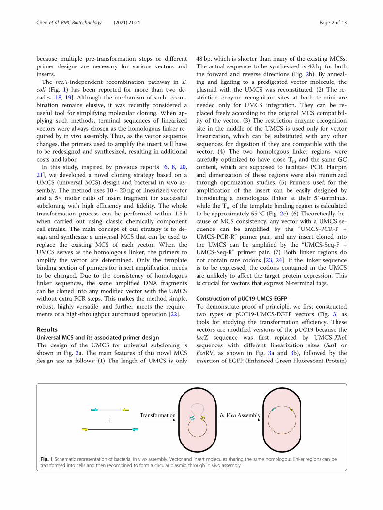
coli (Fig. 1) has been reported for more than two de-cades [18, 19]. Although the mechanism of such recom-bination remains elusive, it was recently considered auseful tool for simplifying molecular cloning. When ap-plying such methods, terminal sequences of linearizedvectors were always chosen as the homologous linker re-quired by in vivo assembly. Thus, as the vector sequencechanges, the primers used to amplify the insert will haveto be redesigned and synthesized, resulting in additionalcosts and labor.In this study, inspired by previous reports [6, 8, 20,
21], we developed a novel cloning strategy based on aUMCS (universal MCS) design and bacterial in vivo as-sembly. The method uses 10 ~ 20 ng of linearized vectorand a 5× molar ratio of insert fragment for successfulsubcloning with high efficiency and fidelity. The wholetransformation process can be performed within 1.5 hwhen carried out using classic chemically componentcell strains. The main concept of our strategy is to de-sign and synthesize a universal MCS that can be used toreplace the existing MCS of each vector. When theUMCS serves as the homologous linker, the primers toamplify the vector are determined. Only the templatebinding section of primers for insert amplification needsto be changed. Due to the consistency of homologouslinker sequences, the same amplified DNA fragmentscan be cloned into any modified vector with the UMCSwithout extra PCR steps. This makes the method simple,robust, highly versatile, and further meets the require-ments of a high-throughput automated operation [22].
ResultsUniversal MCS and its associated primer designThe design of the UMCS for universal subcloning isshown in Fig. 2a. The main features of this novel MCSdesign are as follows: (1) The length of UMCS is only
48 bp, which is shorter than many of the existing MCSs.The actual sequence to be synthesized is 42 bp for boththe forward and reverse directions (Fig. 2b). By anneal-ing and ligating to a predigested vector molecule, theplasmid with the UMCS was reconstituted. (2) The re-striction enzyme recognition sites at both termini areneeded only for UMCS integration. They can be re-placed freely according to the original MCS compatibil-ity of the vector. (3) The restriction enzyme recognitionsite in the middle of the UMCS is used only for vectorlinearization, which can be substituted with any othersequences for digestion if they are compatible with thevector. (4) The two homologous linker regions werecarefully optimized to have close Tm and the same GCcontent, which are supposed to facilitate PCR. Hairpinand dimerization of these regions were also minimizedthrough optimization studies. (5) Primers used for theamplification of the insert can be easily designed byintroducing a homologous linker at their 5′-terminus,while the Tm of the template binding region is calculatedto be approximately 55 °C (Fig. 2c). (6) Theoretically, be-cause of MCS consistency, any vector with a UMCS se-quence can be amplified by the “UMCS-PCR-F +UMCS-PCR-R” primer pair, and any insert cloned intothe UMCS can be amplified by the “UMCS-Seq-F +UMCS-Seq-R” primer pair. (7) Both linker regions donot contain rare codons [23, 24]. If the linker sequenceis to be expressed, the codons contained in the UMCSare unlikely to affect the target protein expression. Thisis crucial for vectors that express N-terminal tags.
Construction of pUC19-UMCS-EGFPTo demonstrate proof of principle, we first constructedtwo types of pUC19-UMCS-EGFP vectors (Fig. 3) astools for studying the transformation efficiency. Thesevectors are modified versions of the pUC19 because thelacZ sequence was first replaced by UMCS-XhoIsequences with different linearization sites (SalI orEcoRV, as shown in Fig. 3a and 3b), followed by theinsertion of EGFP (Enhanced Green Fluorescent Protein)
Fig. 1 Schematic representation of bacterial in vivo assembly. Vector and insert molecules sharing the same homologous linker regions can betransformed into cells and then recombined to form a circular plasmid through in vivo assembly
Chen et al. BMC Biotechnology (2021) 21:24 Page 2 of 13

through XhoI-based digestion and ligation. Among them,SalI represented the linearized product with sticky ends,while EcoRV represented blunt ends. Together withvectors linearized by PCR, the effect of differentlinearization methods on transformation can be clarified.During in vivo assembly, if the mCherry CDS is success-fully cloned into the UMCS, the colony will appear or-ange. Otherwise, it will be green or colorless (throughnonspecific assembly). By calculating orange coloniesagainst the total number of colonies, the positive ratio oftransformation can be determined.
Effects of the linearization method and homologouslinker lengthNext, we evaluated the effects of different linearizationmethods and the length of the homologous linker interms of the number of transformants and the corre-sponding positive ratio. As shown in Fig. 4a, when PCRwas used as a linearization method, the transformationefficiency showed a positive correlation with the lengthof the linker sequence. When a 6-bp linker was used,less than 100 CFU/plate and a positive ratio below 10%were confirmed. In contrast, the efficiency of
Fig. 2 Principle of the UMCS and corresponding primer design. a The UMCS sequence used in this study contains two homologous linkerregions that are connected by the SalI recognition sequence. The flanking HindIII and XbaI recognition sequences were designed for replacingthe original MCS. All three recognition sites can be freely substituted for other sequences. b The UMCS was first synthesized as two ssDNAs (eachwith a length of 42 bp). After annealing and ligating, the UMCS was incorporated into the vector, thus replacing the original MCS. c Therecommended primer design to maximize assembly efficiency. The homologous linker region (12 ~ 15 bp) is included in the 5′-terminus of eachprimer, and the Tm of the template binding region was designed to be approximately 55 °C
Fig. 3 Schematic depicting of pUC19-based plasmid models. Two different designs of the pUC19-UMCS-EGFP plasmid with SalI or EcoRVrecognition sequence were used for sticky- or blunt-end digestion
Chen et al. BMC Biotechnology (2021) 21:24 Page 3 of 13

transformation increased to 259 ± 34, 742 ± 132, 1306 ±123, and 1525 ± 165 CFU/plate, and the positive ratio in-creased dramatically to 78 ± 4.2%, 96 ± 1.5%, 95 ± 1.7%,and 97 ± 1.9% when 9-, 12-, 15-, and 18-bp linkers weretested, respectively. It is evident that a 6-bp linker is notenough for bacterial in vivo assembly, and a length lon-ger than 9 bp is necessary when performing such experi-ments. Moreover, there were no significant differencesbetween the 15- and 18-bp linker. Considering both thecolony number and positive ratio, we concluded that thelength of the homologous linker for the PCR methodwas 12 ~ 15 bp. On the other hand, when linearizationwas carried out by restriction digestion, it was shown(Fig. 4b and 4c) that the digested vectors with blunt ends(EcoRV) yielded much more positive clones than stickyends at any of the linker lengths we tested. According tothe data collected, when a restriction enzyme was usedfor linearization, a 12 ~ 15-bp linker also ensuredenough transformants and positive clones to screen. Inthese cases, further increase the linker length is notnecessary.
Effects of insert/vector ratio on transformationThe molar ratio of insert/vector is considered a criticalfactor for high-efficiency transformation [25]. Thus, wefurther investigated the influence of PCR, SalI andEcoRV digestion on transformation efficiency at multipleinsert/vector levels. The yields of transformants and thepositive ratio at the linker length of 15 bp were deter-mined (Fig. 5 and Supplementary Figure S1) with molarratios of 1, 5, 10, and 15. According to the data shown,regardless of the linearization method used, the optimalmolar ratio remained at 5. A further increase in insertusage will not improve the results, while a decreased in-sert quantity shows a negative effect on transformationefficiency. Therefore, we used a molar ratio of 5 in allsubsequent studies, which is consistent with the findingof Kostylev et al. [26]
Effects of the digestive linearization method on thefidelity of assemblyWe were interested in the assembly fidelity of the digest-ive linearization method mainly based on three reasons:
Fig. 4 Effects of the linearization method and homologous length on assembly efficiency. a Transformation efficiency (shown as CFU/plate andpositive ratio of colonies) of PCR-linearized pUC19-UMCS-S-EGFP with the mCherry sequence flanked by 6-, 9-, 12-, 15-, and 18-bp homologouslinkers (n = 4). b and c Transformation efficiency of SalI- and EcoRV-digested pUC19-UMCS-S-EGFP and pUC19-UMCS-E-EGFP with the mCherrysequence flanked by 9-, 12-, 15-, and 18-bp homologous linkers (n = 4)
Chen et al. BMC Biotechnology (2021) 21:24 Page 4 of 13

(1) Although high fidelity enzymes such as Q5, Phusion,or KOD have extremely low mismatch rates [27, 28]during the reaction, PCR products could still suffer fromrandom errors introduced by DNA polymerase. More-over, the possibility of introducing random mutationsduring the PCR process increases with PCR cycles andthe length of vectors, and it is laborious to verify by se-quencing. (2) Some vectors are difficult to amplify byPCR. For example, the GC content of the vector is toohigh or too low, or the vector contains too many repeti-tive sequences, which will affect PCR and even renderthe reaction impossible to continue [29–31]. (3) As pre-viously mentioned, if vectors cannot be linearized byPCR or fidelity is vital for the experiment, the methodsshown in Fig. 6 must be applied. Under such circum-stances, only the sequences flanking the linearization siteare used as homologous linkers to ensure the versatilityof UMCS. However, when the flanking sequences (15bp) were used as homologous linkers, the post-digestionresidual bases then served as nonhomologous sequences.This means the residual bases might displace part of theinsert and cause mutations at its 5′-terminus, 3′-terminus (confirmed during our pilot study), or both.The frequency of such events is a major concern forUMCS applications, since the UMCS is only universalwhen this frequency is low enough.Therefore, SalI (Fig. 6a-d) and EcoRV (Fig. 6e-h) diges-
tion were taken as sticky- and blunt-end examples toevaluate sequence replacement between insert andresidual bases after assembly. As shown in Fig. 6, whenthe mCherry CDS was used as an insert, five possible
products could be expected: (1) if the assembly fails, thecolony will appear green (only EGFP expression); (2) ifthe fragments assembled successfully and the mCherryCDS remains intact, the colony will appear orange (onlymCherry expression); (3) if assembled successfully, butthe 5′-terminus of the mCherry CDS is partiallyreplaced, the colony will appear orange (only mCherryexpression); (4) if assembled successfully, but the 3′-terminus of the mCherry CDS is partially replaced, thecolony appears yellow for the loss of the mCherry stopcodon (mCherry-EGFP fusion protein is expressed); and(5) if assembled successfully, while both the 5′- and 3′-terminus of the mCherry CDS are partially replaced, thecolony also appears yellow (mCherry-EGFP fusion pro-tein is expressed). For subcloning, we assume that the5′- and 3′-terminus of the insert will be replaced at thesame frequency, then the frequency can be derived bycalculating the number of yellow colonies against the or-ange ones. Thus, Fig. 6d strongly suggests that the fre-quency of mutation at the 3′-terminus of the insert islow when SalI is used for vector linearization. In fact,less than 0.75% of positive clones contained sequence re-placement at the 3′-terminus or at both the 5′- and 3′-terminus. This indicates that the possibility of having atleast one mutated terminus is less than 1.5% when thelength of the linker is 12 or 15 bp. Therefore, it will bevery unlikely for anyone to pick up a mutated productby chance. Moreover, when EcoRV was used forlinearization, the frequency of replacement eventsseemed less than that of SalI by having a statistical valueof less than 0.3% on both 12- and 15-bp linkers. In
Fig. 5 Effects of insert/vector ratio on assembly efficiency. Effects of insert/vector molar ratio on the recombination of pUC19-UMCS-S-EGFPlinearized by PCR (a) or SalI digestion (b) with the mCherry sequence flanked by 15-bp homologous linkers (n = 4)
Chen et al. BMC Biotechnology (2021) 21:24 Page 5 of 13
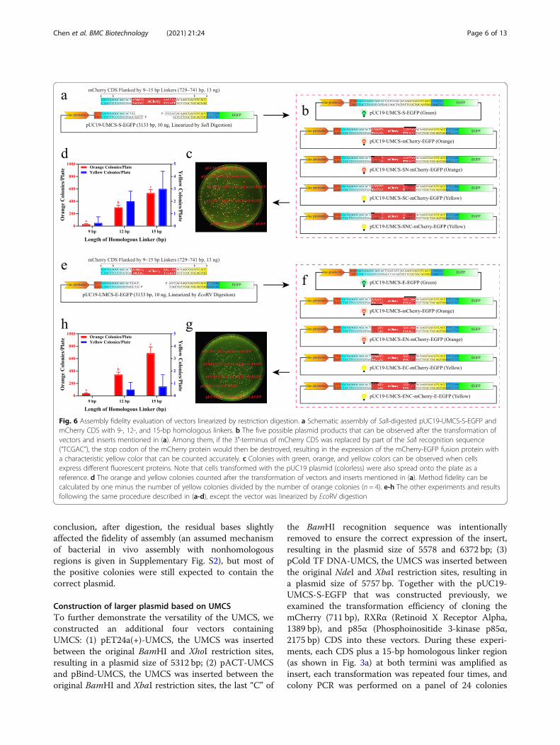
conclusion, after digestion, the residual bases slightlyaffected the fidelity of assembly (an assumed mechanismof bacterial in vivo assembly with nonhomologousregions is given in Supplementary Fig. S2), but most ofthe positive colonies were still expected to contain thecorrect plasmid.
Construction of larger plasmid based on UMCSTo further demonstrate the versatility of the UMCS, weconstructed an additional four vectors containingUMCS: (1) pET24a(+)-UMCS, the UMCS was insertedbetween the original BamHI and XhoI restriction sites,resulting in a plasmid size of 5312 bp; (2) pACT-UMCSand pBind-UMCS, the UMCS was inserted between theoriginal BamHI and XbaI restriction sites, the last “C” of
the BamHI recognition sequence was intentionallyremoved to ensure the correct expression of the insert,resulting in the plasmid size of 5578 and 6372 bp; (3)pCold TF DNA-UMCS, the UMCS was inserted betweenthe original NdeI and XbaI restriction sites, resulting ina plasmid size of 5757 bp. Together with the pUC19-UMCS-S-EGFP that was constructed previously, weexamined the transformation efficiency of cloning themCherry (711 bp), RXRα (Retinoid X Receptor Alpha,1389 bp), and p85α (Phosphoinositide 3-kinase p85α,2175 bp) CDS into these vectors. During these experi-ments, each CDS plus a 15-bp homologous linker region(as shown in Fig. 3a) at both termini was amplified asinsert, each transformation was repeated four times, andcolony PCR was performed on a panel of 24 colonies
Fig. 6 Assembly fidelity evaluation of vectors linearized by restriction digestion. a Schematic assembly of SalI-digested pUC19-UMCS-S-EGFP andmCherry CDS with 9-, 12-, and 15-bp homologous linkers. b The five possible plasmid products that can be observed after the transformation ofvectors and inserts mentioned in (a). Among them, if the 3′-terminus of mCherry CDS was replaced by part of the SalI recognition sequence(“TCGAC”), the stop codon of the mCherry protein would then be destroyed, resulting in the expression of the mCherry-EGFP fusion protein witha characteristic yellow color that can be counted accurately. c Colonies with green, orange, and yellow colors can be observed when cellsexpress different fluorescent proteins. Note that cells transformed with the pUC19 plasmid (colorless) were also spread onto the plate as areference. d The orange and yellow colonies counted after the transformation of vectors and inserts mentioned in (a). Method fidelity can becalculated by one minus the number of yellow colonies divided by the number of orange colonies (n = 4). e-h The other experiments and resultsfollowing the same procedure described in (a-d), except the vector was linearized by EcoRV digestion
Chen et al. BMC Biotechnology (2021) 21:24 Page 6 of 13
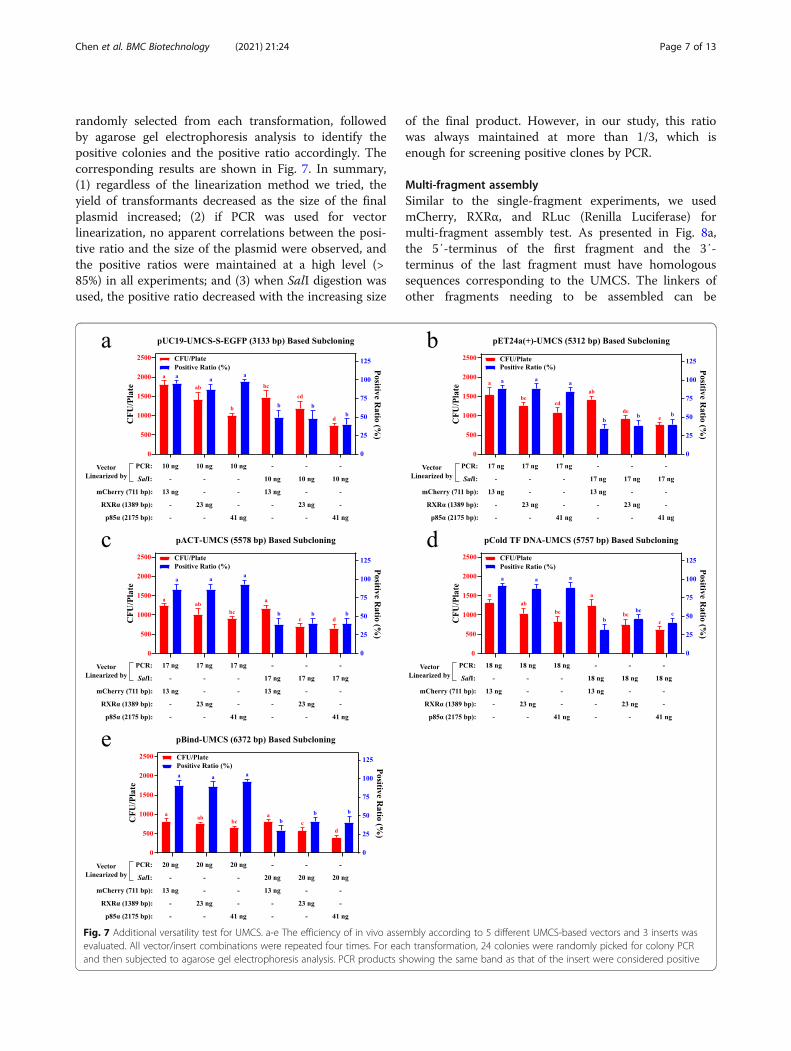
randomly selected from each transformation, followedby agarose gel electrophoresis analysis to identify thepositive colonies and the positive ratio accordingly. Thecorresponding results are shown in Fig. 7. In summary,(1) regardless of the linearization method we tried, theyield of transformants decreased as the size of the finalplasmid increased; (2) if PCR was used for vectorlinearization, no apparent correlations between the posi-tive ratio and the size of the plasmid were observed, andthe positive ratios were maintained at a high level (>85%) in all experiments; and (3) when SalI digestion wasused, the positive ratio decreased with the increasing size
of the final product. However, in our study, this ratiowas always maintained at more than 1/3, which isenough for screening positive clones by PCR.
Multi-fragment assemblySimilar to the single-fragment experiments, we usedmCherry, RXRα, and RLuc (Renilla Luciferase) formulti-fragment assembly test. As presented in Fig. 8a,the 5′-terminus of the first fragment and the 3′-terminus of the last fragment must have homologoussequences corresponding to the UMCS. The linkers ofother fragments needing to be assembled can be
Fig. 7 Additional versatility test for UMCS. a-e The efficiency of in vivo assembly according to 5 different UMCS-based vectors and 3 inserts wasevaluated. All vector/insert combinations were repeated four times. For each transformation, 24 colonies were randomly picked for colony PCRand then subjected to agarose gel electrophoresis analysis. PCR products showing the same band as that of the insert were considered positive
Chen et al. BMC Biotechnology (2021) 21:24 Page 7 of 13

designed according to previous studies [6]. Since multi-fragment assembly significantly reduces the yield oftransformants and positive clones (Fig. 8b), PCR as alinearization method or dephosphorylation after enzymedigestion is recommended. Meanwhile, further increas-ing the amount of DNA and the number of componentcells might be necessary for the assembly of more than 3insert fragments.
DiscussionIn most cases, inserts are required to be placed some-where within a MCS to function properly. The UMCS-based cloning, like the MCS-based cloning, is not aseamless technique because it relies on synthetic hom-ologous regions to work. However, it provides key ad-vantages over current plasmid cloning methods:First, we successfully constructed tens of plasmids with
single or multiple insertions in a very efficient way byusing this method. Although we used DH5α cellsthroughout this study, other strains such as Top10,BL21(DE3), XL-10 Gold and JM109(DE3) were alsotested (as shown in Supplementary Figure S3 and Sup-plementary Table S2) and had similar success, highlight-ing the versatility of this protocol.Second, the DpnI enzyme, which is widely used in
other reports [6, 7, 20, 26, 32] for the digestion of tem-plate DNA, is not necessary when using our protocol be-cause only a few nanograms of linearized vector DNAand a 5× molar concentration of the insert fragment areneeded. Thus, a single tube of carefully stored linearized
vector or insert can be used several times without losingefficiency. Therefore, the cost of applying this protocol iswell controlled.Third, the UMCS protocol contains mostly mixing
steps when both the vector and insert are ready, makingit extremely automation friendly. Instruments for high-throughput transformations are much easier to designand build according to this approach, making this proto-col promising in modern molecular biology studies.Finally, and especially, because of the consistency of
homologous linkers and the associated primers, any line-arized UMCS-based vector and insert can be mixed dir-ectly and is ready for transformation. This is importantfor those who need to optimize protein expression orswitch epitope tags by cloning the same insert into dif-ferent vectors. In addition, the protocol uses only puri-fied DNA, which means the high-fidelity DNApolymerases used to amplify vectors or inserts are ex-pected to have little or no impact on the transformation.This further guarantees the versatility of UMCS becausededicated vectors or inserts are easier to share betweenresearchers from laboratory to laboratory, regardless oftheir amplification conditions and without additionalPCR steps.In this study, by comparing different linearization
methods, we found that even though the number oftransformants was occasionally higher when using thedigestion method, the yield of positive clones was morethan 20% lower than that of PCR (Fig. 4). Interestingly,the DNA ligase activity inside E. coli has shown a great
Fig. 8 Multi-fragment assembly. a Schematic representation of multi-fragment assembly assessment. We used up to 3 different insert fragmentsfor multi-fragment assembly experiments. Each insert has a 5× molar concentration of the PCR linearized vector. b Effects of the fragment counton transformation. The numbers of transformants and positive clones decreases with an increasing number of fragments. For eachtransformation, 24 colonies were randomly picked for colony PCR and then subjected to agarose gel electrophoresis analysis. PCR productsshowing the same band as that of the assembled insert(s) were considered positive
Chen et al. BMC Biotechnology (2021) 21:24 Page 8 of 13

impact on the positive ratio. When comparing Fig. 4aand 4c, we found that when an 18-bp linker was used,the colonies formed after transformation were 1525 ±165 and 1483 ± 121 without a significant difference,while the positive ratio was reduced significantly from96.5 ± 1.9% to 75.4 ± 4.2%, implying the linearized vectorwith the phosphorylated 5′-terminus is more easilyrecircularized than the vector without phosphorylation.Additionally, recircularization by DNA ligase is more ef-fective for vectors with sticky ends, which explains whythe blunt-ended vector yields a higher positive ratio thanthe vector with sticky ends, and the PCR-amplified vec-tors work the best for in vivo assembly. Thus, if the vec-tor itself is small enough or the fidelity of PCR is not avital concern, PCR is a better choice for linearization,whereas if the vector is large enough that the randommutation introduced by PCR is problematic or the vec-tor is simply resistant to PCR amplification, linearizationwith the blunt-end enzyme is also an effective strategy.Finally, if no suitable blunt-end enzymes’ recognition se-quence could be found, digestion by sticky-end enzymesis still a feasible alternative. Furthermore, we believethat if restriction digestion was used for linearization,dephosphorylation might be helpful to further enhancetransformation efficiency, especially for multi-fragmentassembly [33].During our research, nonspecific assembly within vec-
tors was observed occasionally. In rare cases, nonspecificassembly between the vector and insert could also beidentified. As shown in Fig. 9, we have suggested threenonspecific assembly models within the vector moleculeand six models between the vector and insert. When lin-earized pUC19-UMCS-S-EGFP or pUC19-UMCS-E-
EGFP was involved, the results of nonspecific assemblyusually appeared as nonfluorescent (the fluorescent pro-tein or promoter sequence was lost or mutated) col-onies. Such cases usually made up less than 5% of thetotal colonies, regardless of the linearization method. Itwas also reported previously [34] that the PCR productscan be transformed directly and circularized within thecell. Therefore, nonspecific assembly and direct trans-formation of PCR products might be the two reasons forpositive ratios seldom exceeding 97%, even when PCRproducts purified by agarose gel electrophoresis wereused.Conley et al. [35] proposed a mechanism for bacterial
in vivo assembly: double-stranded linear DNA moleculesare the substrate for 3′-5′-exonuclease action by ExoIII,which yields single-stranded homologous DNA se-quences. After annealing at homologous sites, the vectorand insert then circularize to form a plasmid moleculewith gaps. The gaps are subsequently filled by DNApolymerase I and ligases as a DNA repair mechanism,thus completing plasmid formation. In this model, non-homologous parts of the ssDNA termini will be dis-placed by newly-repaired duplex after recombination.However, it is hard to explain solely by this assumptionwhy the intervening nonhomologous sequence (such aspost-digestion residual bases) between homologous re-gions and linear DNA termini still exists as part of thefinal product in some cases. According to our experi-ments, such events happened with a probability ofaround 1.5 and 0.3% when using SalI or EcoRV diges-tion, respectively. Although this phenomenon still favorsus, how cells decide which sequence to keep within thefinal plasmid remains unclear. From this perspective,
Fig. 9 Proposed models for nonspecific assembly. Nonspecific assembly occurs within the vector molecule or between the vector and insert. Anyof the marked assemblies (1 ~ 3 within vectors and 1 ~ 6 between vectors and inserts) occurred, a nonspecific assembly was confirmed
Chen et al. BMC Biotechnology (2021) 21:24 Page 9 of 13

despite the significant improvements we have madeupon understanding the recombination mechanism [36,37], the picture is still far from complete.In some cases, the UMCS might affect the function
or expression of the insert. For example, if the vectorhas a start codon located upstream of the UMCS (asshown in Fig. 3 and also typical in pET system forthe expression of fusion proteins with N-terminaltags), the sequence of Linker-F (see Fig. 2 for detailedinformation) will be translated as a short peptide of“DEGST”. If such peptide impairs the insert’s functionor expression, it must be removed when constructingthe plasmid. The same strategy also applies when theexpression of fusion proteins with C-terminal tags(stop codon located downstream of the UMCS) wasaffected by the sequence of Linker-R (translated as“TSDVT”). Consequently, alternative “half-seamless”or “full-seamless” strategies presented in Fig. 10 canbe established. Among them, the “half-seamless” strat-egies (Fig. 10a and b) for 5′- or 3′-end seamless clon-ing still simplify the primer design, which will save
time and cost of the experiments and reduce the sys-tem complexity.
ConclusionsIn this study, we developed and standardized adigestion-ligation-free approach for rapid and efficientplasmid cloning. The method utilizes a unified MCS de-sign scheme, which simplifies primer design and the fol-lowing PCR steps. Most importantly, it exploits thein vivo assembly of E. coli and significantly reduceshands-on time in the preparation of transformation tojust seconds while maintaining excellent reliability.
Materials and methodsBacterial strains and reagentsChemically competent cell strains of E. coli DH5α,Top10, BL21(DE3), XL10-Gold, and JM109(DE3) wereobtained from Weidi Biotechnology (Shanghai, China)with transformation efficiency of ~ 5 × 10⁸, ~ 5 × 10⁸, ~107, ~ 2 × 10⁹, and ~ 108 CFU/μg pUC19, respectively.Bacteria containing plasmids were cultured in LB (Luria-
Fig. 10 Seamless solutions for vectors with UMCS. a-b Alternative “half-seamless” flow chart of plasmid assembly using UMCS based vectors. cSchematic details of “full-seamless” assembly when UMCS must be ruled out of the final product
Chen et al. BMC Biotechnology (2021) 21:24 Page 10 of 13
Bertani) medium with appropriate antibiotics (ampicillinor kanamycin at 100 or 50 μg/mL, respectively), andIPTG (Isopropyl β-D-1-thiogalactopyranoside, 100 μM)was added when necessary. The SanPrep Column Plas-mid Mini-Preps Kit (Sangon Biotech, Shanghai, China)was used for plasmid extraction from bacteria, the San-Prep Column PCR Product Purification Kit (Sangon Bio-tech) was used for PCR product purification, and theSanPrep Column DNA Gel Extraction Kit (Sangon Bio-tech) was used for PCR product or enzyme-digestedDNA purification after agarose gel electrophoresis, allfollowing the manufacturer’s instructions. Both the 4SRed Plus (Sangon Biotech) stained 1% agarose gel andDNA Marker (250 ~ 10,000 bp, Sangon Biotech) wereused for all gel electrophoresis experiments. FastDigestBamHI, EcoRV, NdeI, SalI, XbaI, XhoI, and T4 DNA lig-ase from Thermo Fisher Scientific (Shanghai, China)were used for vector construction or linearization. Q5®High-Fidelity DNA Polymerase from NEB (Ipswich, MA,USA) was used to amplify the vector and insert, whileTaq PCR Master Mix (Sangon Biotech) was used for col-ony PCR.
Primers and plasmidsPrimers were designed using Oligo 7, the Tm of each pri-mer was calculated according to the “Nearest Neighbor”method. All primers used in this study were designedthen synthesized (as shown in Supplementary Table S3)and obtained from Sangon Biotech. The pUC19 plasmidwas purchased from Weidi Biotechnology. ThepET24a(+) vector was purchased from Invitrogen (Carls-bad, CA, USA). pCold TF DNA was purchased fromTakara (Beijing, China). pACT and pBind were pur-chased from Promega (Madison, WI, USA).pCDNA3.1(+)-EGFP, pCDNA3.1(+)-mCherry, pCMV-RXRα and pCMV-p85α were all gifts from XiaokunZhang.
PCR conditions and product analysisUnless otherwise stated, 50 μL PCR was performed usingQ5 for vector and insert amplification. The PCR condi-tions were: 0.02 U/μL polymerase, 0.5 μM each primer,200 μM dNTPs, and 1 ng prelinearized template DNA.According to the following protocol: 98 °C (30s)→ [98 °C (10 s)→ 60 °C (30 s)→ 72 °C (2500 bp/min,20 cycles for vector, 30 cycles for insert)]→ 72 °C (2min)→ 10 °C (hold). After amplification, the PCR prod-ucts were purified and recovered for further use. Add-itionally, 20 μL PCR was performed using Taq PCRMaster Mix for each colony PCR following the manufac-turer’s instructions. The corresponding PCR conditionwas: 95 °C (30 s)→ [95 °C (15 s)→ 55 °C (15 s)→ 72 °C(1500 bp/min, 25 cycles)]→ 72 °C (2 min)→ 10 °C (hold).Agarose gel electrophoresis was used for identifying
positive clones. Colonies that showed the same band asthe insert were considered positive.
Transformation evaluationUnless otherwise stated, a 1 μL (4 μL for BL21(DE3))mixture of linearized vector and insert was transformedinto 25 μL (100 μL for BL21(DE3)) of competent cells,and then the mixture was first incubated for 25 min onice, followed by heat shock at 42 °C for 60 s and trans-ferred to ice for 2 min. After adding 1000 μL of LBmedium, cells were subsequently incubated at 37 °C andshaking vigorously (220 rpm) for 45 min. After incuba-tion, cells were centrifuged at 6000 rpm (~ 2500 g) for 1min, pellets were resuspended in 75 μL of fresh LBmedium then spread onto LB agar plates containing an-tibiotics and IPTG (only for pUC19-based plasmids thatexpress fluorescent protein) for 16 h at 37 °C. Colonieswere manually counted, and the positive ratio wasassessed by colony PCR (with the “UMCS-Seq-F +UMCS-Seq-R” primer pair) or counting under a 485 nmlight source through a 535 nm filter after an additionalincubation at 4 °C for 48 h. Part of the positive cloneswere further confirmed by Sanger sequencing (SangonBiotech).
Data analysesData are expressed as the mean ± SD from four or moreexperiments. Statistical analyses were performed usingone-way ANOVA with the comparison methods of theTukey test (equal variances) or the Games-Howell test(unequal variances). Data sharing a letter in the samegroup were not considered to be significantly different atthe 5% level.
AbbreviationsMCS: Multiple Cloning Site; PCR: Polymerase Chain Reaction; IVA: In VivoAssembly; ELIC: Exonuclease and Ligation-Independent Cloning;LIC: Ligation-Independent Cloning; SLIC: Sequence and Ligation-IndependentCloning; HAC: Homologous Alignment Cloning; SLiCE: Seamless LigationCloning Extract; IPTG: Isopropyl β-D-1-thiogalactopyranoside; EGFP: EnhancedGreen Fluorescent Protein; RXRα: Retinoid X Receptor Alpha;p85α: Phosphoinositide 3-kinase p85α; RLuc: Renilla Luciferase; LBmedium: Luria-Bertani medium
Supplementary InformationThe online version contains supplementary material available at https://doi.org/10.1186/s12896-021-00679-6.
Additional file 1 Table S1. An overview of some existing methodsdeveloped for plasmid cloning.
Additional file 2 Table S2. A selection of UMCS based cloning for XL-10 Gold and JM109(DE3).
Additional file 3 Table S3. List of the primers and oligonucleotidesused throughout this study.
Additional file 4 Figure S1. Effects of insert/vector ratio on assemblyefficiency for vector linearized by EcoRV digestion.
Chen et al. BMC Biotechnology (2021) 21:24 Page 11 of 13
Additional file 5 Figure S2. Assumed mechanism of bacterial in vivoassembly with nonhomologous regions.
Additional file 6 Figure S3. Efficiency evaluation of UMCS basedsubcloning for Top10 and BL21(DE3).
Additional file 7. Supplementary image file. Vector format of Figure S1,S2, S5.
Additional file 8. Supplementary image file. Uncropped original imageof Fig. 6c.
Additional file 9. Supplementary image file. Uncropped original imageof Fig. 6g.
Additional file 10. Supplementary data file. Raw data points as used forFigs. 4, 5, 6, 7, 8, S1, S3.
Additional file 11. Supplementary protocol description. Standardprotocols for UMCS based cloning.
AcknowledgementsThe authors thank professor Xiaokun Zhang and his team for providing vitalmaterials.
Authors’ contributionsFC and YYL conceived and supervised the study. FC, YYL and YLY performedthe DNA amplification, purification, and transformation. JD, JLH, carried outthe evaluation of transformation efficiency. FC and JL analyzed the data; FCdrafted and finalized the manuscript. YYL, YLY reviewed and commented onthe manuscript. All authors read and approved the final manuscript.
FundingThis work was financially supported by Natural Science Foundation of FujianProvince of China (2018J01464).
Availability of data and materialsThe datasets used and analyzed in the current study are available from thecorresponding author on reasonable request or as in supplementary files.
Ethics approval and consent to participateNot applicable.
Consent for publicationNot applicable.
Competing interestsThe authors declare that they have no competing interests.
Received: 28 November 2020 Accepted: 17 February 2021
References1. Tao QZ, Zhang HB. Cloning and stable maintenance of DNA fragments over
300 kb in Escherichia coli with conventional plasmid-based vectors. NucleicAcids Res. 1998;26(21):4901–9. Available from. https://doi.org/10.1093/nar/26.21.4901.
2. Cohen SN. DNA cloning: a personal view after 40 years. Proc Natl Acad Sci.2013;110(39):15521–9. Available from. https://doi.org/10.1073/pnas.1313397110.
3. Lesley SA, Wilson IA. Protein production and crystallization at the jointcenter for structural genomics. J Struct Funct Genomics. 2005;6:71–9.Available from. https://doi.org/10.1007/s10969-005-2897-2.
4. Chao R, Yuan Y, Zhao H. Recent advances in DNA assembly technologies.FEMS Yeast Res. 2015;15(1):1–9. Available from. https://doi.org/10.1111/1567-1364.12171.
5. Green M, Sambrook DWJ. Molecular cloning: a laboratory manual. 4th ed;2012.
6. Javier GN, Jake WF, Ingo HG. IVA cloning : a single-tube universal cloningsystem exploiting bacterial in vivo assembly. Sci Rep. 2016;(6):1–12. Availablefrom. https://doi.org/10.1038/srep27459.
7. Li C, Wen A, Shen B, et al. FastCloning: a highly simplified, purification-free,sequence- and ligation-independent PCR cloning method. BMC Biotechnol.2011;11(1):92. Available from. https://doi.org/10.1186/1472-6750-11-92.
8. Koskela EV, Frey AD. Homologous recombinatorial cloning without thecreation of single-stranded ends: exonuclease and ligation-independentcloning (ELIC). Mol Biotechnol. 2015;57(3):233–40. Available from. https://doi.org/10.1007/s12033-014-9817-2.
9. Li MZ, Elledge SJ. Harnessing homologous recombination in vitro togenerate recombinant DNA via SLIC. Nat Methods. 2007;4(3):251–6.Available from. https://doi.org/10.1038/nmeth1010.
10. Tan L, Strong EJ, Woods K, et al. Homologous alignment cloning: a rapid,flexible and highly efficient general molecular cloning method. PeerJ. 2018;6:e5146. Available from. https://doi.org/10.7717/peerj.5146.
11. Jeong JY, Yim HS, Ryu JY, et al. One-step sequence- and ligation-independent cloning as a rapid and versatile cloning method for functionalgenomics studies. Appl Environ Microbiol. 2012;78(15):5440–3. Availablefrom. https://doi.org/10.1128/AEM.00844-12.
12. Zhu B, Cai G, Hall EO, et al. In-fusion assembly: seamless engineering ofmultidomain fusion proteins, modular vectors, and mutations. BioTechniques.2007;43(3):354–9. Available from. https://doi.org/10.2144/000112536.
13. Walhout AJ, Temple GF, Brasch MA, et al. GATEWAY recombinationalcloning: application to the cloning of large numbers of open readingframes or ORFeomes. Methods Enzymol. 2000;328:575–92. Available from.https://doi.org/10.1016/s0076-6879(00)28419-x.
14. Motohashi K. Evaluation of the efficiency and utility of recombinantenzyme-free seamless DNA cloning methods. Biochem Biophys Rep. 2017;9:310–5. Available from. https://doi.org/10.1016/j.bbrep.2017.01.010.
15. Motohashi K. A simple and efficient seamless DNA cloning method usingSLiCE from Escherichia coli laboratory strains and its application to SLiP site-directed mutagenesis. BMC Biotechnol. 2015;15(47) Available from. https://doi.org/10.1186/s12896-015-0162-8.
16. Gibson DG, Young L, Chuang RY, et al. Enzymatic assembly of DNAmolecules up to several hundred kilobases. Nat Methods. 2009;6(5):343–5Available from: https://www.nature.com/articles/nmeth.1318.
17. Engler C, Gruetzner R, Kandzia R, et al. Golden gate shuffling: a one-pot DNAshuffling method based on type IIs restriction enzymes. PLoS One. 2009;4(5):e5553.Available from. https://doi.org/10.1371/journal.pone.0005553.
18. Jones DH, Howard BH. A rapid method for recombination and site-specificmutagenesis by placing homologous ends on DNA using polymerase chainreaction. Biotechniques. 1991;10(1):62–6.
19. Bi X, Liu LF. recA-independent and recA-dependent intramolecular plasmidrecombination: differential homology requirement and distance effect. J MolBiol. 1994;235(2):414–23. Available from. https://doi.org/10.1006/jmbi.1994.1002.
20. Benoit RM, Ostermeier C, Geiser M, et al. Seamless insert-plasmid assemblyat high efficiency and low cost. PLoS One. 2016;11(4):e0153158. Availablefrom. https://doi.org/10.1371/journal.pone.0153158.
21. Huang F, Spangler JR, Huang AY. In vivo cloning of up to 16 kb plasmids inE. coli is as simple as PCR. PLoS One. 2017;12(8):e0183974. Available from.https://doi.org/10.1371/journal.pone.0183974.
22. Zhu D, Zhong X, Tan R, et al. High-throughput cloning of human livercomplete open reading frames using homologous recombination inEscherichia coli. Anal Biochem. 2010;397(2):162–7. Available from. https://doi.org/10.1016/j.ab.2009.10.018.
23. Zhang S, Zubay G, Goldman E. Low-usage codons in Escherichia coli, yeast,fruit fly and primates. Gene. 1991;105(1):61–72. Available from. https://doi.org/10.1016/0378-1119(91)90514-C.
24. Chen DQ, Texada DE. Low-usage codons and rare codons of Escherichia coli.Gene Ther Mol Biol. 2006;10:1–12.
25. Zhang Y, Werling U, Edelmann W. SLiCE: a novel bacterial cell extract-basedDNA cloning method. Nucleic Acids Res. 2012;40:e55. Available from.https://doi.org/10.1093/nar/gkr1288.
26. Kostylev M, Otwell AE, Richardson RE, et al. Cloning should be simple:Escherichia coli DH5α-mediated assembly of multiple DNA fragments withshort end homologies. PLoS ONE. 2015;10(9):e0137466. Available from.https://doi.org/10.1371/journal.pone.0137466.
27. Potapov V, Ong JL. Examining sources of error in PCR by single-moleculesequencing. PLoS ONE. 2017;12(1):e0169774. Available from. https://doi.org/10.1371/journal.pone.0169774.
28. McInerney P, Adams P, Hadi MZ. Error rate comparison during polymerasechain reaction by DNA polymerase. Mol Biol Int. 2014;2014:287430. Availablefrom. https://doi.org/10.1155/2014/287430.
29. Henke W, Herdel K, Jung K, et al. Betaine improves the PCR amplification ofGC-rich DNA sequences. Nucleic Acids Res. 1997;25(19):3957–8. Availablefrom. https://doi.org/10.1093/nar/25.19.3957.
Chen et al. BMC Biotechnology (2021) 21:24 Page 12 of 13
30. Kumar S. Molecular cloning and expression of high GC-rich novel tumorsuppressor gene HIC-1. Mol Biotechnol. 2014;56(11):1040–8. Available from.https://doi.org/10.1007/s12033-014-9783-8.
31. Riet J, Ramos LRV, Lewis RV, et al. Improving the PCR protocol to amplify arepetitive DNA sequence. Genet Mol Res. 2017;16(3):gmr16039796. Availablefrom. https://doi.org/10.4238/gmr16039796.
32. Li J, Li C, Xiao W, et al. Site-directed mutagenesis by combination ofhomologous recombination and DpnI digestion of the plasmid template inEscherichia coli. Anal Biochem. 2008;373(2):389–91. Available from. https://doi.org/10.1016/j.ab.2007.10.034.
33. Jacobus AP, Gross J. Optimal cloning of PCR fragments by homologousrecombination in Escherichia coli. PLoS One. 2015;10(3):e0119221. Availablefrom:. https://doi.org/10.1371/journal.pone.0119221.
34. You C, Zhang XZ, Zhang YHP. Simple cloning via direct transformation ofPCR product (DNA multimer) to Escherichia coli and Bacillus subtilis. ApplEnviron Microbiol. 2012;78(5):1593–5. Available from. https://doi.org/10.1128/AEM.07105-11.
35. Conley EC, Saunders VA, Jackson V, et al. Mechanism of intramolecularrecyclizatlon and deletion formation following transformation of Escherichiacoli with linearized plasmid DNA. Nucleic Acids Res. 1986;14(22):8919–32.Available from. https://doi.org/10.1093/nar/14.22.8919.
36. Jake FW, Javier GN. In vivo DNA assembly using common laboratorybacteria : a re-emerging tool to simplify molecular cloning. J Biol Chem.2019;294:15271–81 Available from: https://doi.org/10.1074/jbc.REV119.009109.
37. Nozaki S, Niki H. Exonuclease III (XthA) enforces in vivo DNA cloning ofEscherichia coli to create cohesive ends. J Bacteriol. 2019;201(5):e00660–18.Available from. https://doi.org/10.1128/JB.00660-18.
Publisher’s NoteSpringer Nature remains neutral with regard to jurisdictional claims inpublished maps and institutional affiliations.
Chen et al. BMC Biotechnology (2021) 21:24 Page 13 of 13