Embed Size (px)
Citation preview
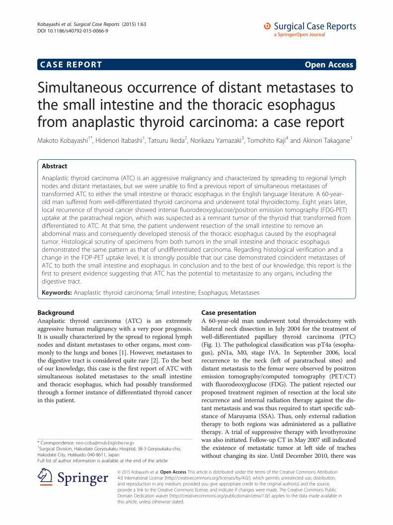
Kobayashi et al. Surgical Case Reports (2015) 1:63 DOI 10.1186/s40792-015-0066-9
CASE REPORT Open Access
Simultaneous occurrence of distant metastases tothe small intestine and the thoracic esophagusfrom anaplastic thyroid carcinoma: a case reportMakoto Kobayashi1*, Hidenori Itabashi1, Tatsuru Ikeda2, Norikazu Yamazaki3, Tomohito Kaji4 and Akinori Takagane1
Abstract
Anaplastic thyroid carcinoma (ATC) is an aggressive malignancy and characterized by spreading to regional lymphnodes and distant metastases, but we were unable to find a previous report of simultaneous metastases oftransformed ATC to either the small intestine or thoracic esophagus in the English language literature. A 60-year-old man suffered from well-differentiated thyroid carcinoma and underwent total thyroidectomy. Eight years later,local recurrence of thyroid cancer showed intense fluorodeoxyglucose/positron emission tomography (FDG-PET)uptake at the paratracheal region, which was suspected as a remnant tumor of the thyroid that transformed fromdifferentiated to ATC. At that time, the patient underwent resection of the small intestine to remove anabdominal mass and consequently developed stenosis of the thoracic esophagus caused by the esophagealtumor. Histological scrutiny of specimens from both tumors in the small intestine and thoracic esophagusdemonstrated the same pattern as that of undifferentiated carcinoma. Regarding histological verification and achange in the FDP-PET uptake level, it is strongly possible that our case demonstrated coincident metastases ofATC to both the small intestine and esophagus. In conclusion and to the best of our knowledge, this report is thefirst to present evidence suggesting that ATC has the potential to metastasize to any organs, including thedigestive tract.
Keywords: Anaplastic thyroid carcinoma; Small intestine; Esophagus; Metastases
BackgroundAnaplastic thyroid carcinoma (ATC) is an extremelyaggressive human malignancy with a very poor prognosis.It is usually characterized by the spread to regional lymphnodes and distant metastases to other organs, most com-monly to the lungs and bones [1]. However, metastases tothe digestive tract is considered quite rare [2]. To the bestof our knowledge, this case is the first report of ATC withsimultaneous isolated metastases to the small intestineand thoracic esophagus, which had possibly transformedthrough a former instance of differentiated thyroid cancerin this patient.
* Correspondence: [email protected] Division, Hakodate Goryoukaku Hospital, 38-3 Goryoukaku-cho,Hakodate City, Hokkaido 040-8611, JapanFull list of author information is available at the end of the article
© 2015 Kobayashi et al. Open Access This ar4.0 International License (http://creativecomand reproduction in any medium, providedprovide a link to the Creative Commons liceDomain Dedication waiver (http://creativecothis article, unless otherwise stated.
Case presentationA 60-year-old man underwent total thyroidectomy withbilateral neck dissection in July 2004 for the treatment ofwell-differentiated papillary thyroid carcinoma (PTC)(Fig. 1). The pathological classification was pT4a (esopha-gus), pN1a, M0, stage IVA. In September 2006, localrecurrence to the neck (left of paratracheal sites) anddistant metastasis to the femur were observed by positronemission tomography/computed tomography (PET/CT)with fluorodeoxyglucose (FDG). The patient rejected ourproposed treatment regimen of resection at the local siterecurrence and internal radiation therapy against the dis-tant metastasis and was thus required to start specific sub-stance of Maruyama (SSA). Thus, only external radiationtherapy to both regions was administered as a palliativetherapy. A trial of suppressive therapy with levothyroxinewas also initiated. Follow-up CT in May 2007 still indicatedthe existence of metastatic tumor at left side of tracheawithout changing its size. Until December 2010, there was
ticle is distributed under the terms of the Creative Commons Attributionmons.org/licenses/by/4.0/), which permits unrestricted use, distribution,you give appropriate credit to the original author(s) and the source,nse, and indicate if changes were made. The Creative Commons Publicmmons.org/publicdomain/zero/1.0/) applies to the data made available in
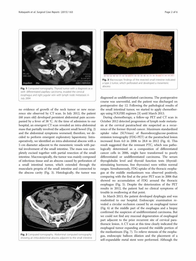
Fig. 1 Computed tomography. Thyroid tumor with a diagnosis as awell- differentiated papillary carcinoma, invaded the cervicalesophagus and right jugular vein with lymph node metastasis inJuly 2004
Fig. 3 Macroscopic findings of the resected small intestine indicateda type 2 tumor, which perforated and developed a mesentericabscess
Kobayashi et al. Surgical Case Reports (2015) 1:63 Page 2 of 6
no evidence of growth of the neck tumor or new recur-rence site observed by CT scan. In July 2012, the patient(68 years old) developed persistent abdominal pain accom-panied by a fever of 38 °C. At the time of admission to ourhospital, an emergent CT scan revealed an intra-abdominalmass that partially involved the adjacent small bowel (Fig. 2)and the abdominal symptoms worsened; therefore, we de-cided to perform emergent exploratory laparotomy. Intra-operatively, we identified an intra-abdominal abscess with a5 cm diameter adjacent to the mesenteric vessels with par-tial involvement of the small intestine. The mass was com-pletely excised together with partial resection of the smallintestine. Macroscopically, the tumor was mainly composedof infectious tissue and an abscess caused by perforation ofa small intestinal tumor, which extended through themuscularis propria of the small intestine and connected tothe abscess cavity (Fig. 3). Histologically, the tumor was
Fig. 2 Computed tomography. Abdominal computed tomographyshowing an intra-abdominal abscess adjacent to the small intestine
diagnosed as undifferentiated carcinoma. The postoperativecourse was uneventful, and the patient was discharged onpostoperative day 12. Following the pathological results ofthe small intestinal tumor, we started to apply chemother-apy using FOLFIRI regimen [3] until March 2013.During chemotherapy, a follow-up PET and CT scan in
October 2012 detected progression of lymph node metasta-sis at the cervical paratracheal site suspected as a recur-rence of the former thyroid cancer. Maximum standardizeduptake value (SUVmax) of fluorodeoxyglucose-positronemission tomography (FDG-PET) at the paratracheal lesionincreased from 6.0 in 2006 to 28.8 in 2012 (Fig. 4). Thisresult suggested that the remnant PTC, which was patho-logically determined as a composition of differentiatedcancer cells in 2006, might have transformed to poorlydifferentiated or undifferentiated carcinoma. The serumthyroglobulin level and thyroid function tests (thyroid-stimulating hormone, free thyroxine) were within normalranges. Simultaneously, FDG uptake of the thoracic esopha-gus at the middle mediastinum was observed positively,comparing with the find at the prior PET scan in 2006 thatshowed no accumulation of FDG around the thoracicesophagus (Fig. 5). Despite the deterioration of the PETresults in 2012, the patient had no clinical symptoms oftrouble in swallowing at that point.In March 2013, the patient developed dysphagia and was
readmitted to our hospital. Endoscopic examination re-vealed a circular occlusion caused by an esophageal tumor(Fig. 6) at the middle part of the esophagus and a biopsyconfirmed the suspicion of undifferentiated carcinoma. Butwe could not find any mucosal degeneration of esophagealpart adjacent to the prior recurrent site of cervical para-thoracic lesion. A CT scan at this time clearly indicated theesophageal tumor expanding around the middle portion ofthe mediastinum (Fig. 7). To relieve stenosis of the esopha-gus, endoscopic balloon dilation and the placement of aself-expandable metal stent were performed. Although the

Fig. 4 PET/CT depicting increased of FDG uptake at the paratracheal site. a PET/CT in 2006. b PET/CT in 2012
Fig. 5 PET/CT depicting increased of FDG uptake at the intrathoracic esophagus site. a PET/CT in 2006 showed no accumulation of FDG aroundthe middle mediastinum. b PET/CT in 2012 indicated FDG uptake on the thoracic esophagus and adjacent lymph node
Kobayashi et al. Surgical Case Reports (2015) 1:63 Page 3 of 6
patient was able to swallow food, he died 9 days afterendoscopic treatment due to dyspnea caused by trachealstenosis. Permission for an autopsy was denied.Histological scrutiny of both specimens from the small
intestine (Fig. 8) and esophagus (Fig. 9) revealed that thetumors were similarly composed of large anaplastic polyg-onal cells that were discohesive and tended to dissociatefrom each other with a prominent neutrophilic infiltrate.There were no patterns specific of adenocarcinoma, squa-mous cell carcinoma, or PTC. The antibodies used in thisstudy for immunohistochemical analysis included panker-atin (clone AE1/AE3), cytokeratin 7 (CK7), cytokeratin 20
Fig. 6 Endoscopic findings of esophageal stenosis showing acircular occlusion caused by the esophageal tumor
(CK20), high molecular weight keratin (HMW-CK, clone34Ebeta12), transcription termination factor 1 (TTF-1,clone 8G7G3/1), p53 (clone DO-7), and caudal typehomeobox 2 (CDX-2, clone DAX-CDX-2), which were allobtained from DakoCytomation (Glostrup, Denmark),and thyroglobulin (polyclonal; Nichirei Biosciences, Inc.,Tokyo, Japan). Cells from both tumors were stronglypositive for pankeratin and p53 and negative for CK7,CK20, HMW-CK, CDX-2, TTF-1, and thyroglobulin(Table 1).The pathological findings of the esophageal tumor were
almost identical to those of the small intestinal tumor,which were both diagnosed as ATC. Considering the clin-ical progression of disease in this patient, we concludedthat both tumors were derived from the same origin—a
Fig. 7 Computed tomography scan of thoracic esophageal tumorgrowing in the middle portion of the mediastinum at the timewhen the patient suffered from severe dysphagia
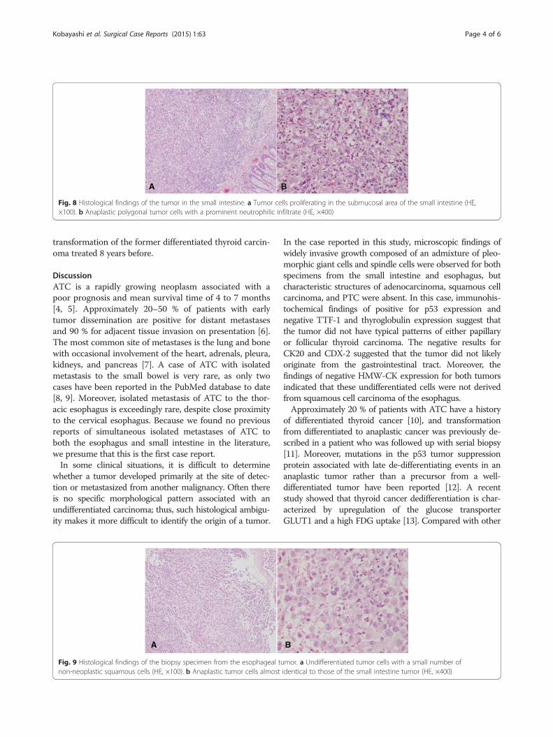
Fig. 8 Histological findings of the tumor in the small intestine. a Tumor cells proliferating in the submucosal area of the small intestine (HE,×100). b Anaplastic polygonal tumor cells with a prominent neutrophilic infiltrate (HE, ×400)
Kobayashi et al. Surgical Case Reports (2015) 1:63 Page 4 of 6
transformation of the former differentiated thyroid carcin-oma treated 8 years before.
DiscussionATC is a rapidly growing neoplasm associated with apoor prognosis and mean survival time of 4 to 7 months[4, 5]. Approximately 20–50 % of patients with earlytumor dissemination are positive for distant metastasesand 90 % for adjacent tissue invasion on presentation [6].The most common site of metastases is the lung and bonewith occasional involvement of the heart, adrenals, pleura,kidneys, and pancreas [7]. A case of ATC with isolatedmetastasis to the small bowel is very rare, as only twocases have been reported in the PubMed database to date[8, 9]. Moreover, isolated metastasis of ATC to the thor-acic esophagus is exceedingly rare, despite close proximityto the cervical esophagus. Because we found no previousreports of simultaneous isolated metastases of ATC toboth the esophagus and small intestine in the literature,we presume that this is the first case report.In some clinical situations, it is difficult to determine
whether a tumor developed primarily at the site of detec-tion or metastasized from another malignancy. Often thereis no specific morphological pattern associated with anundifferentiated carcinoma; thus, such histological ambigu-ity makes it more difficult to identify the origin of a tumor.
Fig. 9 Histological findings of the biopsy specimen from the esophageal tunon-neoplastic squamous cells (HE, ×100). b Anaplastic tumor cells almost
In the case reported in this study, microscopic findings ofwidely invasive growth composed of an admixture of pleo-morphic giant cells and spindle cells were observed for bothspecimens from the small intestine and esophagus, butcharacteristic structures of adenocarcinoma, squamous cellcarcinoma, and PTC were absent. In this case, immunohis-tochemical findings of positive for p53 expression andnegative TTF-1 and thyroglobulin expression suggest thatthe tumor did not have typical patterns of either papillaryor follicular thyroid carcinoma. The negative results forCK20 and CDX-2 suggested that the tumor did not likelyoriginate from the gastrointestinal tract. Moreover, thefindings of negative HMW-CK expression for both tumorsindicated that these undifferentiated cells were not derivedfrom squamous cell carcinoma of the esophagus.Approximately 20 % of patients with ATC have a history
of differentiated thyroid cancer [10], and transformationfrom differentiated to anaplastic cancer was previously de-scribed in a patient who was followed up with serial biopsy[11]. Moreover, mutations in the p53 tumor suppressionprotein associated with late de-differentiating events in ananaplastic tumor rather than a precursor from a well-differentiated tumor have been reported [12]. A recentstudy showed that thyroid cancer dedifferentiation is char-acterized by upregulation of the glucose transporterGLUT1 and a high FDG uptake [13]. Compared with other
mor. a Undifferentiated tumor cells with a small number ofidentical to those of the small intestine tumor (HE, ×400)
Table 1 Results of tumor location and immunohistochemicalmarkers
Tumor location/markers
Pankeratin p53 CK7 CK20 HMW-CK
CDX-2
TTF-1
Tg
Esophagus + + − − − − − −
Small intestine + + − − − − − −
pankeratin AE1/AE3, CK cytokeratin, HMW high molecular weight,Tg thyroglobulin
Kobayashi et al. Surgical Case Reports (2015) 1:63 Page 5 of 6
imaging modalities, PET may improve disease detectionand better facilitate the management of patients with ATC[14]. The American Thyroid Association recommendsFDG-PET/CT for the evaluation of metastatic disease andsuggests that this modality may be useful to distinguishATC from PTC according to the SUVmax level because ofthe higher FDG uptake in the former [15]. In the presentcase, the patient underwent treatment for advanced PTC8 years before presentation, after which local recurrencewith bone metastasis from the original PTC was identified.The latest FGD-PET showed intense uptake in a region oflocal recurrence of pre-existing differentiated thyroid can-cer. The finding of increased activity, as indicated by aSUVmax value from 6 to 29, clearly suggests a possibletransformation from differentiated thyroid cancer to ATC.The uptake of PET around the tumors of the intestine andesophagus also exhibited the same level as that of a dedif-ferentiated lesion.
ConclusionsTechnically, it is very difficult to confirm whether thetumor developed whether primarily or secondarily fromanother malignancy, if both tumors show the same patho-logical character. Unfortunately, we could not get permis-sion for an autopsy and were compelled to make a finaldiagnosis from clinical evidence. But in conclusion, basedon the histological verification and the degree of FDPuptake on PET, it is very likely that in our case, coincidentmetastases of ATC to both the small intestine and thoracicesophagus occurred. This case is unique because therewere no previous reports on simultaneous metastases tothese two organs from transformed ATC. Therefore, thisreport presents the first evidence suggesting that ATC hasthe potential to metastasize to any organs, including thedigestive tract.
ConsentWritten informed consent was obtained from the patient’sfamily for this case report and any accompanying images.A copy of the written consent is available for review bythe Editor-in-Chief of this journal.
AbbreviationsATC: anaplastic thyroid carcinoma; CDX-2: caudal type homeobox 2;CK: cytokeratin; FDG-PET: fluorodeoxyglucose-positron emission tomography;
HMW-CK: high molecular weight cytokeratin; PTC: papillary thyroidcarcinoma; TTF-1: transcription terminal factor 1.
Competing interestsThe authors declare that they have no competing interests.
Authors’ contributionMK have made substantial contributions and is the corresponding author. HIparticipated in the acquisition of clinical data and images of the patient. TIcarried out the histological verification and gave pathology advice. NY is adoctor of otorhinolaryngology and was involved in the first surgicaltreatment, which was a total thyroidectomy and an additional neckdissection. TK, a radiologist, carried out scrutiny using PET/CT and evaluatedthe date and image in this article. AT has been involved in revising themanuscript for important intellectual content. All authors read and approvedthe final manuscript.
AcknowledgementsThe authors would like to thank Enago (www.enago.jp) for the Englishlanguage review.
Author details1Surgical Division, Hakodate Goryoukaku Hospital, 38-3 Goryoukaku-cho,Hakodate City, Hokkaido 040-8611, Japan. 2Pathological Division, HakodateGoryoukaku Hospital, 38-3 Goryoukaku-cho, Hakodate City, Hokkaido040-8611, Japan. 3Division of Otorhinolaryngology, Hakodate GoryoukakuHospital, 38-3 Goryoukaku-cho, Hakodate City, Hokkaido 040-8611, Japan.4Division of Radiology, Hakodate Goryoukaku Hospital, 38-3 Goryoukaku-cho,Hakodate City, Hokkaido 040-8611, Japan.
Received: 18 December 2014 Accepted: 29 July 2015
References1. Carcangiu ML, Steeper T, Zampi G, Rosai J. Anaplastic thyroid carcinoma. A
study of 70 cases. Am J Clin Pathol. 1985;83(2):135–58.2. Yazaki N, Naitoh T, Miura K, Ogawa H, Kinouchi M, Tanaka N, et al. A case
report of anaplastic thyroid carcinoma metastasized to the small intestinecausing intussusception. Jpn J Gastroenterol Surg. 2011;44(11):1426–33.
3. Zaanan A, Gauthier M, Malka D, Locher C, Gornet JM, Thirot-Bidault A, et al.Second-line chemotherapy with fluorouracil, leucovorin, and irinotecan(FOLFIRI regimen) in patients with advanced small bowel adenocarcinomaafter failure of first-lineplatinum-based chemotherapy: a multicenter AGEO study. Cancer.2011;117(7):1422–8.
4. Nel CJ, van Heerden JA, Goellner JR, Gharib H, McConahey WM, Taylor WF,et al. Anaplastic carcinoma of the thyroid: a clinicopathologic study of 82cases. Mayo Clin Proc. 1985;60(1):51–8.
5. Venkatesh YS, Ordonez NG, Schultz PN, Hickey RC, Goepfert H, Samaan NA.Anaplastic carcinoma of the thyroid. A clinicopathologic study of 121 cases.Cancer. 1990;66(2):321–30.
6. O’Neill JP, Shaha AR. Anaplastic thyroid cancer. Oral Oncol. 2013;49(7):702–6.7. Silverberg SG, Hutter RV, Foote Jr FW. Fatal carcinoma of the thyroid:
histology, metastases, and causes of death. Cancer. 1970;25(4):792–802.8. Phillips DL, Benner KG, Keeffe EB, Traweek ST. Isolated metastasis to small
bowel from anaplastic thyroid carcinoma. With a review of extra-abdominalmalignancies that spread to the bowel. J Clin Gastroenterol.1987;9(5):563–7.
9. Ricciardelli L, Rapicano G, Pinto A, Napolitano G, Feleppa C, Martino G, et al.Small bowel inussusception caused by metastasis from anaplastic thyroidcarcinoma: case report and literature review. Ann Ital Chir. 2006;77(1):63–7.
10. McIver B, Hay ID, Giuffrida DF, et al. Anaplastic thyroid carcinoma: a 50-yearexperience at a single institution. Surgery. 2001;130(6):1028–34.
11. Moore Jr JH, Bacharach B, Choi HY. Anaplastic transformation of metastaticfollicular carcinoma of the thyroid. J Surg Oncol. 1985;29(4):216–21.
12. Moretti F, Farsetti A, Soddu S, Misiti S, Crescenzi M, Filetti S, et al. p53re-expression inhibits proliferation and restores differentiation of humanthyroid anaplastic carcinoma cells. Oncogene. 1997;14(6):729–40.
13. Grabellus F, Nagarajah J, Bockisch A, Schmid KW, Sheu SY. Glucosetransporter 1 expression, tumor proliferation, and iodine/glucose uptake in

Kobayashi et al. Surgical Case Reports (2015) 1:63 Page 6 of 6
thyroid cancer with emphasis on poorly differentiated thyroid carcinoma.Clin Nucl Med. 2012;37(2):121–7.
14. Bogsrud TV, Karantanis D, Nathan MA, Mullan BP, Wiseman GA, KasperbauerJL, et al. 18F-FDG PET in the management of patients with anaplasticthyroid carcinoma. Thyroid. 2008;18(7):713–9.
15. Smallridge RC, Ain KB, Asa SL, Bible KC, Brierley JD, Burman KD, et al.American association guidelines for management of patients withanaplastic thyroid cancer. Thyroid. 2012;22(11):1104–39.
Submit your manuscript to a journal and benefi t from:
7 Convenient online submission
7 Rigorous peer review
7 Immediate publication on acceptance
7 Open access: articles freely available online
7 High visibility within the fi eld
7 Retaining the copyright to your article
Submit your next manuscript at 7 springeropen.com


![University of Groningen Modern view on multimodality ... · cancer (EC), distant metastases are common and seem to be associated with venous inva-sion (VI) [1, 2]. Current TNM classifications](https://img.pdfslide.net/doc/110x75/5f2fdadd770424727d70ba4b/university-of-groningen-modern-view-on-multimodality-cancer-ec-distant-metastases.jpg)


![Promoter hypermethylation profiling of distant breast ... · phenotype of distant breast cancer metastases [14–16]. Extensive knowledge of the hypermethylation status of tumor suppressor](https://img.pdfslide.net/doc/110x75/5d21f00788c993722e8c67ea/promoter-hypermethylation-profiling-of-distant-breast-phenotype-of-distant.jpg)