Embed Size (px)
Citation preview
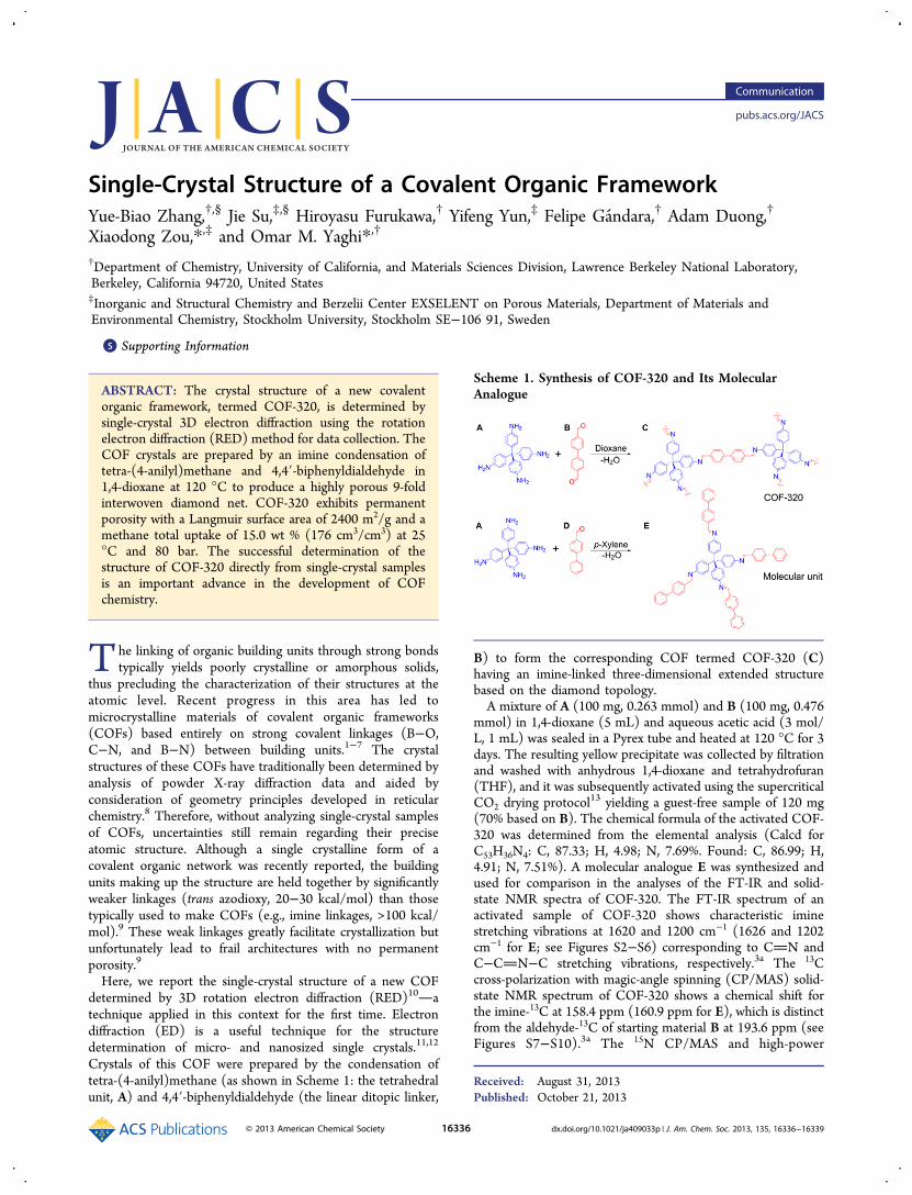
Single-Crystal Structure of a Covalent Organic FrameworkYue-Biao Zhang,†,§ Jie Su,‡,§ Hiroyasu Furukawa,† Yifeng Yun,‡ Felipe Gandara,† Adam Duong,†
Xiaodong Zou,*,‡ and Omar M. Yaghi*,†
†Department of Chemistry, University of California, and Materials Sciences Division, Lawrence Berkeley National Laboratory,Berkeley, California 94720, United States‡Inorganic and Structural Chemistry and Berzelii Center EXSELENT on Porous Materials, Department of Materials andEnvironmental Chemistry, Stockholm University, Stockholm SE−106 91, Sweden
*S Supporting Information
ABSTRACT: The crystal structure of a new covalentorganic framework, termed COF-320, is determined bysingle-crystal 3D electron diffraction using the rotationelectron diffraction (RED) method for data collection. TheCOF crystals are prepared by an imine condensation oftetra-(4-anilyl)methane and 4,4′-biphenyldialdehyde in1,4-dioxane at 120 °C to produce a highly porous 9-foldinterwoven diamond net. COF-320 exhibits permanentporosity with a Langmuir surface area of 2400 m2/g and amethane total uptake of 15.0 wt % (176 cm3/cm3) at 25°C and 80 bar. The successful determination of thestructure of COF-320 directly from single-crystal samplesis an important advance in the development of COFchemistry.
The linking of organic building units through strong bondstypically yields poorly crystalline or amorphous solids,
thus precluding the characterization of their structures at theatomic level. Recent progress in this area has led tomicrocrystalline materials of covalent organic frameworks(COFs) based entirely on strong covalent linkages (B−O,C−N, and B−N) between building units.1−7 The crystalstructures of these COFs have traditionally been determined byanalysis of powder X-ray diffraction data and aided byconsideration of geometry principles developed in reticularchemistry.8 Therefore, without analyzing single-crystal samplesof COFs, uncertainties still remain regarding their preciseatomic structure. Although a single crystalline form of acovalent organic network was recently reported, the buildingunits making up the structure are held together by significantlyweaker linkages (trans azodioxy, 20−30 kcal/mol) than thosetypically used to make COFs (e.g., imine linkages, >100 kcal/mol).9 These weak linkages greatly facilitate crystallization butunfortunately lead to frail architectures with no permanentporosity.9
Here, we report the single-crystal structure of a new COFdetermined by 3D rotation electron diffraction (RED)10atechnique applied in this context for the first time. Electrondiffraction (ED) is a useful technique for the structuredetermination of micro- and nanosized single crystals.11,12
Crystals of this COF were prepared by the condensation oftetra-(4-anilyl)methane (as shown in Scheme 1: the tetrahedralunit, A) and 4,4′-biphenyldialdehyde (the linear ditopic linker,
B) to form the corresponding COF termed COF-320 (C)having an imine-linked three-dimensional extended structurebased on the diamond topology.A mixture of A (100 mg, 0.263 mmol) and B (100 mg, 0.476
mmol) in 1,4-dioxane (5 mL) and aqueous acetic acid (3 mol/L, 1 mL) was sealed in a Pyrex tube and heated at 120 °C for 3days. The resulting yellow precipitate was collected by filtrationand washed with anhydrous 1,4-dioxane and tetrahydrofuran(THF), and it was subsequently activated using the supercriticalCO2 drying protocol13 yielding a guest-free sample of 120 mg(70% based on B). The chemical formula of the activated COF-320 was determined from the elemental analysis (Calcd forC53H36N4: C, 87.33; H, 4.98; N, 7.69%. Found: C, 86.99; H,4.91; N, 7.51%). A molecular analogue E was synthesized andused for comparison in the analyses of the FT-IR and solid-state NMR spectra of COF-320. The FT-IR spectrum of anactivated sample of COF-320 shows characteristic iminestretching vibrations at 1620 and 1200 cm−1 (1626 and 1202cm−1 for E; see Figures S2−S6) corresponding to CN andC−CN−C stretching vibrations, respectively.3a The 13Ccross-polarization with magic-angle spinning (CP/MAS) solid-state NMR spectrum of COF-320 shows a chemical shift forthe imine-13C at 158.4 ppm (160.9 ppm for E), which is distinctfrom the aldehyde-13C of starting material B at 193.6 ppm (seeFigures S7−S10).3a The 15N CP/MAS and high-power
Received: August 31, 2013Published: October 21, 2013
Scheme 1. Synthesis of COF-320 and Its MolecularAnalogue
Communication
pubs.acs.org/JACS
© 2013 American Chemical Society 16336 dx.doi.org/10.1021/ja409033p | J. Am. Chem. Soc. 2013, 135, 16336−16339
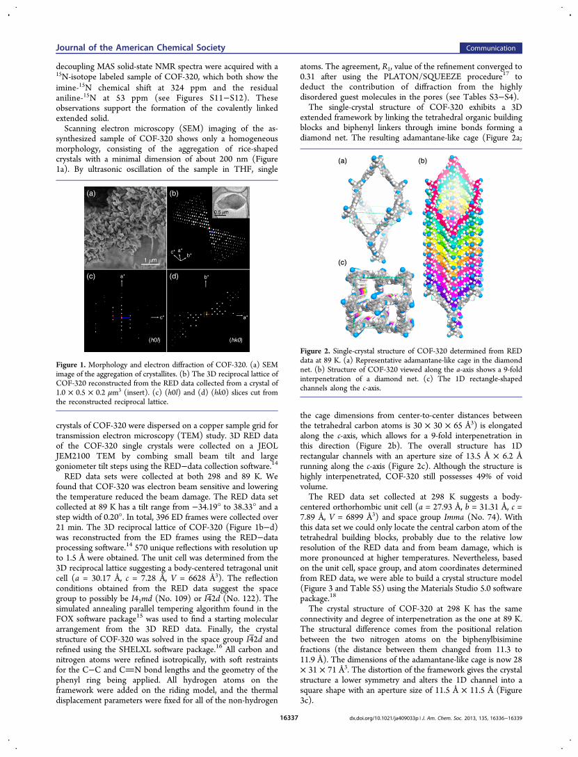
decoupling MAS solid-state NMR spectra were acquired with a15N-isotope labeled sample of COF-320, which both show theimine-15N chemical shift at 324 ppm and the residualaniline-15N at 53 ppm (see Figures S11−S12). Theseobservations support the formation of the covalently linkedextended solid.Scanning electron microscopy (SEM) imaging of the as-
synthesized sample of COF-320 shows only a homogeneousmorphology, consisting of the aggregation of rice-shapedcrystals with a minimal dimension of about 200 nm (Figure1a). By ultrasonic oscillation of the sample in THF, single
crystals of COF-320 were dispersed on a copper sample grid fortransmission electron microscopy (TEM) study. 3D RED dataof the COF-320 single crystals were collected on a JEOLJEM2100 TEM by combing small beam tilt and largegoniometer tilt steps using the RED−data collection software.14RED data sets were collected at both 298 and 89 K. We
found that COF-320 was electron beam sensitive and loweringthe temperature reduced the beam damage. The RED data setcollected at 89 K has a tilt range from −34.19° to 38.33° and astep width of 0.20°. In total, 396 ED frames were collected over21 min. The 3D reciprocal lattice of COF-320 (Figure 1b−d)was reconstructed from the ED frames using the RED−dataprocessing software.14 570 unique reflections with resolution upto 1.5 Å were obtained. The unit cell was determined from the3D reciprocal lattice suggesting a body-centered tetragonal unitcell (a = 30.17 Å, c = 7.28 Å, V = 6628 Å3). The reflectionconditions obtained from the RED data suggest the spacegroup to possibly be I41md (No. 109) or I4 2d (No. 122). Thesimulated annealing parallel tempering algorithm found in theFOX software package15 was used to find a starting moleculararrangement from the 3D RED data. Finally, the crystalstructure of COF-320 was solved in the space group I4 2d andrefined using the SHELXL software package.16 All carbon andnitrogen atoms were refined isotropically, with soft restraintsfor the C−C and CN bond lengths and the geometry of thephenyl ring being applied. All hydrogen atoms on theframework were added on the riding model, and the thermaldisplacement parameters were fixed for all of the non-hydrogen
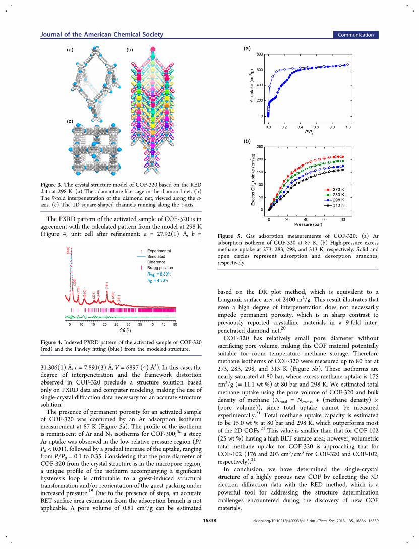
atoms. The agreement, R1, value of the refinement converged to0.31 after using the PLATON/SQUEEZE procedure17 todeduct the contribution of diffraction from the highlydisordered guest molecules in the pores (see Tables S3−S4).The single-crystal structure of COF-320 exhibits a 3D
extended framework by linking the tetrahedral organic buildingblocks and biphenyl linkers through imine bonds forming adiamond net. The resulting adamantane-like cage (Figure 2a;
the cage dimensions from center-to-center distances betweenthe tetrahedral carbon atoms is 30 × 30 × 65 Å3) is elongatedalong the c-axis, which allows for a 9-fold interpenetration inthis direction (Figure 2b). The overall structure has 1Drectangular channels with an aperture size of 13.5 Å × 6.2 Årunning along the c-axis (Figure 2c). Although the structure ishighly interpenetrated, COF-320 still possesses 49% of voidvolume.The RED data set collected at 298 K suggests a body-
centered orthorhombic unit cell (a = 27.93 Å, b = 31.31 Å, c =7.89 Å, V = 6899 Å3) and space group Imma (No. 74). Withthis data set we could only locate the central carbon atom of thetetrahedral building blocks, probably due to the relative lowresolution of the RED data and from beam damage, which ismore pronounced at higher temperatures. Nevertheless, basedon the unit cell, space group, and atom coordinates determinedfrom RED data, we were able to build a crystal structure model(Figure 3 and Table S5) using the Materials Studio 5.0 softwarepackage.18
The crystal structure of COF-320 at 298 K has the sameconnectivity and degree of interpenetration as the one at 89 K.The structural difference comes from the positional relationbetween the two nitrogen atoms on the biphenylbisiminefractions (the distance between them changed from 11.3 to11.9 Å). The dimensions of the adamantane-like cage is now 28× 31 × 71 Å3. The distortion of the framework gives the crystalstructure a lower symmetry and alters the 1D channel into asquare shape with an aperture size of 11.5 Å × 11.5 Å (Figure3c).
Figure 1. Morphology and electron diffraction of COF-320. (a) SEMimage of the aggregation of crystallites. (b) The 3D reciprocal lattice ofCOF-320 reconstructed from the RED data collected from a crystal of1.0 × 0.5 × 0.2 μm3 (insert). (c) (h0l) and (d) (hk0) slices cut fromthe reconstructed reciprocal lattice.
Figure 2. Single-crystal structure of COF-320 determined from REDdata at 89 K. (a) Representative adamantane-like cage in the diamondnet. (b) Structure of COF-320 viewed along the a-axis shows a 9-foldinterpenetration of a diamond net. (c) The 1D rectangle-shapedchannels along the c-axis.
Journal of the American Chemical Society Communication
dx.doi.org/10.1021/ja409033p | J. Am. Chem. Soc. 2013, 135, 16336−1633916337
The PXRD pattern of the activated sample of COF-320 is inagreement with the calculated pattern from the model at 298 K(Figure 4; unit cell after refinement: a = 27.92(1) Å, b =
31.306(1) Å, c = 7.891(3) Å, V = 6897 (4) Å3). In this case, thedegree of interpenetration and the framework distortionobserved in COF-320 preclude a structure solution basedonly on PXRD data and computer modeling, making the use ofsingle-crystal diffraction data necessary for an accurate structuresolution.The presence of permanent porosity for an activated sample
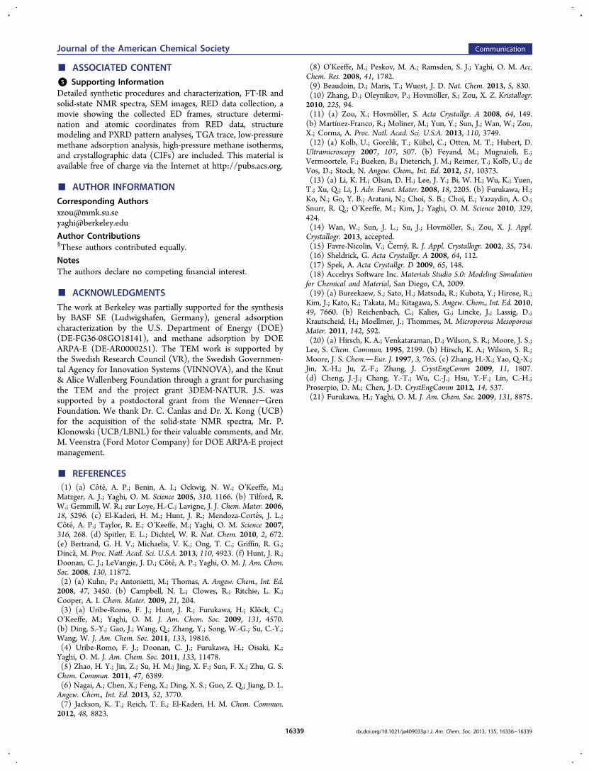
of COF-320 was confirmed by an Ar adsorption isothermmeasurement at 87 K (Figure 5a). The profile of the isothermis reminiscent of Ar and N2 isotherms for COF-300;3a a steepAr uptake was observed in the low relative pressure region (P/P0 < 0.01), followed by a gradual increase of the uptake, rangingfrom P/P0 = 0.1 to 0.35. Considering that the pore diameter ofCOF-320 from the crystal structure is in the micropore region,a unique profile of the isotherm accompanying a significanthysteresis loop is attributable to a guest-induced structuraltransformation and/or reorientation of the guest packing underincreased pressure.19 Due to the presence of steps, an accurateBET surface area estimation from the adsorption branch is notapplicable. A pore volume of 0.81 cm3/g can be estimated
based on the DR plot method, which is equivalent to aLangmuir surface area of 2400 m2/g. This result illustrates thateven a high degree of interpenetration does not necessarilyimpede permanent porosity, which is in sharp contrast topreviously reported crystalline materials in a 9-fold inter-penetrated diamond net.20
COF-320 has relatively small pore diameter withoutsacrificing pore volume, making this COF material potentiallysuitable for room temperature methane storage. Thereforemethane isotherms of COF-320 were measured up to 80 bar at273, 283, 298, and 313 K (Figure 5b). These isotherms arenearly saturated at 80 bar, where excess methane uptake is 175cm3/g (= 11.1 wt %) at 80 bar and 298 K. We estimated totalmethane uptake using the pore volume of COF-320 and bulkdensity of methane (Ntotal = Nexcess + (methane density) ×(pore volume)), since total uptake cannot be measuredexperimentally.21 Total methane uptake capacity is estimatedto be 15.0 wt % at 80 bar and 298 K, which outperforms mostof the 2D COFs.21 This value is smaller than that for COF-102(25 wt %) having a high BET surface area; however, volumetrictotal methane uptake for COF-320 is approaching that forCOF-102 (176 and 203 cm3/cm3 for COF-320 and COF-102,respectively).21
In conclusion, we have determined the single-crystalstructure of a highly porous new COF by collecting the 3Delectron diffraction data with the RED method, which is apowerful tool for addressing the structure determinationchallenges encountered during the discovery of new COFmaterials.
Figure 3. The crystal structure model of COF-320 based on the REDdata at 298 K. (a) The adamantane-like cage in the diamond net. (b)The 9-fold interpenetration of the diamond net, viewed along the a-axis. (c) The 1D square-shaped channels running along the c-axis.
Figure 4. Indexed PXRD pattern of the activated sample of COF-320(red) and the Pawley fitting (blue) from the modeled structure.
Figure 5. Gas adsorption measurements of COF-320: (a) Aradsorption isotherm of COF-320 at 87 K. (b) High-pressure excessmethane uptake at 273, 283, 298, and 313 K, respectively. Solid andopen circles represent adsorption and desorption branches,respectively.
Journal of the American Chemical Society Communication
dx.doi.org/10.1021/ja409033p | J. Am. Chem. Soc. 2013, 135, 16336−1633916338
■ ASSOCIATED CONTENT*S Supporting InformationDetailed synthetic procedures and characterization, FT-IR andsolid-state NMR spectra, SEM images, RED data collection, amovie showing the collected ED frames, structure determi-nation and atomic coordinates from RED data, structuremodeling and PXRD pattern analyses, TGA trace, low-pressuremethane adsorption analysis, high-pressure methane isotherms,and crystallographic data (CIFs) are included. This material isavailable free of charge via the Internet at http://pubs.acs.org.
■ AUTHOR INFORMATIONCorresponding [email protected]@berkeley.edu
Author Contributions§These authors contributed equally.
NotesThe authors declare no competing financial interest.
■ ACKNOWLEDGMENTSThe work at Berkeley was partially supported for the synthesisby BASF SE (Ludwigshafen, Germany), general adsorptioncharacterization by the U.S. Department of Energy (DOE)(DE-FG36-08GO18141), and methane adsorption by DOEARPA-E (DE-AR0000251). The TEM work is supported bythe Swedish Research Council (VR), the Swedish Governmen-tal Agency for Innovation Systems (VINNOVA), and the Knut& Alice Wallenberg Foundation through a grant for purchasingthe TEM and the project grant 3DEM-NATUR. J.S. wassupported by a postdoctoral grant from the Wenner−GrenFoundation. We thank Dr. C. Canlas and Dr. X. Kong (UCB)for the acquisition of the solid-state NMR spectra, Mr. P.Klonowski (UCB/LBNL) for their valuable comments, and Mr.M. Veenstra (Ford Motor Company) for DOE ARPA-E projectmanagement.
■ REFERENCES(1) (a) Cote, A. P.; Benin, A. I.; Ockwig, N. W.; O’Keeffe, M.;Matzger, A. J.; Yaghi, O. M. Science 2005, 310, 1166. (b) Tilford, R.W.; Gemmill, W. R.; zur Loye, H.-C.; Lavigne, J. J. Chem. Mater. 2006,18, 5296. (c) El-Kaderi, H. M.; Hunt, J. R.; Mendoza-Cortes, J. L.;Cote, A. P.; Taylor, R. E.; O’Keeffe, M.; Yaghi, O. M. Science 2007,316, 268. (d) Spitler, E. L.; Dichtel, W. R. Nat. Chem. 2010, 2, 672.(e) Bertrand, G. H. V.; Michaelis, V. K.; Ong, T. C.; Griffin, R. G.;Dinca, M. Proc. Natl. Acad. Sci. U.S.A. 2013, 110, 4923. (f) Hunt, J. R.;Doonan, C. J.; LeVangie, J. D.; Cote, A. P.; Yaghi, O. M. J. Am. Chem.Soc. 2008, 130, 11872.(2) (a) Kuhn, P.; Antonietti, M.; Thomas, A. Angew. Chem., Int. Ed.2008, 47, 3450. (b) Campbell, N. L.; Clowes, R.; Ritchie, L. K.;Cooper, A. I. Chem. Mater. 2009, 21, 204.(3) (a) Uribe-Romo, F. J.; Hunt, J. R.; Furukawa, H.; Klock, C.;O’Keeffe, M.; Yaghi, O. M. J. Am. Chem. Soc. 2009, 131, 4570.(b) Ding, S.-Y.; Gao, J.; Wang, Q.; Zhang, Y.; Song, W.-G.; Su, C.-Y.;Wang, W. J. Am. Chem. Soc. 2011, 133, 19816.(4) Uribe-Romo, F. J.; Doonan, C. J.; Furukawa, H.; Oisaki, K.;Yaghi, O. M. J. Am. Chem. Soc. 2011, 133, 11478.(5) Zhao, H. Y.; Jin, Z.; Su, H. M.; Jing, X. F.; Sun, F. X.; Zhu, G. S.Chem. Commun. 2011, 47, 6389.(6) Nagai, A.; Chen, X.; Feng, X.; Ding, X. S.; Guo, Z. Q.; Jiang, D. L.Angew. Chem., Int. Ed. 2013, 52, 3770.(7) Jackson, K. T.; Reich, T. E.; El-Kaderi, H. M. Chem. Commun.2012, 48, 8823.
(8) O’Keeffe, M.; Peskov, M. A.; Ramsden, S. J.; Yaghi, O. M. Acc.Chem. Res. 2008, 41, 1782.(9) Beaudoin, D.; Maris, T.; Wuest, J. D. Nat. Chem. 2013, 5, 830.(10) Zhang, D.; Oleynikov, P.; Hovmoller, S.; Zou, X. Z. Kristallogr.2010, 225, 94.(11) (a) Zou, X.; Hovmoller, S. Acta Crystallgr. A 2008, 64, 149.(b) Martínez-Franco, R.; Moliner, M.; Yun, Y.; Sun, J.; Wan, W.; Zou,X.; Corma, A. Proc. Natl. Acad. Sci. U.S.A. 2013, 110, 3749.(12) (a) Kolb, U.; Gorelik, T.; Kubel, C.; Otten, M. T.; Hubert, D.Ultramicroscopy 2007, 107, 507. (b) Feyand, M.; Mugnaioli, E.;Vermoortele, F.; Bueken, B.; Dieterich, J. M.; Reimer, T.; Kolb, U.; deVos, D.; Stock, N. Angew. Chem., Int. Ed. 2012, 51, 10373.(13) (a) Li, K. H.; Olsan, D. H.; Lee, J. Y.; Bi, W. H.; Wu, K.; Yuen,T.; Xu, Q.; Li, J. Adv. Funct. Mater. 2008, 18, 2205. (b) Furukawa, H.;Ko, N.; Go, Y. B.; Aratani, N.; Choi, S. B.; Choi, E.; Yazaydin, A. O.;Snurr, R. Q.; O’Keeffe, M.; Kim, J.; Yaghi, O. M. Science 2010, 329,424.(14) Wan, W.; Sun, J. L.; Su, J.; Hovmoller, S.; Zou, X. J. Appl.Crystallogr. 2013, accepted.(15) Favre-Nicolin, V.; Cerny, R. J. Appl. Crystallogr. 2002, 35, 734.(16) Sheldrick, G. Acta Crystallgr. A 2008, 64, 112.(17) Spek, A. Acta Crystallgr. D 2009, 65, 148.(18) Accelrys Software Inc. Materials Studio 5.0: Modeling Simulationfor Chemical and Material, San Diego, CA, 2009.(19) (a) Bureekaew, S.; Sato, H.; Matsuda, R.; Kubota, Y.; Hirose, R.;Kim, J.; Kato, K.; Takata, M.; Kitagawa, S. Angew. Chem., Int. Ed. 2010,49, 7660. (b) Reichenbach, C.; Kalies, G.; Lincke, J.; Lassig, D.;Krautscheid, H.; Moellmer, J.; Thommes, M. Microporous MesoporousMater. 2011, 142, 592.(20) (a) Hirsch, K. A.; Venkataraman, D.; Wilson, S. R.; Moore, J. S.;Lee, S. Chem. Commun. 1995, 2199. (b) Hirsch, K. A.; Wilson, S. R.;Moore, J. S. Chem.Eur. J. 1997, 3, 765. (c) Zhang, H.-X.; Yao, Q.-X.;Jin, X.-H.; Ju, Z.-F.; Zhang, J. CrystEngComm 2009, 11, 1807.(d) Cheng, J.-J.; Chang, Y.-T.; Wu, C.-J.; Hsu, Y.-F.; Lin, C.-H.;Proserpio, D. M.; Chen, J.-D. CrystEngComm 2012, 14, 537.(21) Furukawa, H.; Yaghi, O. M. J. Am. Chem. Soc. 2009, 131, 8875.
Journal of the American Chemical Society Communication
dx.doi.org/10.1021/ja409033p | J. Am. Chem. Soc. 2013, 135, 16336−1633916339