Embed Size (px)
Citation preview

Single krone påtannimplantat & estetikk
Asbjørn JokstadInstitutt for klinisk odontologi
UiT Norges arktiske universitet

A satisfactory esthetic outcome?
1. Evaluation of esthetic outcomes

A satisfactory esthetic outcome?High smile line A.K.A. “Gummy smile”
Low smile line

A satisfactory esthetic outcome?
Fava et al. COIR 2015

A satisfactory esthetic outcome?
Fava et al. COIR 2015

Evaluation systems to appraise the qualities of the soft tissues in patients having received a single crown
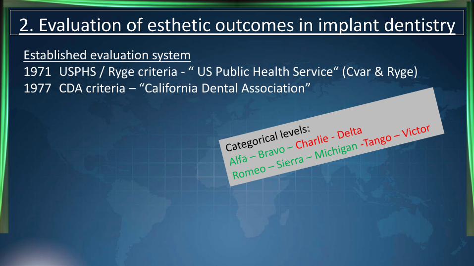
Established evaluation system 1971 USPHS / Ryge criteria - “ US Public Health Service“ (Cvar & Ryge)1977 CDA criteria – “California Dental Association”
2. Evaluation of esthetic outcomes in implant dentistry

“PINK” criteria
Established categorical evaluation system 1971 USPHS criteria - “ US Public Health Service“ (Cvar & Ryge)1977 CDA criteria – “California Dental Association”Specifically to implant-retained reconstructions in the esthetic zones2005 ICAI - “Implant Crown Aesthetic Index” (Meijer et al. COIR)
1. Mesiodistal dimension of the crown: must be in harmony with the adjacent and contralateral tooth, 5-points (gross - slight undercontour- no deviation - slight - gross overcontour)
2. Position of the incisal edge of the crown: must be in harmony with the adjacent and contralateral tooth, 5-points (gross - slight undercontour- no deviation - slight - gross overcontour)
3. Labial convexity of the crown: must be in harmony with the adjacent and contralateral tooth, 5-points (gross - slight undercontour- no deviation - slight - gross overcontour)
4. Colour and translucency of the crown: must be in harmony with the adjacent and contralateral tooth, 3-points (gross -slight -no mismatch)
5. Surface of the crown: characteristics of the crown such as roughness and ridges must be in harmony with the adjacent and contralateral tooth, 3-points (gross -slight -no mismatch)
1&2 Position of mucosa in the approximal embrasures: must be in their natural position, 3-points (deviation ≥1.5 mm- <1.5 mm- no deviation)
3 Position of the labial margin of the peri-implant mucosa: must be at the same level as the contralateral tooth and in harmony with the adjacent teeth, 3-points (deviation ≥1.5 mm- <1.5 mm- no deviation)
4&5 Contour of the labial surface of the mucosa: must be in harmony with the adjacent and contralateral tooth, 5-points (gross - slight undercontoured - no deviation - slight - gross overcontoured)
6&7 Colour and surface of the labial mucosa: must be in harmony with the adjacent and contralateral tooth and must have a natural appearance, 3-points (gross -li h i h)
12 34&5
1
2
3
4 &5
2. Evaluation of esthetic outcomes in implant dentistry6&7

Established categorical evaluation system 1971 USPHS criteria - “ US Public Health Service“ (Cvar & Ryge)1977 CDA criteria – “California Dental Association”Specifically to implant-retained reconstructions in the esthetic zones2005 ICAI - “Implant Crown Aesthetic Index” (Meijer et al. COIR)2005 PES - “Pink esthetic score“ (Fürhauser et al. COIR)
Variable 0 1 2
Mesial papilla Shape vs. referencetooth Absent Incomplete Complete
Distal papilla Shape vs. referencetooth Absent Incomplete Complete
Level of soft-tissue margin Level vs. reference tooth Major discrepancy
>2 mm
Minor discrepancy 1–2 mm
No discrepancy<1 mm
Soft-tissuecontour
Natural, matching reference tooth Unnatural Fairly natural Natural
Alveolar process
Alveolar processdeficiency Obvious Slight None
Soft-tissuecolor Color vs. reference tooth Obvious difference Moderate
difference No difference
2. Evaluation of esthetic outcomes in implant dentistry
From: Fürhauser et al. 2005

Established categorical evaluation system 1971 USPHS criteria - “ US Public Health Service“ (Cvar & Ryge)1977 CDA criteria – “California Dental Association”Specifically to implant-retained reconstructions in the esthetic zones2005 ICAI - “Implant Crown Aesthetic Index” (Meijer et al. COIR)2005 PES - “Pink esthetic score“ (Fürhauser et al. COIR)
2009 PES/WES - “Pink and white esthetic score” (Belser et al. J.Perio)
2. Evaluation of esthetic outcomes in implant dentistry
From: Belser et al. 2009

Established categorical evaluation system 1971 USPHS criteria - “ US Public Health Service“ (Cvar & Ryge)1977 CDA criteria – “California Dental Association”Specifically to implant-retained reconstructions in the esthetic zones2005 ICAI - “Implant Crown Aesthetic Index” (Meijer et al. COIR)2005 PES - “Pink esthetic score“ (Fürhauser et al. COIR) 2009 PES/WES - “Pink and white esthetic score” (Belser et al. J. Perio)2010 CEI – “Complex esthetic index” (Juodzbalys & Wang J. Perio)
2. Evaluation of esthetic outcomes in implant dentistry
(S): soft tissue index(P): predictive index (“Bone”)(R): implant-supported restoration index
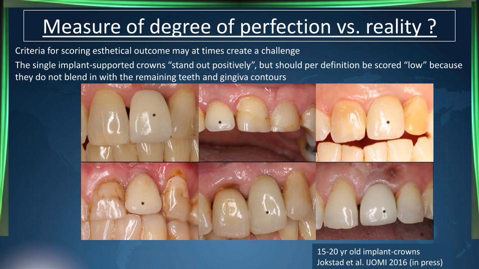
Measure of degree of perfection vs. reality ?Criteria for scoring esthetical outcome may at times create a challengeThe single implant-supported crowns “stand out positively”, but should per definition be scored “low” because they do not blend in with the remaining teeth and gingiva contours
15-20 yr old implant-crowns Jokstad et al. IJOMI 2016 (in press)
**
*
*
* *

Established categorical evaluation system 1971 USPHS criteria - “ US Public Health Service“ (Cvar & Ryge)1977 CDA criteria – “California Dental Association”Specifically to implant-retained reconstructions in the esthetic zones2005 ICAI - “Implant Crown Aesthetic Index” (Meijer et al. COIR)2005 PES - “Pink esthetic score“ (Fürhauser et al. COIR) 2009 PES/WES - “Pink and white esthetic score” (Belser et al. J.Perio)2010 CEI – “Complex esthetic index” (Juodzbalys & Wang J.Perio)
Specifically to implant-retained reconstructions and papillae1997 PI – “(Jemt) Papilla Index“ score (Jemt Int. J. Per. Res. Dent)
i.e., position of the soft-tissue crest relative to the apical location of the tooth:implant-crown contact area
Score: 0 (1 2) (3 4)-/+ ≥ half the height
2. Evaluation of esthetic outcomes in implant dentistry

1. Evaluation systems to appraise the qualities of the soft tissues in patients having received a single crown2. The effects of various clinical variables on peri-implant soft tissue appearance and cortical bone loss

2. Effects of clinical variables on peri-implant soft tissue appearance and cortical bone loss
We may today expect predictable esthetic outcomes due to refinements over the years:
– Alternative surgical and restorative treatment strategies
– Innovative implant system components and biomaterials

Alternative surgical and restorative treatment strategies
Timing of implant placement
+/-Socket preservation
+/-Site enhancement• Bone• Soft-tissues• Keratinized
gingiva
Loading protocol alternatives

1
2
3
4
5 Implant placed +/- augment & Load permanent prosthesis
Implant placed +/- augment
Heal +/-4 mths
Load Temp./permanen
t prosthesis
Implant placed +/- augment & Load temporary
prosthesis
Observation of esthetic outcome
Load Permanent prosthesis
Alternative surgical and restorative treatment strategies for healed sites / missing teeth
Implant placed +/- augment
Heal +/-4 mths
Recovery surgery for esthetics*
Soft-tissue building
Load Temp./permanent prosthesis
Implant placed +/- augment & Load temporary prosthesis
Observation of esthetic outcome
Recovery surgery for esthetics*
Soft-tissue building
Load Permanent prosthesis

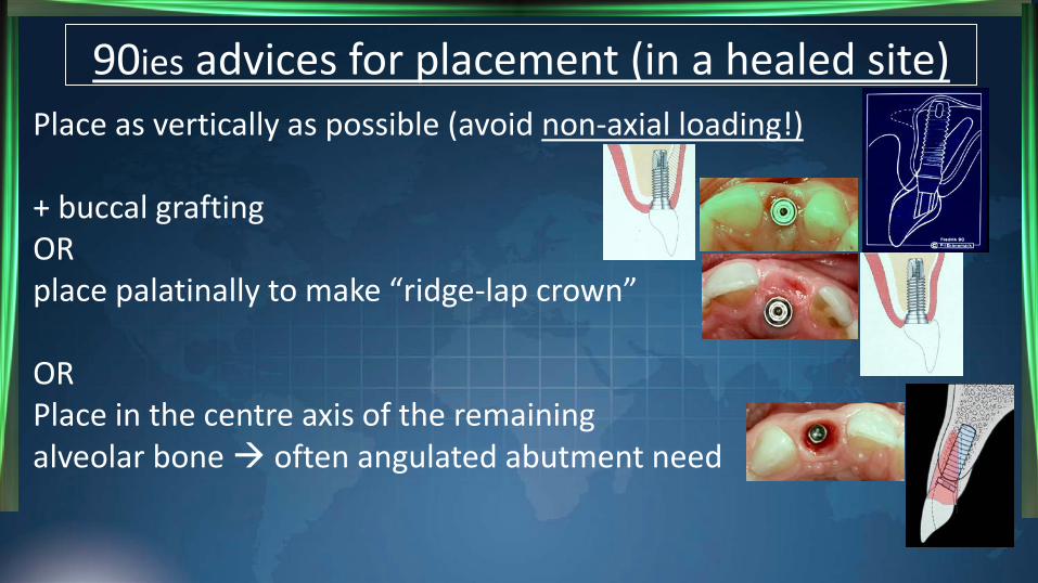
90ies advices for placement (in a healed site)Place as vertically as possible (avoid non-axial loading!)
+ buccal grafting
OR
place palatinally to make “ridge-lap crown”
90ies advices for placement (in a healed site)Place as vertically as possible (avoid non-axial loading!)
+ buccal grafting ORplace palatinally to make “ridge-lap crown”
ORPlace in the centre axis of the remaining alveolar bone often angulated abutment need

1
2
3
Remaining tooth extracted +/- augment
Heal +/-3 mths
Implant placed +/- augment
Heal +/- 4 mths
Recovery surgery for esthetics
Soft-tissue building
Load Temp./perm. prosthesisRemaining tooth
extracted +/- augment
Heal +/-3 mths
Implant placed +/-augment & Load Temp./perm. prosthesis
Remaining tooth extracted +/- augment
Heal +/-3 mths
Implant placed +/-augment
Heal +/-4 mths
Load Temp./perm. prosthesis
Alternative surgical and restorative treatment strategies for remaining hopeless tooth / root

TIME

1
2
3
4
5
6
7
8
Remaining tooth extracted +/- augment
Heal +/-3 mths
Implant placed +/- augment
Heal +/-4 mths Load Temp./perm. prosthesis
Remaining tooth extracted & Implant placed +/- augment
Heal +/- 4 mths
Recovery surgery for esthetics*
Soft-tissue building Load Temp./perm. prosthesis
Garber 1995, Weisgold et al. 1997, Salama et al. 1998, Kois 2001
Remaining tooth extracted +/- augment
Heal +/-3 mths
Implant placed +/-augment
Heal +/- 4 mths
Recovery surgery for esthetics
Soft-tissue building
Load Temp./perm. prosthesis
Remaining tooth extracted +/- augment
Heal +/-3 mths
Implant placed +/- augment & Load Temp./perm. prosthesis
Remaining tooth extracted & Implant placed +/- augment
Heal +/- 4 mths Load Temp./perm. prosthesis
Remaining tooth extracted & Implant placed +/-augment & Load temporary prosthesis
Observation of esthetic outcome Load Perm. prosthesis
Remaining tooth extracted & Implant placed +/- augment & Load permanent prosthesis
Remaining tooth extracted & Implant placed +/-augment & Load temporary prosthesis
Observation of esthetic outcome
Recovery surgery for esthetics
Soft-tissue building
Load Perm. prosthesis
Alternative surgical and restorative treatment strategies for remaining hopeless tooth / root
2. Effects of clinical variables on peri-implant soft tissue appearance and cortical bone loss
We may today expect predictable esthetic outcomes due to refinements over the years:
– Alternative surgical and restorative treatment strategies
– Innovative implant system components and biomaterials

Implant placement strategies – immediate or early?
1. +/- Augmentation2. Auto-/allograft3. +/- membrane4. ((HA-)cylindric)
‘90ies

Implant placement strategies – immediate or early?
Stepped implants
‘90ies

Wide implants(Narrow implants)
Implant placement strategies – immediate or early?
late ‘90ies

1. 4-8 w. healing postextract2. Tissue-level ( bone level)3. Buccal grafts – Auto-
+Xenograft particles4. Collagen membrane5. Submerge 8-12 w.
Implant placement strategies – immediate or early?
2011 pioneered by U.Bern

Amount of bone needed to accommodate circumferential crater without loss of height in buccal mucosal margin; dotted line = original degree of B-L resorption
1. ... the bone thickness should be at least 2 mm, preferably 4 mm
2. If < 2mm bone is available, part of the buccal bone plate will be lost after remodeling, with the consequence of a high risk of soft tissue recession
3. Such a large amount of bone buccally does not exist normally, and has to be created with augmentation procedures in almost every esthetically demanding case
A deductive reasoning approachPremise: A 1.5 mm wide “circumferential crater” exists around all implants, including on the buccal side. Hence,
From: Grunder et al. IJPRD 2005
Thickness that bone on buccal side of implant should have to support gingival margin despite horizontal crater formation.
Influential paperBUT
The evidence of the premise is weaksee: Zhang et al. COIR 2014

BioHorizon 3i Osseotite NT 3i Certain Prevail Innova Endopore Brånemark Std. ITI Std.+ Narrow-Neck4 x 12 mm 4 x 13 mm 3.8 x 11.5 mm 4 x 9 mm 3.75 x 18 mm 3.3 x 12 mm
ReplaceSelect Straight ReplaceSelect Taper Steri-Oss Replace Zimmer ScrewVent-taper Zimmer ScrewVent Zimmer MicroVent4.3 x 15 mm 4.3 x 16 mm 3.3 x 18 mm 4.7x 16 mm 3.8 x 16 mm 4.3 x 16 mm
“Saucerization” – influence by the implant design?

BioHorizon 3i Osseotite NT 3i Certain Prevail Innova Endopore Brånemark Std. ITI Std.+ Narrow-Neck4 x 12 mm 4 x 13 mm 3.8 x 11.5 mm 4 x 9 mm 3.75 x 18 mm 3.3 x 12 mm
ReplaceSelect Straight ReplaceSelect Taper Steri-Oss Replace Zimmer ScrewVent-taper Zimmer ScrewVent Zimmer MicroVent4.3 x 15 mm 4.3 x 16 mm 3.3 x 18 mm 4.7x 16 mm 3.8 x 16 mm 4.3 x 16 mm
“Saucerization” – influence by the implant design or by anatomy?

2. Effects of clinical variables on peri-implant soft tissue appearance and cortical bone loss
We may today expect predictable esthetic outcomes due to refinements over the years:
– Alternative surgical and restorative treatment strategies
– Innovative implant system components and biomaterials
The parameters to achieve the best possible appearance of peri-implant soft-tissues?

1. Tissue biotype / thickness2. Incision / flap design 3. Osteotomy procedure 4. Implant position, vertical & adjacent tissues5. Torque / primary stability6. Flap handling7. Suturing technique8. Cover screw / tenting abutment
Potential effect of site or surgery variables on outcome?
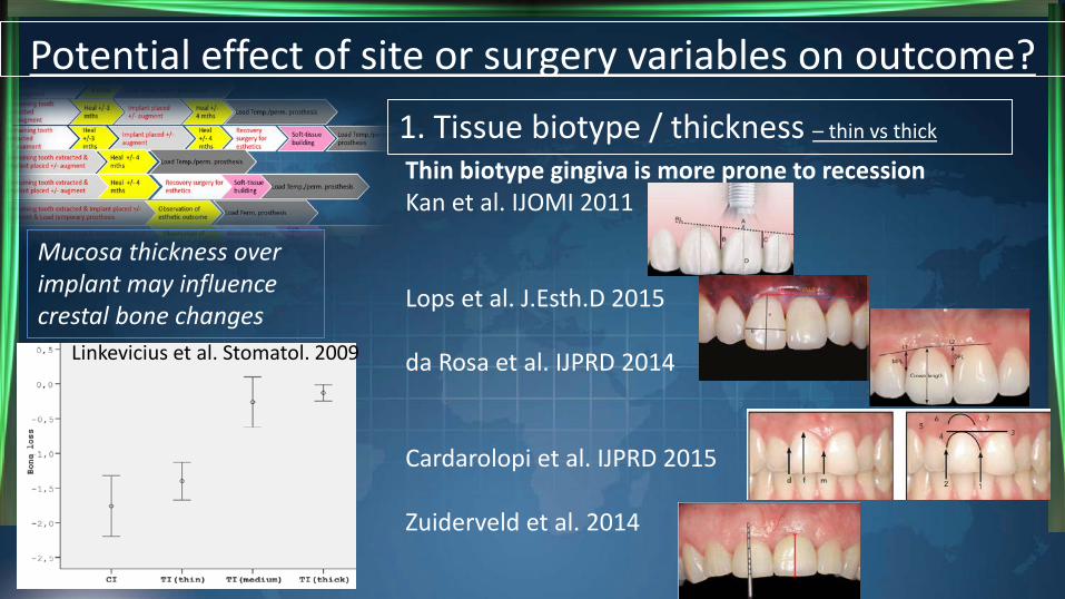
Thin biotype gingiva is more prone to recessionKan et al. IJOMI 2011
Lops et al. J.Esth.D 2015
da Rosa et al. IJPRD 2014
Cardarolopi et al. IJPRD 2015
Zuiderveld et al. 2014
1. Tissue biotype / thickness – thin vs thick
Potential effect of site or surgery variables on outcome?
Mucosa thickness over implant may influence crestal bone changes
Linkevicius et al. Stomatol. 2009

1. Tissue biotype / thickness – thin vs thick
2.Incision / flap design - use1. Trapezoidal instead of intra-sulcularincision (Gomez-Roman IJOMI 2001)
2. Split-finger approach (Misch et al. Imp Dent 2004)
Potential effect of site or surgery variables on outcome?

1. Tissue biotype / thickness – thin vs thick
2. Incision / flap design - papilla-sparing approach
3. Osteotomy procedure 4. Implant position, vertical & adjacent tissues5. Torque / primary stability6. Flap handling7. Suturing technique8. Cover screw / “tenting” abutment
If also immediate placement:Extraction reasonExtraction techniqueSocket debridementSocket preservation
Potential effect of site or surgery variables on outcome?
Evidence is inconclusive
Evidence is conflicting
Evidence is inconclusive
Evidence is inconclusive
Evidence is inconclusive, or conflicting or lacking
Evidence is lacking
Keratinized gingiva - Wennström & Derks COIR 2012
Crown-implant ratio - Gulje et al. IJOMI 2015
“Platform-switching”Abutment connect-disconnect
Evidence is lacking
Not likely
Evidence is conflicting
Evidence is lacking

1. Esthetic Risk
High
Low
2. Complexity of Treatment
Process
High
Moderate
Low
3. Risks of complications
and consequences
High
Moderate
Low
SAC Classification –Straightforward - Advanced - Complex
Modifying Factors
Basis for informed consent to therapy
General determinants

Reduced Immune system
Heavy Smoker (>10 cigs/day)
Ongoing Sub-optimalpreceedingoutcome
Light smoker (<10 cigs/day)
Moderate / Suboptimal outcome
Healthy, co-operative with an intact immune system
Non-smoker Completed Optimal
High Risk
Moderate Risk
Low Risk
1. Compromised
General or Local health
2. Smoking Habits
3. Growth Considerations
4. Iatrogen
ic factors
Modifying Factors

High High High scalloped, thin
Triangular Acute >=7mm to contact point & Restored
>=2 teeth Soft tissue defects
Vertical bone deficiency
Medium Medium Medium scalloped, medium thick
Chronic 5.5-6.5mm to contact point
1 tooth (<= 7mm)
Horizontal bone deficiency
Low Low Low scalloped, thick
Rectangular None <=5mm to contact point & Virgin
1 tooth (>= 7mm)
Intact soft tissue
No bone deficiency
Patient Estheti
c Expectations
1. Lip Line
2. Gingiva
l biotyp
e
3. Tooth Crown Shape
4. Implant site
Infection
nt teeth bone
level & restora
tive status
6. Width
of span
7. Soft tissue anato
my
8. Bone anatomy at
alveolar crestHigh Risk
Moderate Risk
Low Risk
Modifying Factors

Deficient, requiring prior augmentation
High risk of involvement
Yes Thin Insufficient <1mm
Implant placement with staged procedures
High / Severely compromised outcome
Deficient, but allowing simultaneousaugmentation
Moderate risk of involvement
Implant placement with simultaneousprocedures
Moderate / Suboptimal outcome
Adequate Minimal risk of involvement
No Thick Sufficient >1mm
Implant placement withoutadjunctive procedures
Minimal / No adverse effect
Bone volume• Horizo
ntal• Vertica
l
Anatomic Risk
Esthetic Risk• Zone• Biotyp
e• Facial
bone wall
Complexity of
Treatment
Process
Risks of complicati
ons and consequen
ces
High Risk
Moderate Risk
Low Risk
Modifying Factors

a. Virgin b. Periodontal disease or parafunction
a. Adjunctive therapy needed to gain sufficient space b. to achieve satisfactory result c. Full arch d. Required
a. No guidance b. Involved in guidance c. Present
a. Fixed b. Margin > 3mm from crest c. Immediate d. --e. High
a. Restricted b. some reduction required c. Extended space
a. Removable b. Margin <3mm from crest c. --d. PFM e. Moderate
a. Restored teeth b. Caries or Trauma
a. Adequate b. Sufficientc. Single tooth d. Not required
a. Anterior guidance b. minimal involvement c. Absent
a. None b. not required c. Conventional/Early d. Resin-metal e. Low
1. Oral environmen
ta. Adjacent
toothb. Tooth loss
reason
a. Interarchdistance
b. Mesio-distal space
c. Restoration span
d. Saddle volume/ character
Occlusiona. Scheme
b. Bite involvemen
tc.
Parafunction
Restorationa. During healingb. Develop soft
tissuec. Loading protocol
d. Biomaterialse. Anticipated Maintenance
High Risk
Moderate Risk
Low Risk
Modifying Factors

Reduced Immune system
Heavy Smoker (>10 cigs/day)
Ongoing Sub-optimalpreceedingoutcome
Light smoker (<10 cigs/day)
Moderate / Suboptimal outcome
Healthy, co-operative with an intact immune system
Non-smoker Completed Optimal
High High High scalloped, thin
Triangular Acute >=7mm to contact point & Restored
>=2 teeth Soft tissue defects
Vertical bone deficiency
Medium Medium Medium scalloped, medium thick
Chronic 5.5-6.5mm to contact point
1 tooth (<= 7mm)
Horizontal bone deficiency
Low Low Low scalloped, thick
Rectangular None <=5mm to contact point & Virgin
1 tooth (>= 7mm)
Intact soft tissue
No bone deficiency
1. Compromised
General or Local health
2. Smoking Habits
3. Growth Considerations
4. Iatroge
nic factors
Patient Estheti
c Expecta
tions
1. Lip Line
2. Gingiva
l biotype
3. Tooth Crown Shape
4. Implant
site Infectio
n
jnt
teeth bone
level & restora
tive status
6. Width
of span
7. Soft tissue
anatomy
8. Bone anatom
y at alveolar crest
Deficient, requiring prior augmentation
High risk of involvement
Yes /Thin /Insufficient <1mm
Implant placement with staged procedures
High / Severely compromised outcome
Deficient, but allowing simultaneousaugmentation
Moderate risk of involvement
Implant placement with simultaneousprocedures
Moderate / Suboptimal outcome
Adequate Minimal risk of involvement
No /Thick /Sufficient > 1mm
Implant placement withoutadjunctive procedures
Minimal / No adverse effect
a. Virgin b. Periodontal disease or parafunction
a. Adjunctive therapy needed to gain sufficient space b. to achieve satisfactory result c. Full arch d. Required
a. No guidance b. Involved in guidance c. Present
a. Fixed b. Margin > 3mm from crest c. Immediate d. --e. High
a. Restricted b. some reduction required c. Extended space
a. Removable b. Margin <3mm from crest c. --d. PFM e. Moderate
a. Restored teeth b. Caries or Trauma
a. Adequate b. Sufficientc. Single tooth d. Not required
a. Anterior guidance b. minimal involvement c. Absent
a. None b. not required c. Conventional/Early d. Resin-metal e. Low
Bone volume•Horizontal
•Vertical
Anatomic Risk
Esthetic Risk•Zone•Biotype
•Facial bone wall
Complexity of
Treatment
Process
Risks of complications
and consequ
ences
1. Oral environment•a. Adjacent
tooth•b. Tooth
loss reason
2. Restorative volume•a. Interarch distance•b. Mesio-distal
space•c. Restoration span•d. Saddle
volume/character
3. Occlusion•a. Scheme•b. Bite
involvement•c.
Parafunctions
4. Provisional Restoration•a. During healing•b. Development soft
tissue•c. Loading protocol•d. Biomaterials•e. Anticipated
Maintenance
Modifying Factors

1. Evaluation systems to appraise the qualities of the soft tissues in patients having received a single crown2. The effects of various clinical variables on peri-implant soft tissue appearance and cortical bone loss3. Clinical research focused on dimensional relationships between the implant-crown-complex and clinical and radiographical landmarks
Learning objectives of this presentation

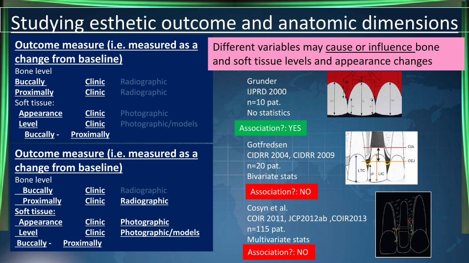
Studying esthetic outcome and anatomic dimensions
Outcome measure (i.e. measured as a change from baseline) Bone level
Buccally Clinic RadiographicProximally Clinic Radiographic
Soft tissue: Appearance Clinic PhotographicLevel Clinic Photographic/models
Buccally - Proximally
Observation studies (i.e., measured at a single point of time)Bone level
Buccally Clinic RadiographicProximally Clinic Radiographic
Soft tissue: Appearance Clinic PhotographicLevel Clinic Photographic/models
Buccally - Proximally
Clinical variables, e.g., Implant hardwareSurgical proceduresAnatomy
Bone and soft tissue levels and appearance may be associated with different variables
Different variables may cause or influence bone and soft tissue levels and appearance changes
Simplistic versus complex (multivariate) statistics1. Generalized estimating equations (GEE)
2. General linear modelling (GLM) 3. Multilevel analyses (AKA mixed / hierarchical / random effects models

Observation (i.e., single point of time)Bone levelBuccally Clinic RadiographicProximally Clinic RadiographicSoft tissue: Appearance Clinic PhotographicLevel Clinic Photographic/models
Buccally - Proximally
Kan et al. J Perio 2003n=45 pat.Bivariate statistics
Gastaldo et al.J Perio 2004n=48 pat.Bivariate statistics
Bone and soft tissue levels and appearance may be associated with different variables
Association?: YES
Association?: YES
Studying esthetic outcome and anatomic dimensions

Observation (i.e., single point of time)Bone level
Buccally Clinic RadiographicProximally Clinic Radiographic
Soft tissue: Appearance Clinic PhotographicLevel Clinic Photographic/models
Buccally - Proximally
Vela et al. IJPRD 2012n=50 pat.Bivariate statistics
Observation (i.e., single point of time)Bone level
Buccally Clinic RadiographicProximally Clinic Radiographic
Soft tissue: Appearance Clinic PhotographicLevel Clinic Photographic/models
Buccally - Proximally
Kourkouta et al. COIR 2009n=15 pat.Bivariate statistics
Bone and soft tissue levels and appearance may be associated with different variables
Association?: YES
Association?: YES
Perez et al. IJPRD 2012n=46 imp..Bivariate statistics
Association?: YES
Studying esthetic outcome and anatomic dimensions

Observation (i.e., single point of time)Bone level
Buccally Clinic RadiographicProximally Clinic Radiographic
Soft tissue: Appearance Clinic PhotographicLevel Clinic Photographic/models
Buccally - Proximally
Choquet et al. J Perio 2001n=26 pat.Bivariate statistics
Kawai & Almeida Cleft P-C J 2008n=40 pat.Bivariate statistics
Lops & Romeo COIR 2008n=46 pat.Bivariate statistics
Bone and soft tissue levels and appearance may be associated with different variables
Association?: YES
Association?: YES
Association?: NO
Studying esthetic outcome and anatomic dimensions

Observation (i.e., single point of time)Bone levelBuccally Clinic RadiographicProximally Clinic RadiographicSoft tissue: Appearance Clinic PhotographicLevel Clinic Photographic/modelsBuccally - Proximally
Nispakultorn et al.COIR 2010n=40 pat.Bivariate stats
+ cbCT
Bone and soft tissue levels and appearance may be associated with different variables
Peng et al. IJPRD 2013n=25 pat.Bivariate stats
Chang & WennstromCOIR 2013n=32 pat.Multivariate stats
Association?: YES
Association?: NO
Association?: YES
Studying esthetic outcome and anatomic dimensions
Outcome measure (i.e. measured as a change from baseline) Bone levelBuccally Clinic RadiographicProximally Clinic RadiographicSoft tissue: Appearance Clinic PhotographicLevel Clinic Photographic/models
Buccally - Proximally
GrunderIJPRD 2000n=10 pat.No statistics
Different variables may cause or influence bone and soft tissue levels and appearance changes
Outcome measure (i.e. measured as a change from baseline) Bone level
Buccally Clinic RadiographicProximally Clinic Radiographic
Soft tissue: Appearance Clinic PhotographicLevel Clinic Photographic/models
Buccally - Proximally
GotfredsenCIDRR 2004, CIDRR 2009n=20 pat.Bivariate stats
Cosyn et al. COIR 2011, JCP2012ab ,COIR2013n=115 pat.Multivariate statsAssociation?: NO
Association?: YES
Association?: NO
Studying esthetic outcome and anatomic dimensions

Outcome measure (i.e. measured as a change from baseline) Bone levelBuccally Clinic RadiographicProximally Clinic RadiographicSoft tissue: Appearance Clinic PhotographicLevel Clinic Photographic/models
Buccally - Proximally
Henriksson&JemtCIDRR 2004n=18 pat.Bivariate stats
Palmer et al. JCP 2007n=66 pat.Bivariate stats
Schropp et al. COIR 2005, 2013, 2014abn=72 pat.Bivariate stats
+ cbCT (2014)
Gallucci et al. JCP 2011n=20 pat.Multivariate stats
Tissue level implants
Ryser et al. JOMS 2005n=40 pat.Multivariate stats
Degidi et al. J Perio 2008n=49 pat.Bivariate stats
Tymstra et al. & vanNimwegen et al. JCP2011 & IJP 2015n=45 pat.Multivariate stats
Association?: NO
Association?: YES
Association?: NOAssociation?: NO
Association?: NO Association?: YES
Association?: NO
Studying esthetic outcome and anatomic dimensions

A satisfactory esthetic outcome as an effect of bone level?

Outcome measure (i.e. measured as a change from baseline) Bone level
Buccally Clinic RadiographicProximally Clinic Radiographic
Soft tissue: Appearance Clinic PhotographicLevel Clinic Photographic/models
Buccally - Proximally
JemtIJP 2008n=38 pat.Bivariate stats
Cardaropoli et al. COIR 2003n=28 pat.Multivariate stats
Chang&WennstromCOIR 2010n=43 pat.Multivariate stats
BICON implantsUrdaneta et al. COIR 2014n=206 pat.Multivariate stats
Association?: NO
Association?: NO
Association?: NO
Association?: NO
Studying bone levels and anatomic dimensions

The advent of use of cbCT, pre- & post-placement
From: Sanz et al. / Tomasi et al. / Ferrus et al. / Multicentre study. COIR 2010
After 3 years: Both the interproximal papilla filling and the midfacial mucosa stability were not influenced by variables such as type of fixture configuration, tooth category, smoke habit, and thickness of buccal bone wall of ≤ 1 mm (thin buccal wall). (Cecchinatoet al. COIR 2015)
Rossi et al. - IJPRD 2013 – 9 pat. Bivariate stats – pre-post- 4 mths
Miyamoto & Obama (2011)Benic et al. (2012-2011e)
Roe et al. (2012)Vera et al. (2012)
Buser et al. (2013a,b)Cortes et al. (2013)Fu et al. (2014-2013e)
Koutouzis et al. (2015, 2014)Kaminaka et al. (2015-2014e)Schropp et al. (2015-2014e)
Hasan et al. (2015)Lemes et al. (2015)Chappuis et al. (2015e)Noelken et al. (2015e)Veltri et al. (2016-2015e)Kuchler et al. (2016-2015e)
Association?: NO
Studying esthetic outcome and anatomic dimensionsOutcome measure (i.e. measured as a change from baseline)

Chappuis et al. COIR 2015N= 61 pat.Bivariate stats, Pre-post 5-9 yrs
Hor. dist. of “saucer” :TL: 1.0 mm BL: 0.6 mm
Graphical display of 1.5 mm wide “saucers” claimed to be present around all implants
From: Grunder et al. IJPRD 2005
Studying esthetic outcome and anatomic dimensionsOutcome measure (i.e. measured as a change from baseline)

Buccal bone vz. gingival thickness vz. esthetics?
From:De Bruyckere et al. JCP 2015Younes et al. COIR 2016
Gingival thickness,Thin vs thick biotype
Correlation between buccal bone & gingival thickness is only moderate
N= 21 pat.

COIR 2016; 27: 956: “Within present limitations, acceptable and stable aesthetics are not jeopardized by a thin or missing buccal bone”
Buccal bone vz. gingival thickness vz. esthetics?
BUT!
cbCT accuracy of ≤1.2 mm peri-implant buccal
bone ?
Poor (Schulze et al. 2001)Poor (Spin-Netto et al. 2011)Poor (Benic et al. 2013)Modest (Gonzales et al. 2016)
BUT!
COIR LAST ISSUE!
N= 12 pat.Association?: NO
1. Evaluation systems to appraise the qualities of the soft tissues in patients having received a single crownPES & PES/WES have been validated and appear to predominate in use
Summarizing – Take home message

1. Evaluation systems to appraise the qualities of the soft tissues in patients having received a single crownPES & PES/WES have been validated and appear to predominate in use
2. The effects of various clinical variables on peri-implant soft tissue appearance and cortical bone lossEffects of many variables singularly and in combination are largely unknown, principally due to small datasets and short study duration
Summarizing – Take home message

1. Evaluation systems to appraise the qualities of the soft tissues in patients having received a single crownPES & PES/WES have been validated and appear to predominate in use
2. The effects of various clinical variables on peri-implant soft tissue appearance and cortical bone lossEffects of many variables singularly and in combination are largely unknown, principally due to small datasets and short study duration3. Clinical research focused on dimensional relationships between the implant-crown-complex and clinical and radiographicallandmarksCross-sectional studies with simplistic statistics indicate associations, while longitudinal studies with adequate multi-level multivariate statistics provide less conclusive data
Summarizing – Take home message