Embed Size (px)
Citation preview

Single Lgr5- or Lgr6-expressing taste stem/progenitorcells generate taste bud cells ex vivoWenwen Rena, Brian C. Lewandowskia, Jaime Watsona, Eitaro Aiharab, Ken Iwatsukic, Alexander A. Bachmanova,Robert F. Margolskeea, and Peihua Jianga,1
aMonell Chemical Senses Center, Philadelphia, PA 19104; bDepartment of Molecular and Cellular Physiology, University of Cincinnati, Cincinnati, OH 45267;and cDepartment of Nutritional Science and Food Safety, Faculty of Applied Bioscience, Tokyo University of Agriculture, Tokyo 156-8502, Japan
Edited by Solomon H. Snyder, Johns Hopkins University School of Medicine, Baltimore, MD, and approved October 8, 2014 (received for review May 19, 2014)
Leucine-rich repeat-containing G protein-coupled receptor 5 (Lgr5)and its homologs (e.g., Lgr6) mark adult stem cells in multipletissues. Recently, we and others have shown that Lgr5 marks adulttaste stem/progenitor cells in posterior tongue. However, theregenerative potential of Lgr5-expressing (Lgr5+) cells and theidentity of adult taste stem/progenitor cells that regenerate tastetissue in anterior tongue remain elusive. In the present work, wedescribe a culture system in which single isolated Lgr5+ or Lgr6+
cells from taste tissue can generate continuously expanding 3Dstructures (“organoids”). Many cells within these taste organoidswere cycling and positive for proliferative cell markers, cytokeratinK5 and Sox2, and incorporated 5-bromo-2’-deoxyuridine. Impor-tantly, mature taste receptor cells that express gustducin, carbonicanhydrase 4, taste receptor type 1 member 3, nucleoside triphos-phate diphosphohydrolase-2, or cytokeratin K8 were present inthe taste organoids. Using calcium imaging assays, we found thatcells grown out from taste organoids derived from isolated Lgr5+
cells were functional and responded to tastants in a dose-depen-dent manner. Genetic lineage tracing showed that Lgr6+ cells gaverise to taste bud cells in taste papillae in both anterior and poste-rior tongue. RT-PCR data demonstrated that Lgr5 and Lgr6 maymark the same subset of taste stem/progenitor cells both anteri-orly and posteriorly. Together, our data demonstrate that func-tional taste cells can be generated ex vivo from single Lgr5+ orLgr6+ cells, validating the use of this model for the study of tastecell generation.
Lgr5 | Lgr6 | taste stem cells | taste progenitor cells
Taste bud cells are heterogeneous and undergo constantturnover (1); however, the origins and generation of taste
buds in adult mammals remain largely unclear. Based on mor-phological and functional characteristics, there are at least threedifferent types of mature taste bud cells [type 1 (glial-like cells),type 2 (receptor cells, including those responsible for sensingsweet, bitter, and umami stimuli), and type 3 (presynaptic cells,including sour sensors)], and well as one type of immature tastebud cell [type 4 (basal cells that are precursors of other types ofmature taste cells)] (2, 3). Mature taste bud cells are postmitoticand short-lived, with average life spans estimated at 8–12 d (4, 5),although distinct subtypes of taste bud cells may have differentlife spans (1, 4, 5). At present, the stem cell population and theregenerative process from adult taste stem/progenitor cells tomature taste bud cells are not well characterized.Lgr5 (leucine-rich repeat-containing G protein-coupled re-
ceptor 5), encoded by a Wnt (wingless-type MMTV integrationsite family) target gene, marks adult stem/progenitor cells intaste tissue in posterior tongue that in vivo give rise to all majortypes of taste bud cells, as well as perigemmal cells (6, 7). Lgr5 isalso known to mark actively cycling stem cells in small intestine,colon, stomach, and hair follicle, as well as quiescent stem cellsin liver, pancreas, and cochlea (8). Isolated Lgr5+ adultstem cells from multiple tissues are able to generate so-calledorganoid structures ex vivo (9–11). For instance, Sato and col-leagues (10) developed a 3D culture system to grow crypt-villus
organoids from single intestinal stem cells; all differentiated celltypes were found in these structures, indicating the multipotentnature of these cells. We hypothesized that Lgr5+ taste stem/pro-genitor cells in a 3D culture system would be capable of expandingand giving rise to taste receptor cells ex vivo. In the present study,we isolated Lgr5+ stem/progenitor cells from taste tissue and cul-tured them in a 3D culture system. Single Lgr5+ cells grew intoorganoid structures ex vivo in defined culture conditions, with thepresence of both proliferating cells and differentiated mature tastecells in which taste signaling components are functionallyexpressed. When organoids were replated onto a 2D surface pre-coated with laminin and polylysine, cells grew out of the organ-oids and attached to the flat surface, and some cells retained theexpressed taste signaling elements and responded to taste stimuli.Lgr5 marks adult taste stem/progenitor cells in posterior
tongue, which was shown using an engineered mouse model inwhich enhanced green fluorescent protein (EGFP) and tamoxi-fen-inducible Cre recombinase (CreERT2) are knocked-in toreplace the coding sequence of Lgr5 and act as surrogatemarkers for Lgr5 (6, 7). Although Lgr5 is present in fungiformpapillae in anterior tongue during embryonic stages and earlylife, based on the intrinsic GFP signal from the Lgr5-EGFPtransgene, Lgr5-EGFP signal could not be detected in fungiformpapillae cells in adult mice (6, 7). Therefore, taste stem/pro-genitor cells remain to be identified in fungiform papillae inanterior tongue. We hypothesized that Lgr6, an Lgr5 homolog,may mark adult taste stem/progenitor cells in anterior tongue,prompted by the finding that Lgr6 is preferentially expressed intaste tissue, but not in the surrounding epithelium devoid of taste
Significance
Taste tissue regenerates continuously throughout the life spanin mammals. Here, using lineage tracing and a culture system,we show that leucine-rich repeat-containing G protein-coupledreceptor 5-expressing and leucine-rich repeat-containing Gprotein-coupled receptor 6-expressing taste stem/progenitorcells generate mature taste cells in vivo and ex vivo. Impor-tantly, our ex vivo studies show that single-progenitor cells cangenerate all mature taste cell types and that differentiatedtaste cells form in the absence of innervation. This ex vivomodel mimics the development of taste bud cells in taste pa-pillae, recapitulates the process of taste renewal from adulttaste stem cells to mature taste cells, and provides a means tostudy the regulation of taste cell generation and to understandthe origins and cell lineage relationships within taste buds.
Author contributions: W.R., R.F.M., and P.J. designed research; W.R., B.C.L., and J.W.performed research; E.A., K.I., and A.A.B. contributed new reagents/analytic tools; W.R.,B.C.L., R.F.M., and P.J. analyzed data; and R.F.M. and P.J. wrote the paper.
The authors declare no conflict of interest.
This article is a PNAS Direct Submission.1To whom correspondence should be addressed. Email: [email protected].
This article contains supporting information online at www.pnas.org/lookup/suppl/doi:10.1073/pnas.1409064111/-/DCSupplemental.
www.pnas.org/cgi/doi/10.1073/pnas.1409064111 PNAS | November 18, 2014 | vol. 111 | no. 46 | 16401–16406
DEV
ELOPM
ENTA
LBIOLO
GY

tissue (12). Using the Lgr6-EGFP-ires-CreERT2 mouse line (13),we here show that Lgr6 is expressed in cells at the basal area oftaste buds in fungiform and circumvallate papillae. By geneticlineage tracing, we show that Lgr6+ cells give rise to taste budcells in taste papillae in both anterior and posterior tongue. RT-PCR shows that Lgr5 and Lgr6 may mark the same subset oftaste stem/progenitor cells both anteriorly and posteriorly. Sim-ilar to Lgr5+ cells, isolated Lgr6+ cells can build taste organoidsthat generate mature taste cells.
ResultsSingle Isolated Lgr5+ Cells Generate Taste Organoids. To determinewhether Lgr5+ taste stem/progenitor cells are capable ofexpanding and generating taste cells in vitro, and to establisha taste culture system, we purified Lgr5+ taste stem/progenitorcells (Fig. 1A) from Lgr5-EGFP-ires-CreERT2 mice, using fluo-rescence-activated cell sorting (FACS), based on the greenfluorescence signal of Lgr5-EGFP+ cells (Fig. 1 B and C). Thecell sorting gates for isolating GFP-expressing cells were set suchthat no cells from wild-type littermate controls were isolated(Fig. 1B). All sorted cells expressed EGFP, as demonstrated bythe green fluorescence signal (Fig. 1C). Single isolated Lgr5+
taste progenitor cells were embedded in Matrigel containingClevers’ organoid culture factors for intestinal stem cells andthen cultured in defined medium (14). FACS-sorted single Lgr5+
cells proliferated and formed sphere-like structures similar tothose obtained from liver Lgr5+ progenitor cells (9) (Fig. 1D).The plating efficiency was about 3–10%. Cultures can be pas-saged at least six times and maintained for at least 2 mo (thelongest time tested to date). After culturing for >3 d, most
organoids showed no detectable EGFP signal; the EGFP signaldecreased rapidly and became undetectable in 2–3 d. However,organoids were occasionally found to retain a strong EGFP sig-nal: in two of 12 preparations, organoids with GFP fluorescencewere seen in 43 (12%) of 374 organoids and 61 (5%) of 1201organoids examined. Presumably this continued expression ofGFP-fluorescence resulted from Lgr5-EGFP cells that were ex-panded from single isolated Lgr5-EGFP+ cells (Fig. 1E, Bottom).To assess the clonality of isolated Lgr5+ cells and to determine
whether organoids truly grow out from single Lgr5+ cells, asopposed to small aggregates of cells, we crossed Lgr5-EGFP-ires-CreERT2 mice with Rosa26-tdTomato mice to generate Lgr5-EGFP-ires-CreERT2+/−; Rosa26-tdTomato+/− mice in whichtamoxifen-induced Cre generates expression of tdTomatofluorescence protein (red) to mark cells from the Lgr5+ lineage.Half the Lgr5-EGFP-ires-CreERT2+/−; Rosa26-tdTomato+/− micewere injected with tamoxifen 1 d before cell sorting to markLgr5+ progeny, using tdTomato fluorescence. Then we purifiedcells that were positive for EGFP (green) via FACS from bothtamoxifen-treated and untreated mice and cultured them inmixture. Using this strategy, tamoxifen-induced Cre generatedexpression of tdTomato fluorescence protein (red) in only afraction of single isolated Lgr5+ cells, as well as their progeny.Whole organoids grown from these isolated Lgr5+ cellswere composed entirely of either tdTomato+ cells (31 and 34organoids from two separate experiments) or tdTomato− cells(∼300 organoids each from two preparations) (Fig. 1F), nevermixed populations of tdTomato+ and tdTomato− cells, indicatingthey are clonally expanded from single isolated Lgr5+ cells.R-spondins have been found to be essential for culturing
Lgr5+ stem cells from multiple types of tissues (15–17). To de-termine whether R-spondins are also critical for growth andexpansion of Lgr5+ cells into taste organoids, we cultured Lgr5+
cells from taste tissue in the same medium formula withoutR-spondin-1 supplement. Fig. S1A shows representative fields oforganoids in the presence or absence of R-spondin-1 and dem-onstrates that R-spondin-1 enhances the growth and expansionof organoids derived from taste Lgr5+ cells.
Organoids Derived from Single Lgr5+ Cells Contain Actively CyclingCells. Because of the continuous expansion of organoids underour defined culture conditions, we reasoned that organoids mustcontain progenitor cells. To determine the proliferating capa-bilities of cells in cultured organoids, we performed 5-bromo-2′-deoxyuridine (BrdU) tracing. At day 10, individual organoidswere examined for incorporation of BrdU after overnight in-cubation in BrdU-containing culture medium. As expected, nu-merous cells in organoids showed BrdU immunoreactivity (Fig.2A). BrdU+ cells were typically scattered within organoid struc-tures (Fig. 2A). Sox2 is implicated in taste cell development andtaste cell renewal (18) and is a general stem cell marker (19). Asubset of BrdU+ cells were also Sox2+, confirming the pro-liferating properties, as well as potential “stemness,” of thesecells (Fig. 2A). K8 is a general marker for intragemmal taste budcells (20). We tested cultures for the presence of K8+ cells thatwere distinct from proliferating cells by double immunostainingwith cytokeratin K14 and K5, two additional progenitor cellmarkers for basal epithelial cells, including taste tissue (21). K8immunoreactivity was detected in many organoids [23 (53%) of43], albeit in varying numbers of cells, and these cells did notshow any staining for proliferating markers such as K5 and K14(Fig. 2 B and C), mirroring the segregation of these two cellpopulations in taste tissue. Organoids with detectable EGFPsignal showed the presence of K5+ cells as well (Fig. S2).Intrigued by the infrequent presence (two of 12 preparations)
of detectable Lgr5-EGFP signal after 2–3 d in culture in onlya fraction of cultured organoids (5–11%, as detailed earlier) andby the apparent growth-promoting effects of R-spondin-1, the
Fig. 1. Single isolated Lgr5+ cells generate taste-bud-like “organoids” in 3Dculture. (A) Confocal images of Lgr5-EGFP+ cells (green) in a circumvallatepapilla section from an Lgr5-EGFP-ires-CreERT2+/− mouse. (B) Representativeresults of FACS sorting of cells isolated from taste tissue from wild-type (WT)and Lgr5-EGFP-ires-CreERT2+/− mice. Boxed areas depict the gating param-eters to sort only Lgr5-EGFP+ cells. (C) Single dissociated cells from digestedtongue epithelium from Lgr5-EGFP-ires-CreERT2+/− mice before (Top) andafter (Bottom) FACS sorting for Lgr5-EGFP+ cells. All sorted cells expressedEGFP (green). (D) Representative bright-field images of cultured organoidsderived from single isolated Lgr5+ cells at indicated days in culture. At days1 and 2, lower panels show representative fluorescence images of culturedorganoids derived from Lgr5+ cells with detectable EGFP signals. (E) Repre-sentative bright-field (BF) and fluorescence images of organoids with orwithout the intrinsic GFP signal. (F) Representative bright-field (BF) andfluorescence images of cultured organoids derived from sorted Lgr5-EGFP+
(arrowhead) and Lgr5-EGFP+/tdTomato+ cells (arrow) at the indicated points.[Scale bars: A, C–E, 100 μm; F, 50 μm (day 1) and 100 μm (day 10).] All panelsrepresent data from at least three independent preparations with the ex-ception of E (two preparations).
16402 | www.pnas.org/cgi/doi/10.1073/pnas.1409064111 Ren et al.

ligand for Lgr5, Lgr4, and Lgr6 (15–17, 22), in cultured organoids,we performed RT-PCR to determine whether Lgr5 and/or itshomologs were present in cultured organoids. All three Lgrtranscripts were detected in cultured organoids, despite the oc-casional detection of an Lgr5-EGFP signal in only a small fractionof culture organoids (Fig. S3A).
Mature Taste Cells Are Generated in Vitro from Taste Organoids.Single Lgr5+ stem cells from intestine can build organoids con-taining differentiated cells (10). However, in other cases, such asLgr5+ cells isolated from pancreatic ducts, no insulin-producingcells are found in long-term organoid cultures, indicating therequirement for additional extrinsic factors or intrinsic repro-gramming (11). To determine whether any mature taste receptorcells are produced in organoids derived from Lgr5+ cells,we performed double or triple immunostaining of culturedorganoids, using antibodies against the taste signaling compo-nents gustducin (G protein for sweet, bitter, and umami sig-naling), T1R3 (taste receptor type 1 member 3, a commonsubunit of the sweet T1R2/T1R3 and the umami T1R1/T1R3taste receptor) (23–25), and carbonic anhydrase 4 (CA4; a markerof sour taste cells) (26). In multiple organoids that showed nodetectable EGFP signal, we identified cells immunopositive forgustducin (green) and T1R3 (red) (Fig. 3A and Table S1).Gustducin-expressing cells (mostly bitter taste cells in posteriortongue) generally did not overlap with T1R3-expressing cells (Fig.3A), reminiscent of their segregation in the circumvallate papillain posterior tongue (27). Of particular note, these cells displayedmorphology typical of mature taste receptor cells, with a char-acteristic large, round nucleus and a slender cell body (Fig. 3A).Nevertheless, many K8+ cells (Fig. 3A, blue) were not immu-nopositive for taste receptor cell markers, suggesting many cellscontain additional types of taste cells, such as type 1 nucleosidetriphosphate diphosphohydrolase-2+ (NTPDase2+) cells (Fig.S3B), or cells that are differentiated but not yet committed toa particular cell type. To further assess the expression of tastesignaling components in the cultured organoids, we performed
RT-PCR, using taste-gene-specific primers. Transcripts forT1R1, T1R2, T1R3, gustducin, and NTPDase2 were all ampli-fied from cDNA prepared from cultured organoids (Fig. S3C).To determine the lineage potential of single Lgr5+ stem/pro-
genitor cells, whether they are multipotent and give rise to multipletaste bud cell subtypes or unipotent and committed only to onetaste bud cell subtype (e.g., type 2), we stained organoids withoutintrinsic GFP fluorescence for markers for type 2 (gustducin,green), type 3 (CA4, red), and intragemmal (K8+, blue) taste cells(Fig. 3B). Interestingly, in a subset of organoids [18 (56%) of 32;Table S1] derived from single Lgr5+ cells, both type 2 (gustducin+)and type 3 (CA4+) taste cells were present in a nonoverlappingfashion, indicating that Lgr5+ cells are indeed multipotent.To determine whether organoids that expressed EGFP in-
dicative of Lgr5 expression differed in their ability to generatemature taste cells from organoids without EGFP/Lgr5, we per-formed immunostaining with antibodies against T1R3 (blue),gustducin (red), and CA4 (blue). All organoids with intrinsicEGFP signal (green) showed immunoreactivity to two markersamong those we examined (10 of 10 for gustducin and CA4, andseven of seven for gustducin and T1R3; Fig. 3 C and D and TableS1). In contrast, only a subset of organoids without EGFP ac-tivity stained with both gustducin and CA4 markers [18 (56%) of32; Table S1]. Thus, it would appear that organoids that continueto express Lgr5 are multipotent, whereas those without EGFPhave more limited potency.
Mature Taste Cells Derived from Lgr5+ Cells Respond to Tastants. To de-termine whether apparent taste cells (based on immunohistochemistry
Fig. 2. Organoids derived from single Lgr5+ cells contain actively cyclingcells. (A) Whole-mount immunostaining of culture day 10 organoids lackingdetectable EGFP green fluorescence. Reactivity with anti-Sox2 (red) and anti-BrdU (green) antibodies shows that most actively dividing organoid cells thatincorporated BrdU were also immunoreactive for the stem cell marker Sox2.(B) Whole-mount immunostaining of culture day 10 organoids with anti-K5(basal cell marker, red) and anti-K8 (taste bud cell marker, green) antibodies.These two types of cells segregated. (C) Whole-mount immunostaining ofculture day 30 organoids with anti-K14 (basal cell marker, red) and anti-K8(taste bud cell marker, green) antibodies. These two types of cells segregated.All organoids were counterstained with DAPI provided in the mounting me-dium (Vector Labs) to show the cellular content and size of organoids. (Scalebars: A and B, 50 μm; C, 100 μm.) All experiments were performed in triplicate.
Fig. 3. Mature taste-like cells are generated in cultured organoids derivedfrom single Lgr5+ cells. (A) The first panel on the left is a whole-mount ofa day 32 organoid without detectable EGFP green fluorescence immuno-stained with anti-gustducin (green), anti-T1R3 (red), and anti-K8 (blue)antibodies. The remainder of the images are higher magnifications of theboxed area. (B) The first panel on the left is a whole-mount of a day 32organoid without detectable EGFP green fluorescence immunostained withanti-gustducin (green), anti-CA4 (red), and anti-K8 (blue) antibodies. Theremainder of the images are higher magnifications of the boxed area. Bothtype 2 cells (gustducin+, green) and type 3 cells (CA4+, red) were present inthe organoid and segregated. (C ) Whole-mount immunostaining of day14 organoids with anti-gustducin (red) and anti-T1R3 (blue) antibodiesand intrinsic EGFP fluorescence (green). (D) Whole-mount immuno-staining of day 14 organoids with anti-gustducin (red) and anti-CA4(blue) antibodies and intrinsic EGFP fluorescence (green). (Scale bars:A and B, 100 μm; C and D, 20 μm.) The number of organoids examined forantibody staining is presented in Table S1. At least three independentexperiments were performed.
Ren et al. PNAS | November 18, 2014 | vol. 111 | no. 46 | 16403
DEV
ELOPM
ENTA
LBIOLO
GY

and morphology) produced from progenitor cultures are func-tional and respond to taste stimuli, we performed calcium im-aging. Because of technical difficulties in imaging or stimulatingcells within 3D structures, we reseeded cultured organoids ontolaminin-coated coverslips to allow cells to grow and differentiateinto a 2D structure for up to 2 wk in the same taste culturemedium, as described for 3D cultures. The presence of pre-sumptive taste cells that express taste cell markers in suchstructures was confirmed by immunostaining and RT-PCR (Fig.S4). On the basis of immunostaining, it appeared that moregustducin+ cells than T1R3+ cells were generated under 2Dculture conditions (Fig. S4 A and B). Calcium imaging of cul-tured cells grown on laminin-coated coverslips showed thata small number of cells responded to sweet-tasting compounds(acesulfame-K and sucralose), suggesting the presence of func-tional T1R2/T1R3-expressing sweet taste cells derived fromLgr5+ cells (Fig. 4A and Fig. S5). Some cells responded to thebitter compound denatonium benzoate in a dose-dependentmanner (Fig. 4B and Fig. S5). The responses to denatonium ben-zoate were inhibited by a brief preincubation with the phospholi-pase C β2 (Plcβ2) blocker U73122, and that effect was reversibleafter prolonged recovery (∼30 min; Fig. 4B), suggesting theresponses were mediated by the Plcβ pathway, consistent with theknown mechanism underlying bitter taste transduction in vivo (28).Similarly, we observed that some salts (e.g., 200 and 250 mM NaCland 50 mM KCl) activated a subset of cells, which is indicative ofthe presence of salt-responsive cells (Fig. S5A). We did not findcells that responded to only 50 mM monosodium glutamate, aprototypical umami compound (Fig. S5B). However, one cell(of 153 cells tested) was tuned to multiple taste qualities andresponded to monosodium glutamate, denatonium, sucralose,and acesulfame-K (Fig. S5 B and C). We also tested 50 mM citricacid for sour-responsive cells. Unexpectedly, more than 90% ofcells showed responses to citric acid, presumably as a result ofthe presence of proton-sensitive channels in different cell types,
which prevented us from specifically identifying potential sour-responsive cells (Fig. S5 B and D).
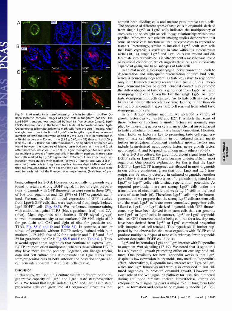
Lgr6+ Cells Are Progenitor Cells in Anterior and Posterior Tongue.Our RT-PCR results show that in addition to Lgr5, Lgr6 canbe amplified from our cultured organoids. Lgr5 marks adult tastestem/progenitor cells only in posterior tongue (6, 7). Promptedby this finding, as well as the result that Lgr6 is preferentiallyexpressed in fungiform taste tissue but not in the surroundingepithelium devoid of taste tissue (12), we set out to determinewhether Lgr6 marks adult taste stem/progenitor cells in anteriortaste fields (fungiform papillae). We used heterozygous micewith one wild-type Lgr6 allele and one allele in which EGFP hasbeen inserted into the Lgr6 gene (a knockout/knockin model)(13). Thus, EGFP serves as a surrogate marker for Lgr6 ex-pression. EGFP green fluorescence was detected at the basalarea of taste buds of the fungiform and circumvallate papillae(Fig. 5A and Fig. S6A). Occasionally, weak EGFP green fluo-rescence was detected in intragemmal taste cells as well. Theexpression pattern of Lgr6-EGFP in the circumvallate papillaeresembles that of Lgr5-EGFP, which indicates that Lgr6 mayalso mark stem/progenitor cells in taste tissue. The low frequencyof Lgr6-marked cells is likely a result of the mosaic expressionof Lgr6-EGFP in taste tissue, which is known to occur as wellin other tissues (13). No EGFP signal was detected in tongueepithelium devoid of taste tissue.We next performed lineage tracing to visualize the progeny of
Lgr6+ taste cells. Lgr6-EGFP-ires-CreERT2 mice were crossedwith Rosa26-tdTomato mice to generate Lgr6+/−; Rosa26-tdTo-mato+/− mice in which tamoxifen-induced Cre generates tdTo-mato fluorescent protein (red) to mark cells from the Lgr6+
lineage. We examined the distribution of tdTomato+ cells atdifferent points after a single tamoxifen injection. If the numberof tdTomato+ cells increased for prolonged periods after ta-moxifen induction, this would provide strong evidence that Lgr6+
cells may serve as taste progenitor cells that give rise to othertypes of taste cells. Indeed, at 1 d after tamoxifen induction, weobserved that basal cells in the fungiform and circumvallatepapillae were tdTomato+ (Fig. 5B and Fig. S6B). Occasionally,we found that intragemmal cells were tdTomato+. At 2 wk aftertamoxifen induction, in both fungiform and circumvallate pa-pillae, multiple intragemmal cells were tdTomato+. At 1 mo aftertamoxifen induction in both circumvallate and fungiform papil-lae, the number of tdTomato+ cells within taste buds was similarto that at 2 wk (Fig. 5B and Fig. S6B). By immunostaining tastetissue from Lgr6+/−; tdTomato+/− mice 1 mo after tamoxifen in-duction, we found that the progeny of Lgr6+ cells included atleast type 2 [transient receptor potential cation channel sub-family M member 5 (Trpm5+)] and type 3 (serotonin+) tastereceptor cells (Fig. 5C and Fig. S6C).Lgr6+ cells in posterior tongue showed a pattern of localiza-
tion similar to that of Lgr5+ cells. To determine whether thesetwo populations of cells are related, we sorted Lgr5+ cells fromposterior tongue and Lgr6+ cells from posterior and from ante-rior tongue, generated cDNA from the different sets of sortedcells, and performed RT-PCR using intron-spanning primers todetermine whether we could detect Lgr6 in Lgr5+ cells and viceversa. Interestingly, all three Lgr transcripts Lgr4–Lgr6 weredetected in sorted Lgr5+ cells from posterior tongue and insorted Lgr6+ cells from either posterior or anterior tongue (Fig.S7). In contrast, no or barely detectable Lgr5 and Lgr6 wereamplified from cells negative for GFP expression sorted fromLgr5-EGFP-ires-CreERT2+/− tongue tissues (Fig. S7).To determine whether Lgr6+ cells, similar to Lgr5+ cells, can
proliferate and differentiate into taste-like cells ex vivo, we sortedLgr6-EGFP cells on the basis of their EGFP fluorescence.Similar to Lgr5+ cells, Lgr6+ cells grew into 3D organoids (Fig.S8A), and most organoids had no intrinsic EGFP signal after
Fig. 4. Mature taste cells derived from single Lgr5+ cells are functional. (A)Representative trace of Ca2+ responses of an organoid-derived taste cell tosucralose and acesulfame-K sweeteners. (B) Representative trace of Ca2+
responses of an organoid-derived taste cell responding to bitter denatoniumbenzoate in a dose-dependent fashion (5, 10, and 20 mM). The phospholi-pase C β2 inhibitor U73122 (10 μM) inhibited denatonium-induced calciumresponses, consistent with bitter signaling pathways of taste cells in vivo. Theeffect of U73122 was reversible after washout for ∼30 min. Imagingexperiments were performed with 2D cultures independently grown from 15organoids (see Fig. S5 for details).
16404 | www.pnas.org/cgi/doi/10.1073/pnas.1409064111 Ren et al.
being cultured for 2–3 d. However, occasionally, organoids werefound to retain a strong EGFP signal: In two of eight prepara-tions, organoids with GFP fluorescence were seen in three (3%)of 106 total organoids and 129 (9%) of 1447 organoids exam-ined. Presumably, this continued expression of GFP resultedfrom Lgr6-EGFP cells that were expanded from single isolatedLgr6-EGFP+ cells (Fig. S8B). We performed immunostainingwith antibodies against T1R3 (blue), gustducin (red), and CA4(blue). Most organoids with intrinsic EGFP signal (green)showed immunoreactivity to two markers (∼80–89%: eight of 10for gustducin and CA4 and eight of nine for gustducin andT1R3, Fig. S8 C and D and Table S1). In contrast, a smallersubset of organoids without EGFP activity stained with bothmarkers (∼19–45%: five of 27 for gustducin and T1R3 and 13 of29 for gustducin and CA4; Fig. S8 E and F and Table S1). Thus,it would appear that organoids that continue to express Lgr6-EGFP are more often multipotent, whereas those without EGFPmay have more limited potency. Together, our lineage tracingdata and cell culture data demonstrate that Lgr6 marks tastestem/progenitor cells in both anterior and posterior tongue andcan generate apparent mature taste cells ex vivo.
DiscussionIn this study, we used a 3D culture system to determine the re-generative capacity of Lgr5+ and Lgr6+ taste stem/progenitorcells. We found that single isolated Lgr5+ and Lgr6+ taste stem/progenitor cells can grow into 3D “organoid” structures that
contain both dividing cells and mature presumptive taste cells.The presence of different types of taste cells in organoids derivedfrom single Lgr5+ or Lgr6+ cells indicates the multipotency ofsuch cells and sheds light on cell lineage relationships within tastepapillae. Moreover, our calcium imaging studies demonstrate thatsome of these cells function as taste receptor cells to respond totastants. Interestingly, similar to intestinal Lgr5+ adult stem cellsthat build crypt-villus structures in vitro without a mesenchymalniche (10, 14), single Lgr5+ and Lgr6+ cells can expand and dif-ferentiate into taste-like cells in vitro without a mesenchymal nicheor neuronal connection, which suggests these cells are intrinsicallycapable of giving rise to all subtypes of taste cells.In animal models, glossopharyngeal nerve transection leads to
degeneration and subsequent regeneration of taste bud cells,which is neuronally dependent, as taste cells start to regenerateonly after transected nerves reenter taste tissue (7, 29). There-fore, neuronal factors or direct neuronal contact may promotethe differentiation of taste cells generated from Lgr5+ or Lgr6+
stem/progenitor cells. Given the fact that single Lgr5+ or Lgr6+
taste stem/progenitor cells can give rise to taste cells in vitro, it islikely that neuronally secreted extrinsic factors, rather than di-rect neuronal contact, trigger taste cell renewal from adult tastestem/progenitor cells.In our defined culture medium, we included a variety of
growth factors, as well as N2 and B27. It is likely that some ofthese factors or functionally similar factors are normally sup-plied by innervating nerves or cells in mesenchymal tissue adjacentto taste epithelium to maintain taste tissue homeostasis. However,which factor or factors is key to promoting taste cell regenera-tion or taste cell differentiation in our cultured system meritsfurther investigation. Prominent candidate growth factors mayinclude brain-derived neurotrophic factor, nerve growth factor,neurotrophin-4, and R-spondins, among many others (30–34).After 2–3 d in culture, the green fluorescent signal from Lgr5-
EGFP cells or Lgr6-EGFP cells became undetectable in mostorganoids. One possible explanation for this is that the Lgr5-EGFP or Lgr6-EGFP transgenes are silenced in most organoidsin our culture conditions, given that both Lgr5 and Lgr6 tran-scripts can be readily detected in cultured organoids. Anotherpossibility is that at least two types of organoids originated fromLgr5+ or Lgr6+ cells, with distinct regeneration potentials. Asreported previously, there are strong Lgr5+ cells under thetrench areas of circumvallate and weak Lgr5+ cells in the basalareas of taste buds (6). Therefore, Lgr5+ cells may be hetero-geneous, and we propose that the strong Lgr5+ cells are stem cellsand the weak Lgr5+ cells are more committed progenitor cells.Likewise, Lgr5+- or Lgr6+-derived organoids with GFP fluores-cence may have been derived from stem cells that can generatenew Lgr5+ or Lgr6+ cells. In contrast, Lgr5+ or Lgr6+ organoidsthat lack GFP fluorescence after being cultured for a few days mayhave been derived from Lgr5+ or Lgr6+ committed progenitorcells incapable of self-renewal. This hypothesis is further sup-ported by the observation that most organoids with EGFP couldproduce multiple subtypes of taste cells, whereas fewer organoidswithout detectable EGFP could do so.Lgr5 and its homologs Lgr4 and Lgr6 interact with R-spondins
to augment Wnt signaling (17–19). We noted that R-spondin-1has a substantial growth-promoting effect on our organoid cul-tures. One possibility for how R-spondin works is that Lgr5,despite its low expression in organoids, may mediate R-spondin’seffect. Alternatively, R-spondin may interact with Lgr4 or Lgr6,which are Lgr5 homologs and were also expressed in our cul-tured organoids, to promote organoid growth. However, theexact role of the Wnt signaling pathway for taste tissue renewalduring adulthood remains unclear. Nevertheless, during de-velopment, Wnt signaling plays a major role in fungiform tastepapillae formation and seems to be regionally specific (35, 36).
Fig. 5. Lgr6 marks taste stem/progenitor cells in fungiform papillae. (A)Representative confocal images of Lgr6+ cells in fungiform papillae. TheLgr6-EGFP transgene was detected by intrinsic fluorescence (green). Lgr6-EGFP cells were found at the base of taste buds. (B) Tamoxifen-induced Lgr6-Cre generates tdTomato activity to mark cells from the Lgr6+ lineage. Aftera single tamoxifen induction of Lgr6-Cre in fungiform papillae, increasednumbers of taste bud cells were labeled at 2 wk (3.59 ± 0.44 per taste bud ina 10-μM section; n = 32) and 1 mo (4.66 ± 0.60; n = 38) than at 1 d (1.09 ±0.20; n = 34) (P < 0.0001 for both comparisons). No significant difference wasfound between the numbers of labeled taste bud cells at 1 mo and 2 wkafter tamoxifen induction (P = 0.17). (C) Lgr6+ stem/progenitor cells gener-ate multiple subtypes of taste bud cells in fungiform papillae. Mature tastebud cells marked by Lgr6-Cre-generated tdTomato 1 mo after tamoxifeninduction were stained with markers for type 2 (Trpm5) and type 3 (5-HT,serotonin) taste cells in fungiform papillae. Arrows depict tdTomato+ cellsthat are immunopositive for a specific taste cell marker. Three mice wereused for each point of the lineage tracing experiments. (Scale bars: 40 μm.)
Ren et al. PNAS | November 18, 2014 | vol. 111 | no. 46 | 16405
DEV
ELOPM
ENTA
LBIOLO
GY

Single Lgr5+ or Lgr6+ cells can give rise to different types oftaste cells in our 3D organoid cultures, indicating that some ofthese cells are multipotent. It remains to be determined whetherindividual taste Lgr5+ or Lgr6+ cells (e.g., the strongly Lgr5+
cells from under the trench areas) are multipotent in vivo. Twocontrasting models for taste cell lineages have been proposed(37): one posits existing lineage-specific stem/progenitor cells foreach subtype of taste cell, and the other posits multipotent tastestem cells that can give rise to all subtypes of taste cells. How-ever, these two models do not necessarily contradict each other:multipotent stem cells can give rise to lineage-specific progenitorcells. Our organoid culture data are consistent with both models.It is likely that some Lgr5+ or Lgr6+ cells are multipotent, suchas those having EGFP activity, whereas other Lgr5+ or Lgr6+
cells are committed progenitor cells. The multipotency of tasteprogenitor cells is further supported by the finding that Skn-1aknockout mice have a complete loss of sweet/bitter/umami type 2cells with a concomitant expansion of polycystic kidney disease2-like 1 protein–expressing sour taste receptor cells, which sug-gests at least the binary differentiation potential of progenitorcells into type 2 and type 3 cells (38) and is consistent with thehypothesis proposed by Miura and coworkers (39, 40).Lgr5+ cells give rise to all three major types of cells in taste
tissues in posterior tongue (6, 7). Similarly, we found that Lgr6+
cells can give rise to multiple types of cells in taste tissues in boththe anterior and posterior tongue, including type 2 receptor cellsand type 3 presynaptic cells. Similar to Lgr5-EGFP+ cells inposterior tongue, Lgr6-EGFP+ cells sit deep in the trench area ofthe circumvallate papilla, as well as at the base of taste buds.In contrast to Lgr5-EGFP+ cells, Lgr6-EGFP+ cells also aredetected in anterior tongue at the base of taste buds in fungiformpapillae. Our lineage tracing study showed that the progeny of
Lgr6+ progenitor cells can persist for 1 mo (the longest timetested to date). Although we are not certain whether Lgr6+ cellsrepresent bona fide stem cells in fungiform papillae, they areundoubtedly progenitor cells that give rise to other types ofmature taste cells both in vivo and ex vivo. Furthermore, thesimilar distribution pattern of Lgr5+ and Lgr6+ cells in posteriortongue; the detection of Lgr6 in Lgr5+ posterior cells and Lgr5 inLgr6+ cells in both anterior and posterior tongue, despite noLgr5-EGFP signal in fungiform papillae cells in adult mice,which could be a result of low expression of Lgr5 or regionalsilencing of Lgr5-EGFP transgene (6, 7, 12); and their similarregenerative capacity suggest they mark the same set of pro-genitor cells in both anterior and posterior tongue.
Materials and MethodsGenetically engineered mice (Lgr5-EGFP-ires-CreERT2 [stock # 008875], Lgr6-EGFP-ires-CreERT2 [stock # 016934], Rosa26-tdTomato [stock # 007905]) wereobtained from the Jackson Laboratory. All experiments were performedunder National Institutes of Health guidelines for the care and use of ani-mals in research and approved by the Institutional Animal Care and UseCommittee of the Monell Chemical Senses Center (protocol 1150). Detailsabout lineage tracing, cell sorting, 3D organoid cultures, immuno-staining, BrdU labeling, and calcium imaging are described in SI Materialsand Methods.
ACKNOWLEDGMENTS. We thank all members of the P.J. and R.F.M.laboratories for their input. Research reported in this publication wassupported by institutional funds (P.J.) from the Monell Chemical SensesCenter, as well as by NIH Grants DC010842 (to P.J.), DC000882 (to A.A.B.),DC012980 (to B.C.L.), DK081421 (to R.F.M.), and DC003055 (to R.F.M.), andan industry consortium funding Monell research (P.J., B.C.L., A.A.B., and R.F.M.).Imaging was performed at the Monell Histology and Cellular Localization Core,which is supported in part by NIH–National Institute on Deafness and OtherCommunication Disorders Core Grant DC011735 (to R.F.M.).
1. Beidler LM, Smallman RL (1965) Renewal of cells within taste buds. J Cell Biol 27(2):263–272.
2. Kapsimali M, Barlow LA (2013) Developing a sense of taste. Semin Cell Dev Biol 24(3):200–209.
3. Miura H, Scott JK, Harada S, Barlow LA (2014) Sonic hedgehog-expressing basal cellsare general post-mitotic precursors of functional taste receptor cells. Dev Dyn 243(10):1286–1297.
4. Hamamichi R, Asano-Miyoshi M, Emori Y (2006) Taste bud contains both short-livedand long-lived cell populations. Neuroscience 141(4):2129–2138.
5. Perea-Martinez I, Nagai T, Chaudhari N (2013) Functional cell types in taste buds havedistinct longevities. PLoS ONE 8(1):e53399.
6. Yee KK, et al. (2013) Lgr5-EGFP marks taste bud stem/progenitor cells in posteriortongue. Stem Cells 31(5):992–1000.
7. Takeda N, et al. (2013) Lgr5 Identifies Progenitor Cells Capable of Taste Bud Re-generation after Injury. PLoS ONE 8(6):e66314.
8. Barker N, Tan S, Clevers H (2013) Lgr proteins in epithelial stem cell biology. De-velopment 140(12):2484–2494.
9. Huch M, et al. (2013) In vitro expansion of single Lgr5+ liver stem cells induced byWnt-driven regeneration. Nature 494(7436):247–250.
10. Sato T, et al. (2009) Single Lgr5 stem cells build crypt-villus structures in vitro withouta mesenchymal niche. Nature 459(7244):262–265.
11. Huch M, et al. (2013) Unlimited in vitro expansion of adult bi-potent pancreas pro-genitors through the Lgr5/R-spondin axis. EMBO J 32(20):2708–2721.
12. Hevezi P, et al. (2009) Genome-wide analysis of gene expression in primate taste budsreveals links to diverse processes. PLoS ONE 4(7):e6395.
13. Snippert HJ, et al. (2010) Lgr6 marks stem cells in the hair follicle that generate all celllineages of the skin. Science 327(5971):1385–1389.
14. Sato T, Clevers H (2013) Primary mouse small intestinal epithelial cell cultures.Methods Mol Biol 945:319–328.
15. Carmon KS, Gong X, Lin Q, Thomas A, Liu Q (2011) R-spondins function as ligands ofthe orphan receptors LGR4 and LGR5 to regulate Wnt/beta-catenin signaling. ProcNatl Acad Sci USA 108(28):11452–11457.
16. de Lau W, et al. (2011) Lgr5 homologues associate with Wnt receptors and mediateR-spondin signalling. Nature 476(7360):293–297.
17. Ruffner H, et al. (2012) R-Spondin potentiates Wnt/β-catenin signaling through or-phan receptors LGR4 and LGR5. PLoS ONE 7(7):e40976.
18. Okubo T, Pevny LH, Hogan BL (2006) Sox2 is required for development of taste budsensory cells. Genes Dev 20(19):2654–2659.
19. Avilion AA, et al. (2003) Multipotent cell lineages in early mouse development de-pend on SOX2 function. Genes Dev 17(1):126–140.
20. Okubo T, Clark C, Hogan BL (2009) Cell lineage mapping of taste bud cells and ker-atinocytes in the mouse tongue and soft palate. Stem Cells 27(2):442–450.
21. Feng P, Huang L, Wang H (2014) Taste bud homeostasis in health, disease, and aging.Chem Senses 39(1):3–16.
22. de Lau W, Peng WC, Gros P, Clevers H (2014) The R-spondin/Lgr5/Rnf43 module:Regulator of Wnt signal strength. Genes Dev 28(4):305–316.
23. Max M, et al. (2001) Tas1r3, encoding a new candidate taste receptor, is allelic to thesweet responsiveness locus Sac. Nat Genet 28(1):58–63.
24. Nelson G, et al. (2002) An amino-acid taste receptor. Nature 416(6877):199–202.25. Nelson G, et al. (2001) Mammalian sweet taste receptors. Cell 106(3):381–390.26. Chandrashekar J, et al. (2009) The taste of carbonation. Science 326(5951):443–445.27. Kim MR, et al. (2003) Regional expression patterns of taste receptors and gustducin in
the mouse tongue. Biochem Biophys Res Commun 312(2):500–506.28. Zhang Y, et al. (2003) Coding of sweet, bitter, and umami tastes: Different receptor
cells sharing similar signaling pathways. Cell 112(3):293–301.29. King CT, Travers SP, Rowland NE, Garcea M, Spector AC (1999) Glossopharyngeal
nerve transection eliminates quinine-stimulated fos-like immunoreactivity in thenucleus of the solitary tract: Implications for a functional topography of gustatorynerve input in rats. J Neurosci 19(8):3107–3121.
30. Ganchrow D, Ganchrow JR, Verdin-Alcazar M, Whitehead MC (2003) Brain-derivedneurotrophic factor-, neurotrophin-3-, and tyrosine kinase receptor-like immunore-activity in lingual taste bud fields of mature hamster. J Comp Neurol 455(1):11–24.
31. Mistretta CM, Goosens KA, Farinas I, Reichardt LF (1999) Alterations in size, number, andmorphology of gustatory papillae and taste buds in BDNF null mutantmice demonstrateneural dependence of developing taste organs. J Comp Neurol 409(1):13–24.
32. Nosrat CA, Blomlöf J, ElShamy WM, Ernfors P, Olson L (1997) Lingual deficits in BDNFand NT3 mutant mice leading to gustatory and somatosensory disturbances, re-spectively. Development 124(7):1333–1342.
33. Oakley B, et al. (1998) The morphogenesis of mouse vallate gustatory epithelium andtaste buds requires BDNF-dependent taste neurons. Brain Res Dev Brain Res 105(1):85–96.
34. Patel AV, Huang T, Krimm RF (2010) Lingual and palatal gustatory afferents eachdepend on both BDNF and NT-4, but the dependence is greater for lingual thanpalatal afferents. J Comp Neurol 518(16):3290–3301.
35. Iwatsuki K, et al. (2007) Wnt signaling interacts with Shh to regulate taste papilladevelopment. Proc Natl Acad Sci USA 104(7):2253–2258.
36. Liu F, et al. (2007) Wnt-beta-catenin signaling initiates taste papilla development. NatGenet 39(1):106–112.
37. Stone LM, Tan SS, Tam PP, Finger TE (2002) Analysis of cell lineage relationships intaste buds. J Neurosci 22(11):4522–4529.
38. Matsumoto I, Ohmoto M, Narukawa M, Yoshihara Y, Abe K (2011) Skn-1a (Pou2f3)specifies taste receptor cell lineage. Nat Neurosci 14(6):685–687.
39. Miura H, Kusakabe Y, Harada S (2006) Cell lineage and differentiation in taste buds.Arch Histol Cytol 69(4):209–225.
40. Miura H, et al. (2003) Co-expression pattern of Shh with Prox1 and that of Nkx2.2with Mash1 in mouse taste bud. Gene Expr Patterns 3(4):427–430.
16406 | www.pnas.org/cgi/doi/10.1073/pnas.1409064111 Ren et al.