Embed Size (px)
Citation preview
RESEARCH COMMUNICATION
Single mesodermal cells guideoutgrowth of ectodermal tubularstructures in DrosophilaChristian Wolf and Reinhard Schuh1
Max Planck Institut fur biophysikalische Chemie, AbteilungMolekulare Entwicklungsbiologie, Am Fassberg, D-37077Gottingen, Germany
The Drosophila tracheal system, a tubular network, isformed from isolated ectodermal metameres by guidedbranch outgrowth and branch fusion. Branch outgrowthis triggered by the localized and transient activity ofBranchless (Bnl/dFGF). Here, we report the discovery ofa mesodermal cell that links the leading cells of out-growing main branches 2.5 hr before they fuse. Thisbridge-cell serves as an essential guidance post and needsHunchback (Hb) activity to exert its function. Thebridge-cell provides cues acting in concert with Bnl/dFGF signaling to mediate directed branch outgrowththat ultimately leads to position-specific branch fusion.
Received May 16, 2000; revised version accepted July 5, 2000.
Formation of three-dimensional tubular structures, suchas the insect tracheal system (Manning and Krasnow1993; Samakovlis et al. 1996), the vertebrate vascularsystem (Risau 1997), and the lung (Hogan et al. 1997),involves the guided outgrowth of epithelial cells. In Dro-sophila, the tracheal system is generated from 10 iso-lated lateral cell clusters on each side of the embryo (Fig.1A). These cell clusters, which are each composed ofabout 80 ectodermal cells, invaginate in a strictly coor-dinated manner into the underlying mesoderm, wherethey establish a pattern of six primary tubular branches(Fig. 1B). Some of these branches grow along the dorso-ventral body axis to form the dorsal, the lateral, and theganglionic branches. Additional primary branches ex-tend along the anteroposterior axis to generate the vis-ceral and dorsal trunk anterior and posterior branches.The individual tracheal cell clusters connect by fusion ofthe dorsal trunk and the lateral trunk branches (Fig. 1C).The two halves of the network interconnect by anasto-mosis formation, and the three-dimensional systemstarts with the transport of gases during larval develop-ment (for details, see Manning and Krasnow 1993; Sama-kovlis et al. 1996).
Tubular branch outgrowth is guided by the local andcomplex expression pattern of a Drosophila FGF homo-log, Branchless (Bnl/dFGF), emanating from cell clusters
surrounding each tracheal metamere (Sutherland et al.1996; Metzger and Krasnow 1999). However, althoughmutant analysis shows that Bnl/dFGF is necessary forprimary branch outgrowth, the restricted Bnl/dFGF ex-pression seems not to be essential for the directed out-growth of all primary branches. This conclusion is basedon the observation that the constitutive activation ofBnl/dFGF signaling in bnl mutant embryos partially re-stores outgrowth of the main tracheal tube, the dorsaltrunk, whereas the other primary branches are not gen-erated. Thus, it was proposed that additional guidancecues might be necessary for the outgrowth of dorsaltrunk branches (Sutherland et al. 1996).
Results and Discussion
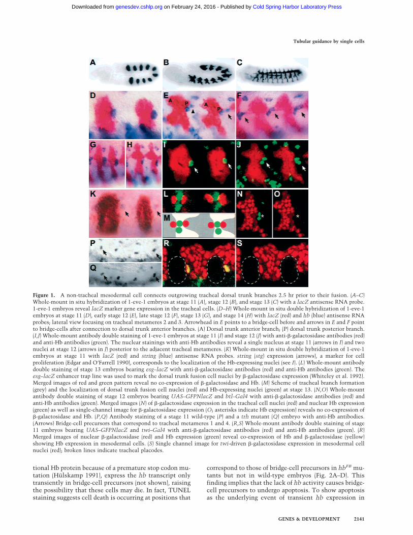
We noted a single cell that is marked by expression of thegene hunchback (hb; Lehmann 1985; Tautz et al. 1987;Hulskamp 1991) at the posterior lateral margin of eachtracheal metamere (Fig. 1D,I). This cell gives rise todaughter cells that maintain hb expression (Fig. 1E,J,K).The more ventrally located daughter cell maintains around morphology and remains in position, whereas thedorsal daughter cell connects to the posterior bud of thetracheal metamere, termed the dorsal trunk posteriorbranch (Fig. 1E). Subsequently, the dorsal daughter cellelongates and extends posteriorly and thereby contactsto the anterior bud, termed the dorsal trunk anteriorbranch, of the adjacent posterior tracheal metamere (Fig.1E,F). In this way, the dorsal daughter cell bridges theleading cells of the dorsal trunk anterior and posteriorbranches of two adjacent metameres (Fig. 1F), whichthen fuse about 2.5 hr later to form the continuous dorsaltrunk. Thus, we refer to the dorsal daughter cell as thebridge-cell. The cell remains at this position until fusionbetween the dorsal trunk anterior and posterior branchesoccurs (Fig. 1G). During this fusion process, the bridge-cell becomes displaced and hb expression starts to fade(Fig. 1H).
To trace the origin of the bridge-cell, we performeddouble-staining experiments with tracheal-specific mark-ers and hb. �-Galactosidase expression in nuclei of dorsaltrunk fusion cells and in nuclei of tracheal cellsrevealed a lack of colocalization with bridge-cell hb ex-pression (Fig. 1L–1O). Furthermore, trachealess (trh;Isaac and Andrew 1996; Wilk et al. 1996) mutant em-bryos, which lack tracheal cell identity, show hb-ex-pressing bridge-cells as found in wild-type embryos (Fig.1P,Q). Thus, these results indicate that the bridge-cell isof nontracheal origin. Finally, double-staining of hb anda mesodermal marker (Greig and Akam 1993) revealedcoexpression of hb and the marker in bridge-cell precur-sors (Fig. 1R,S). Therefore, the bridge-cell is a nontra-cheal cell and of mesodermal origin.
To understand the function of bridge-cells in dorsaltrunk formation, we first asked whether bridge-cell de-velopment is affected in hb mutant embryos. Homozy-gous hbFB mutant embryos, which express a nonfunc-
[Key Words: hunchback; FGF; tracheal system; Drosophila; guidance]1Corresponding author.E-MAIL [email protected]; FAX 49-551-201-1755.Article and publication are at www.genesdev.org/cgi/doi/10.1101/gad.180900.
2140 GENES & DEVELOPMENT 14:2140–2145 © 2000 by Cold Spring Harbor Laboratory Press ISSN 0890-9369/00 $5.00; www.genesdev.org
Cold Spring Harbor Laboratory Press on February 24, 2016 - Published by genesdev.cshlp.orgDownloaded from
tional Hb protein because of a premature stop codon mu-tation (Hulskamp 1991), express the hb transcript onlytransiently in bridge-cell precursors (not shown), raisingthe possibility that these cells may die. In fact, TUNELstaining suggests cell death is occurring at positions that
correspond to those of bridge-cell precursors in hbFB mu-tants but not in wild-type embryos (Fig. 2A–D). Thisfinding implies that the lack of hb activity causes bridge-cell precursors to undergo apoptosis. To show apoptosisas the underlying event of transient hb expression in
Figure 1. A non-tracheal mesodermal cell connects outgrowing tracheal dorsal trunk branches 2.5 hr prior to their fusion. (A–C)Whole-mount in situ hybridization of 1-eve-1 embryos at stage 11 (A), stage 12 (B), and stage 13 (C) with a lacZ antisense RNA probe.1-eve-1 embryos reveal lacZ marker gene expression in the tracheal cells. (D–H) Whole-mount in situ double hybridization of 1-eve-1embryos at stage 11 (D), early stage 12 (E), late stage 12 (F), stage 13 (G), and stage 14 (H) with lacZ (red) and hb (blue) antisense RNAprobes; lateral view focusing on tracheal metameres 2 and 3. Arrowhead in E points to a bridge-cell before and arrows in E and F pointto bridge-cells after connection to dorsal trunk anterior branches. (A) Dorsal trunk anterior branch; (P) dorsal trunk posterior branch.(I,J) Whole-mount antibody double staining of 1-eve-1 embryos at stage 11 (I) and stage 12 (J) with anti-�-galactosidase antibodies (red)and anti-Hb antibodies (green). The nuclear stainings with anti-Hb antibodies reveal a single nucleus at stage 11 (arrows in I) and twonuclei at stage 12 (arrows in J) posterior to the adjacent tracheal metameres. (K) Whole-mount in situ double hybridization of 1-eve-1embryos at stage 11 with lacZ (red) and string (blue) antisense RNA probes. string (stg) expression (arrows), a marker for cellproliferation (Edgar and O’Farrell 1990), corresponds to the localization of the Hb-expressing nuclei (see I). (L) Whole-mount antibodydouble staining of stage 13 embryos bearing esg–lacZ with anti-�-galactosidase antibodies (red) and anti-Hb antibodies (green). Theesg–lacZ enhancer trap line was used to mark the dorsal trunk fusion cell nuclei by �-galactosidase expression (Whiteley et al. 1992).Merged images of red and green pattern reveal no co-expression of �-galactosidase and Hb. (M) Scheme of tracheal branch formation(grey) and the localization of dorsal trunk fusion cell nuclei (red) and Hb-expressing nuclei (green) at stage 13. (N,O) Whole-mountantibody double staining of stage 12 embryos bearing UAS–GFPNlacZ and btl–Gal4 with anti-�-galactosidase antibodies (red) andanti-Hb antibodies (green). Merged images (N) of �-galactosidase expression in the tracheal cell nuclei (red) and nuclear Hb expression(green) as well as single-channel image for �-galactosidase expression (O; asterisks indicate Hb expression) reveals no co-expression of�-galactosidase and Hb. (P,Q) Antibody staining of a stage 11 wild-type (P) and a trh mutant (Q) embryo with anti-Hb antibodies.(Arrows) Bridge-cell precursors that correspond to tracheal metameres 1 and 4. (R,S) Whole-mount antibody double staining of stage11 embryos bearing UAS–GFPNlacZ and twi–Gal4 with anti-�-galactosidase antibodies (red) and anti-Hb antibodies (green). (R)Merged images of nuclear �-galactosidase (red) and Hb expression (green) reveal co-expression of Hb and �-galactosidase (yellow)showing Hb expression in mesodermal cells. (S) Single channel image for twi-driven �-galactosidase expression in mesodermal cellnuclei (red); broken lines indicate tracheal placodes.
Tubular guidance by single cells
GENES & DEVELOPMENT 2141
Cold Spring Harbor Laboratory Press on February 24, 2016 - Published by genesdev.cshlp.orgDownloaded from
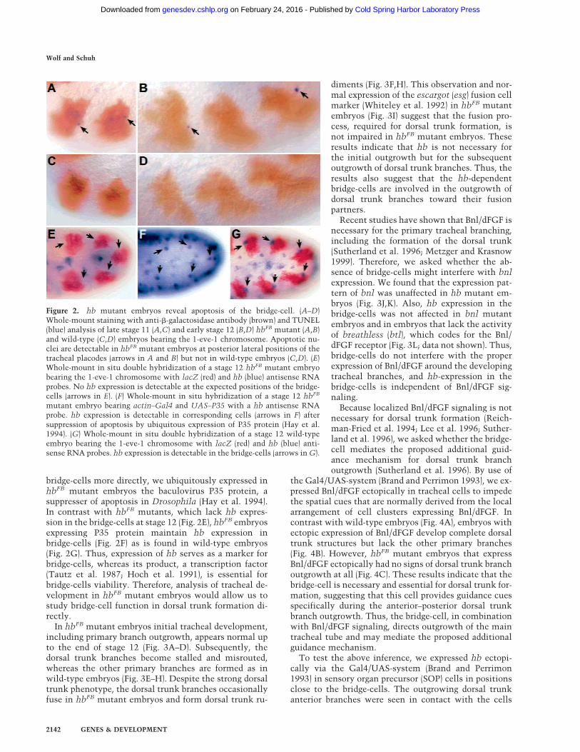
bridge-cells more directly, we ubiquitously expressed inhbFB mutant embryos the baculovirus P35 protein, asuppresser of apoptosis in Drosophila (Hay et al. 1994).In contrast with hbFB mutants, which lack hb expres-sion in the bridge-cells at stage 12 (Fig. 2E), hbFB embryosexpressing P35 protein maintain hb expression inbridge-cells (Fig. 2F) as is found in wild-type embryos(Fig. 2G). Thus, expression of hb serves as a marker forbridge-cells, whereas its product, a transcription factor(Tautz et al. 1987; Hoch et al. 1991), is essential forbridge-cells viability. Therefore, analysis of tracheal de-velopment in hbFB mutant embryos would allow us tostudy bridge-cell function in dorsal trunk formation di-rectly.
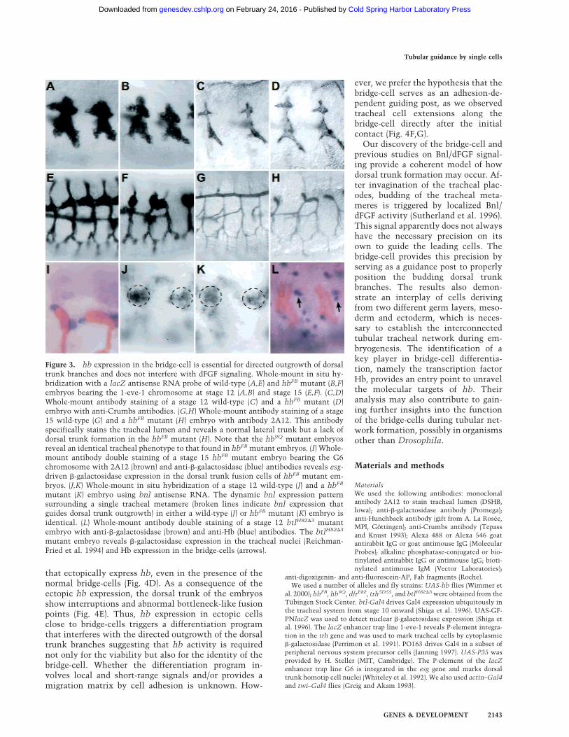
In hbFB mutant embryos initial tracheal development,including primary branch outgrowth, appears normal upto the end of stage 12 (Fig. 3A–D). Subsequently, thedorsal trunk branches become stalled and misrouted,whereas the other primary branches are formed as inwild-type embryos (Fig. 3E–H). Despite the strong dorsaltrunk phenotype, the dorsal trunk branches occasionallyfuse in hbFB mutant embryos and form dorsal trunk ru-
diments (Fig. 3F,H). This observation and nor-mal expression of the escargot (esg) fusion cellmarker (Whiteley et al. 1992) in hbFB mutantembryos (Fig. 3I) suggest that the fusion pro-cess, required for dorsal trunk formation, isnot impaired in hbFB mutant embryos. Theseresults indicate that hb is not necessary forthe initial outgrowth but for the subsequentoutgrowth of dorsal trunk branches. Thus, theresults also suggest that the hb-dependentbridge-cells are involved in the outgrowth ofdorsal trunk branches toward their fusionpartners.
Recent studies have shown that Bnl/dFGF isnecessary for the primary tracheal branching,including the formation of the dorsal trunk(Sutherland et al. 1996; Metzger and Krasnow1999). Therefore, we asked whether the ab-sence of bridge-cells might interfere with bnlexpression. We found that the expression pat-tern of bnl was unaffected in hb mutant em-bryos (Fig. 3J,K). Also, hb expression in thebridge-cells was not affected in bnl mutantembryos and in embryos that lack the activityof breathless (btl), which codes for the Bnl/dFGF receptor (Fig. 3L; data not shown). Thus,bridge-cells do not interfere with the properexpression of Bnl/dFGF around the developingtracheal branches, and hb-expression in thebridge-cells is independent of Bnl/dFGF sig-naling.
Because localized Bnl/dFGF signaling is notnecessary for dorsal trunk formation (Reich-man-Fried et al. 1994; Lee et al. 1996; Suther-land et al. 1996), we asked whether the bridge-cell mediates the proposed additional guid-ance mechanism for dorsal trunk branchoutgrowth (Sutherland et al. 1996). By use of
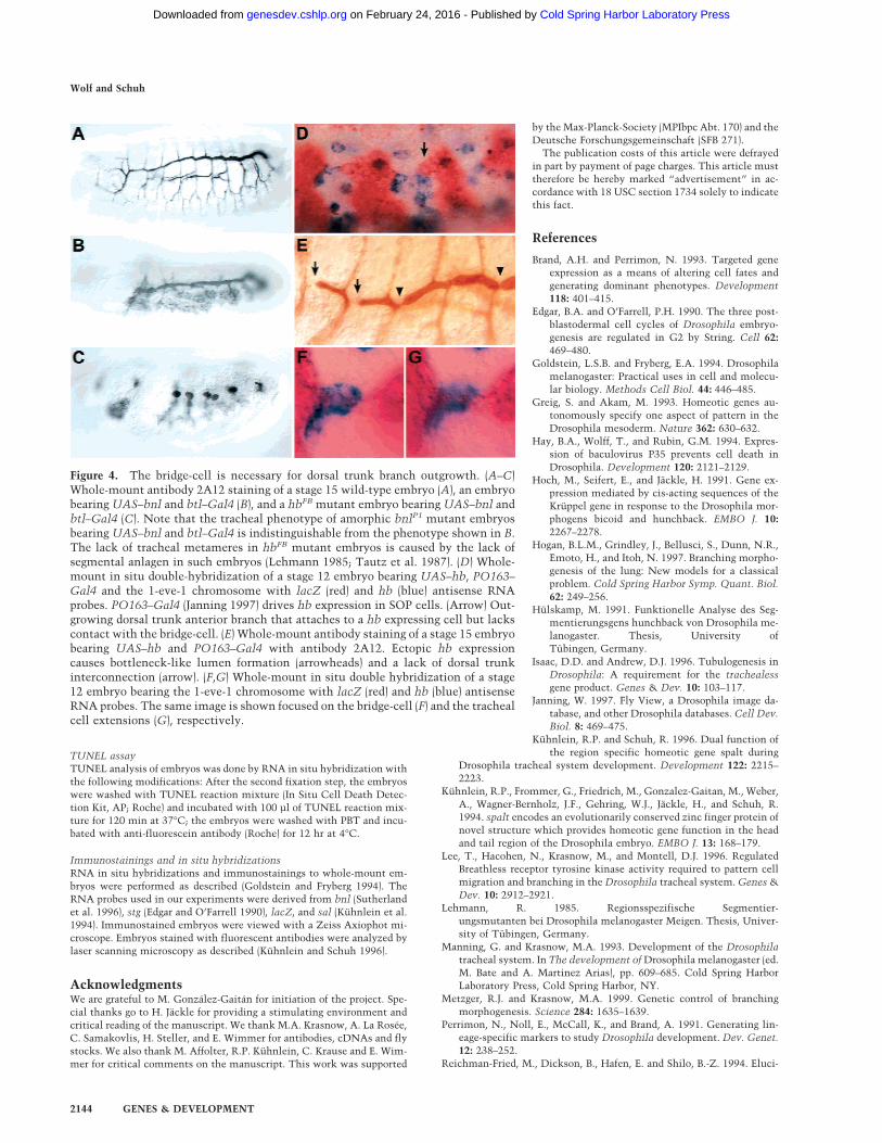
the Gal4/UAS-system (Brand and Perrimon 1993), we ex-pressed Bnl/dFGF ectopically in tracheal cells to impedethe spatial cues that are normally derived from the localarrangement of cell clusters expressing Bnl/dFGF. Incontrast with wild-type embryos (Fig. 4A), embryos withectopic expression of Bnl/dFGF develop complete dorsaltrunk structures but lack the other primary branches(Fig. 4B). However, hbFB mutant embryos that expressBnl/dFGF ectopically had no signs of dorsal trunk branchoutgrowth at all (Fig. 4C). These results indicate that thebridge-cell is necessary and essential for dorsal trunk for-mation, suggesting that this cell provides guidance cuesspecifically during the anterior–posterior dorsal trunkbranch outgrowth. Thus, the bridge-cell, in combinationwith Bnl/dFGF signaling, directs outgrowth of the maintracheal tube and may mediate the proposed additionalguidance mechanism.
To test the above inference, we expressed hb ectopi-cally via the Gal4/UAS-system (Brand and Perrimon1993) in sensory organ precursor (SOP) cells in positionsclose to the bridge-cells. The outgrowing dorsal trunkanterior branches were seen in contact with the cells
Figure 2. hb mutant embryos reveal apoptosis of the bridge-cell. (A–D)Whole-mount staining with anti-�-galactosidase antibody (brown) and TUNEL(blue) analysis of late stage 11 (A,C) and early stage 12 (B,D) hbFB mutant (A,B)and wild-type (C,D) embryos bearing the 1-eve-1 chromosome. Apoptotic nu-clei are detectable in hbFB mutant embryos at posterior lateral positions of thetracheal placodes (arrows in A and B) but not in wild-type embryos (C,D). (E)Whole-mount in situ double hybridization of a stage 12 hbFB mutant embryobearing the 1-eve-1 chromosome with lacZ (red) and hb (blue) antisense RNAprobes. No hb expression is detectable at the expected positions of the bridge-cells (arrows in E). (F) Whole-mount in situ hybridization of a stage 12 hbFB
mutant embryo bearing actin–Gal4 and UAS–P35 with a hb antisense RNAprobe. hb expression is detectable in corresponding cells (arrows in F) aftersuppression of apoptosis by ubiquitous expression of P35 protein (Hay et al.1994). (G) Whole-mount in situ double hybridization of a stage 12 wild-typeembryo bearing the 1-eve-1 chromosome with lacZ (red) and hb (blue) anti-sense RNA probes. hb expression is detectable in the bridge-cells (arrows in G).
Wolf and Schuh
2142 GENES & DEVELOPMENT
Cold Spring Harbor Laboratory Press on February 24, 2016 - Published by genesdev.cshlp.orgDownloaded from
that ectopically express hb, even in the presence of thenormal bridge-cells (Fig. 4D). As a consequence of theectopic hb expression, the dorsal trunk of the embryosshow interruptions and abnormal bottleneck-like fusionpoints (Fig. 4E). Thus, hb expression in ectopic cellsclose to bridge-cells triggers a differentiation programthat interferes with the directed outgrowth of the dorsaltrunk branches suggesting that hb activity is requirednot only for the viability but also for the identity of thebridge-cell. Whether the differentiation program in-volves local and short-range signals and/or provides amigration matrix by cell adhesion is unknown. How-
ever, we prefer the hypothesis that thebridge-cell serves as an adhesion-de-pendent guiding post, as we observedtracheal cell extensions along thebridge-cell directly after the initialcontact (Fig. 4F,G).
Our discovery of the bridge-cell andprevious studies on Bnl/dFGF signal-ing provide a coherent model of howdorsal trunk formation may occur. Af-ter invagination of the tracheal plac-odes, budding of the tracheal meta-meres is triggered by localized Bnl/dFGF activity (Sutherland et al. 1996).This signal apparently does not alwayshave the necessary precision on itsown to guide the leading cells. Thebridge-cell provides this precision byserving as a guidance post to properlyposition the budding dorsal trunkbranches. The results also demon-strate an interplay of cells derivingfrom two different germ layers, meso-derm and ectoderm, which is neces-sary to establish the interconnectedtubular tracheal network during em-bryogenesis. The identification of akey player in bridge-cell differentia-tion, namely the transcription factorHb, provides an entry point to unravelthe molecular targets of hb. Theiranalysis may also contribute to gain-ing further insights into the functionof the bridge-cells during tubular net-work formation, possibly in organismsother than Drosophila.
Materials and methods
MaterialsWe used the following antibodies: monoclonalantibody 2A12 to stain tracheal lumen (DSHB,Iowa); anti-�-galactosidase antibody (Promega);anti-Hunchback antibody (gift from A. La Rosee,MPI, Gottingen); anti-Crumbs antibody (Tepassand Knust 1993); Alexa 488 or Alexa 546 goatantirabbit IgG or goat antimouse IgG (MolecularProbes); alkaline phosphatase-conjugated or bio-tinylated antirabbit IgG or antimouse IgG; bioti-nylated antimouse IgM (Vector Laboratories);
anti-digoxigenin- and anti-fluorescein-AP, Fab fragments (Roche).We used a number of alleles and fly strains: UAS-hb flies (Wimmer et
al. 2000); hbFB, hb9Q, dfrE82, trh5D55, and btlH82�3 were obtained from theTubingen Stock Center. btl-Gal4 drives Gal4 expression ubiquitously inthe tracheal system from stage 10 onward (Shiga et al. 1996). UAS-GF-PNlacZ was used to detect nuclear �-galactosidase expression (Shiga etal. 1996). The lacZ enhancer trap line 1-eve-1 reveals P-element integra-tion in the trh gene and was used to mark tracheal cells by cytoplasmic�-galactosidase (Perrimon et al. 1991). PO163 drives Gal4 in a subset ofperipheral nervous system precursor cells (Janning 1997). UAS-P35 wasprovided by H. Steller (MIT, Cambridge). The P-element of the lacZenhancer trap line G6 is integrated in the esg gene and marks dorsaltrunk homotip cell nuclei (Whiteley et al. 1992). We also used actin–Gal4and twi–Gal4 flies (Greig and Akam 1993).
Figure 3. hb expression in the bridge-cell is essential for directed outgrowth of dorsaltrunk branches and does not interfere with dFGF signaling. Whole-mount in situ hy-bridization with a lacZ antisense RNA probe of wild-type (A,E) and hbFB mutant (B,F)embryos bearing the 1-eve-1 chromosome at stage 12 (A,B) and stage 15 (E,F). (C,D)Whole-mount antibody staining of a stage 12 wild-type (C) and a hbFB mutant (D)embryo with anti-Crumbs antibodies. (G,H) Whole-mount antibody staining of a stage15 wild-type (G) and a hbFB mutant (H) embryo with antibody 2A12. This antibodyspecifically stains the tracheal lumen and reveals a normal lateral trunk but a lack ofdorsal trunk formation in the hbFB mutant (H). Note that the hb9Q mutant embryosreveal an identical tracheal phenotype to that found in hbFB mutant embryos. (I) Whole-mount antibody double staining of a stage 15 hbFB mutant embryo bearing the G6chromosome with 2A12 (brown) and anti-�-galactosidase (blue) antibodies reveals esg-driven �-galactosidase expression in the dorsal trunk fusion cells of hbFB mutant em-bryos. (J,K) Whole-mount in situ hybridization of a stage 12 wild-type (J) and a hbFB
mutant (K) embryo using bnl antisense RNA. The dynamic bnl expression patternsurrounding a single tracheal metamere (broken lines indicate bnl expression thatguides dorsal trunk outgrowth) in either a wild-type (J) or hbFB mutant (K) embryo isidentical. (L) Whole-mount antibody double staining of a stage 12 btlH82�3 mutantembryo with anti-�-galactosidase (brown) and anti-Hb (blue) antibodies. The btlH82�3
mutant embryo reveals �-galactosidase expression in the tracheal nuclei (Reichman-Fried et al. 1994) and Hb expression in the bridge-cells (arrows).
Tubular guidance by single cells
GENES & DEVELOPMENT 2143
Cold Spring Harbor Laboratory Press on February 24, 2016 - Published by genesdev.cshlp.orgDownloaded from
TUNEL assayTUNEL analysis of embryos was done by RNA in situ hybridization withthe following modifications: After the second fixation step, the embryoswere washed with TUNEL reaction mixture (In Situ Cell Death Detec-tion Kit, AP; Roche) and incubated with 100 µl of TUNEL reaction mix-ture for 120 min at 37°C; the embryos were washed with PBT and incu-bated with anti-fluorescein antibody (Roche) for 12 hr at 4°C.
Immunostainings and in situ hybridizationsRNA in situ hybridizations and immunostainings to whole-mount em-bryos were performed as described (Goldstein and Fryberg 1994). TheRNA probes used in our experiments were derived from bnl (Sutherlandet al. 1996), stg (Edgar and O’Farrell 1990), lacZ, and sal (Kuhnlein et al.1994). Immunostained embryos were viewed with a Zeiss Axiophot mi-croscope. Embryos stained with fluorescent antibodies were analyzed bylaser scanning microscopy as described (Kuhnlein and Schuh 1996).
AcknowledgmentsWe are grateful to M. Gonzalez-Gaitan for initiation of the project. Spe-cial thanks go to H. Jackle for providing a stimulating environment andcritical reading of the manuscript. We thank M.A. Krasnow, A. La Rosee,C. Samakovlis, H. Steller, and E. Wimmer for antibodies, cDNAs and flystocks. We also thank M. Affolter, R.P. Kuhnlein, C. Krause and E. Wim-mer for critical comments on the manuscript. This work was supported
by the Max-Planck-Society (MPIbpc Abt. 170) and theDeutsche Forschungsgemeinschaft (SFB 271).
The publication costs of this article were defrayedin part by payment of page charges. This article musttherefore be hereby marked “advertisement” in ac-cordance with 18 USC section 1734 solely to indicatethis fact.
References
Brand, A.H. and Perrimon, N. 1993. Targeted geneexpression as a means of altering cell fates andgenerating dominant phenotypes. Development118: 401–415.
Edgar, B.A. and O’Farrell, P.H. 1990. The three post-blastodermal cell cycles of Drosophila embryo-genesis are regulated in G2 by String. Cell 62:469–480.
Goldstein, L.S.B. and Fryberg, E.A. 1994. Drosophilamelanogaster: Practical uses in cell and molecu-lar biology. Methods Cell Biol. 44: 446–485.
Greig, S. and Akam, M. 1993. Homeotic genes au-tonomously specify one aspect of pattern in theDrosophila mesoderm. Nature 362: 630–632.
Hay, B.A., Wolff, T., and Rubin, G.M. 1994. Expres-sion of baculovirus P35 prevents cell death inDrosophila. Development 120: 2121–2129.
Hoch, M., Seifert, E., and Jackle, H. 1991. Gene ex-pression mediated by cis-acting sequences of theKruppel gene in response to the Drosophila mor-phogens bicoid and hunchback. EMBO J. 10:2267–2278.
Hogan, B.L.M., Grindley, J., Bellusci, S., Dunn, N.R.,Emoto, H., and Itoh, N. 1997. Branching morpho-genesis of the lung: New models for a classicalproblem. Cold Spring Harbor Symp. Quant. Biol.62: 249–256.
Hulskamp, M. 1991. Funktionelle Analyse des Seg-mentierungsgens hunchback von Drosophila me-lanogaster. Thesis, University ofTubingen, Germany.
Isaac, D.D. and Andrew, D.J. 1996. Tubulogenesis inDrosophila: A requirement for the trachealessgene product. Genes & Dev. 10: 103–117.
Janning, W. 1997. Fly View, a Drosophila image da-tabase, and other Drosophila databases. Cell Dev.Biol. 8: 469–475.
Kuhnlein, R.P. and Schuh, R. 1996. Dual function ofthe region specific homeotic gene spalt during
Drosophila tracheal system development. Development 122: 2215–2223.
Kuhnlein, R.P., Frommer, G., Friedrich, M., Gonzalez-Gaitan, M., Weber,A., Wagner-Bernholz, J.F., Gehring, W.J., Jackle, H., and Schuh, R.1994. spalt encodes an evolutionarily conserved zinc finger protein ofnovel structure which provides homeotic gene function in the headand tail region of the Drosophila embryo. EMBO J. 13: 168–179.
Lee, T., Hacohen, N., Krasnow, M., and Montell, D.J. 1996. RegulatedBreathless receptor tyrosine kinase activity required to pattern cellmigration and branching in the Drosophila tracheal system. Genes &Dev. 10: 2912–2921.
Lehmann, R. 1985. Regionsspezifische Segmentier-ungsmutanten bei Drosophila melanogaster Meigen. Thesis, Univer-sity of Tubingen, Germany.
Manning, G. and Krasnow, M.A. 1993. Development of the Drosophilatracheal system. In The development of Drosophila melanogaster (ed.M. Bate and A. Martinez Arias), pp. 609–685. Cold Spring HarborLaboratory Press, Cold Spring Harbor, NY.
Metzger, R.J. and Krasnow, M.A. 1999. Genetic control of branchingmorphogenesis. Science 284: 1635–1639.
Perrimon, N., Noll, E., McCall, K., and Brand, A. 1991. Generating lin-eage-specific markers to study Drosophila development. Dev. Genet.12: 238–252.
Reichman-Fried, M., Dickson, B., Hafen, E. and Shilo, B.-Z. 1994. Eluci-
Figure 4. The bridge-cell is necessary for dorsal trunk branch outgrowth. (A–C)Whole-mount antibody 2A12 staining of a stage 15 wild-type embryo (A), an embryobearing UAS–bnl and btl–Gal4 (B), and a hbFB mutant embryo bearing UAS–bnl andbtl–Gal4 (C). Note that the tracheal phenotype of amorphic bnlP1 mutant embryosbearing UAS–bnl and btl–Gal4 is indistinguishable from the phenotype shown in B.The lack of tracheal metameres in hbFB mutant embryos is caused by the lack ofsegmental anlagen in such embryos (Lehmann 1985; Tautz et al. 1987). (D) Whole-mount in situ double-hybridization of a stage 12 embryo bearing UAS–hb, PO163–Gal4 and the 1-eve-1 chromosome with lacZ (red) and hb (blue) antisense RNAprobes. PO163–Gal4 (Janning 1997) drives hb expression in SOP cells. (Arrow) Out-growing dorsal trunk anterior branch that attaches to a hb expressing cell but lackscontact with the bridge-cell. (E) Whole-mount antibody staining of a stage 15 embryobearing UAS–hb and PO163–Gal4 with antibody 2A12. Ectopic hb expressioncauses bottleneck-like lumen formation (arrowheads) and a lack of dorsal trunkinterconnection (arrow). (F,G) Whole-mount in situ double hybridization of a stage12 embryo bearing the 1-eve-1 chromosome with lacZ (red) and hb (blue) antisenseRNA probes. The same image is shown focused on the bridge-cell (F) and the trachealcell extensions (G), respectively.
Wolf and Schuh
2144 GENES & DEVELOPMENT
Cold Spring Harbor Laboratory Press on February 24, 2016 - Published by genesdev.cshlp.orgDownloaded from
dation of the role of breathless, a Drosophila FGF receptor homolog,in tracheal cell migration. Genes & Dev. 8: 428–439.
Risau, W. 1997. Mechanism of angiogenesis. Nature 386: 671–674.Samakovlis, C., Hacohen, N., Manning, G., Sutherland, D.C., Guillemin,
K., and Krasnow, M.A. 1996. Development of the Drosophila trachealsystem occurs by a series of morphologically distinct but geneticallycoupled branching events. Development 122: 1395–1407.
Shiga, Y., Tanaka-Matakatsu, M., and Hayashi, S. 1996. A nuclear GFP/�-galactosidase fusion protein as a marker for morphogenesis in liv-ing Drosophila. Dev. Growth Differ. 38: 99–106.
Sutherland, D., Samakovlis, C., and Krasnow, M.A. 1996. branchlessencodes a Drosophila FGF homolog that controls tracheal cell migra-tion and the pattern of branching. Cell 87: 1091–1101.
Tautz, D., Lehmann, R., Schnurch, H., Schuh, R., Seifert, E., Kienlin, A.,Jones, K., and Jackle, H. 1987. Finger protein of novel structure en-coded by hunchback, a second member of the gap class of Drosophilasegmentation genes. Nature 327: 383–389.
Tepass, U. and Knust, E. 1993. Crumbs and stardust act in a geneticpathway that controls the organization of epithelia in Drosophilamelanogaster. Dev. Biol. 159: 311–326.
Whiteley, M., Noguchi, P.D., Sensabaugh, S.M., Odenwald, W.F., andKassis, J.A. 1992. The Drosophila gene escargot encodes a zinc fingermotif found in snail-related genes. Mech. Dev. 36: 117–127.
Wilk, R., Weizman, I., and Shilo, B.-Z. 1996. trachealess encodes a bHLH-PAS protein that is an inducer of tracheal cell fates in Drosophila.Genes & Dev. 10: 93–102.
Wimmer, E.A., Carleton, A., Harjes, P., Turner, T., and Desplan, C. 2000.bicoid-independent formation of thoracic segments in Drosophila.Science 278: 2476–2479.
Tubular guidance by single cells
GENES & DEVELOPMENT 2145
Cold Spring Harbor Laboratory Press on February 24, 2016 - Published by genesdev.cshlp.orgDownloaded from
10.1101/gad.180900Access the most recent version at doi: 2000 14: 2140-2145 Genes Dev.
Christian Wolf and Reinhard Schuh
Drosophilastructures in Single mesodermal cells guide outgrowth of ectodermal tubular
References
http://genesdev.cshlp.org/content/14/17/2140.full.html#ref-list-1
This article cites 21 articles, 10 of which can be accessed free at:
ServiceEmail Alerting
click here.right corner of the article orReceive free email alerts when new articles cite this article - sign up in the box at the top
http://genesdev.cshlp.org/subscriptionsgo to: Genes & Development To subscribe to
Cold Spring Harbor Laboratory Press
Cold Spring Harbor Laboratory Press on February 24, 2016 - Published by genesdev.cshlp.orgDownloaded from